Vol. 6, No. 3 (2016): 1480-1489 Research Article
Open Access
I
ISSSSNN::22332200--6688110 0
Evaluation of the Protective Role of Flaxseed Oil on
Inflammatory Mediators, Antioxidant Defense System
and Oxidative Stress of Liver Tissue in
Hypercholesterolemic Rats
Samy Ali Hussein*, Yakout Abdel Fatah El Senosi, Mohamed Ragaa Hassanien and
Mohamed- Magdy F. Hammad
Biochemistry Department, Faculty of Vet. Med. Moshtohor, Benha University, Egypt.
* Corresponding author: Samy Ali Hussein, e-mail: [email protected]
ABSTRACT
Hypercholesterolemia is one of the major risk factors that precipitate coronary heart disease, atherosclerosis, peripheral vascular and ischemic cerebrovascular diseases. Protective effects of flaxseed oil on hepatic oxidative stress and antioxidant status, liver and cardiac marker enzymes in addition to pro-inflammatory cytokines in hypercholesterolemia-induced in rats were evaluated. Forty male rats were divided into four equal groups. Group Ι :(Control) rats were fed on normal diet. Group Π: hypercholesterolemic diet (HCD) rats were fed [4% cholesterol
(w/w) and 1% cholic acid]. Group III: rats were fed HCD and received flaxseed oil(270 mg/kg b.wt/day, orally).
Group IV: rats were fed normal diet and administer flaxseed oil(270 mg/kg b.wt/day, orally).Blood and liver tissue
samples were collected at 2, 4 and 6 weeks from the onset of treatment with flaxseed oil. The obtained results showed marked increase in serum tumor necrosis factor-alpha (TNF-α) and interleukin-6 (IL-6), LDH, CK-MB, ALT, AST and GGT activities in addition to liver nitric oxide (NO) and L-MDA levels in hypercholesterolemic rats. Meanwhile, liver CAT, SOD and GPx activities were significantly decreased. Treatment with flaxseed oil in hypercholesterolemic rats lowered serum TNF-α, IL-6, liver marker enzymes, NO and L-MDA and ameliorate antioxidant enzymatic status in liver tissue. These results suggest that, flaxseed oil may be effective in controlling cholesterolemic status and has the potential in reducing cardiovascular complications due to hypercholesterolemia. Also, administration of flaxseed oil enhanced the antioxidant defense system in liver tissues and have a meliorating effect in hypercholesterolemia induced hepatic oxidative stress.
Keywords:
Flaxseed oil; Hypercholesterolemia; oxidative stress; pro-inflammatory cytokines; lipid peroxidation; Cardiac marker enzymes.1. INTRODUCTION
Hypercholesterolemia is the main risk factor of cardiovascular diseases such as atherosclerosis, myocardial infarction, stroke and cerebrovascular diseases. These diseases are diagnosed through natural increasing of lipids (triglycerides and cholesterol) and lipoprotein in blood [1]. Hypercholesterolemia has been associated with enhanced oxidative stress related to increased lipid peroxidation. Increased generation of oxidized LDL is a major factor in the vascular damage associated with high cholesterol levels. Hence, the inhibition of oxidative stress under
hypercholesterolemic conditions is considered to be an important therapeutic approach and efforts have been made to identify the antioxidative functions of various medicinal plants [2].
Hypercholesterolemia and Hyperlipidemia has been ranked as one of the greatest risk factors contributing to the prevalence and severity of coronary heart diseases. In developing countries, the incidence of cardiovascular disease is increasing alarmingly especially; India is on the verge of a cardiovascular epidemic [3]. Feeding animals with cholesterol has
often been used to elevate serum or tissue cholesterol levels to study the etiology of hypercholesterolemia-related metabolic disturbances [4]. Exogenous hypercholesterolemia causes fat deposition in the liver and depletion of the hepatocyte population; it can also cause malfunctioning of the liver, which apparently follows micro vesicular stenosis due to the intracellular accumulation of lipids [5]. Furthermore, free-radical-mediated peroxidative modification of polyunsaturated fatty acids of LDL and very-low-density lipoprotein (VLDL) is thought to contribute to the progression of atherosclerotic lesions. Oxidative stress is an early event in the evolution of hyperlipidemia, and it has been suggested that appropriate support for enhancing antioxidant supply in subjects with abnormally elevated lipid levels can attenuate the course of the disease [6]. It has been reported that high levels of fat increase fat-mediated oxidative stress and decrease antioxidative enzyme activity. Hence, HFD-induced oxidative damage may result in many chronic health problems, such as impairment of liver function [7]. Reactive oxygen species (ROS) have detrimental effects on hepatocytes, by damaging DNA, lipids and proteins, leading to a disruption in cellular homeostasis and aggravation in metabolic syndrome features [8]. Recently, hypercholesterolemia has been associated with enhanced oxidative stress related to increased lipid peroxidation. Increased generation of oxidized LDL is a major factor in the vascular damage associated with high cholesterol levels. Hence, the inhibition of oxidative stress under hypercholesterolemic conditions is considered to be an important therapeutic approach and efforts have been made to identify the antioxidative functions of various medicinal plants [2]. The potential therapeutic and preventive benefits of plant-based medications have been the subject of extensive studies, and many natural constituents have been uncovered with significant pharmacologic activity including agents with antiglycemic, hypolipidemic and antihypertensive properties [9].
Flaxseed (Linum usitatissimum) is the richest dietary source of omega-3 fatty acids among plant sources. Flaxseed is widely used for its edible oil in many parts of the world. A number of investigations have demonstrated that diet supplemented with flaxseed oil has profound beneficial health effects in various pathologies. Flaxseed is also the richest source of lignans, which have been reported to have antioxidant and hypolipidemic effects [10]. Moreover, Vijaimohan et al., (2006) [11] demonstrate that, flaxseed oil present in flaxseeds may be developed as a useful therapy for hyperlipidemia through reducing hepatic lipids, thereby proving its hypolipidemic activity. Addition, of flaxseed in the diet in animal studies has shown inhibit atherogenesis [12] and protect during hypercholesterolemic conditions [13]. Accordingly, the purpose of the present study was to investigate the effect of flaxseed oil against high cholesterol diet induced hypercholesterolemia in rats. Also, to determine whether flaxseed when administered to hypercholesterolemic induced-rats beneficial for
prevention and treatment of hypercholesterolemia complications.
2. MATERIALS AND METHODS
2.1 Experimental animals
Sixty male albino rats, 12-16 weeks old and average body weight 180-220 g were used in the experimental investigation of this study. Rats were obtained from Laboratory Animals Research Center, Faculty of Veterinary Medicine, Moshtohor, Benha University. Animals were housed in separate metal cages, fresh and clean drinking water was supplied ad-libitum. Rats were kept at constant environmental and nutritional conditions throughout the period of experiment. The animals were left 14 days for acclimatization before the beginning of the experiment.
2. 2 Flaxseed oil
Flaxseed oil manufactured by (South Egypt Drug Industries Co. (SEDICO), 6 October City-Egypt) and it had a light yellow color. The concentration of flaxseed oil 1000mg and present in the soft gelatine capsulated form. Flaxseed oil was dissolved in propylene glycol and was administered orally in a daily dose of 270 mg/kg body weight using stomach tube.
2. 3 Induction of Hypercholesterolemia Hypercholesterolemia was induced in rat by feeding
high cholesterol diet [4% cholesterol (w/w) and 1% cholic acid (w/w)] for 8-weeks [14].
2. 4 Experimental design
Rats were randomly divided into four main equal groups, 15 rats each, placed in individual cages and classified as follows:
Group 1: Control Normal group: Rats fed an ordinary diet only.
Group 2: High cholesterol diet (HCD) group: Rats fed with hypercholesterolemic diet (HCD) [4% cholesterol (w/w) and 1% cholic acid] and received no drug all over the period of the experiment.
Group 3: High cholesterol diet (HCD) + flaxseed oil treated group: Rats fed with HCD and received flaxseed oil(270 mg/kg. body weight/day, orally) after two weeks (from the onset of induction of hypercholesterolemia).
Group 4: Normal flaxseed oil group: Rats fed with normal diet and administered with flaxseed oil (270 mg/kg. body weight/day, orally).
2.5 Sampling
Random blood sample and liver tissue specimens were collected from all animals groups (control and experimental groups) three times along the duration of experiment after 2, 4 and 6 weeks from the onset of treatment with flaxseed oil.
2.5.1 Blood samples
pipette and received in dry sterile samples tube, processed directly for determination of cardiac and hepatic marker enzymes {alanine aminotransaminase (ALT), aspartate aminotransaminase (AST), lactate dehydrogenase (LDH), creatine phosphokinase-MB (CPK-MB) and gamma glutamyl transferase (GGT) } activities, then kept in a deep freeze at -20°C until used for determination of tumor necrosis factor-alpha (TNF-α) and interleukin-6 (IL-6) concentrations.
2.5.2. Liver tissue specimens:
Rats killed by decapitation. The livers specimens were quickly removed, rinsed in ice-cold 0.9% sodium chloride solution, quick frozen in a deep freeze at -20°C for subsequent biochemical analyses.
Preparation of liver tissues
Briefly, liver tissues was minced into small pieces, homogenized with ice cold 0.05 M potassium phosphate buffer (pH 7.4) to make 10% homogenates. The homogenates were be centrifuged at 6000 r.p.m for 15 minute at 4°C until used for subsequent biochemical analysis. All liver samples were analyzed for the determination of L-malondialdehyde (L-MDA), Nitric
oxide (NO), antioxidant enzymes {(Catalase (CAT), superoxide dismutase(SOD), Glutathione peroxidase(GPX)} and reduced Glutathione (GSH).
2. 6. Biochemical analysis:
Serum ALT and AST, GGT, LDH, CPK-MB, TNF-α and IL-6 levels and liver tissue L-MDA, NO, GSH, CAT, SOD, GPx activities were analyzed according to the methods described by Murray, (1984)[15]; Beleta and Gella, (1990) [16]; Dito, (1979) [17]; Rat Creatine Kinase MB Isoenzyme (CKMB) ELISA (Kamiya Biomedical Company, Cat. No. KT-12247); Beyaert and Fiers, (1998)[18]; Chan and Perlstein, (1987)[19]; Mesbah et al., (2004) [20]; Montgomey and Dymock, (1961)[21]; Moron et al., (1979) [22]; Luck, (1974) [23]; Kakkar et al., (1984) [24]; Gross et al., (1967) [25].
2.7 Statistical analysis
The obtained data were statistically analyzed by one-way analysis of variance (ANOVA) followed by the Duncan multiple test. All analyses were performed using the statistical package for social science (SPSS, 13.0 software, 2009). Values of P<0.05 were considered to be significant.
Table 1: Effect of flaxseed oil on serum TNF-α and IL-6 concentrations in normal and hypercholesterolemic rats.
Parameters
Exp.Groups
TNF-α (pg/ml) IL-6 (pg/ml)
2Weeks 4 Weeks 6 Weeks 2Weeks 4 Weeks 6 Weeks
Normal control 16.46±3.44a 14.49±2.61c 13.97±1.43b 14.97±1.58a 10.29±1.24c 14.30±1.27b High cholesterol diet 23.18±1.30a 29.97±4.96a 31.95±6.78a 17.18±1.44a 25.52±1.16a 37.33±1.01a Flaxseed Oil treated
(270 mg/kg b.wt/day) 12.95±2.88b 24.65±1.66a,b 21.36±0.61a,b 13.52±0.37a 19.40±0.55b 18.95±4.24b Flaxseed Oil Normal
(270 mg/kg b.wt/day) 17.41±0.65a,b 11.92±2.74c 20.35±3.91a,b 14.44±3.24a 9.97±0.77 c 14.59±2.40b Data are presented as (Mean ± S.E).S.E = Standard error. Mean values with different superscript letters in the same column are significantly
different at (P≤0.05).
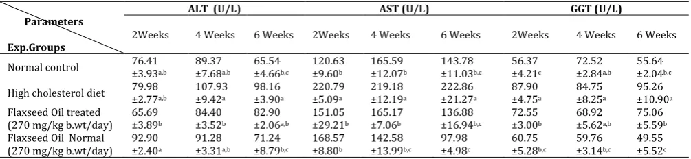
Table 2: Effect of flaxseed oil on serum AST, ALT and GGT activities in normal and hypercholesterolemic rats.
Parameters
Exp.Groups
ALT (U/L) AST (U/L) GGT (U/L)
2Weeks 4 Weeks 6 Weeks 2Weeks 4 Weeks 6 Weeks 2Weeks 4 Weeks 6 Weeks
Normal control 76.41 ±3.93a,b 89.37 ±7.68a,b 65.54 ±4.66b,c 120.63 ±9.60b ±12.07165.59 b 143.78 ±11.03b,c 56.37 ±4.21c 72.52 ±2.84a,b 55.64 ±2.04b,c
High cholesterol diet 79.98 ±2.77a,b 107.93 ±9.42a 98.16 ±3.90a 220.79 ±5.09a ±12.19219.18 a 222.86 ±21.27a 87.90 ±4.75a 84.75 ±8.25a 95.26 ±10.90a Flaxseed Oil treated
(270 mg/kg b.wt/day) 65.69 ±3.89b
84.40 ±3.52b
82.90 ±2.06a,b
151.05 ±29.21b
165.17 ±7.06b
136.88 ±16.94b,c
72.55 ±3.00b
68.92 ±5.62a,b
75.06 ±5.59b Flaxseed Oil Normal
(270 mg/kg b.wt/day) 92.90 ±2.40a
91.28 ±3.31a,b
71.24 ±8.79b,c
168.57 ±8.80b
142.58 ±13.99b,c
97.98 ±4.98c
60.75 ±5.28b,c
59.76 ±3.14b,c
49.55 ±5.52c Data are presented as (Mean ± S.E).S.E = Standard error. Mean values with different superscript letters in the same column are significantly
different at (P≤0.05)
3. RESULTS AND DISCUSSION
3.1. Effect of flaxseed oil on serum TNF-α and IL-6
concentrations in normal and
hypercholesterolemic rats
A significant increase in serum TNF-α and IL-6 concentrations were observed in cholesterol fed rats after four and six weeks of the experiment as compared with rats fed normal control diet. Flaxseed oil treatment in rats fed high cholesterol diet resulted in a significant decrease in serum TNF-α level after two
weeks of the experiments. However, serum IL-6 level was significantly decreased after two and six weeks as compared to untreated high cholesterol -fed rats (Table 1).
3.2. Effect of flaxseed oil on serum AST, ALT and GGT activities in normal and hypercholesterolemic rats
significantly increased all over the periods of the experiment associated with significant increase in serum GGT activity after two and six weeks as compared with rats fed normal control diet. Flaxseed oil treatment in rats fed high cholesterol diet resulted in a significant decrease in serum ALT activity after four with associated with significant decrease in serum AST and GGT activities all over the periods of the experiments as compared to untreated cholesterol -fed rats.
3.3. Effect of flaxseed oil on serum LDH and CK-MB activities in normal and hypercholesterolemic rats The obtained data demonstrated in table (3) revealed that, a significant increase in serum LDH and CK-MB activities were observed in cholesterol fed rats all over the periods of the experiment when compared with rats fed normal control diet. Flaxseed oil treatment in rats fed high cholesterol diet resulted in a significant decrease in serum LDH activity after two and sex weeks, associated with a significant decrease in serum CK-MB activity after four and sex weeks as compared to untreated cholesterol -fed rats.
Table 3: Effect of flaxseed oil on serum LDH and CK-MB activities in normal and hypercholesterolemic rats.
Parameters
Exp.Groups
LDH (nmol/ml) CK-MB (nmol/ml)
2Weeks 4 Weeks 6 Weeks 2Weeks 4 Weeks 6 Weeks
Normal control 333.59 ±26.85c 381.44±87.46b 700.60±68.99b 201.01±17.70b,c 205.63±11.13c 259.76±23.99b High cholesterol diet 1120.47±92.39a 1096.99±113.23a 1050.73±132.62a 441.27±58.47a 467.70±33.41a 443.38±57.28a Flaxseed Oil treated
(270 mg/kg b.wt/day) 606.46±8.07b 922.59±67.73a 354.70±21.92c 378.43±24.88a 341.24±41.13b 108.57±29.47c Flaxseed Oil Normal
(270 mg/kg b.wt/day) 481.03±98.41b,c 545.65±114.07b 403.54±19.58c 129..36±13.69 c 250.48±13.91c 173.30±16.00b,c Data are presented as (Mean ± S.E).S.E = Standard error. Mean values with different superscript letters in the same column are significantly
different at (P≤0.05)
3.4. Effect of flaxseed oil on liver tissue NO, L-MDA and GSH concentrations in normal and hypercholesterolemic rats
A significant increase in liver tissue NO concentration was observed in cholesterol fed rats after two and four weeks of the experiment, associated with a significant increase in liver L-MDA concentration all over the periods of the experiment. However, liver tissue GSH concentration was non significantly decreased when
compared with rats fed normal control diet. Flaxseed oil treatment in rats fed high cholesterol diet resulted in a significant decrease in liver NO concentration after two and six weeks and in liver L-MDA concentration after two and four weeks of the experiments. Also, a significant decrease in liver tissue GSH concentration was observed after two weeks of the experiment when compared to untreated cholesterol -fed rats (Table 4).
Table 4: Effect of flaxseed oil administration on liver tissue NO, L-MDA and GSH concentrations in normal and
hypercholesterolemic rats.
Parameters
Exp.Groups
NO (ng/g tissue) L-MDA (mmol/g tissue) GSH (ng/g tissue)
2Weeks 4 Weeks 6 Weeks 2Weeks 4 Weeks 6 Weeks 2Weeks 4 Weeks 6 Weeks
Normal control 110.68 ±3.94 b 90.84 ±7.78 b,c 104.10 ±6.56 a,b 19.71 ±2.43c ±1.6115.98 c 17.04 ±0.99b 11.60 ±2.11 a 11.76 ±3.77 a 8.66 ±1.13 a
High cholesterol diet 134.98 ±5.68 a 126.05 ±5.69 a 126.91 ±1.64 a 48.04 ±4.08a ±6.4865.33 a 40.86 ±5.77a 10.59 ±1.42a 7.65 ±0.66 a,b 7.26 ±1.22 a Flaxseed Oil treated
(270 mg/kg b.wt/day) 98.12 ±4.90 b
116.90 ±4.14 a,b
90.14 ±14.20 b,c
33.74 ±2.91b
29.44 ±9.26b,c
31.13 ±5.71a,b
2.11 ±0.67 c
2.10 ±0.21 b,c
5.75 ±0.55 a Flaxseed Oil Normal
(270 mg/kg b.wt/day) 97.24 ±10.47 b
96.24 ±6.75 b,c
84.62 ±8.26 b,c
19.23 ±1.97 c
20.87 ±1.79c
21.97 ±2.43 b
6.63 ±0.33 b
6.27 ±1.29 a,b,c
5.79 ±1.15 a Data are presented as (Mean ± S.E).S.E = Standard error. Mean values with different superscript letters in the same column are significantly
different at (P≤0.05)
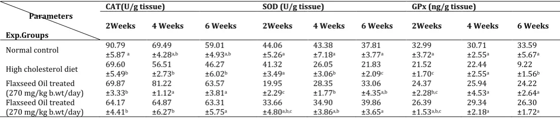
Table 5: Effect of flaxseed oil administration on liver tissue CAT, SOD and GPX activities in normal and hypercholesterolemic
rats.
Parameters
Exp.Groups
CAT(U/g tissue) SOD (U/g tissue) GPx (ng/g tissue)
2Weeks 4 Weeks 6 Weeks 2Weeks 4 Weeks 6 Weeks 2Weeks 4 Weeks 6 Weeks
Normal control 90.79 ±5.87 a 69.49 ±4.28a,b 59.01 ±4.93a,b 44.06 ±5.26a ±7.1843.38 a 37.81 ±3.77a 32.99 ±3.72a 30.71 ±2.55a 33.59 ±5.67a
High cholesterol diet 69.60 ±5.49b 56.51 ±2.73b 46.27 ±6.02b 41.32 ±3.49a ±3.0626.05 b 21.83 ±2.09c 21.52 ±1.70c 22.44 ±2.55a 9.22 ±1.56b Flaxseed Oil treated
(270 mg/kg b.wt/day)
69.87 ±3.33b
81.22 ±1.12a
63.57 ±3.81a
19.95 ±2.29c
28.35 ±1.77b
33.06 ±4.35a,b
24.37 ±2.28b,c
25.94 ±4.53a
24.22 ±2.64a Flaxseed Oil treated
(270 mg/kg b.wt/day) 64.17 ±4.41b
64.87 ±6.27b
63.31 ±5.75a
33.66 ±4.80a,b,c
34.90 ±3.86a,b
39.86 ±3.65a
26.39 ±1.53a,b,c
29.34 ±2.18a
26.30 ±1.72a Data are presented as (Mean ± S.E).S.E = Standard error. Mean values with different superscript letters in the same column are significantly
3.5. Effect of flaxseed oil on liver tissue CAT, SOD and GPX activities in normal and hypercholesterolemic rats.
The obtained results in table (5) revealed that, a significant decrease in liver CAT activity was observed in cholesterol fed rats after two weeks of the experiment. A significant decrease in liver SOD activity was observed in cholesterol fed rats after four and six weeks. However, liver GPX activity was significantly decreased after two and six weeks when compared with rats fed normal control diet. Flaxseed oil treatment in rats fed high cholesterol diet resulted in a significant increase in liver CAT activity after four and six weeks. Also, a significant increase in liver tissue SOD and GPX activities were observed after six weeks. Meanwhile, liver SOD activity was significantly decreased after two weeks of the experiment as compared to untreated cholesterol-fed rats.
Hypercholesterolemia is a lipoprotein metabolic disorder characterized by high serum levels of low density lipoprotein (LDL-C) and blood cholesterol. Alteration in cholesterol and triglycerides metabolism as a result of hypercholesterolemia has been shown to negatively affect oxidative stress biomarkers and promotes production of reactive oxygen species (ROS) by various mechanisms that lead to increased lipid peroxidation [26]. A significant increase in serum TNF-α and IL-6 concentrations were observed in high cholesterol fed rats when compared with rats fed normal control diet. These results are nearly similar to those recorded by Howard and Culley, (2006)[27] and Rajamannan et al., (2005)[28] who reported that, a significant increase inflammatory markers (CRP, TNFα) level was establish in rabbits fed cholesterol enriched diet as compared with the normal control group. Elevated serum cholesterol concentrations may trigger inflammatory responses in the blood circulation and locally in vascular walls [29]. The present results also showed that, high cholesterol diet results in gross systemic inflammatory responses, as indicated by markedly elevated cytokine concentrations. Because of the in vivo nature of current experimental model, the elevated serum cytokines concentrations could be derived either from circulating white blood cells or from any tissue source, such as endothelium [30]. The mechanisms underlying the different responses over time of different cytokines are not clear. It is possible that the high IL-6 and TNF-α response, which are mainly produced by macrophages and monocytes [31]. Prolonged increases in the concentrations of these cytokines are most likely associated with either severe damage of tissues after trauma or invasion of the body by pathogenic organisms [32].
Previous studies have found that dietary high cholesterol intake can increase the productions of atherogenic inflammatory cytokines such as IL-6 and TNF-α [33]. In addition, excessive intake of cholesterol may cause vascular inflammation, and pro-inflammatory cytokines such as TNF-α and IL-6 may stimulate the expression of adhesion molecules and
chemokines such as Vascular Cell Adhesion Molecule-1 (VCAM-1), Intracellular Adhesion Molecule-1 (ICAM-1), and fibronectin in aorta tissue [34,35]. VCAM-1 and ICAM-1 are thought to play an important role in the process of atherosclerosis by recruiting inflammatory cells, and they are both are up-regulated by pro-atherogenic factors [35]. A single cell layer lining the vascular wall, called the vascular endothelium, plays an important role in maintaining the structure and function of vessels. Besides, being the vascular endothelium a mechanical barrier between blood and vessel wall, it is also the origin of production for different bioactive factors that regulate vascular tone, coagulation, cell proliferation, cell death, and inflammation [36]. The cytokines can be represented in two shapes including the anti-inflammatory markers such as IL-1β, IL-6 and TNF-α, the second shape is the adhesion molecules such as ICAM-1 and VCAM-1 [37]. Reactive oxygen species are well documented to function as signaling molecules, stimulating cellular activities ranging from cytokine secretion to cell proliferation, and at higher concentrations, they can induce cell injury and death by oxidative modification of proteins and carbohydrates, lipid peroxidation, and DNA[38]. This is combined with, oxidants lead to the activation of endothelial cells, the action that may results in a wide range of functional changes such as the increase in expression of VCAM-1, ICAM-1, and E-selectin, and the production of chemokines, such as monocyte chemo attractant peptide-1 (AP-1) [39]. In a parallel results, Ceriello et al., (1998)[40] reported that, ICAM-1 is one of the most important intercellular adhesion molecules involved in atherogenesis and noticed increased circulating ICAM-1 plasma levels in NIDDM patients that may be resulted from the acute increase of plasma glucose which leads to produce an oxidative stress and induce cellular expression of ICAM-1. In addition, Ozawa et al., (2003)[41] illustrated the induction of ICAM-1 through pro-inflammatory cytokines (IL-1β, TNF-α, IFN-γ and IL-2) and also, VCAM-1 can be induced by IL-1β [42]. These cytokines have a probable anti-inflammatory action on the artery wall by producing an improvement in endothelial dysfunction in addition to their lipid-controlling action [43].
cytokine production are not clear. Induction of cytokine gene expression is regulated by a common pathway, i e, activation of nuclear factor-κB (NF-κB) signaling [48,49]. The presence of NF-κB binding sites has been found in the promoter regions of the IL-6, IL-1, and TNF-α genes [50]. Studies have shown that oleic acid inhibit inflammatory responses within the atherosclerotic lesions by inhibiting NF-κB activity in vascular smooth muscle cells and mononuclear cells [51].
Serum ALT, AST and GGT activities were significantly increased in cholesterol fed rats when compared with rats fed normal control diet. These results are nearly similar to those recorded by Suanarunsawat et al., (2011)[52] and Elmhdwi et al., (2014)[53] who reported that, plasma ALT and AST activities were increased in rats fed high fat diet as compared with normal diet group. High cholesterol diet was reported in several studies to cause hepatotoxicity and fatty liver [54,55]. Also, Alkhamees, (2013)[56], indicated that, high cholesterol diet significantly induced elevation in plasma liver enzymes ALT and AST activities. These parameters are known to be markers for hepatotoxicity. Who added that, histopathological findings revealed several impairments in liver sections from high cholesterol diet supplemented rats by showing moderate degree of fat accumulation, degeneration, fibrosis and inflammatory infiltrates. In the present study, the increase of ALT, AST and GGT activities in blood serum indicate that tissue impairment caused by dyslipidemia may be led to adverse effect by increasing lipid peroxidation which in turn produce damage to liver tissue so outflow of these enzymes from the liver cytosol to the blood stream which indicate that inability of liver to metabolize the ALT and AST [57].
Flaxseed oil treatment in rats fed high cholesterol diet resulted in a significant decrease in serum ALT, AST and GGT activities as compared to untreated cholesterol -fed rats. Liver is the most important organ, which plays a vital role in regulating various physiological processes in the body. It is involved in several vital functions, such as metabolism, secretion and storage. It has great capacity to detoxicate toxic substances and synthesizes useful principles [58]. The parameters such as AST and ALT in blood have been found to be of great importance in the assessment of liver damage [59]. A study on omega-3 suggests that, omega-3 polyunsaturated fatty acid (one component of flaxseed oil) supplementation may decrease liver fat [60]. Possible mechanisms that may be responsible for the protection liver damage by omega-3 fatty acids includes membrane stabilizing action on the hepatocytes [59]. Also, Hatzitolios et al., (2004)[61] reported that, the administration of ώ-3 fatty acids (ώ-3 FA) to the patients with hyperlipidemia reduced the values of AST and ALT activities and prevented the occurrence of fatty liver in 35% of the affected patients. Moreover, Alwayn et al., (2005)[62] recorded that, in a nonalcoholic fatty-liver model, ώ-3 FA administration
in murine, receiving high carbohydrate intake, led to reduce AST and ALT activities compared with the group without ώ-3 FA treatment. Similarly, Atakisi et al., (2013)[63] demonstrated that, the activity of ALT was decreased in diethylnitrosamine induced toxicity in rats treated with ώ-3 FA compared with diethylnitrosamine non -treated group.
The current study showed significant increase in serum LDH and CK-MB activities in cholesterol fed rats when compared with rats fed normal control diet. These results are nearly similar to those recorded by Suanarunsawat et al., (2011)[52] who reported that, high cholesterol diet markedly suppressed hepatic and cardiac functions as expressed by an augmentation of serum levels of AST, ALT, LDH and CK-MB activities. Increased activities of these enzymes in serum are indicative of cellular damage, loss of functional integrity, and/ or permeability of cell membrane. Flaxseed oil treatment in rats fed high cholesterol diet resulted in a significant decrease in serum LDH activity after two and sex weeks and in serum CK-MB activity after four and sex weeks of experiment as compared to untreated cholesterol -fed rats. Similarly Yu et al., (2013)[64] reported that, α-Linoleic acid pretreatment significantly inhibited doxorubicin-induced increases of CK-MB and LDH activities, thereby further supporting that ALA has cardioprotective properties. Also, Xie et al., (2011)[65] established that, ALA intake confers cardioprotection in myocardial ischemia/reperfusion by exerting anti-inflammatory and anti-oxidative stress effects in diabetic rats. In addition to these anti-oxidative cardioprotective effects, ALA also has antiapoptotic properties. For instance, pretreatment with ALA has been reported to protect against myocardial cell apoptosis by inhibition of reactive oxygen species (ROS) generation, thereby contributing to reducing myocardial infarction size [66].
Liver tissue L-MDA concentration was markedly increased in cholesterol fed rats all over the periods of the experiment. In the current work, hypercholesterolemia, a major cause for atherosclerosis was associated with increases in the levels of the lipid peroxidation product L-MDA, and decrease in the level of GSH in Liver tissue suggesting an increase in the levels or activity of oxygen radicals. MDA and GSH have been considered as specific indicators of oxidative stress [75]. L-MDA level can be used as a marker of lipid peroxidation and its measurement gives a direct evidence for LDL oxidation and is leading in predicting free radical-induced injury. Therefore, the observed elevation in tissue L-MDA may be attributed to hyperlipidemia that enhances the processes of lipid peroxidation. Hypercholesterolemia could increase the levels of ROS through stimulation of polymorphonuclear leukocytes (PMNLs) and dysfunction of endothelial cells [12,76]. Flaxseed oil treatment in rats fed high cholesterol diet resulted in a significant decrease in liver L-MDA concentration after two and four weeks of the experiments. Similarly, Prasad, (2000) [77] reported that, oral administration of flaxseed oil reduced lipid peroxidation in rats. The protective activity of flaxseed oil could be attributed to its antioxidant effect. The antioxidant activity of flaxseed oil could be attributed to its high content of omega-3 polyunsaturated fatty acid (PUFA) as reported by Khan et al., (2012) [78] who concluded that, the vegetarians who cannot consume fish oil can have similar health benefits from flaxseed oil which contains omega-3 PUFA. However, Lee and Prasad, (2003) [79] concluded that, flaxseed oil (Omega Nutrition) suppresses oxygen radical production by white blood cells and improves cardiovascular health due to its powerful antioxidant activity.
A non-significant decrease in liver tissue GSH concentration was observed in cholesterol fed rats all over the periods of the experiment when compared with rats fed normal control diet. Intracellular reduction oxidation (redox) state is regulated in part by the presence of GSH[80]. The GSH/GSSG ratio may be sensitive indicator of oxidative stress. Significant decrease in the level of total glutathione observed on HCD feeding might be due to impaired GSH biosynthesis and constant on slaught of ONOO- formed by reactions of O2- and NO, both of which increased in hypercholesterolemia. GSH plays critical role in the detoxification process against reactive nitrogen species e.g. NO, NO2 and ONOO- [81]. Oxidized glutathione (GSSG) is formed by the linking of two tripeptides by disulfide bridge. The generation of GSSG takes place during the oxidation of GSH by glutathione peroxides in the following reaction to maintain the sufficient level of GSH. The increased oxidized glutathione levels in hypercholesterolemic rats can be attributed to spontaneous non-enzymatic GSH oxidation [82]. Flaxseed oil treatment in rats fed high cholesterol diet resulted in a significant decrease in liver tissue GSH concentration after two weeks of the experiment.
Glutathione and glutathione-related enzymes play a key role in protecting the cells against the damaging effects of reactive oxygen species. Intracellular GSH can act as a reductant, reducing hydrogen peroxide and lipid hydroperoxides directly to H2O, a reaction catalyzed by GSH-Px. Depletion of intracellular GSH, under conditions of continuous intracellular oxidative stress, leads to oxidation and damage of lipids, proteins and DNA by the reactive oxygen species[83]. In the present study, the observed decrease in GSH concentration effect flaxseed oil administrations may be due to an increase in the free radical associated with hypercholesterolemia. Hypercholesterolemia induces not only atherosclerosis but also produces a lot of free radicals in blood and tissues [84].
reaction catalyzed by SOD. Thus, SOD and CAT acts mutually supportive anti-oxidative enzymes, which provide protective defense against reactive oxygen species [95]. It is commonly accepted that SOD protects against the free radical injury by converting O2- and prevent the formation of OH radicals, and the H2O2 can be removed by catalase. The main physiological benefits of flaxseeds are attributed primarily to the high linoleic acid content which contributes to their antioxidant properties [96]. Flaxseed oil might be ameliorating the damage caused by high cholesterol diet in two possible ways: First, flaxseed oil supplementation increases the levels of SOD, catalase and GPx in the liver tissue resulting in enhanced defense against ROS. Second, the constituent omega-3 PUFA of dietary flaxseed oil may have replaced the polyunsaturated fatty acid components of the cell membrane that had been attacked by oxygen free radicals [97,98]. Also, flaxseed oil a major source of omega-3 fatty acids by altering membrane fatty acid composition appeared to affect membrane organization and functions. Flaxseed oil appears to accelerate repair and/or regeneration of injured organelles e.g., mitochondria and lysosomes and plasma membrane by activating endogenous antioxidant defense mechanism [99]. Thus, flaxseed oil provided protection from high cholesterol diet-induced free radical attack. These results suggest that, flaxseed oil may be effective in controlling cholesterolemic status and improving dyslipidemia and has the potential in reducing
cardiovascular complications due to
hypercholesterolemia.
4. CONCLUSION
This study suggest that flaxseed oil possess antioxidant activity and produces potent antiatherogenic and an effective treatment against hypercholesterolemia. Administration of flaxseed oil significantly reduced lipid peroxidation and have a modulating role in hypercholesterolemia induced oxidative stress as well as it improved the antioxidant defense system in hepatic tissues. Hence, dietary consumption of flaxseed oil may provide benefits for patients with hypercholesterolemia-induced oxidative stress. We recommended that, administration of diet rich in the natural antioxidant is very important for protection of different body tissue, against oxidative stress or hypercholesterolemia and cardiovascular disease and may be beneficial for patients who suffer from hyperlipidemia, hypercholesterolemia and/or arteriosclerosis.
5. REFERENCES
1. Maruthappan, V.G., Shree, K.S. (2010). Blood cholesterol lowering effect of adenanthera pavonina seed extract on atherogenic diet induced hyperlipidemia in rats. Int J Pharm Sci Res, 1(7).87-94.
2. Adaramoye, O.A., Akintayo, O., Achem, J. et al., (2008). Lipid-lowering effects of methanolic extract of vernonia amygdalina leaves in rats fed on high cholesterol diet. Vascular health and Risk Management, 4(1). 235--241. 3. Okrainec, K., Banerjee, D.K., Eisenberg, M.J. (2004). Coronary
artery disease in the developing world. Am. Heart J, 148: 7-15.
4. Bocan, T.M. (1998). Animal models of atherosclerosis and interpretation of drug intervention studies.Curr.Pharm.Des, 4: 37–52.
5. Assy, N., Kaita, K., Mymin, D. et al., (2000). Fatty infiltration of liver in hyperlipidemic patients. Dig. Dis. Sci, 45: 1929–1934. 6. Yang, R.L., Shi, Y.H., Hao, G. et al., (2008). Increasing oxidative stress with progressive hyperlipidemia in human: relation between malondialdehyde and atherogenic index. J. Clin. Biochem.Nutr. 43: 154–158.
7. Sreekumar, R., Unnikrishnan, J., Fu, A. et al., (2002). Impact of high-fat diet and antioxidant supplement on mitochondrial functions and gene transcripts in rat muscle. Am J Physiol Endocrinol Metab. ,282(5).E1055-61.
8. Raval, J., Lyman, S., Nitta, T. et al., (2006). Basal reactive oxygen species determine the susceptibility to apoptosis in cirrhotic hepatocytes. Free Radical Biology and Medicine, 41, 1645–1654.
9. Kumari, K., Augusti, K.T. (2007). Lipid lowering effect of S-methyl cysteine sulfoxide from Allium cepa Linn in high cholesterol diet fed rats. J. Ethnopharmacol. 109: 367–371. 10.Newairy, A.S., Abdou, H.M. (2009). Protective role of flax
lignans against lead acetate induced oxidative damage and hyperlipidemia in rats. Food and Chemical Toxicology, 47, 813–818.
11.Vijaimohan, K., Jainu, M., Sabitha, K.E. et al., (2006). Beneficial effect of alpha linolenic acid rich flaxseed oil on growth performance and hepatic cholesterol in high fat diet rats. Life Sci, 79(5).448-454.
12.Prasad, K. (2005). Hypocholesterolemic and antiatherosclerotic effect of flax lignan complex isolated from flaxseed. Atherosclerosis, 179: 269-275.
13.Dupasquier, C.M., Weber, A.M., Ander, B.P. et al., (2006). Effects of dietary flaxseed on vascular contractile function and atherosclerosis during prolonged hypercholesterolemia in rabbits. American Journal of Physiology.Heart and Circulatory Physiology, 291, 2987–2996.
14.Kamesh, V.K., Sumathi, T. (2012). “Antihypercholesterolemic effect of Bacopa monniera linn. On high cholesterol diet induced hypercholesterolemia in rats,” Asian Pacific Journal of Tropical Medicine, vol. 5, no. 12, pp. 949–955.
15.Murray, R. (1984). Aminotransferases. Kaplan, A., Rubaltelli, F. F., Hammerman, C. Clin. Chem, The C. V. Mosby Co. St Louis. Toronto. Princeton, p: 1154- 1162.
16.Beleta, J., Gella, F. J. (1990). Metodo recomendado para la determinacion en rutina de la concentacion catalitica de la gamma glutamil transferase en suero sanguineo humano. Quim. Clin. (9). 58- 61.
17.Dito, W.R. (1979). Lactatedehydrogenase: Abreif review. In: Griffith JC, ED .Clinical Enzymology.New York: masson-publishing USA, p: 1-8.
18.Beyaert, R., Fiers, W. (1998). Tumor Necrosis Factor and Lymphotoxin. In Cytokines, A.R.M.S. a. R. Thorpe, eds. Academic Press, San Diego, p. 335-360.
19.Chan, D.W., Perlstein, N.T. (1987). Immunoassay: A Practical Guide, Eds, Academic Press: New York, p71.
20.Mesbah, L., Soraya, B., Narimane, S. et al., (2004). protective effect of flavonides against the toxicity of vinblastine cyclophosphamide and paracetamol by inhibition of lipid peroxydation and increase of liver glutathione. Haematol.7 (1). 59 67.
21.Montgomey, H.A.C., Dymock, J.F. (1961). Colorimetric determination of nitrite. Analyst, 86,414.
22.Moron, M.S., Depierre, J.W., Mannervik, B. (1979). Levels of glutathione, glutathione reductase and glutathione S-transferase activities in rat lung and liver. Biochimica et Biophysica ACTA 582, 67-78.
23.Luck, H. (1974). Estimation of catalase. In: methods in enzymatic analysis. 2nd edition, Bergmeyer, Academic Press, New York. Pp. 885-890.
24.Kakkar, P., Das, B., Viswanathan, P.N. (1984). A modified spectrophotometric assay of superoxide dismutase. Indian J. Biochem Biophys, 21: 130-132.
25.Gross, R.T., Bracci, R., Rudolph, N. et al., (1967). Hydrogen peroxide toxicity and detoxification in the erythrocytes of newborn infants. Blood, 29: 481–493.
rats. Afr J Pharm Pharmacol , 7(6).280–288.
27.Howard, H.T., Culley, N.C. (2006). Accumulation of low density lipoprotein associated cholesterol in calcifying vesicle fractions correlates with intimal thickening in thoracic aortas of juvenile rabbits fed a supplemental cholesterol diet. Lipids Health Dis 5: 5-25.
28.Rajamannan, N.M., Subramaniam, M., Stock, S.R. et al., (2005). Atorvastatin inhibits calcification and enhances nitric oxide synthase production in the hypercholesterolaemic aortic valve. Heart 91: 806-810.
29.Grainger, D.J., Mosedale, D.E., Metcalfe, J.C. et al., (2000). Dietary fat and reduced levels of TGFbeta1 act synergistically to promote activation of the vascular endothelium and formation of lipid lesions.J Cell Sci, 113:2355– 2361. 30.Libby, P., Ridker, P.M., Maseri, A. (2002). Inflammation and
atherosclerosis.Circulation, 105:1135– 43.
31.Gabay, C., Kushner, I. (1999). Acute-phase proteins and other systemic responses to inflammation.N Engl J Med, 340:448 – 54.
32.Grimble, R.F. (2003). Inflammatory response in the elderly. Curr Opin Clin Nutr Metab Care, 6:21–29.
33.Rahman, S.M., Van Dam, A.M., Schultzberg, M. et al., (2005). High cholesterol diet results in increased expression of interleukin-6 and caspase-1 in the brain of apolipoprotein E knockout and wild type mice. J Neuroimmunol, 169:59-67. 34.Sparks, D.L., Kuo, Y.M., Roher, A. et al., (2000). Alterations of
Alzheimer’s disease in the cholesterol-fed rabbit, including vascular inflammation.Preliminary observations. Ann N Y Acad Sci, 903:335–344.
35.Kim, S.J., Park, J.H., Kim, K.H. et al., (2011). Melittin inhibits atherosclerosis in LPS/high-fat treated mice through atheroprotective actions. J Atheroscler Thromb, 18(12).1117–1126.
36.Gibbons, G.H., Dzau, V.J. (1994). The emerging concept of vascular remodeling. N. Engl. J. Med, 330:1431-1439. 37.Inoue, I., Katayama, S. (2004). The possible therapeutic
actions of peroxisome proliferator-activated receptor alpha (PPAR alpha) agonists, PPAR gamma agonists, 3-hydroxy-3-methylglutaryl coenzyme A (HMG-CoA) reductase inhibitors, angiotensin converting enzyme (ACE) inhibitors and calcium (Ca)-antagonists on vascular endothelial cells. Curr. Drug Targets: Cardiovasc. Haematol. Disord, 4, 35-52.
38.Lander, H.M. (1997). An essential role for free radicals and derived species in signal transduction. FASEB J, 11, 118-124. 39.Amran, A.A., Zakaria, Z., Nordin, N.M.M. (2011). Changes in
the vascular cell adhesion molecule-1, intercellular adhesion molecule-1 and c-reactive protein following administration of aqueous extract of piper sarmentosum on experimental rabbits fed with cholesterol diet. Lipid Health Dis, 10:2. 40.Ceriello, A., Falleti, E., Motz, E. et al., (1998).
Hyperglycemia-induced circulating ICAM-1 increase in diabetes mellitus: the possible role of oxidative stress. Horm Metab Res, 30:14614-9.
41.Ozawa, A., Tada, H., Tamai, R. et al., (2003). Expression of IL-2 receptor beta and gamma chains by human gingival fibroblasts and up-regulation of adhesion to neutrophils in response to IL-2. J Leukoc Biol, 74:352-359.
42.Joe, B.H., Borke, J.L., Keskintepe, M. et al., (2001). Interleukin-1beta regulation of adhesion molecules on human gingival and periodontal ligament fibroblasts. J Periodontol, 72:865-870.
43.Tsimihodimos, V., Miltiadous, G., Daskalopoulou, S.S. et al., (2005). Fenofibrate: metabolic and pleiotropic effects. Curr. Vasc. Pharmacol, 3, 87-98.
44.Skoog, T., Dichtl, W., Boquist, S. et al., (2002). Plasma tumour necrosis factor-alpha and early carotid atherosclerosis in healthy middle-aged men. Eur Heart J, 23:376–383.
45.James, M.J., Gibson, R.A., Cleland, L.G. (2000). Dietary polyunsaturated fatty acids and inflammatory mediator production.AmJ Clin Nutr, 71(suppl). 343S–348S.
46.Kelley, D.S., Taylor, P.C., Nelson, G.J. et al., (1999). Docosahexaenoic acid ingestion inhibits natural killer cell activity and production of inflammatory mediators in young healthy men. Lipids, 34:317–324.
47.Zhao, G., Etherton, T.D., Martin, K.R. et al., (2007). Dietaryα-linolenic acid inhibits proinflammatory cytokine production by peripheral blood mononuclear cells in hypercholesterolemic subjects. American Society for Clinical
Nutrition.85: 385–391.
48.Barnes, P.J., Karin, M. (1997). Nuclear factor-kappaB: a pivotal transcription factor in chronic inflammatory diseases. N Engl J Med, 336: 1066 –1071.
49.Hajra, L., Evans, A.I., Chen, M. et al., (2000). The NF-kappa B signal transduction pathway in aortic endothelial cells is primed for activation in regions predisposed to atherosclerotic lesion formation. Proc Natl Acad Sci U S A, 97:9052–9057.
50.Siebenlist, U., Franzoso, G., Brown, K. (1994). Structure, regulation and function of NF-kappa B. Annu Rev Cell Biol, 10:405–455.
51.Carluccio, M.A., Massaro, M., Bonfrate, C. et al., (1999). Oleic acid inhibits endothelial activation: a direct vascular antiatherogenic mechanism of a nutritional component in the Mediterranean diet. Arterioscler Thromb Vasc Biol, 19(2). 220–228.
52.Suanarunsawat, T., Ayutthaya, W.D., Songsak, T. et al., (2011). Lipid-lowering and antioxidative activities of aqueous extracts of Ocimum sanctum L. leaves in rats fed with a high-cholesterol diet.Oxid Med Cell Longev.962025.
53.Elmhdwi, M.F., Eltaib, F.I., Fatma A. Elslimani et al., (2014). The prophylactic effect of oil extracted from Syzygium aromaticum on blood lipid level in mice fed high fat diet Journal of Biochemistry Research Vol. 2(2), pp.9-17. 54.Hirako, S., Kim, H.J., Shimizu, S. et al., (2011). Lowdose fish oil
consumption prevents hepatic lipid accumulation in high cholesterol diet fed mice. J Agric Food Chem, 59: 13353-9. 55.Wang, X., Hasegawa, J., Kitamura, Y. et al., (2011a). Effects of
hesperidin on the progression of hypercholesterolemia and fatty liver induced by high-cholesterol diet in rats. J Pharmacol Sci, 117: 129-138.
56.Alkhamees, O.A. (2013). Protective Effects of Vitamin-P and Vitamin-C on Hypercholesterolemia-induced Oxidative Hepatic Damage and Lipid Profile Changes in Female Rats: A Comparative Study. Journal of Applied Pharmaceutical Science 3 (04). 099-105.
57.Cinar, M., Yigit, A.A., Eraslan, G. (2010). Effects of vitamin C or vitamin E supplementation on Cadmium induced oxidative stress and anaemia in broilers. Revue Méd.Vét, 161: 449-454. 58.Sgro, C., Clinard, F., Ouazir, K. et al., (2002). Incidence of
drug-induced hepatic injuries: a French population-based study. Hepatology, 36:451–455.
59.Maganathan, M., Madhana-Gopal, K., Sasikala, P. et al., (2011). Evaluation of Hepato-protecttive Effect of Omega 3-Fatty Acid against Paracetamol Induced Liver Injury In Albino Rats. Global J. of Pharmacology, 5 (1). 50-53.
60.Parker, H.M., Johnson, N.A., Burdon, C.A. et al., (2011). Omega-3 supplementation and non-alcoholic fatty liver disease: A systematic review and meta-analysis. J. Hepatol., 56 (4). 944-951.
61.Hatzitolios, A., Savopoulos, C., Lazaraki, G. et al., (2004). Efficacy of omega-3 fatty acids, atorvastatin and orlistat in non-alcoholic fatty liver disease with dyslipidemia. Indian J Gastroenterol, 23: 131-134.
62.Alwayn, I.P., Gura, K., Nose, V. et al., (2005). Omega-3 fatty acid supplementation prevents hepatic steatosis in a murine model of nonalcoholic fatty liver disease. Pediatr Res, 57: 445-452.
63.Atakisi, O., Atakisi, E., Ozcan, A. et al., (2013). Protective effect of omega-3 fatty acids on diethyl-nitrosamine toxicity in rats. European Review for Medical and Pharmacological Sciences, 17: 467-471.
64.Yu, X., Cui, L., Zhang, Z. et al., (2013). α-Linolenic acid attenuates doxorubicin-induced cardiotoxicity in rats through suppression of oxidative stress and apoptosis. Acta Biochim Biophys Sin (Shanghai), 45(10).817-826.
65.Xie, N., Zhang, W., Li, J. et al., (2011). alpha-Linolenic acid intake attenuates myocardial ischemia/reperfusion injury through anti-inflammatory and anti-oxidative stress effects in diabetic but not normal rats. Arch Med Res, 42(3). 171– 181.
66.Oh, S.K., Yun, K.H., Yoo, N.J. et al., (2009). Cardioprotective effects of alpha-lipoic acid on myocardial reperfusion injury: suppression of reactive oxygen species generation and activation of mitogen-activated protein kinase. Korean Circ J, 39: 359–366.
inhibits hyperlipidemiainduced platelet aggregation via attenuating oxidative/nitrative stress. Physiol. Res., 60(2). 347-354.
68.Setorki, M., Asgary, S., Haghjooyjavanmard, S. et al., (2011). Reduces cholesterol induced atherosclerotic lesions in aorta artery in hypercholesterolemic rabbits. J. Med. Plants Res., 5(9). 1518-1525.
69.Kanazawa, K., Kawashima, S., Mikami, S. et al., (1996). Endothelial constitutive nitric oxide synthase protein and mRNA increased in rabbit atherosclerotic aorta despite impaired endothelium-dependent vascular relaxation. Am. J. Pathol., 148(6). 1949-1956.
70.Cai, H., Harrison, D.G. (2000). Endothelial dysfunction in cardiovascular diseases: The role of oxidant stress.Circulat. Res., 87(10). 840-844.
71.Minor, R.L., Myers, P.R., Guerra Jr, R. et al., (1990). Diet-induced atherosclerosis increases the release of nitrogen oxides from rabbit aorta. J. Clin.Invest, 86(6). 2109-2116. 72.Rahman, M.M., Varghese, Z., Moorhead, J.F. (2001).
Paradoxical increase in nitric oxide synthase activity in hypercholesterolaemic rats with impaired renal function and decreased activity of nitric oxide. Nephrol.Dial.Transpl, 16(2). 262-268.
73.Ho, S.C., Lin, C.C. (2008). Investigation of heat treating conditions for enhancing the anti-inflammatory activity of citrus fruit (Citrus reticulata) peels. J. Agric. Food Chem. 56(17). 7976-7982.
74.Pan, M.H., Chang, Y.H., Tsai, M.L. et al., (2008). Pterostilbene suppressed lipopolysaccharide-induced up-expression of iNOS and COX-2 in murine macrophages. J. Agric.Food Chem. 56(16). 7502-7509.
75.Mayne, S.T. (2003). Antioxidant nutrients and chronic disease: use of biomarkers of exposure and oxidative stress status in epidemiologic research. J Nutr 133.Suppl 3: 933S-940S.
76.Prasad, K. (2008). Regression of hypercholesterolemic atherosclerosis in rabbits by secoisolariciresinol diglucoside isolated from flaxseed. Atherosclerosis 197: 34-42.
77.Prasad, K. (2000). Flaxseed: a source of hypocholesterolemic and antiatherogenic agents.Drug News Perspect.13(2).99- 104.
78.Khan, M.W., Privamvada, S., Khan, S.A. et al., (2012). Protective effect of -3 polyunsaturated fatty acids (PUFAs) on sodium nitroprusside-induced nephrotoxicity and oxidative damage in rat kidney. Hum. Exp. Toxicol. 31(10). 1035-1049. 79.Lee, P., Prasad, K. (2003). Effects of flaxseed oil on serum
lipids and atherosclerosis in hypercholesterolemic rabbits. J. Cardiovasc. Pharmacol.Therp. 8(3). 227-235.
80.Kosower, N.S., Kosower, E.M. (1978). The glutathione status of the cell, International Review of Cytology, 1978, 54, 109-160.
81.Ma, X.L., Lopez, B.L., Liu, G.L. et al., (1997). Hyperchoesterolemic impairs a detoxification mechanism against peroxynitrite and renders the vascular tissue more susceptible to oxidative injury, Circulation Research, 80(6), 894-901.
82.Meister, A., Anderson, M.E. (1983). Glutathione metabolism and functions. Kroc found series 6, Raven Press New York, 1983, 1-382.
83.Nordberg, J., Arner, E.S. (2001). “Reactive oxygen species, antioxidants and the mammalian thioredoxin system”. Free.Radic. Biol., 31: 1287-1312.
84.Suanarunsawat, T., Devakul Na Ayutthaya, W., Songsak, T. et al., (2010). “Antioxidant activity and lipid-lowering effect of essential oils extracted from Ocimum sanctum L. leaves in
rats fed with a high cholesterol diet,” Journal of Clinical Biochemistry and Nutrition, vol. 46, no. 1, pp. 52–59. 85.Ahmed, S., Gul, S., Gul, H. et al., (2014). Cyclooxygenase-2
inhibition improves antioxidative defense during experimental hypercholesterolemia. Bosn J Basic Med Sci 2014, 14 (2). 63-69.
86.Meilhac, O., Ramachandran, S., Chiang, K. et al., (2001). Role of arterial wall antioxidant defense in beneficial effects of exercise on atherosclerosis in mice. Arterioscler Thromb Vasc Biol, 21: 1681-8.
87.Forstermann, U. (2008). Oxidative stress in vascular disease: causes, defense mechanisms and potential therapies. Nat Clin Pract Cardiovasc Med, 5:338–349.
88.Sudhahar, V., Kumar, S.A., Varalakshmi, P. (2006). Role of lupeol and lupeol linoleate on lipemic-oxidative stress in experimental hypercholesterolemia. Life Sci, 78:1329–1335. 89.Anila, L., Vijayalakshmi, N.R. (2003). Antioxidant action of
flavonoids from Mangifera indica and Emblica officinalis in hypercholesterolemic rats. Food Chem, 83:569–574. 90.Ma, J., Qiao, Z., Xiang, X. (2011). Aqueous extract of Astragalus
mongholicus ameliorates high cholesterol diet induced oxidative injury in experimental rats models. J. Med. Plants Res, 5, 855–858.
91.Jones, D.P. (2006). Redefining oxidative stress. Antioxid Redox Signal, 8:1865–1879.
92.Seifried, H.E., Anderson, D.E., Fishera, E.I. et al., (2007). A review of the interaction among dietary antioxidants and reactive oxygen species. J Nutr Biochem, 18:567–579. 93.Makni, M., Fetoui, H., Gargouri, N. et al., (2008).
Hypolipidemic and hepatoprotective effects of flax and pumpkin seed mixture rich in omega-3 and omega-6 fatty acids in hypercholesterolemic rats. Food and Chemical Toxicology, 46(12). 3714−3720.
94.Jangale, N.M., Devarshi, P.P., Dubal, A.A. et al., (2013). Dietary flaxseed oil and fish oil modulates expression of antioxidant and inflammatory genes with alleviation of protein glycation status and inflammation in liver of streptozotocin– nicotinamide induced diabetic rats. Food Chemistry 141: 187–195.
95.Moselhy, S.S., Ali, H.K. (2009). Hepatoprotective effect of cinnamon extracts against carbon tetrachloride induced oxidative stress and liver injury in mice. Biol. Res, 42(1). 93-98.
96.Simopoulos, A.P. (1991). Omega-3 fatty acids in health and disease and in growth and development. American Journal Of clinical Nutrition, 54: 438−463.
97.Masters, C. (1996). Omega-3 fatty acids and the peroxisome. Molecular and Cell Biochemistry, 165, 83–93.
98.Ozgocmen, S., Atalay Catal, S., Ardicoglu, O. et al., (2000). Effect of omega-3 fatty acids in the management of fibromyalgia syndrome. International Journal of Clinical Pharmacology and Therapeutics, 38, 362–363.
99.Naqshbandi, A., Rizwan, S., Khan, F. (2012). Dietary supplementation of flaxseed oil ameliorates the effect of cisplatin on rat kidney. Journal of Functional Foods.p, 316-326.
*****
© 2016; AIZEON Publishers; All Rights Reserved