R E S E A R C H
Open Access
Resistance of
Staphylococcus aureus
to
antimicrobial agents in Ethiopia: a
meta-analysis
Serawit Deyno
1*, Sintayehu Fekadu
2and Ayalew Astatkie
3Abstract
Background:
Emergence of antimicrobial resistance by
Staphylococcus aureus
has limited treatment options against
its infections. The purpose of this study was to determine the pooled prevalence of resistance to different antimicrobial
agents by
S. aureus
in Ethiopia.
Methods:
Web-based search was conducted in the databases of PubMed, Google Scholar, Hinari, Scopus and the Directory
of Open Access Journals (DOAJ) to identify potentially eligible published studies. Required data were extracted and entered
into Excel spread sheet. Statistical analyses were performed using Stata version 13.0. The
metaprop
Stata command was used
to pool prevalence values. Twenty-one separate meta-analysis were done to estimate the pooled prevalence of the resistance
of
S. aureus
to twenty-one different antimicrobial agents. Heterogeneity among the studies was assessed using the I
2statistic
and chi-square test. Publication bias was assessed using Egger
’
s test. Because of significant heterogeneity amongst the studies,
the random effects model was used to pool prevalence values.
Results:
The electronic database search yielded 1317 studies among which 45 studies met our inclusion criteria. Our analyses
demonstrated very high level of resistance to amoxicillin (77% [95% confidence interval (CI): 68%, 0.85%]), penicillin (76%
[95% CI: 67%, 84%]), ampicillin (75% [95% CI: 65%, 85%]), tetracycline (62% [95% CI: 55%, 68%]), methicillin (47%
[95% CI: 33%, 61%]), cotrimoxaziole (47% [95% CI: 40%, 55%]), doxycycline (43% [95% CI: 26%, 60%]), and erythromycin
(41% [95% CI: 29%, 54%]). Relatively low prevalence of resistance was observed with kanamycin (14% [95% CI: 5%,
25%]) and ciprofloxacin (19% [95% CI: 13%, 26%]). The resistance level to vancomycin is 11% 995% CI: (4%,
20%). High heterogeneity was observed for each of the meta-analysis performed (I
2ranging from 79.36% to
95.93%; all
p
-values
≤
0.01). Eggers
’
test did not show a significant publication bias for all antimicrobial agents
except for erythromycin and ampicillin.
Conclusions:
S. aureus
in Ethiopia has gotten notoriously resistant to almost to all of antimicrobial agents in
use including, penicillin, cephalosporins, tetracyclines, chloramphenicol, methicillin, vancomycin and sulphonamides.
The resistance level to vancomycin is bothersome and requires a due attention. Continued and multidimensional efforts
of antimicrobial stewardship program promoting rational use of antibiotics, infection prevention and containment of AMR
are urgently needed.
Keywords:
Antimicrobial resistance,
Staphylococcus aureus
, Meta-analysis, Ethiopia
* Correspondence:dserawit@gmail.com
1Department of Pharmacology, School of Medicine, College of Medicine and
Health Sciences, Hawassa University, P. O. Box 1560, Hawassa, Ethiopia Full list of author information is available at the end of the article
Background
Staphylococcus aureus
(
S. aureus
) infection is a major
cause of skin, soft tissue, respiratory, bone, joint, and
car-diovascular disorders [1].
S. aureus
remains a versatile and
dangerous pathogen in humans. The frequencies of both
community-acquired and hospital-acquired staphylococcal
infections have increased steadily. Treatment of these
in-fections has become more difficult because of the
emer-gence of multidrug-resistant strains [2].
Various mechanisms are responsible for
S. aureus
anti-microbial resistance (AMR). Penicillin is inactivated by
β
-lactamase. AMR to methicillin confers resistance to all
β
-lactamase-resistant
penicillin
’
s
and
cephalosporins
which require the presence of the mec gene that encodes
penicillin-binding protein [3]. The enterococcal
plasmid-bearing gene for resistance to vancomycin has been
transferred by conjugation to
S. aureus
in vitro [4]. Both
increased cell-wall synthesis and alterations in the cell
wall that prevent vancomycin from reaching sites of
cell-wall synthesis have been suggested as mechanisms [4].
Increase in vancomycin use has led to the emergence of
two types of glycopeptide-resistant
S. aureus
. The first
one, designated vancomycin intermediate-resistant
S.
aureus
(VISA), is associated with a thickened and poorly
cross-linked cell wall is due to continuous exposure to
glycopeptide. The second type, vancomycin-resistant
S.
aureus
(VRSA), is due to acquisition from
Enterococcus
species of the
vanA
operon resulting in high-level
resist-ance and is a rare phenomenon [5].
In Ethiopia the first published antimicrobial
prelimin-ary report on AMR was published by Plorde et al. in
1970 for different microbial agents [6]. Beginning from
that time AMR report were made by different
antimicro-bial surveillances and studies, it showed rapid rise and
spread of resistant strains.
Facilitating more appropriate choices of treatment,
minimizing the morbidity and mortality due to resistant
infections, and preserving the effectiveness of
antimicro-bials requires summarization and synthesis of the
evidence regarding AMR in a country. Appropriately
summarized and synthesized evidence is mandatory for
updating national treatment guidelines. To our
know-ledge, no previous meta-analysis or systematic review
has been conducted on
S. aureus
AMR to all
antimicro-bial commonly in use in Ethiopia. The purpose of this
study was, therefore, to determine pooled prevalence of
S. aureus
resistance to common antimicrobial agents in
Ethiopia based on the best available studies.
Methods
Study design
This study did a meta-analysis of prevalence of
S. aures
resistance to different antimicrobial agents in Ethiopia
using the best available studies.
Literature search strategy
Web-based search using PubMed, Google Scholar,
Hinari, Scopus and the Directory of Open Access
Journals (DOAJ) was conducted in June 2016. Google
search was used for unpublished works and government
documents. Two of the authors (SD and SF)
independ-ently searched for relevant studies to be included in this
meta-analysis. The PubMed search was carried out via
the EndNote software. Relevant search results from
Google scholar, Embase, Scopus and the DOAJ were
individually downloaded and manually entered into
EndNote. The reference lists of the identified studies
were used to identify other relevant studies.
The search was done using various key words:
Staphylococcus,
antimicrobial
resistance,
antibiotic
resistance, drug resistance, drug susceptibility,
anti-bacterial resistance, Ethiopia. These key terms were
used in various combinations using Boolean search
technique. We did not limit the search by year or
language of publication.
Study selection procedures and criteria
Study selection was performed in two stages
independ-ently by two of the authors (SD and SF). First, the titles
and abstracts of all retrieved articles were reviewed and
then grouped as
“
eligible for inclusion
”
if they did
ad-dress the study question and
“
ineligible for inclusion
”
if they did not. Second, articles which were grouped
under
“
eligible for inclusion
”
were reviewed in full
detail for decision.
All available studies and data were included based on
the following predefined inclusion criteria. 1) Studies
that were original journal articles, short
communica-tions, or unpublished works; 2) Studies that did the
anti-microbial susceptibility test according to the criteria
of the Clinical Laboratory Standards Institute (CLSI)
and defined antimicrobial resistance range according
to CLSI manual [7], 3) Studies which used human
in-fection sample.
Studies that 1) were duplicates, 2) were based on small
number of isolates (1
–
10), 3) were conducted on
non-human samples like on foods, food handlers
’
belongings,
health workers belongings or health workers carriage
and 4) which were based on non-infectious carriage
were excluded from this meta-analysis.
Data extraction
of drug sensitive isolates (q) was reported, the number
of resistant isolates was calculated by multiplying the
number of isolates (n) by one minus the proportion of
drug sensitive isolates (1-q) and if the proportion of drug
resistant isolates was given the number of resistant
iso-lates was found by multiplying the proportion (p) with
total number of isolates (n).
Statistical analysis and reporting
Statistical analyses were performed using Stata version
13.0 (Statacorp, LP, college station, TX). The prevalence
values from the different studies were pooled using the
metaprop
command in Stata [8]. We did twenty-one
separate meta-analyses to estimate the pooled prevalence
of the resistance of
S. aureus
to twenty one different
antimicrobial agents. The number of studies included in
each of the meta-analyses ranged from 4 to 39.
Hetero-geneity amongst the studies was assessed using the I
2statistic. Because of significant heterogeneity amongst
the studies the random-effects model (REM) was used
to estimate the pooled prevalence and 95% CIs using the
DerSimonian and Laird method [9]. The Freeman-Tukey
double arcsine transformation was used so that studies
reporting proportions near or at 0 and 1 would not be
excluded from the meta-analysis. The possible presence
of publication bias was checked using Egger
’
s test [10].
For studies that appeared to report unusually higher
prevalence of resistance compared to others, we did
sen-sitivity analysis after dropping the study which we
sus-pected of reporting a higher-than-usual result. If the
point estimate of pooled prevalence after dropping a
study lies within the 95% CI of the overall pooled
esti-mate for all studies combined, we considered the given
study as having non-significant influence on the overall
pooled estimate. Otherwise, the study was considered as
having significantly influencing the overall estimate.
Results of the current meta-analysis are reported as
per the Preferred Reporting Items for Systematic
Reviews and Meta-Analysis (PRISMA) guideline. The
PRISMA checklist was used to ensure inclusion of
relevant information (the filled checklist is included
as Additional file 1: S1) [11].
Results
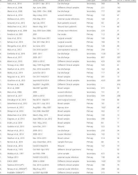
Included studies and characteristics
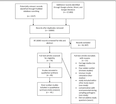
The electronic database search yielded 1317 from
PubMed and 17,400 from Google scholar, Hinari, and
Google search of which 16,083 articles remained after
removing duplicate articles. Title and abstract screening
reduced eligible articles to 76 for full text evaluation.
After reading the full texts, 31 studies were excluded for
various reasons. Thirteen studies were excluded as their
report is based on small number of isolates (less than or
equal to10) [12
–
24], four studies reported crude resistance
for all bacterial pathogen isolated [25
–
28], eleven did not
address our study question [29
–
36], six studies were based
on samples taken from of healthy carriers [37
–
42], one
study [43] was part of another study [44], and one study
[45] suffered from environmental contamination of the
samples during processing. Thus, 45 studies met our
inclusion criteria (Fig. 1). Forty-one of the studies were
journal articles, three were unpublished works [46
–
48]
and one was an official government document from the
Drug Administration and Control Authority (DACA) of
Ethiopia [49].
S. aureus
isolates from a total of 4570 patients were
tested for their antimicrobial resistance. The isolates were
from ear discharge [50
–
57], eye discharge [47, 58
–
60],
blood [61
–
68], wound infection [69
–
74], surgical site
infection [30, 73, 75
–
78], mixed samples [6, 46, 48, 49,
79
–
83], leprosy ulcer [84, 85], and urine sample [86, 87].
Twenty nine studies used primary data while nineteen
studies used records from hospitals or regional
laborator-ies (the characteristics of each included study is
summa-rized Table 1).
Publication bias and heterogeneity
Evidence of high heterogeneity was observed for each of
the meta-analyses performed (I
2ranging from 79.36% to
95.93%; all
p
-values
≤
0.01). Eggers
’
test did not suggest
any significant publication bias except for erythromycin
and ampicillin (see Additional file 2: S2).
Prevalence of
S. aureus
resistance to different
antimicrobial agents
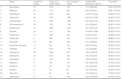
Summary of the pooled prevalence of
S. aureus
AMR
prevalence for twenty-one different antimicrobial agents
and the number of studies included in the meta-analysis
for each agent are presented in Table 2. Prevalence of
S.
aureus
resistance for each antimicrobial agent based on
pharmacological classification of the agents is given below.
As new anti-MRSA agents such as linezolid, daptomycin,
tigecycline, telavancin and ceftaroline are rarely available
in Ethiopia and no published studies available on
resist-ance to this agents, our results do not cover such agents.
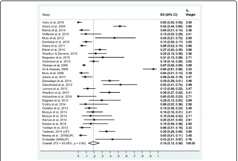
Prevalence of resistance to glycopeptides (vancomycin)
separately was 0.09, (95% CI: 0.03, 0.17). When, both Guta
et al. and Desalegn et al. were excluded, vancomycin
re-sistance was 0.07 (95% CI: 0.02, 0.14). All the three pooled
values lie within the overall pooled estimate.
Prevalence of resistance to penicillin
’
s
Here, the pooled prevalence of
S. aureus
resistance to
penicillin G, amoxicillin, ampicillin, and
amoxacilin-caluvanic acid was estimated. Resistance to penicillin G
was estimated based on 33 studies, to amoxicillin based
on 18 studies, to ampicillin based on 27 studies and to
amoxacilin-caluvanic acid based on 12 studies. Pooled
resistance rates were highest for
β
-lactamase-sensitive
penicillin
’
s. Resistance to amoxicillin was 77% (95% CI:
68%, 85%), to penicillin G 75% (95% CI: 65%, 85%) and
to ampicillin 76% (95% CI: 67%, 84%). Resistance to
carbencilin (
β
-lactam-sensitive antibiotic) was relatively
lower than other
β
-lactam-antibiotics (34% [95% CI:
17%, 54%]).
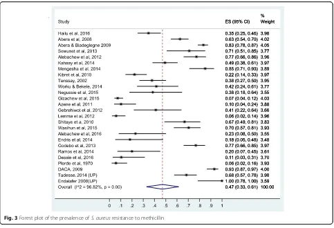
Relatively lower resistance rate was observed to
β
-lactamase-resistant penicillin
’
s: methicillin (47% [95% CI:
33%, 61%]) and amoxicillin-clavulanic acid (30% [95% CI:
19%, 43%]). The forest plots for methicillin and amoxacilin
resistance are presented in Figs. 3 and 4, respectively while
the forest plots for penicillin G, ampicillin,
amoxicillin-clavulanic acid, and carbencillin resistance are presented in
Additional file 3: S3, Additional file 4: S4, Additional file 5:
S5 and Additional file 6: S6.
Prevalence of resistance to cephalosporins
Table 1
Characteristics of included studies
No Study Study period Sample source Data type Sample size No ofS. aureusisolate
1. Hailu et al.,2016 Jan.2013 -Apr. 2015. Ear discharge Secondary 368 78
2. Abera et al.,2008 Apr -June 2006 Different clinical samples Primary 221 142
3. Abera et al.,2009 Sep. 2000 - Dec. 2008. Ear discharge Secondary 777 260
4. Mama et al.,2014 May toSep. 2013. wound infection Primary 150 47
5. Shiferaw et al.,2015 Feb.-May 2014 External ocular infections Primary 160 21
6. Sewunet, et al.,2013 Apr-July 2010 Burn patients wound Primary 50 24
7. Alebachew et al.,2012 March–May 2011 Wound burn patient’s Primary 114 66
8. Biadglegne, et al.,2009 Sep. 2003-June 2008. Urinary tract infections Secondary 529 31
9. Ferede et al.,2001 2001 Ear swabs Primary 112 28
10. Guta et al.,2014 Nov 2010-June 2011 surgical wounds Primary 100 45
11. Kahsay et al.,2014 Dec 2011-Mar 2012 surgical wounds Primary 184 73
12. Mengesha et al.,2014 Jan-June 2012 Surgical wounds Primary 128 40
13. Mulu et al.,2012 Oct 2010-Jan2011 post-operative wounds Primary 294 11
14. Denboba et al.,2016 2001–2011 Ear discharge Secondary 1225 241
15. Abera et al.,2011 2003–2011 Ear discharge Secondary 897 207
16. Kibret et al.,2010 2003 to 2010 Different clinical samples Secondary 429 429
17. Tenssay et al.,2002 May 1997-Aug1998 Different Clinical samples Primary 545 61
18. Wasihun et al.,2015 Nov 2014-June2015. Ear discharege Primary 162 46
19. Worku et al.,2014 June-Oct 2013 Ear discharge Primary 117 24
20. Negussie et al.,2015 Oct 2011-Feb2012 Blood samples Primary 201 13
21. Gizachew et al.,2015 Sep-Feb2013/2014 Different Clinical samples Secondary 4321 309
22. Yismaw et al.,2008 Sep2001- Aug2005 Different Clinical samples Secondary - 616
23. Ali et al.,2008 Mar2001-apr2005 Blood sample Secondary 472 34
24. Mulu et al.,2006 2005 wound infection Secondary 151 51
25. Azene et al.,2011 2003 to 2010 wound infection Secondary 599 208
26. Dessalegn et al.,2014 Nov2010 - Mar2011 post-surgical wound Primary 194 66
27. Gebrehiwot et al.,2012 July 2011–July 2012 Blood sample Primary 181 17
28. Lemma et al.,2012 Aug2006–May 2007 leprosy ulcer Primary 1827 68
29. Shitaye et al.,2010 Oct 2006 -Mar2007 Blood samples Primary 302 17
30. Alebachew et al. 2016 March–May, 2013 Blood samples Primary 100 13
31. Dagnew, et al. 2013 Sept 2006 to Jan 2012 Blood sample Secondary 390 17
32. Endris, et al. 2014 Feb - May, 2012. Blood samples Primary 83 11
33. Godebo et al.,2013 June-Dec., 2011 Wound Primary 322 73
34. Muluye et al.,2013 2009–2012 Ear discharge Secondary 250 54
35. Muluye et al.,2014 2009–2012 ocular infection Secondary 102 13
36. Wasihun et al.,2015 Nov 2014–2015 Blood culture Primary 514 54
37. Ramos et al.,2014 July–December, 2013 Sample from pus Primary 68 15
38. Dessie et al.,2016 Oct2013-Mar2014 Wound Primary 107 19
39. Plorde et al.,1970 Oct1969- Apr.1970 different clinical specimens Primary - 52
40. Wolday et al.,1997 Jan1992–1994 Urine sample Secondary 672 16
41. Tesfaye 2013 Feb2012-Oct2012. external ocular infection Primary 198 42
42. DACA 2009 2004 to 2008 Different clinical samples Secondary 1422 722
43. Tadesse 2014 (UPa) Dec 2013-Jun2014 Different clinical samples Primary 188 79
44. Neway et al.,2006(UPa) May-Aug. 2015 external ocular infection Primary 288 63
45. Endalafer 2008(UPa) Jun2007-Apr2008 Different clinical samples Primary 215 14
Table 2
Pooled prevalence of
S. aureus
resistance to different antimicrobial agents in Ethiopia
Agent Number of
studies
Total no of isolate tested
No of resistant isolate
Pooled AMR prevalence (95% CI)
I2(p-value)
1. Vancomycin 19 1750 132 0.11 (0.04,0.20) 95.34 (P≤0.01)
2. Methicilin 26 1179 843 0.47 (0.33,0.61) 96.82 (P≤0.01)
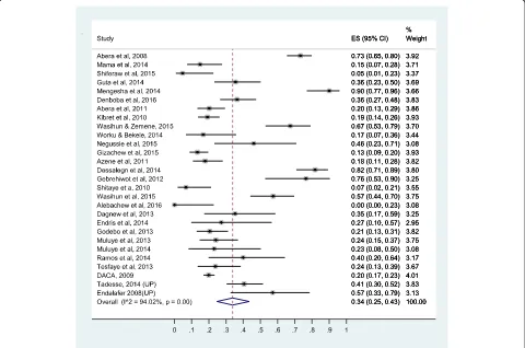
3. Ciprofloxacin 31 2254 400 0.19 (0.13, 0.26) 93.06(P≤0.01)
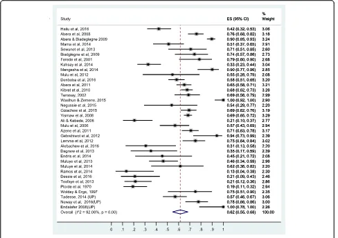
4. Tetracycline 36 3019 1982 0.62 (0.55, 0.68) 92.06(P≤0.01)
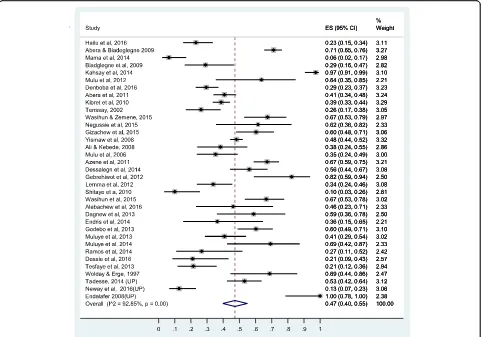
5. Cotrimoxazole 35 2825 1364 0.47 (0.40, 0.55) 92.85(P≤0.01)
6. Chloramphenicol 34 2763 1128 0.37 (0.29,0.45) 94.28 (P≤0.01)
7. Erythromycin 37 3828 2222 0.41 (0.29, 0.54) 98.28(P≤0.01)
8. Penicillin 33 2271 1627 0.76 (0.67, 0.84) 95.05(P≤0.01)
9. Clindamycin 14 1445 414 0.24 (0.12,0.37) 95.48(P≤0.01)
10. Carbencillin 6 398 184 0.34 (0.17,0.54) 86.89(P≤0.01)
11. Amoxicillin 18 870 660 0.77 (0.68,0.85) 87.44(P≤0.01)
12. Amoxicillin-clavulanic 12 524 166 0.30 (0.19,0.43) 88.64(P≤0.01)
13. Ampicillin 27 1814 1181 0.75 (0.65,0.85) 95.21(P≤0.01)
14. Gentamycin 39 3348 892 0.26 (0.18,0.34) 95.93(P≤0.01)
15. Ceftriaxone 28 2032 626 0.34 (0.25,0.43) 94.02(P≤0.01)
16. Cephalothin 15 2330 785 0.30 (0.18,0.43) 94.02(P≤0.01)
17. Cefoxitine 6 374 101 0.27 (0.06, 0.54) 95.74(P≤0.01)
18. Doxycline 14 541 239 0.43 (0.26,0.60) 93.32(P≤0.01)
19. Amikacin 4 211 62 0.23 (0.07,0.44) 90.42(P≤0.01)
20. Kanamycin 7 66 40 0.14 (0.05,0.25) 79.36(P≤0.01)
21. Norfloxacilin 11 751 186 0.25 (0.14,0.38) 92.79(P≤0.01)
L
Fig. 3Forest plot of the prevalence ofS. aureusresistance to methicillin
β
-lactamase-resistant penicillin
’
s (amoxaclin-clavulanic
acid). The prevalence of resistance to cephalothin is 30%
(95% CI: 18%, 43%), to ceftriaxone 34% (95% CI: 25%,
43%) and to cefoxitine 27% (95% CI: 6%, 54%). The
for-est plot for ceftriaxone resistance is presented in Fig. 5
while the forest plots for cephalotine and cefoxitine
re-sistance are presented respectively in Additional file 7:
S7 and Additional file 8: S8.
Prevalence of resistance to floroquinolones
Two antimicrobial agents were tested from the
floroqui-nolones: ciprofloxacin and norfloxacilin. Thirty one
studies were used to estimate the prevalence of
cipro-floxacin resistance and eleven studies were included for
the estimation of norfloxacilin resistance. The pooled
prevalence of
S. aureus
resistance to ciprofloxacin was
19% (95% CI: 13%, 26%) and to norfloxacillin 25% (95%
CI: 14%, 38%). The forest plot for ciprofloxacin
resist-ance is presented in Fig. 6 while the forest plot for
nor-floxacilin included as Additional file 9: S9.
Prevalence of resistance to protein synthesis inhibitors
Higher rates of resistance were observed with reversible
inhibitors of protein synthesis compared to aminoglycosides
(irreversible inhibitors of protein synthesis). Tetracycline
showed the highest resistance rate (62% [95% CI: 55%,
68%]) followed by doxycycline 43% (95% CI: 26%, 60%),
erythromycin (41% [95% CI: 29%, 54%]), and
chlorampheni-col (37% [95% CI: 29%, 54%]). Clindamycin and
aminoglyco-sides showed relatively lower level of resistance (Table 2).
The prevalence of resistance to gentamycin is 26%
(95% CI: 18%, 34%), to amikacin 23% (95% CI: 7%, 44%)
and to kanamycin 14% (95% CI: 5%, 25%). The forest
plot for gentamycin resistance is presented in Fig. 7
while the forest plots for erythromycin,
chlorampheni-col, doxycycline, amikacin, clindamycin, and
kanamy-cin resistance are presented as the Additional file 10:
S10, Additional file 11: S11, Additional file 12: S12,
Additional file 13: S13, Additional file 14: S14 and
Additional file 15: S15.
Prevalence of resistance to antimetabolites
Thirty five studies were included for estimation of pooled
prevalence of
S. aureus
resistance to
sulphametaxozole-trimethoprim and found to be 47% (95% CI: 40%, 55%).
The forest plot for sulphametaxozole- trimethoprim
resistance is presented in Fig. 8.
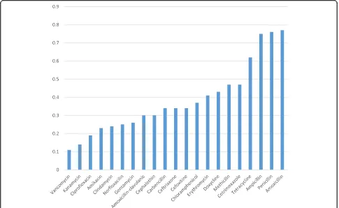
Comparison of the prevalence of
S. aures
resistance to
different antimicrobial agents
Comparison of the prevalence of
S. aures
resistance to
different antimicrobial agents addressed by this
meta-analysis is given in Fig. 9. It is found that the magnitude
of
S.aureus
resistance to the different antimicrobial
agents ranges from 11% to vancomycin to 77% to
amoxicil-lin. Accordingly, invitro antimicrobial effectiveness in
de-creasing order believed to be vancomycin, kanamycin,
ciprofloxacilin, amikacin, clindamycin, amoxacilin-clavulanic
acid, cephalothin, carbencilin, ceftriaxone, cefoxitine,
chlor-amphenicol, erythromycin, doxycycline, methicillin,
cotri-moxazole, tetracycline, ampicillin, pencilin, and amoxacilin.
Discussion
In this meta-analysis, we estimated the pooled
preva-lence of
S. aureus
resistance to 21 different antimicrobial
agents commonly used in Ethiopia. Generally 45 studies
were included for the meta-analysis, however the
num-ber of studies included in each meta-analyses ranged
from 4 to 39. Overall, the 45 studies provided evidence
regarding the level of
S. aureus
resistance to different
antimicrobial agents based on 4530 isolates. It was
found that
S. aureus
resistance to commonly available
antimicrobial agents in Ethiopia was alarmingly high
ran-ging from 11% to vancomycin to 77% to amoxicillin.
The pooled estimate of the prevalence of
S. aureus
re-sistance particularly to methicillin (MRSA) in Ethiopia is
similar to 2014 global surveillance reports of the World
Health Organization (WHO) 2014 [88], which showed
MRSA prevalence between 33% to 95% in Africa. The
pooled prevalence of MRSA in Ethiopia 47% (95% CI:
33%
–
61%) is within the range of the global WHO report
for Africa.
caused by transmission of resistant strains within
hospi-tals by cross colonization of patients via hands of
health-care staff and direct patient to patient contact and
subsequent spread [91].
Global pattern of AMR shows variation among
differ-ent geographic, socioeconomic strata and among studies
[49, 88, 92]. Variation may be to differences in time,
place, design, and population involved in the study. This
may be due to healthcare facilities conditions like
imple-mentation and monitoring of infection prevention
policies and rational antibiotic usage which varies in
dif-ferent facilities. The most important reason is due to
character of the study. Studies are conducted within a
specified time and locality. It is reasonable to assume
population under study might be infected by the same
strains of agent at specified period of time and location.
This could be a good reason why heterogeneity tests
showed significant variability (
p
-value
≤
0.01) among
studies included in this meta-analysis for 2 l
anti-microbial agents.
S. aureus
acquires resistance by various mechanisms:
formation of alternative pathways for sulphonamides
[93, 94], production of
β
-lactamase to
β
-lactam-sensitive
antibiotics, increased efflux to tetracycline [95, 96], presence
of acetyltransferase to chloramphenicol, decrease in
accu-mulation to macrolide antibiotics [97],
aminoglycoside-modifying enzymes production to aminoglycosides, altered
topoisomerase IV and DNA gyrase expression for
fluoroqui-nolones, and expression of mec gene altering penicillin
binding protein to
β
-lactam antibiotics [98]. Since the AMR
for
β
-lactam sensitive
β
-lactam antibiotics is very high, it
can be speculated that most strains of
S. aureus
found in Ethiopia produce the
β
-lactamase enzyme.
However, there is no molecular study conducted to
identify the type of resistant strains and mechanism
responsible for resistance in Ethiopia.
Lower rate of resistance was seen with
β
-lactamase-resistant antibiotics (amoxicillin-clavulanic acid,
methi-cillin, ceftriaxone, cefoxtine, and cephalothin) compared
to
β
-lactamase-sensitive penicillins. Unlike
β
-lactamase
sensitive penicillin
’
s, resistance to carbeniciln is
signifi-cantly lower. The lower rate of resistance observed with
carbencilin and clindamycin may be due to their
infre-quent use in Ethiopia [99].
high level of resistance requires the presence of the mec
gene that encodes penicillin-binding protein [98]. The
implication of high prevalence of MRSA for suspected
or verified
S. aureus
infections such as common skin
and wound infections and surgical prophylaxis is that
there is a need for better alternatives drugs. Alternative
drugs needed to treat or prevent
S. aureus
infections are
more expensive and, because of their adverse effects,
monitoring during treatment is advisable which
in-creases the costs even further.
The prevalence of resistance
S. aureus
to vancomycin
11% 995% CI: (4%, 20%) in this study is bothersome and
higher compared to global prevalence estimate [100].
The prevalence of VISA was 2.05% before, 2.63% in
2006
–
2009, and 7.93% in 2010
–
2014. Vancomycin
resist-ance is erasing all possible treatment options in Ethiopia
for MRSA. The higher prevalence of vancomycin in
Ethiopia compared to global estimate may be due to larger
and irrational use of antimicrobial agents in Ethiopia,
The prevalence estimates of glycopeptides/vancomycin
resistance from Guta et al.
,
and Desalegn et al.
,
were
unusually high, however sensitivity analysis showed
non-significant influence on the overall pooled prevalence
estimate. The prevalence estimates from Guta et al. and
Desalegn et al. were unusually high, however sensitivity
analysis showed non-significant influence on the overall
pooled prevalence estimate. Larger exposure probability
to resistant strains due to larger use of vancomycin in
hospital settings might have resulted in a relatively
higher prevalence of vancomycin resistance in the two
studies [73, 77].
In four of the twenty studies (published in 2014 and
after) [48, 51, 73, 77], the prevalence of
S. aureus
resist-ance to vancomycin is higher than 40%. In contrast, in
studies published before 2014 the prevalence of
S.
aur-eus
resistance to vancomycin in Ethiopia is much lower
(0% to 16%). This may indicate a rapid rise and spread
of vancomycin resistant
S. aureus
strains in Ethiopia as
the rate of vancomycin use and exposure in Ethiopia
increases. This calls for inclusion for effective new
anti-MRSA antimicrobial agents for treatment of
staphylococcal infections in the national medicine list
and effective antimicrobial stewardship programs for
prevention and containment of antimicrobial resistance.
based on the finding of our invitro finding. However,
clin-ical effectiveness study had not yet proved it. Resistance to
vancomycin, the only choice for MRSA in Ethiopia, is of a
great concern. It is bothersome due to lack of alternative
agents in Ethiopia for the treatment of
S. aureus
infections.
Making things worse, alternative new anti-MRSA agents
(like linezolid, daptomycin, tigecycline, telavancin, and
cef-taroline are rarely available in Ethiopia for treatment of
vancomycin resistant
S. aureus.
Many factors contribute to AMR. First, lack of
infec-tion preveninfec-tion contributes to recurrent infecinfec-tion then
to spread of resistant strains. Second, misuse of
antimi-crobials from prescription
–
dispensing-to patient use
[101]. In Ethiopia, it is a common practice that
antibi-otics can be purchased without prescription, which leads
to misuse of antibiotics by the public [102]. Third factor
could be misuse of antibiotics by health professionals
and non-standardized practice [101]. The fourth factor
could be poor hospital hygienic conditions [103]. A last
contributing factor could be lack of routine
antimicro-bial susceptibility testing which diverts to empiric
therapy [49]. In line to strategies for prevention and
con-tainment of
S. aureus
there is a need for innovative way
of halting AMR. Combination therapy and availability of
new anti-MRSA agents will play vital role in fighting
against AMR to
S. aureus
.
However, interpretation of the findings of this
meta-analysis requires considering the limitations thereof. The
limitations arise from the inherent characteristics of the
included individual studies. First, this is invitro
anti-microbial resistance testing and its direct translation to
clinical effectiveness requires caution. Second, many
studies involved very limited localities and were done
mainly in teaching hospitals in bigger cities where
pa-tients with advanced, severe stages, recurrent
infec-tions are treated. Hence, the resistance level could
have overestimated.
Conclusions
This meta-analysis demonstrates that
S. aureus
has
got-ten alarmingly resistant to many of common
antimicro-bials used in Ethiopia. It is highly resistant to penicillin,
cephalosporin, tetracyclines, chloramphenicol,
methicil-lin, sulphonamides, and vancomycin. Resistance to
vancomycin is of a great concern and bothersome due to
unavailability of treatment options for
S. aureus
infec-tions in Ethiopia.
treat resistant strains. Combination therapy, effective in
battling AMR in many infectious diseases model,
may prove significant advantage in battling resistance
to
S. aureus
.
Therapeutic
options
are
urgently
needed for patients infected with resistant
S. aureus
.
Further researches focusing on clinical treatment
outcome and identifying dynamics promoting
resist-ance, high risk strains and molecular genetic basis of
resistance are needed.
Additional files
Additional file 1: S1.RISMA Checklist. (DOC 63 kb)
Additional file 2: S2.Egger’s test of publication bias. (DOCX 18 kb)
Additional file 3: S3.Forest plot of the prevalence ofS. aureusresistance to penicillin G. (DOCX 22 kb)
Additional file 4: S4.Forest plot of the prevalence ofS. aureusresistance to Ampicillin. (DOCX 20 kb)
Additional file 5: S5.Forest plot of the prevalence ofS. aureusresistance to amoxacilin-clavulanate. (DOCX 19 kb)
Additional file 6: S6.Forest plot of the prevalence ofS. aureusresistance to carbencilin. (DOCX 16 kb)
Additional file 7: S7.Forest plot of the prevalence ofS. aureusresistance to cephalothin. (DOCX 18 kb)
Additional file 8: S8.Forest plot of the prevalence ofS. aureusresistance to cefoxitine. (DOCX 16 kb)
Additional file 9: S9.Forest plot of the prevalence ofS. aureusresistance to norfloxacilin. (DOCX 17 kb)
Additional file 10: S10.Forest plot of the prevalence ofS. aureusresistance to erythromycin. (DOCX 21 kb)
Additional file 11: S11.Forest plot of the prevalence ofS. aureusresistance to chloramphenicol. (DOCX 22 kb)
Additional file 12: S12.Forest plot of the prevalence ofS. aureusresistance to doxycycline. (DOCX 18 kb)
Additional file 13: S13.Forest plot of the prevalence ofS. aureusresistance to amikacin. (DOCX 16 kb)
Additional file 14: S14.Forest plot of the prevalence ofS. aureusresistance to clindamycin. (DOCX 18 kb)
Additional file 15: S15.Forest plot of the prevalence ofS. aureusresistance to kanamycin. (DOCX 15 kb)
Abbreviations
AMR:Antimicrobial resistance; MRSA: Methicillin resistantStaphylococcus aureus; NCCLs: National Committee for clinical Laboratory Standard;S. aureus:Staphylococcus
aureus; VISA: Vancomycin intermediateStaphylococcus aureus; VRSA: Vancomycin resistantStaphylococcus aureus
Acknowledgements Not applicable.
Funding
There was no funding support to conduct this meta-analysis.
Availability of data and materials
The data supporting the conclusions of this article are included within the article and its supporting information.
Authors’contributions
SD, SF and AA conceptualized the research idea. SD and SF conducted literature search, selection and data extraction. AA performed the statistical analyses. SD prepared the draft manuscript. All authors revised, edited and approved the final manuscript.
Ethics approval and consent to participate Not applicable.
Consent for publication Not applicable.
Competing interests
The authors declare that they have no competing interests.
Publisher
’
s Note
Springer Nature remains neutral with regard to jurisdictional claims in published maps and institutional affiliations.
Author details
1Department of Pharmacology, School of Medicine, College of Medicine and
Health Sciences, Hawassa University, P. O. Box 1560, Hawassa, Ethiopia.
2Department of Microbiology, Faculty of Medicine, Shimane University,
Shimane, Japan.3School of Public and Environmental Health, College of Medicine and Health Sciences, Hawassa University, Hawassa, Ethiopia.
Received: 28 June 2017 Accepted: 15 August 2017
References
1. Lowy FD. Staphylococcus aureus infections. N Engl J Med. 1998;339(8):520–32. 2. McGuinness WA, Malachowa N, FR DL. Vancomycin resistance in Staphylococcus
Aureus. Yale J Biol Med. 2017;90(2):269–81.
3. Hiramatsu K. Mechanism of methicillin resistance and genetic background of Staphylococcus Aureus. Nihon Naika Gakkai Zasshi. 1992;81(10):1592–8. 4. Hiramatsu K, Hanaki H, Ino T, Yabuta K, Oguri T, Tenover FC.
Methicillin-resistant Staphylococcus Aureus clinical strain with reduced vancomycin susceptibility. J Antimicrob Chemother. 1997;40:135–6.
5. Périchon B, Courvalin P. VanA-type vancomycin-resistant Staphylococcus Aureus. Antimicrob Agents Chemother. 2009;53(11):4580–7.
6. Plorde JJ, Eng SC, Wright LJ, Debesai A. Antibiotic sensitivities of common bacterial pathogens isolated in Addis Ababa; a preliminary report. Ethiop Med J. 1970;8(3):107–18.
7. Stephen J, Ronald J, Yvette S, José H, Ivonne D, Robert L, Susan E, Carol A: Manual of antimicrobial susceptibility testing. In.USA; 2005.
8. Nyaga VN, Arbyn M, Aerts M. Metaprop: a Stata command to perform meta-analysis of binomial data. Archives of Public Health. 2014;72:39.
9. DerSimonian R, Laird N. Meta-analysis in clinical trials. Control Clin Trials. 1986;7:177–88.
10. Egger M, Davey SG, Schneider M, Minder C. Bias in meta-analysis detected by a simple, graphical test. BMJ. 1997;315(7109):629–34.
11. Moher D, Liberati A, Tetzlaff J, Altman DG, The PRISMA Group. Perferred Reporting items for Systematic Review and Meta-analysis:The PRISMA statement. PLoS Med. 2009;6:e1000097.
12. Abrha A, Abdissa A, Beyene G, Getahun G, Girma T. Bacteraemia among severely malnourished children in jimma university hospital, ethiopia. Ethiopian journal of health sciences. 2011;21(3):175–82.
13. Alemu A, Moges F, Shiferaw Y, Tafess K, Kassu A, Anagaw B, Agegn A. Bacterial profile and drug susceptibility pattern of urinary tract infection in pregnant women at University of Gondar Teaching Hospital, Northwest Ethiopia. BMC research notes. 2012;5:197.
14. Beyene G, Tsegaye W. Bacterial uropathogens in urinary tract infection and antibiotic susceptibility pattern in jimma university specialized hospital, southwest ethiopia. Ethiopian journal of health sciences. 2011;21(2):141–6. 15. Demilie T, Beyene G, Melaku S, Tsegaye W. Urinary bacterial profile and antibiotic susceptibility pattern among pregnant women in north west ethiopia. Ethiopian journal of health sciences. 2012;22(2):121–8.
16. Tesfahunegn Z, Asrat D, Woldeamanuel Y, Estifanos K. Bacteriology of surgical site and catheter related urinary tract infections among patients admitted in Mekelle hospital, Mekelle, Tigray, Ethiopia. Ethiop Med J. 2009;47(2):117–27. 17. Assefa Y, Moges F, Endris M, Zereay B, Amare B, Bekele D, Tesfaye S, Mulu A,
Belyhun Y. Bacteriological profile and drug susceptibility patterns in dacryocystitis patients attending Gondar University Teaching Hospital, Northwest Ethiopia. BMC Ophthalmol. 2015;15:34.
19. Kibret M, Abera B. Prevalence and antibiogram of bacterial isolates from urinary tract infections at Dessie Health Research Laboratory, Ethiopia. Asian Pac J Trop Biomed. 2014;4(2):164–8.
20. Tadesse E, Teshome M, Merid Y, Kibret B, Shimelis T. Asymptomatic urinary tract infection among pregnant women attending the antenatal clinic of Hawassa referral hospital, Southern Ethiopia. BMC research notes. 2014;7:155. 21. Wondimeneh Y, Muluye D, Alemu A, Atinafu A, Yitayew G, Gebrecherkos T,
Damtie D, Ferede G. Urinary tract infection among obstetric fistula patients at Gondar University Hospital, northwest Ethiopia. BMC Womens Health. 2014;14:12.
22. Yeshitela B, Gebre-Selassie S, Feleke Y. Asymptomatic bacteriuria and symptomatic urinary tract infections (UTI) in patients with diabetes mellitus in Tikur Anbessa specialized university hospital, Addis Ababa, Ethiopia. Ethiop Med J. 2012;50(3):239–49.
23. Gebre-Selassie S. Asymptomatic bacteriuria in pregnancy: epidemiological, clinical and microbiological approach. Ethiop Med J. 1998;36(3):185–92. 24. Derese B, Kedir H, Teklemariam Z, Weldegebreal F, Balakrishnan S. Bacterial
profile of urinary tract infection and antimicrobial susceptibility pattern among pregnant women attending at antenatal Clinic in Dil Chora Referral Hospital, Dire Dawa, eastern Ethiopia. Ther Clin Risk Manag. 2016;12:251–60. 25. Moges AF, Genetu A, Mengistu G. Antibiotic sensitivities of common bacterial
pathogens in urinary tract infections at Gondar hospital, Ethiopia. East Afr Med J. 2002;79(3):140–2.
26. Muluye D, Wondimeneh Y, Ferede G, Nega T, Adane K, Biadgo B, Tesfa H, Moges F. Bacterial isolates and their antibiotic susceptibility patterns among patients with pus and/or wound discharge at Gondar university hospital. BMC research notes. 2014;7:619.
27. Melaku S, Gebre-Selassie S, Damtie M, Alamrew K. Hospital acquired infections among surgical, gynaecology and obstetrics patients in Felege-Hiwot referral hospital, Bahir Dar, northwest Ethiopia. Ethiop Med J. 2012;50(2):135–44. 28. Kebede A, Adamu Y, Bejiga A. Bacteriological study of dacryocystitis among
patients attending in Menelik II hospital, Addis Ababa, Ethiopia. Ethiop Med J. 2010;48(1):29–33.
29. Melaku S, Kibret M, Abera B, Gebre-Sellassie S. Antibiogram of nosocomial urinary tract infections in Felege Hiwot referral hospital, Ethiopia. Afr Health Sci. 2012;12(2):134–9.
30. Mulu W, Kibru G, Beyene G, Damtie M. Postoperative nosocomial infections and antimicrobial resistance pattern of bacteria isolates among patients admitted at Felege Hiwot referral hospital, Bahirdar, Ethiopia. Ethiopian journal of health sciences. 2012;22(1):7–18.
31. Seboxa T, Amogne W, Abebe W, Tsegaye T, Azazh A, Hailu W, Fufa K, Grude N, Henriksen TH. High mortality from blood stream infection in Addis Ababa, Ethiopia, is due to antimicrobial resistance. PLoS One. 2015;10(12):e0144944. 32. Tilahun B, Worku B, Tachbele E, Terefe S, Kloos H, Legesse W. High load of
multi-drug resistant nosocomial neonatal pathogens carried by cockroaches in a neonatal intensive care unit at Tikur Anbessa specialized hospital, Addis Ababa, Ethiopia. Antimicrob Resist Infect Control. 2012;1:12.
33. Zenebe T, Kannan S, Yilma D, Beyene G. Invasive bacterial pathogens and their antibiotic susceptibility patterns in Jimma University specialized hospital, Jimma, Southwest Ethiopia. Ethiopian journal of health sciences. 2011;21(1):1–8. 34. Tibebu M, Embiyale W. Community acquired multi drug resistant
Staphylococcus Aureus in a rural setting of north western Ethiopia: a tough challenge. Ethiop Med J. 2014;52(3):147–50.
35. Truneh M. Phage types and drug susceptibility patterns of Staphylococcus Aureus from two hospitals in northwest Ethiopia. Ethiop Med J. 1991;29(1):1–6. 36. Teshager L, Asrat D, Gebre-Selassie S, Tamiru S. Catheterized and non-catheterized
urinary tract infections among patients attended at Jimma University teaching hospital, southwest, Ethiopia. Ethiop Med J. 2008;46(1):55–62.
37. Lemma MT, Zenebe Y, Tulu B, Mekonnen D, Mekonnen Z. Methicillin resistant Staphylococcus Aureus among HIV infected pediatric patients in Northwest Ethiopia: carriage rates and antibiotic co-resistance profiles. PLoS One. 2015;10(9):e0137254.
38. Kejela T, Bacha K. Prevalence and antibiotic susceptibility pattern of methicillin-resistant Staphylococcus Aureus (MRSA) among primary school children and prisoners in Jimma town, Southwest Ethiopia. Ann Clin Microbiol Antimicrob. 2013;12:11.
39. Gebreyesus A, Gebre-Selassie S, Mihert A. Nasal and hand carriage rate of methicillin resistant Staphylococcus Aureus (MRSA) among health care workers in Mekelle hospital, North Ethiopia. Ethiop Med J. 2013;51(1):41–7.
40. Dagnew M, Tiruneh M, Moges F, Tekeste Z. Survey of nasal carriage of Staphylococcus Aureus and intestinal parasites among food handlers
working at Gondar University, Northwest Ethiopia. BMC Public Health. 2012;12(1):837.
41. Shibabaw A, Abebe T, Mihret A. Nasal carriage rate of methicillin resistant Staphylococcus Aureus among Dessie referral hospital health care workers; Dessie, Northeast Ethiopia. Antimicrob Resist Infect Control. 2013;2(1):25. 42. Shibabaw A, Abebe T, Mihret A. Antimicrobial susceptibility pattern of nasal
Staphylococcus Aureus among Dessie referral hospital health care workers, Dessie, Northeast Ethiopia. Int J Infect Dis. 2014;25:22–5.
43. Yismaw G, Abay S, Asrat D, Yifru S, Kassu A. Bacteriological profile and resistant pattern of clinical isolates from pediatric patients, Gondar University teaching hospital, Gondar, Northwest Ethiopia. Ethiop Med J. 2010;48(4):293–300. 44. Yismaw G, Tiruneh M, Kassu A, Negeri C, Mulu A. A retrospective analysis of
prevalence and antimicrobial susceptibility patterns of Staphylococcus Aureus in Gondar teaching hospital, 2001-2005. Ethiop Med J. 2008;46(2):143–8. 45. Asrat D, Amanuel YW. Prevalence and antibiotic susceptibility pattern of
bacterial isolates from blood culture in Tikur Anbassa hospital, Addis Ababa, Ethiopia. Ethiop Med J. 2001;39(2):97–104.
46. Endalafer N. Bacterial nosocomial infections and their antimicrobial susceptibility patterns in surgical wards and surgical intensive care unit of Tikur Anbessa university hospital. Addis Ababa: Addis Ababa University; 2008. 47. Neway S, Desta K, Dessie W, Yeshitila B, Lema T. Bacterial profile and antimicrobial
susceptibility pattern of external ocular infections with associated risk factors in Alert cente. Ethiopia: Addis Ababa; 2016.
48. Tadesse S. Antimicrobial resistance profile ofStaphylococcus aureusisolated from clinical specimens and nasal swabs of patients at Tikur Anbessa specialized hospital. Addis Ababa: Addis Ababa University; 2014. 49. DACA. Antimicrobials use, resistance and containment baseline survey:
syntheses of findings. Addis Ababa: DACA; 2009.
50. Hailu D, Mekonnen D, Derbie A, Mulu W, Abera B. Pathogenic bacteria profile and antimicrobial susceptibility patterns of ear infection at Bahir Dar regional Health Research Laboratory center Ethiopia. SpringerPlus. 2016;5:466. 51. Argaw-Denboba A, Abejew AA, Mekonnen AG. Antibiotic-resistant bacteria
are major threats of otitis Media in Wollo Area, Northeastern Ethiopia: A Ten-Year Retrospective Analysis. Int J Microbiol. 2016;2016:8724671. 52. Muluye D, Wondimeneh Y, Ferede G, Moges F, Nega T. Bacterial isolates
and drug susceptibility patterns of ear discharge from patients with ear infection at Gondar University Hospital, Northwest Ethiopia. BMC ear, nose, and throat disorders. 2013;13(1):10.
53. Wasihun AG, Zemene Y. Bacterial profile and antimicrobial susceptibility patterns of otitis media in Ayder Teaching and Referral Hospital, Mekelle University, Northern Ethiopia. SpringerPlus. 2015;4:701.
54. Abera B, Kibret M. Bacteriology and antimicrobial susceptibility of otitis Media at Dessie Regional Health Research Laboratory, Ethiopia. Ethiop J Health Dev. 2011;25(2):161–7.
55. Ferede D, Geyid A, Lulseged S, Melaku A. Drug susceptibility pattern of bacterial isolates from children with chronic suppurative otitis media. Ethiop J Health Dev. 2001;15(2):89–96.
56. Worku M, Bekele M. Bacterial isolate and antibacterial resistance pattern of ear infection among patients attending at Hawassa university referral hospital, Hawassa, Ethiopia. Indian J Otol. 2014;20:155–9.
57. Abera B, Biadeglegne F. Antimicrobial resistance patterns of Staphylococcus Aureus and Proteus spp. isolated from otitis media at Bahir Dar regional laboratory, north West Ethiopia. Ethiop Med J. 2009;47(4):271–6. 58. Shiferaw B, Gelaw B, Assefa A, Assefa Y, Addis Z. Bacterial isolates and their
antimicrobial susceptibility pattern among patients with external ocular infections at Borumeda hospital, Northeast Ethiopia. BMC ophthalmology. 2015;15:103.
59. Muluye D, Wondimeneh Y, Moges F, Nega T, Ferede G. Types and drug susceptibility patterns of bacterial isolates from eye discharge samples at Gondar University Hospital, Northwest Ethiopia. BMC research notes. 2014;7:292.
60. Tesfaye T, Beyene T, Gelaw T, Bekele S, Saravanan M. Bacterial profile and antimicrobial susceptibility pattern of external ocular infections in Jimma University specialized hospital, Southwest Ethiopia. American Journal of Infectious Diseases and Microbiology. 2013;1(1):13–20.
61. Negussie A, Mulugeta G, Bedru A, Ali I, Shimeles D, Lema T, Aseffa A. Bacteriological profile and antimicrobial susceptibility pattern of blood culture isolates among septicemia suspected children in selected hospitals Addis Ababa, Ethiopia. Int J Biol Med Res. 2015;6(1):4709–17.
Living with Human Immunodeficiency Virus at Gondar University Teaching Hospital, Northwest Ethiopia. Biomed Res Int. 2016;2016:5371875. 63. Endris M, Takele Y, Woldeyohannes D, Tiruneh M, Mohammed R, Moges F,
Lynen L, Jacobs J, van Griensven J, Diro E. Bacterial sepsis in patients with visceral leishmaniasis in Northwest Ethiopia. Biomed Res Int. 2014;2014: 361058.
64. Shitaye D, Asrat D, Woldeamanuel Y, Worku B. Risk factors and etiology of neonatal sepsis in Tikur Anbessa university hospital, Ethiopia. Ethiop Med J. 2010;48(1):11–21.
65. Wasihun AG, Wlekidan LN, Gebremariam SA, Dejene TA, Welderufael AL, Haile TD, Muthupandian S. Bacteriological profile and antimicrobial susceptibility patterns of blood culture isolates among febrile patients in Mekelle hospital, Northern Ethiopia. SpringerPlus. 2015;4:314.
66. Ali J, Kebede Y. Frequency of isolation and antimicrobial susceptibility pattern of bacterial isolates from blood culture, Gondar University teaching hospital, Northwest Ethiopia. Ethiop Med J. 2008;46(2):155–61.
67. Dagnew M, Yismaw G, Gizachew M, Gadisa A, Abebe T, Tadesse T, Alemu A, Mathewos B. Bacterial profile and antimicrobial susceptibility pattern in septicemia suspected patients attending Gondar University Hospital, Northwest Ethiopia. BMC research notes. 2013;6:283.
68. Gebrehiwot A, Lakew W, Moges F, Moges B, Anagaw B, Yismaw G, Nega T, Unakal C, Kassu A. Bacterial profile and drug susceptibility pattern of neonatal sepsis in Gondar University hospital, Gondar northwest Ethiopia. Pharm Lett. 2012;4(6):1811–6.
69. Alebachew T, Yismaw G, Derabe A, Sisay Z. Staphylococcus Aureus burn wound infection among patients attending yekatit 12 hospital burn unit, addis ababa, ethiopia. Ethiopian journal of health sciences. 2012;22(3):209–13. 70. Godebo G, Kibru G, Tassew H. Multidrug-resistant bacterial isolates in infected wounds at Jimma University Specialized Hospital, Ethiopia. Ann Clin Microbiol Antimicrob. 2013;12:17.
71. Azene MK, Beyene BA. Bacteriology and antibiogram of pathogens from wound infections at Dessie laboratory, north-east Ethiopia. Tanzania journal of health research. 2011;13(4):68–74.
72. Mulu A, Moges F, Tessema B, Kassu A. Pattern and multiple drug resistance of bacterial pathogens isolated from wound infection at University of Gondar Teaching Hospital, Northwest Ethiopia. Ethiop Med J. 2006;44(2):125–31. 73. Guta M, Aragaw K, Merid Y. Bacteria from infected surgical wounds and
their antimicrobial resistance in Hawassa university referral teaching hospital, southern Ethiopia. Afr J Microbiol Res. 2014;8(11):1118–24.
74. Mama M, Alemseged A, Tsegaye S. Antimicrobial susceptibility pattern of bacterial isolates from wound infection and their sensitivity to alternative topical agents at Jimma University Specialized Hospital, South-West Ethiopia. Annals of Clinical Microbiology and Antimicrobials. 2014;13(1):14. 75. Kahsay A, Mihret A, Abebe T, Andualem T. Isolation and antimicrobial
susceptibility pattern ofStaphylococcus aureusin patients with surgical site infection at Debre Markos Referral Hospital, Amhara Region, Ethiopia. Arch Public Health. 2014;72(1):16.
76. Mengesha RE, Kasa BG, Saravanan M, Berhe DF, Wasihun AG. Aerobic bacteria in post surgical wound infections and pattern of their antimicrobial susceptibility in Ayder teaching and referral hospital, Mekelle, Ethiopia. BMC research notes. 2014;7:575.
77. Dessalegn L, Shimelis T, Tadesse E, Gebre-selassie S. Aerobic bacterial isolates from post-surgical wound and their antimicrobial susceptibility pattern: a hospital based cross-sectional study. Journal of Medical Research. 2014;3(2):18–23.
78. Dessie W, Mulugeta G, Fentaw S, Mihret A, Hassen M, Abebe E. Pattern of bacterial pathogens and their susceptibility isolated from surgical site infections at selected referral hospitals, Addis Ababa, Ethiopia. International journal of microbiology. 2016;2016:2418902.
79. Sewunet T, Demissie Y, Mihret A, Abebe T. Bacterial profile and antimicrobial susceptibility pattern of isolates among burn patients at Yekatit 12 hospital burn center, Addis Ababa, Ethiopia. Ethiopian journal of health sciences. 2013; 23(3):209–16.
80. Gizachew M, Abdella H, Tiruneh M. Antimicrobial Susceptibility Patterns of
Staphylococcus aureusatthe University of Gondar Tertiary Hospital, Northwest Ethiopia: A Retrospective Cross Sectional Study. Bacteriology & Parasitology. 2015;6(3):1000228
81. Kibret M, Abera B. Antimicrobial resistance trend of bacteria from clinical isolates: an 8-year retrospective study at Dessie regional laboratory, Northeast Ethiopia. Ethiopian pharmaceutical journal. 2010;28:39–46.
82. Tenssay ZW. Multiple antimicrobial resistance in bacterial isolates from clinical and environmental sources of Jimma hospital, south West Ethiopia. Ethiopia Journal of Science. 2002;25(2):295–302.
83. Abera B, Alem A, Bezabih B. Methicillin-resistant strains of Staphylococcus Aureus and coagulase-negative staphylococus from clinical isolates at Felege Hiwot Refferal hospital, north West Ethiopia. Ethiop Med J. 2008; 46(2):149–54.
84. Lema T, Woldeamanuel Y, Asrat D, Hunegnaw M, Baraki A, Kebede Y, Yamuah L, Aseffa A. The pattern of bacterial isolates and drug sensitivities of infected ulcers in patients with leprosy in ALERT, Kuyera and Gambo hospitals, Ethiopia. Lepr Rev. 2012;83(1):40–51.
85. Ramos JM, Perez-Tanoira R, Garcia-Garcia C, Prieto-Perez L, Bellon MC, Mateos F, Tisisano G, Yohannes T, Reyes F, Gorgolas M. Leprosy ulcers in a rural hospital of Ethiopia: pattern of aerobic bacterial isolates and drug sensitivities. Ann Clin Microbiol Antimicrob. 2014;13:47.
86. Wolday D, Erge W. Increased incidence of resistance to antimicrobials by urinary pathogens isolated at Tikur Anbessa hospital. Ethiop Med J. 1997; 35(2):127–35.
87. Biadglegne F, Abera B. Antimicrobial resistance of bacterial isolates from urinary tract infections at Felge Hiwot referral hospital, Ethiopia. Ethiop J Health Dev. 2009;23(3):236–8.
88. WHO: Antimicrobial resistance: global report ton surveillance. France; 2014. 89. Li S, Li J, Qiao Y, Ning X, Zeng T, Shen X. Prevalence and invasiveness of
community-acquired methicillin-resistant Staphylococcus Aureus: a meta-analysis. Indian J Pathol Microbiol. 2014;57(3):418–22.
90. Salgado CD, Farr BM, Calfee DP. Community-acquired methicillin-resistant Staphylococcus Aureus: a meta-analysis of prevalence and risk factors. Clin Infect Dis. 2003;36(2):131–9.
91. Struelens MJ. The epidemiology of antimicrobial resistance in hospital acquired infections: problems and possible solutions. BMJ. 1998;317(7159):652–4. 92. Falagas ME, Karageorgopoulos DE, Leptidis J, Korbila IP. MRSA in Africa: filling
the global map of antimicrobial resistance. PLoS One. 2013;8(7):e68024. 93. Sevag MG, Green MN. The mechanism of resistance to sulfonamides: I. Factors
controlling the formation of Arylamine from Tryptophane by Staphylococcus Aureus. J Bacteriol. 1944;48(6):615–22.
94. Steers E, Sevag MG. The mechanism of resistance to sulfonamides; a comparative study of the amino acid metabolism of Staphylococcus Aureus in relation to the mechanism of resistance. Arch Biochem. 1949;24(1):129–43.
95. Sompolinsky D, Zaidenzaig Y, Ziegler-Schlomowitz R, Abramova N. Mechanism of tetracycline resistance in Staphylococcus Aureus. J Gen Microbiol. 1970;62(3): 351–62.
96. Kono M, Sasatsu M, O'Hara K, Shiomi Y, Hayasaka T. Mechanism of resistance to some cephalosporins in Staphylococcus Aureus. Antimicrob Agents Chemother. 1983;23(6):938–40.
97. Yamagishi S, Nakajima Y, Inoue M, Oka Y. Decrease in accumulation of macrolide antibiotics as a mechanism of resistance in Staphylococcus Aureus. Japanese journal of microbiology. 1971;15(1):39–52.
98. Li XF, Fan XJ, Guo XJ, Feng P, Lu XJ, Gao YY, Xiong YL, Yu RJ, Ding X. A study on the mechanism of drug resistance inStaphylococcus aureus. Sichuan Da Xue Xue Bao Yi Xue Ban. 2006;37(3):365–8.
99. Stein M, Komerska J, Prizade M, Sheinberg B, Tasher D, Somekh E. Clindamycin resistance among Staphylococcus Aureus strains in Israel: implications for empirical treatment of skin and soft tissue infections. Int J Infect Dis. 2016;46: 18–21.
100. Zhang S, Sun X, Chang W, Dai Y, Ma X. Systematic review and meta-analysis of the epidemiology of vancomycin-intermediate and heterogeneous vancomycin-intermediate Staphylococcus Aureus isolates. PLoS One. 2015; 10(8):e0136082.
101. Yadesa TM, Gudina EK, Angamo MT. Antimicrobial use-related problems and predictors among hospitalized medical in-patients in Southwest Ethiopia: prospective observational study. PLoS One. 2015;10(12):e0138385. 102. Gebretekle GB, Serbessa MK. Exploration of over the counter sales of
antibiotics in community pharmacies of Addis Ababa,Ethiopia: pharmacy professionals' perspective. Antimicrobial resistance and infection control. 2016;5:2. 103. Abera B, Kibret M, Mulu W. Knowledge and beliefs on antimicrobial resistance