Part 3: Prevention and management of acute complications in children undergoing oncology therapy 1
Part 3: Prevention and management of acute oral complications in children
undergoing oncology therapy
Authors
Dr Gabrielle Allen
BDS, BSciDent (Hons), FRACDS
A/Prof Sam Gue
BDS, MDSc, FRACDS, FRACDS (Paed), FICD A/Prof Tom Revesz
MBBS, MD, PhD, FRACP Professor Richard Logan BDS, MDS FFOP (RCPA), PhD Professor Dorothy Keefe MBBS, MD, FRACP, FRCP
Introduction
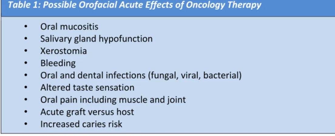
Oral complications are prevalent among childhood oncology patients; ranging from 30 to 100% of patients (Hong & daFonseca 2008). These oral complications can be both acute and severe, often requiring additional supportive therapy. They may delay cancer treatment or become dose limiting, increase hospital admission, increase cost, decrease quality of life and in essence add further burden to the patients, families and clinicians. There are numerous reports describing acute oral complications from cancer therapy resulting in significant morbidity and delays in overall therapy (Walsh 2010). Table 1 lists the potential acute oral complications of oncology therapy.
Table 1: Possible Orofacial Acute Effects of Oncology Therapy • Oral mucositis
• Salivary gland hypofunction • Xerostomia
• Bleeding
• Oral and dental infections (fungal, viral, bacterial) • Altered taste sensation
• Oral pain including muscle and joint • Acute graft versus host
• Increased caries risk
Part 3: Prevention and management of acute complications in children undergoing oncology therapy 2 Management of Acute Oral Manifestations During Oncology Therapy
During periods of immunosuppression resulting from oncology therapy, the following three objectives are recommended by the AAPD (2013):
i) To maintain optimal oral health during cancer therapy;
ii) To manage any oral side effects that may develop as a consequence of the cancer therapy; and iii) To reinforce the patient and parents’ education regarding the importance of optimal oral care
in order to minimise oral problems/discomfort during treatment
Standardised mouth care and continual dental reviews are recommended throughout oncology therapy. The recall and prevention program is individualised to reflect the child’s risk level due to their oncology treatment protocol and caries risk as discussed in Part 1 of this series. The oral care regimen is to be maintained and reinforced during oncology treatment, regardless of their haematological status.
Oral Mucositis
Oral mucositis can be defined as the inflammation of the oral mucosa which results from the direct cytotoxic effects of radiation or antineoplastic agents on the oral mucosa (Little et al 2008). In the past oral mucositis was considered to be inflammation of the mucosa of the mouth, which could range from erythema to severe ulceration (Oral Cancer Foundation 1998). Currently, the pathobiology of mucositis is accepted to be much more complex, involving the epithelial tissues as well as damage to the subepithelial tissues of the mucosa (Sonis et al 2004).
As oral mucositis develops, the oral epithelium shows mucosal erythema, which progresses to mucosal ulceration before a normal clinical appearance returns. However, this normal appearance does not reflect the underlying mucosal environment, which remains altered (Logan et al 2007). The lesions predominately appear in the nonkeratinized epithelium, such as the cheeks, ventral surface of the tongue, and the floor of the mouth. In severe cases of oral mucositis, the keratinized surfaces can be affected (Cheng et al 2002; Fadda et al 2006; Stockman et al 2006).
In addition to the above, oral mucositis can have different clinical presentations dependent upon whether it radiation or chemotherapy induced (Hogan 2009). In patients undergoing radiotherapy, oral mucositis may often initially present as a white mucosal surface lesions which progresses to a deepening erythema following a cumulative dose of 10-20Gy of radiotherapy, and may persist for several weeks depending on the level of radiation and duration of radiotherapy (Hogan 2009; Cheng et al 2004). Children undergoing chemotherapy who develop oral mucositis; are described to start showing symptoms from day 3 to peak on peaked on day 10 and started to resolve on day 14 post chemotherapy (Cheng et al 2004).
Oral mucositis occurs in about 20%–40% of adult patients receiving chemotherapy and up to 50% of adult patients receiving chemotherapy and radiotherapy (GONG 2007). Oral mucositis is reported to be more prevalent in children than in adults, with a reported incidence up to 65% in children (Cheng et al 2002). However, the estimates of the prevalence vary greatly in the literature from 52%–80% (Cheng et al 2004). Reliable data on oral mucositis prevalence rates in the paediatric patient with cancer population are scarce (Figliolia et al 2008). Recent data gained from retrospective and prospective studies on the incidence of oral mucositis in a paediatric population at the Women’s and Children’s hospital indicate a prevalence of 34% of inpatients undergoing oncology therapy (Qutob et al 2013) following the implementation of a standardised oral health care protocol.
The risk of a paediatric patient developing oral mucositis significantly increases with the use of high-dose and multiple chemotherapy agents (Cheng et al 2002). Cytotoxic agents including methotrexate,
Part 3: Prevention and management of acute complications in children undergoing oncology therapy 3 fluorouracil, doxorubicin, paclitaxel, capecitabine, and etoposide are reported to be associated with higher risks of oral mucositis believed to be particularly stomatotoxic (Cheng et al 2004; GONG 2007).
Prevention of Oral Mucositis
Modification of the treatment regimen to lower doses and longer recovery periods between doses remain the most effective methods for limiting the prevalence and severity of mucositis. Therefore, mucositis remains a common primary dose-limiting factor (Cheng et al 2002). However, interruptions to treatment, dose reductions (Cheng et al 2002) and potential cessation (Logan et al 2007) can affect treatment prognosis. Patient survival, cure rates, and length of remission, therefore, can be directly affected (Cheng et al 2002; Fadda et al 2006).
A Cochrane review (2010) found that there was some evidence to support the use of cryotherapy (ice chips) and keratinocyte growth factor (palifermin) in the prevention of oral mucositis. Sucralfate was shown to reduce the severity of oral mucositis and there was weak evidence that aloe vera, amifostine, intravenous glutamine, granulocyte colony stimulating factor (G-CSF), honey, laser and antibiotic lozenges may offer some benefit in the reduction of oral mucositis. The difficulty with applying this knowledge is that certain therapies have been assessed in only particular types of cancers and cancer therapies can vary or be used in combination (Worthington et al 2013).
Qutob et al (2013) in his systematic review explained that there is some evidence to suggest that implementation of a successful oral care protocol can be utilised in an attempt to reduce the frequency and severity of oral mucositis. Implementation of an oral care protocol at the Women’s and Children’s Hospital in South Australia has seen a reduction in the frequency and severity of oral mucositis (Qutob et al 2013).
Treatment of Oral Mucositis
To date there is no effective treatment routinely available for oral mucositis. A Cochrane review 2010 found that there was limited evidence in the form of two trials that low level laser treatments may reduce the severity of oral mucositis. Further research is required (Clarkson et al 2010). Current treatment is focussed on palliation of symptoms and prevention of secondary infection. This currently involves the use of topical, systemic and IV analgesics, good oral hygiene and parenteral nutrition as required (Peterson et al 2012, Keefe et al 2007; AAPD 2013). Once oral mucositis has developed, non medicated mouthrinses such as saline or sodium bicarbonate have been recommended due to the irritation caused by alcohol containing chlorhexidine rinses. Where possible the use of non-alcoholic 0.2% chlorhexidine mouth rinse throughout periods of oral mucositis has been recommended by the protocol in place at the Women’s and Children’s Hospital in Adelaide. This is not as a treatment agent for oral mucositis but due to its antimicrobial benefit in an attempt to prevent secondary infection. There is a clear need for further research in this area in the paediatric population.
Salivary Gland Hypofunction and Xerostomia
Salivary hypofunction refers to the objective reduction in salivary flow. Xerostomia however, refers to the subjective feeling of a dry mouth or lack of saliva. Salivary gland hypofunction can lead to general discomfort, difficulty swallowing, chewing and speaking in addition to possible taste and smell changes (Fischer & Epstein 2008). Saliva has an important lubrication function and intraoral tissues can become irritated and inflamed when deficient (Fischer & Epstein 2008). The oral tissues can become erythematous and the tongue furrowed. Radiation therapy can also result in a change in the composition of the saliva. The IgA content, pH and bicarbonate concentration are all reported to reduce (Anderson et al 1981; Marks et al 1981). Consequently, there is a shift in the oral environment toward cariogenic bacteria, an increased susceptibility to fungal infections and there are also threats to general health due to reduced appetite (Fischer & Epstein 2008).
Part 3: Prevention and management of acute complications in children undergoing oncology therapy 4 Radiotherapy is well recognised to affect salivary gland function in cancer patients (Hong & daFonseca 2008). The major salivary glands that are near or in the irradiated field can become reversibly and irreversibly damaged to varying degree with results in salivary gland hypofunction in children and adolescents (Hong & daFonseca 2008). The level of damage and reversibility of damage is related to the age of the patient, total radiation dosage and field or irradiation. Total body irradiation (TBI) is used in the conditioning process in patients requiring bone marrow transplantation and can also lead to salivary gland changes which may lead to xerostomia. Radiotherapy is also known to lead to permanent salivary gland hypofunction and associated xerostomia. Salivary glands that are included in the field of irradiation can be damaged. These effects are reversible at doses of 20-30Gy and irreversible at total doses greater than 50Gy. The acinar cells undergo inflammatory and degenerative changes, the ductal epithelium can become altered and fibrosis of surrounding connective tissue can occur (Fischer & Epstein 2008; Franzen et al 1992). The serous acini are reported to be more affected than the mucous acini resulting in a thickened saliva secretion (Franzen et al 1992).
Chemotherapeutic agents can also cause transient damage to salivary glands and approximately 40% of adult patients undergoing chemotherapy commonly report transient salivary gland hypofunction and associated xerostomia, however these effects are less significant compared to radiotherapy (Wolff et al 1990; Fischer & Epstein 2008; Hong & daFonseca 2008).
Prevention of Salivary Gland Hypofunction and Xerostomia
Due to the importance in minimising any damage to the salivary glands, shields are utilised during radiotherapy to reduce the exposure of the salivary glands to the radiation. The use of shields has been shown to reduce the actual damage to salivary glands, however it should be noted that it is not always possible to reduce or prevent salivary gland damage and continual assessment and monitoring is recommended prior, during and once treatment has been completed. The effects of chemotherapy on salivary glands are thought to be transient (Jensen et al 2010). However, keeping patients well hydrated and the consumption of a slightly acidic diet have been suggested to help reduce the clinical manifestation of xerostomia in response to the reduced function of the salivary gland (Hong and daFonseca 2008).
Treatment of Salivary Gland Hypofunction and Xerostomia
Saliva stimulating medications such as anticholinergic or parasympathomimetic (pilocarpine) drugs that are recommended in adult populations during and after radiotherapy (Little 2008) to help with salivary flow are not recommended for use in the paediatric population (AAPD 2013). Topical agents such as neutral mouth rinses, oral mucosal coating agents, salivary substitutes can be useful (Hong & daFonseca 2008). Sugar free gum, sipping of water and placement of a humidifier by the bedside can also be beneficial (Hong & daFonseca 2008).
Bleeding
Patients undergoing total body irradiation or high dose chemotherapy or who have bone marrow involvement are susceptible to thrombocytopenia (Little 2008). Oral manifestations of thrombocytopenia include bruising, petechiae, purpura and oozing from mucosal surfaces (daFonseca 1998). Petechiae commonly occur on the palate and purpura on the lateral boarder of the tongue (Little et al 2008). The incidence of oral bleeding in paediatric patients undergoing oncology therapy is reported to range from 6-42% (Hong & da Fonseca 2008). Bleeding commonly occurs from the gingival tissues but can also be seen originating from the tongue and lips but is reported to occur infrequently when the platelet count is above 50,000/mm3 (Hong & daFonseca 2008). These complications can be further enhanced by poor oral hygiene and the presence of plaque, calculus, orthodontic bands and appliances (daFonseca 1998). Gingival bleeding and submucosal haemorrhage as the result of minor trauma can be observed when the platelet count is below 50,000 cells/mm3 (Little et al 2008). Spontaneous gingival bleeding can occur when the platelets reach a level of 20,000 cells/mm3 or less (daFonseca 1998).
Part 3: Prevention and management of acute complications in children undergoing oncology therapy 5
Prevention of Oral Bleeding
Gingival haemorrhage is worse in the presence of poor oral hygiene. Oral hygiene practices are recommended even during periods of thrombocytopenia but soft devices are recommended. Where tooth brushing is not possible wiping the tissues with a sponge soaked in chlorhexidine can be recommended (Little et al 2008, AAPD 2013).
Treatment of Oral Bleeding
Local measures are used to control bleeding such as gauze pressure packs, ice packs and topical agents such as tranexamic acid (Little et al 2008; daFonseca 1998). If unsuccessful and the platelet count is low, the Oncology Team should be contacted and a platelet transfusion considered (Little et al 2008; daFonseca 1998).
Oral and Dental Infections (fungal, viral, bacterial)
The mouth is a frequently documented source of sepsis in the immunosuppressed cancer patient (AAPD 2013). During chemotherapy and radiotherapy patients are more prone to secondary infection. There may be a quantitative decrease in salivary flow and also changes in the composition of the saliva during oncology therapy. This results in several organisms such as bacterial, fungal and viral cells opportunistically infecting the oral cavity. Patients that are receiving chemotherapy can also be significantly immunosuppressed. Many patients are on broad spectrum antibiotics concurrently with their chemotherapy which can further predispose them to certain opportunistic infections (Little et al 2008). Oral candidiasis is an acute complication that commonly occurs in children with leukaemia because of altered cell-mediated immunity. Oral candidiasis also occurs frequently in patients receiving chemotherapy, particularly if they have severe neutropenia (Alberth et al 2006; Gonzalez Gravina et al 2007). Candida albicans is the species that most frequently infects immunosuppressed individuals with hyposalivation. Clinical presentation includes symptoms of oral pain, burning, taste alterations and intolerances to particular foods such as citrus fruits or spicy foods. All types of lesion presentations from denuded epithelium to hyperplastic lesions are possible (Little et al 2008). Candidal infection may lead to life-threatening systemic infection (Gonzalez Gravina et al 2007).
Oral bacterial infections can present with typical signs of infections such as swelling, erythema and fever. In immunocompromised patients there is a microbial shift to include more gram negative organisms that are usually found in the respiratory or gastrointestinal tract. The most common presentation is a non-healing ulcer (Little et al 2008).
Recurrent herpes simplex virus (HSV) eruptions can occur during chemotherapy and BMT if antiviral agents are not prophylactically prescribed. Interestingly they are not common during radiotherapy. The eruptions experienced by the patient undergoing chemotherapy have been found to be larger and take longer to heal than those in non-immunosupressed patients (Little et al 2008; daFonseca 1998).
Prevention of Oral Infections
Excellent oral hygiene including the use of an antimicrobial mouthrinse is recommended to reduce the risk of oral infections. Additionally, areas of irritation that could potentially result in a breach in the oral epithelium are recommended to be eliminated pre-treatment to reduce the risk of oral infections (AAPD 2013). Antiviral and antifungal are to be prescribed at the discretion of their oncologist on an individualised basis.
Part 3: Prevention and management of acute complications in children undergoing oncology therapy 6 Microscopic examination is often performed to allow for definitive diagnosis of oral Candidiasis. Candidiasis is best managed by the use of topical oral antifungal agents (Little et al 2008). The Australian therapeutic guidelines recommend amphotericin lozenges, miconazole 2% gel or nystatin oral drops all to be used four times daily after food (TG 2012).
When a bacterial oral infection is suspected it is recommended to take a swab and perform culture and bacterial sensitivity testing and treat with antimicrobials accordingly (Little et al 2008).
In immunocompromised HSV, the ulcers mimic aphthous ulcers and can occur on non-keratinised epithelium. A swab and laboratory tests are recommended for a definitive diagnosis. This also allows for differentiation between other oral infections such as that form varicella zoster and cytomegalovirus. An appropriate dose of acyclovir or an equivalent antiviral per day is sufficient to keep the HSV suppressed in HSV positive patients during chemotherapy (Little et al 2008). Prophylaxis is also recommended for patients positive to cytomegalovirus undergoing BMT during periods of immunosuppression (daFonseca 1998). If the lesions are unresolving or extensive antiviral sensitivity is suggested (Little et al 2008).
Altered Sensation
A number of patients that are receiving radiation therapy experience a diminished sense of taste. This is thought to be due to damage to the microvilli of the taste cells (Little et al 2008). Additionally the decrease in salivary flow can affect the taste of and lead to avoidance of particular foods (daFonseca 1998). Patients on chemotherapy also notice an alteration in smell and taste with foods becoming bitter and unpleasant odours more noticeable. In most cases taste has been reported to be restored 1-4 months post therapy with 3-4 months being needed for resolution following radiotherapy (Hong & daFonseca 2008; Little et al 2008).
Neurotoxicity can occur as an adverse effect of chemotherapy. Vincristine and vinblastine are more commonly the causative agents and it usually affects peripheral nerves. Patients may present with odontogenic pain mimicking pulpitis. It is more common in the molar region and may be bilateral (Little et al 2008; daFonseca 1998).
Management of Altered Sensation
Diagnosis and management of neurotoxicity requires the clinician to be familiar with the chemotherapy regimen and also to have ensured the absence of clinical and radiographic pathology (Little et al 2008). The dental team must ensure a potential odontogenic or mucosal source of the discomfort has been eliminated. Consultation with a dietician can also be recommended to identify foods that have pleasant aromas and therefore can stimulate an appetite and maintain nutritional balance (daFonseca 1998).
Trismus
Trismus can result during radiation therapy to the head and neck that involves the masticatory muscles (AAPD 2013). This complication develops due to the fibrosis of the involved masticatory muscles (Hong & daFonseca 2008). Oral function and oral hygiene becomes very difficult when trismus is severe (Hong & daFonseca 2008).
Prevention and treatment of trismus
Daily exercises and stretches can be beneficial and are recommended to commence prior to treatment and continue throughout and for 3-6 months post radiation therapy (AAPD 2013; Hong & daFonseca 2008). Therapy for trismus is focussed on the reduction of fibrosis with the use of prosthetic aids, trigger point injections, analgesics, muscle relaxants and other pain management strategies (AAPD 2013; Little et al 2008; Scully & Epstein 1996).
Part 3: Prevention and management of acute complications in children undergoing oncology therapy 7 With allogenic transplants, GVHD can develop in which the transplanted T-Lymphocyte cells recognise the histocompatibility of the host cells as foreign and attack the healthy cells of the recipient resulting in damage to organs such as the skin, liver and intestines. This can develop within a few weeks (acute) or much later (chronic) (NCI 2013).
Acute graft versus host disease has a median onset of day 19 post transplant but can occur at any stage in the first 100 days post transplant (daFonseca 1998; Krause & Gratz 2009; NCI 2013; Nicolatou-Galitis et al 2001). Acute graft versus host disease should be suspected when oral mucosal changes such as erythema and lichenoid changes appear, worsen and persist past day 21 post transplant (daFonseca 1998). Particularly if the patient is reporting pain and dryness beyond 4-5 weeks post transplant (daFonseca 1998). Mucosal changes present as erythema most commonly seen on the dorsal and ventral of the tongue, floor of the mouth, gingiva and labial mucosa. Lichenoid changes are also reported and affect the labial and buccal mucosa and lateral of the tongue (daFonseca 1998). Biopsies are often required to confirm the diagnosis.
Treatment of Graft Versus Host Disease (GVHD)
The development of GVHD is serious and it is routinely treated with immunosuppressive agents. Treatment is difficult and clinical trials are being performed in this area (NCI 2013). Acute GVHD can develop into chronic GVHD. Oral lesions are managed with systematic relief.
Increased Caries and Gingivitis
There is mixed reports in the literature with regards to increased caries rates in children undergoing oncology therapy and patterns tend to reflect the presence or lack of successful oral care protocol. However, during periods of active treatment patients can have salivary gland hypofunction often combined with high carbohydrate diets and regular consumption of oral medications containing sucrose. All these factors increase caries rate and individualised caries prevention programs are recommended as necessary. It is known that cancer and its treatments can be associated with mineralisation disorders and impaired salivary secretions, both risk factors for dental caries (Wogelius et al., 2008). Radiation induced hyposalivation has been reported to lead to a shift in the microflora to a highly cariogenic bacteria which when combined with a reduction in the protective features of saliva leads to an increased caries risk (Hong & daFonseca 2008). It is essential that the dentist recognises this increased risk and advises patients accordingly (Hong & daFonseca 2008).
Conclusion
Acute oral complications can occur frequently throughout the course of oncology therapy. Patients can be predisposed to opportunistic infections as a result of their particular malignancy or due to the effects of the myelosuppressive therapy itself. Odontogenic infections can have serious consequence in an immunosuppressed patient and oral mucositis can develop following exposure to cytotoxic cancer therapies further increasing one’s susceptibility to oral infections of an oral origin. It is crucial that an oral care protocol is utilised during these periods to optimise oral health. The paediatric dentist has a role in identifying and diagnosing oral complications. It is recommended that management is co-ordinated with the treating oncology team, particularly where pharmacological approaches are being utilised.
Part 3: Prevention and management of acute complications in children undergoing oncology therapy 8 References
Alberth, M., Majoros, L., Kovalecz, G et al., Significance of oral Candida infections in children with cancer.
Pathology Oncology Research, 2006; vol 12: pp 237–241.
American Academy of Pediatric Dentistry (AAPD)., Guideline on dental management of pediatric patients receiving chemotherapy, hematopoietic cell transplantation, and/or radiation, 2013.
Cheng KK, Chang AM, & Yuen MP., Prevention of oral mucositis in paediatric patients treated with chemotherapy: A randomised crossover trial comparing two protocols of oral care. European Journal of Cancer, 2004; vol: 40: pp1109–1010.
Cheng KK, Molassiotis A, & Chang AM., An oral care protocol intervention to prevent chemotherapy-induced oral mucositis in paediatric cancer patients: A pilot study. European Journal of Oncology Nursing,2002; vol 6: pp 66–73.
Clarkson JE, Worthington HV, Furness et al., Interventions for treating oral mucositis for patients with cancer receiving treatment [Review]. Cochrane Database of Systematic Reviews, 2010.
daFonseca MA, Pediatric bone marrow transplantation: oral complications and recommendations for care,
Pediatric Dentsitry, 1998; vol 20, pp386-399.
Fadda G, Campus G & Luglie P., Risk factors for oral mucositis in paediatric oncology patients receiving alkylant chemotherapy. BioMed Central Oral Health,2006; vol 6: pp 1–8.
Figliolia SL, Oliveira DT, Pereira MC., et al, Oral mucositis in acute lymphoblastic leukaemia. Oral Diseases,
2008; vol 14: pp 761–766.
Fischer & Epstein., Management of patients who have undergone head and neck cancer therapy, Dental Clinics of North America, 2008; vol 52: pp39-60.
Franzen L, Funegard U, Ericson T & Henriksson R., Parotid gland function during and following radiotherapy of malignancies in the head and neck: A consecutive study of salivary flow and patient discomfort,
European Journal of Cancer, 1992; vol 28: pp 457-462.
Gippsland Oncology Nurses Group, Cancer related mucositis management: GONG cancer care guidelines. Retrieved from http://www.gha.net.au/Uploadlibrary/393586894mucosit
is_management_guidelines0606.pdf, 2007.
Gonzalez Gravina H, Gonzalez de Moran E, Zambrano O, et al., Oral candidiasis in children and adolescents with cancer. Identification of Candida spp. Medicina Oral Patologia Oral Cirugia Bucal , 2007; vol 12: pp
419–423.
Hogan R., Implemental of an oral care protocol and its effects on oral mucositis, J Paediatr Oncol Nurs,
2009; vol 26: pp125-135.
Hong CH & daFonseca., Consideration in the paediatric population with cancer, Dent Clin N Am, 2008; vol 52: pp155-81.
Part 3: Prevention and management of acute complications in children undergoing oncology therapy 9 Jensen SB, Pedersen AML, Vissink A, Andersen E et al., A systematic review of salivary gland hypofunction and xerostomia induced by cancer therapies: prevalence, severity and impact on quality of life, Support
Care Cancer, 2010, vol 18: pp1039-1060.
Keefe DM, Schubert MM & Elting LS., Updated clinical practice guidelines for the prevention and treatment of oral mucositis, Cancer, 2007, vol: 109, pp820-31.
Little JW, Falace DA, Miller CS & Rhodus NL., Dental management of the medically compromised patient, Seventh Ed. 2008, Mosby Elsevier, St Louis.
Logan RM, Stringer AM, Bowen JM et al., The role of pro-inflammatory cytokines in treatment-induced alimentary tract mucositis: Pathobiology, animal models and cytotoxic drugs, Cancer Treatment Reviews,
2007; vol 33: pp448-460.
Nicolatou-Galitis Q, Kitra V, Vliet-Constantinidou et al., The oral manifestations of chronic veruss graft disease in paediatric allogenic bone marrow transplant recipients, J Oral Pathol Med, 2001; vol 30, pp148-53.
Oral Cancer Foundation, Prevention and treatment of oral mucositis in cancer patients, Best Practice; 1998; vol 2: pp1-6.
Peterson DE, Bensaduon RJ & Roila F, ESMO Guidelines working group. Management of oral and gastrointestinal mucositis: ESMO Clinical Guidelines, Ann Oncol, 2012; vol: 22, pp74-74.
Qutob AF, Allen GJ, Gue S, ReveszT et al., Implementation of a hospital oral care protocol and recording of oral mucositis in children receiving cancer treatment : A retrospective and a prospective study. Support
Care Cancer, 2013; vol 21: pp 1113-20.
Resende RG, Guimaraes Abreu MH, de Souza LN, Silva RS et al., Association Between IL1B (+3954) Polymorphisms and IL -1B Levels in Blood and Saliva together with Acute Graft-Versus-Host Disease, J
Interferon Cytokine Res, 2013;DOI: 10.1089/jir.2012.0111.
Rojas de Morales T, Zambrano O, Rivera et al., Oral-disease prevention in children with cancer: Testing preventive protocol effectiveness. Medicina Oral, 2001; vol 6: pp326–234.
Scully C & Epstein JB., Oral health care for the cancer patient, Eur J cancer B Oral Oncol, 1996; vol 32: pp281-92.
Sonis ST, Elting LS, Keefe DM, et al., Perspectives on cancer therapy-induced mucosal injury: Pathogenesis, measurement, epidemiology, and consequences for patients. Cancer Supplement,100(9, Suppl.), 2004; pp 1995–2025.
Therapeutic Guidelines (TG), Therapeutic Guidelines Oral and Dental Version 2, 2012, Australia.
Walsh LJ., Clinical assessment and management of the oral environment in the oncology patient, Australian Dental Journal, 2010, vol 55: pp 66-77.
Wogelius P, Dahllof G, Gorst-Rasmussen AG, Sorensen HT, et al., A population-based observational study of dental caries among survivors of childhood cancer, Pediatric Blood & Cancer, 2008,vol: 50,no:6, pp: 1221-1226.
Part 3: Prevention and management of acute complications in children undergoing oncology therapy 10 Wolff A, Fox PC, Ship JA, Atkinson JC et al., Oral mucosal status and major salivary gland function, Oral Surg
Oral Med Oral Pathol, 1990; vol 70: pp49-54.
Worthington HV, Clarkson JE, Bryan et al., Interventions for preventing oral mucositis for patients with cancer receiving treatment. Cochrane Database of Systematic Reviews,2013.