Gingival bleeding on
probing: relationship to
change in periodontal pocket
depth and effect of sodium
hypochlorite oral rinse
Gonzalez S., Cohen C. L., Galvan M., Alonaizan F. A., Rich S. K., Slots J. Gingival bleeding on probing: relationship to change in periodontal pocket depth and effect of sodium hypochlorite oral rinse. J Periodont Res 2014; doi: 10.1111/ jre.12219.©2014 John Wiley & Sons A/S. Published by John Wiley & Sons Ltd Background and Objective:This study evaluated the potential of gingival bleed-ing on probbleed-ing to serve as a predictor of future periodontal breakdown. It also assessed the ability of 0.25% sodium hypochlorite twice-a-week oral rinse to convert periodontal pockets showing bleeding on probing to nonbleeding sites. Material and Methods:The study was performed as a randomized, single-blinded, clinical trial in parallel groups. Seven periodontitis patients rinsed twice-weekly for 3 mo with 15 mL of a fresh solution of 0.25% sodium hypochlorite, and five periodontitis patients rinsed with water. The 12 study patients received no subgingival or supragingival scaling. Cloroxâ Regular-Bleach was the source of sodium hypochlorite. At baseline and 3-mo visits, gingival bleeding was assessed within 30 s after probing to full pocket depth using an approximate force of 0.75 N.
Results:A total of 470 (38%) of 1230 periodontal pockets in the bleach-rinse group revealed bleeding on probing at the initial visit but not at the 3-mo visit; only 71 (9%) of 828 pockets in the control group became bleeding-negative during the study (p<0.001). Bleeding on probing in 4- to 7-mm-deep pockets decreased by 53% in the bleach-rinse group but increased by 6% in the water-rinse group (p<0.001). Ninety-seven pockets showed depth increases of ≥2 mm after 3 mo: 60 (62%) of those pockets exhibited bleeding on probing at both the initial and the 3-mo visits; 24 (25%) bled at only one of the two visits; and 13 (13%) never demonstrated gingival bleeding (p<0.001).
Conclusions:Persistent gingival bleeding on probing was associated with an increased risk for periodontal breakdown, and the absence of gingival bleeding seemed to be a useful, although not perfect, indicator of disease stability. Twice-weekly oral rinsing with dilute bleach (0.25% sodium hypochlorite) produced a significant reduction in bleeding on probing, even in deep unscaled pockets. Sodium hypochlorite constitutes a valuable antiseptic in periodontal self-care.
S. Gonzalez1, C. L. Cohen1, M. Galvan1, F. A. Alonaizan2, S. K. Rich1, J. Slots1
1Graduate Periodontology Clinic, Ostrow School of Dentistry of USC, Los Angeles, CA, USA and2Graduate Endodontic Clinic, Ostrow School of Dentistry of USC, Los Angeles, CA, USA
Jørgen Slots, DDS, DMD, PhD, MS, MBA, Department of Periodontology and Microbiology, Ostrow School of Dentistry of USC, Los Angeles, CA 90089-0641, USA Tel: +1 213 740 1091
e-mail: jslots@usc.edu
Key words:dental plaque; gingival bleeding on
probing; household bleach; mouthrinse; periodontal treatment; sodium hypochlorite Accepted for publication June 21, 2014
All rights reserved Published by John Wiley & Sons Ltd
JOURNAL OF PERIODONTAL RESEARCH doi:10.1111/jre.12219
Periodontitis remains a diagnostic and therapeutic challenge. Dental profes-sionals lack accurate biomarkers to predict ongoing or future progression of periodontal disease, and practitio-ners often resort to surgery to prevent an anticipated scenario of continued attachment loss. Successful treatment of aggressive/active periodontitis usu-ally requires surgical intervention and/or adjunctive systemic antibiotic therapy (1). On the other hand, as many as 85% of patients with chronic periodontitis may remain disease-sta-ble for 5–6 years after only a one-time scaling (2) or no treatment at all (3), and may derive relatively little benefit from surgical treatment. The avail-ability of a reliable test to assess risk for periodontal disease activity would help to optimize treatment of individ-ual patients and specific periodontal sites.
Gingival bleeding on probing com-prises one of the more promising diagnostic predictability tests in peri-odontics. Claffey et al. (4) found loss of probing attachment in 41% of periodontal sites that bled on probing at 75% of recall visits during a 3.5-year observation period. Lang et al. (5) reported that periodontal sites which bled on probing at four consec-utive maintenance visits showed a 30% risk of losing attachment, whereas sites with bleeding on prob-ing at one out of four consecutive recall visits had only a 3% risk of breakdown, and periodontitis patients who demonstrated gingival bleeding in fewer than 10% of sites in the den-tition were at low risk for progressive disease. A meta-analysis showed that persistent bleeding on probing after treatment was associated with pro-gression of periodontitis with an odds ratio of 2.8 (6). Charalampakis et al. (7) found that bleeding on probing yielded a better prediction of progres-sive periodontal disease if combined with quantified bacterial markers. Also, repeated mucosal bleeding on probing around implants may serve as a risk indicator of peri-implantitis (8), and the absence of mucosal bleeding may be an indicator of low risk for implant failure as a result of infection (9).
Other types of diagnostic tests to predict periodontal breakdown have also been evaluated. A high score of 4 in the Community Periodontal Index of Treatment Needs screening system yielded a low positive-predictive value for progressive periodontitis, but low sextant scores of 0–2 provided a pre-sumptive identification of nonprogres-sive sites (10). The absence of a radiographic lamina dura in angular periodontal defects (11) and in peri-implantitis lesions (12) was an indica-tor of progressive disease, although with low positive predictability, but periodontal sites with a radiographi-cally intact crestal lamina dura exhib-ited literally no risk of disease progression for at least 2 years (13). The lack of periodontal disease pro-gression at Ramfjord’s six index teeth was suggestive of a low risk of pro-gressive disease in the entire dentition (14). A clinically based periodontal risk calculator failed, largely because of false-positive high-risk scores, to reli-ably predict progression of periodontal disease during a 3-year post-treatment period (15). Periodontopathic bacteria in subgingival plaque (7,16–18) or saliva (19), or gingival crevice fluid biomarkers (20,21), may also serve as indicators of periodontal disease sta-tus. The modest to moderate positive-predictive value of diagnostic tests for progressive periodontal disease are the result of a low incidence of dis-ease-active periodontitis and a rela-tively high number of false-positive assessments (22). Diagnostic tests may be improved by combining several independent variables from different aspects of the periodontal disease pro-cess (7,23), but such an approach remains to be defined and validated in periodontics.
As persistent bleeding on probing provides one of the most robust pre-dictors of periodontal breakdown, and because little information exists on the potential of periodontal self-care to resolve gingival bleeding, this study examined the ability of 0.25% sodium hypochlorite oral rinse, used twice-weekly for 3 mo, to convert gin-gival bleeding sites to nonbleeding sites in periodontitis patients. The study subjects received no subgingival
or supragingival scaling before or dur-ing the study in order to stress-test the boundary of effectiveness of sodium hypochlorite oral rinse.
Material and methods
The details of the material and meth-ods are outlined elsewhere and are only briefly summarized below (24). The study included 12 periodontitis patients, who completed a controlled clinical trial with sodium hypochlorite oral rinse. The study patients had a mean age of 41 years and an average of 28.6 teeth. Each patient exhibited at least four separate teeth with a pocket depth of≥6 mm. The patients were medically healthy and required no emergency dental care. Excluded from the study were individuals who were unable to comply with the research protocol, smoked >10 ciga-rettes daily or had received periodon-tal therapy or systemic antibiotics during the 6 mo before entering the study. The University of Southern California Health Sciences Campus Institutional Review Board approved the study (# HS-10-00509). All patients understood and signed informed con-sent and HIPAA documents before enrolling in the study.
All study patients received a com-prehensive clinical examination and conventional oral hygiene instruction at baseline, but no subgingival or supragingival scaling. Standard peri-odontal therapy was performed at the conclusion of the study. By random assignment, seven study participants rinsed with sodium hypochlorite (test group) and five rinsed with water (control group). The source of sodium hypochlorite was Cloroxâ
Regular-Bleach (The Clorox Company,
Oakland, CA, USA), which contains
6% sodium hypochlorite and is
registered as a bacteriocide, virucide and fungicide agent with the United States Environmental Protection
Agency (CAS number 5813-100).
Participants in the sodium hypochlorite rinse group were provided with Clorox Regular-Bleach and instructed to mix 5 mL (one teaspoonful) of bleach with 120 mL (one half-glass) of tap water to yield a sodium hypochlorite
concentration of 0.25%. A fresh bleach solution was to be made up at each time of rinsing. The study patients were requested to rinse their mouth for 30 s every Wednesday and Sunday for 3 mo, with either 15 mL of a fresh solution of 0.25% sodium hypochlorite or 15 mL of water, and were given a rinse log to record the exact date and time of rinsing.
A calibrated blinded examiner determined pocket depth and gingival bleeding within 30 s after probing to full pocket depth using a Marquis CP-12 probe (Hu-Friedy Mfg. Co., Chicago, IL, USA) and a probing force of approximately 0.75 N (76 gram-force). Mid-facial, mid-lingual, mesiofacial, distofacial, mesiolingual and distolingual surfaces were exam-ined, and a total of 1230 pockets in the bleach-rinse group and 828 pockets in the water-rinse group were studied.
The statistical analysis was per-formed using the SPSS-19.0 software program (SPSS Inc., Chicago, IL, USA). The individual pockets were treated as independent statistical units, based on the nonspecific and wide-ranging antimicrobial action of sodium hypochlorite and the observa-tions that pockets with a large range of depths responded positively to the bleach treatment and that residual bleeding on probing sites showed no tendency to cluster in particular patients or around specific teeth. Based on the percentage of periodontal pock-ets with bleeding on probing and pocket depth changes, the chi-square test was used to identify significant differences between the bleach-rinse and the water-rinse groups. The Spear-man’s rank correlation coefficient test assessed the relationship between the frequency of bleeding on probing and increased pocket depth. A p-value of ≤0.05 was considered statistically significant.
Results
The post-study interview and the patient rinse log revealed that the study participants fully understood the instructions for mixing the bleach rinse solution and complied with the Table
1. Bleeding o n probing (BOP) in rela tion to periodo ntal pocket de pth in patients rec eiving 0.25% sodiu m hyp ochlorite rins e (test) or wat er rins e (contr ol ) Valu es are given as no . o f ble eding pockets/ no. of total pock ets at the specific pock et depth (per centag e o f bleed ing pockets at the specifi c pocket dept h) at baselin e and at 3 mo, in contro l and test gro ups.
twice-weekly rinsing guidelines. No adverse events were identified in any of the participants, except for minor complaints about the taste of bleach.
Table 1 details bleeding on probing in relation to periodontal pocket depth. Both the bleach-rinse group with 1230 pockets and the water-rinse group with 828 pockets showed posi-tive correlations at baseline between frequency of gingival bleeding and increased pocket depth (p<0.001). No periodontal site of 1 mm depth bled on probing at any study time point. At baseline, bleeding on prob-ing occurred in 37% of 2- to 3-mm-deep pockets and in 88% of pockets with a depth of ≥6 mm. At 3 mo, bleeding on probing in pockets of 4– 7 mm depth showed a 53% decrease in the bleach-rinse group but a 6% increase in the water-rinse group (p<0.001). At baseline, 27 pockets in the bleach-rinse group had a probing depth of ≥8 mm and all bled on probing. At the 3-mo visit, the bleach-rinse group revealed the pres-ence of only three pockets of≥8 mm, and none showed bleeding. In com-parison, 127 pockets in the control group had probing depths of ≥8 mm at baseline and 108 (85%) bled on probing. At 3 mo, 114 control pock-ets of≥ 8 mm depth were present and 91 (80%) bled on probing. The differ-ence between the bleach-rinse and the
water-rinse groups in decrease of per-centage of≥8 mm pockets and bleed-ing sites was statistically significant (p<0.001).
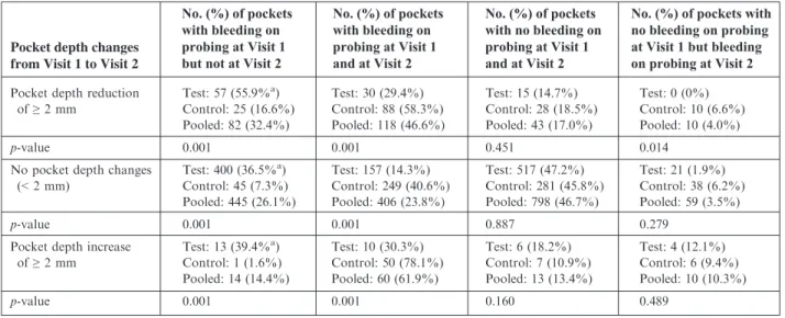
Table 2 describes how changes in bleeding on probing affected periodon-tal pocket depth. A toperiodon-tal of 470 (38%) periodontal pockets in the bleach-rinse group revealed bleeding on probing at the initial visit but not at the 3-mo visit; only 71 (9%) pockets in the con-trol group became bleeding-negative during the study (p<0.001). Con-versely, 197 (16%) periodontal pockets in the bleach-rinse group, but as many as 387 (47%) pockets in the control group, showed persistent bleeding throughout the study (p<0.001). Ninety-seven pockets (5% of the total pockets in the study) increased in prob-ing depth by ≥2 mm: 60 (62%) of those pockets exhibited bleeding on probing at both the initial and the 3-mo visits; 24 (25%) bled at only one of the two visits; and 13 (13%) never demon-strated gingival bleeding (p<0.001). A total of 25 pockets in the bleach-rinse group (2% of test pockets) developed bleeding on probing during the study; none of those pockets showed a depth reduction of ≥2 mm, and four (0.3%) pockets experienced a depth increase of ≥2 mm. No statistically significant changes were found in gingival reces-sion, furcation involvement or tooth mobility, within or between the
bleach-rinse and control groups, during the 3-mo study.
Discussion
This study sought to assess the profi-ciency of bleeding on probing to pre-dict progression of periodontal disease and the capability of sodium hypochlo-rite oral rinse to resolve bleeding on probing in periodontal pockets of dif-ferent depths. Bleeding on probing is a potentially attractive risk indicator for periodontal breakdown because it is a relatively simple, quick, all-or-none visual testing method that is not greatly dependent on special skills or excep-tional tactile sensitivity on the part of a clinical observer. Also, probing of the periodontal pocket depth is already a standard assessment technique in dental practice, requiring no extra training for experienced operators. However, to avoid underestimating gingival bleeding, we probed to the base of periodontal pockets and not just around the gingival margin (25) and used a probing force of approximately 0.75 N instead of the con-ventional 0.50 N (26). Lang et al. (27) showed that an increase in probing force from 0.50 to 0.75 N doubled the percent-age of periodontal pockets that bled on probing.
Bleeding on probing occurred in 55% of untreated periodontal pockets in our study compared with 71% (28),
Table 2. Periodontal pocket depth changes in relation to changes in bleeding on probing
aPercentage of total pockets in the particular Table row. ‘Visit 1’ denotes baseline and ‘Visit 2’ denotes termination of the study at 3 mo. Test patients rinsed with 0.25% sodium hypochlorite and control patients rinsed with water.
63% (29) and 57% (30) pockets in
pre-vious studies. We found that
untreated, 3-mm-deep pockets exhib-ited a frequency of bleeding on prob-ing of 43%, whereas Farinaet al.(31) reported a frequency of bleeding on probing of 18%. Bleeding on probing scores are elevated with deep pockets and heavy plaque accumulation, and are reduced by cigarette smoking, which may explain some of the differ-ences in the findings across studies (28,31,32). Rather than merely deter-mine an overall correlation between bleeding on probing and progression of periodontal disease, we aimed to quantify, in more detail, the relation-ship between bleeding on probing and periodontal disease activity. Of the 97 pockets (5% of the total pockets in the study) that showed increased probing depth of ≥2 mm, disease progression was detected 2.5-fold more frequently in pockets that bled on probing at both the initial and the 3-mo visits (60 pockets) than in pockets that bled on probing at only one of the two visits (24 pockets). Disease progression occurred in only 13 (1.5%) of the 854 pockets that never demonstrated bleeding on probing. The increased rate of periodontal breakdown in periodontal sites having repeat bleed-ing on probbleed-ing and the low occur-rence of false-negative readings show the benefits of maintaining the periodontium in a nonbleeding state.
Bleeding on probing can decrease substantially after mechanical depura-tion but, as also shown in the control group of this study, not after conven-tional oral hygiene alone (28,29,33). Scaling and root planing of 4- to 7-mm-deep pockets has been reported to reduce bleeding on probing scores by approximately 50%, with reduc-tions of 6–64% occurring after 1 mo and reductions of 12–80% occurring after 3 mo (33). However, our study showed that twice-weekly oral rinsing with 0.25% sodium hypochlorite for 3 mo, in the absence of subgingival scaling, also resulted in a reduction in bleeding on probing, of 53%, in pockets of 4–7 mm depth. Kalkwarf et al. (32) found that bleeding on probing returned in 35–60% of pockets ≥5 mm deep during the first
3 mo following nonsurgical or surgical therapy. However, our study revealed that 3 mo of twice-weekly rinsing with sodium hypochlorite/dilute bleach gave rise to a 49% decrease in gingival bleeding in pockets≥5 mm deep. The finding that bleeding on probing was resolved in sites with deep probing depths, exposed to sodium hypochlo-rite oral rinsing, which has not been reported previously, lends substantial credence to the use of sodium hypo-chlorite in periodontal maintenance care. Further studies are needed to determine if periodontitis patients may experience even less disease progression if rinsing with dilute bleach is com-bined with scaling and root planing.
The marked decrease in gingival bleeding occurred despite oral rinse only penetrates 0.1–0.2 mm into the subgingival area (34). The improved gingival health after rinsing with sodium hypochlorite was most proba-bly a result of the very low level of supragingival plaque, causing a decrease in the subgingival counts of pathogenic bacteria (35–37). Lobene et al. (38) and De Nardo et al. (39) found anti-plaque and anti-gingivitis effects of sodium hypochlorite oral rinse similar to those reported here. An attractive feature of the present rinsing protocol is the minimal extra time (30 s, twice weekly) that is imposed on patients’ usual oral hygiene effort.
Sodium hypochlorite is a powerful bactericidal and antiviral agent (40), which can even neutralize hard-to-destroy pathogenic prions (41). The antimicrobial action of sodium hypo-chlorite relies on oxidation or uncou-pling of electrons from proteins and nucleotides, and the resulting wide-range damage of infectious agents virtually precludes the development of resistance. Properties of sodium hypo-chlorite other than plaque removal may also have contributed to the reduction of gingival bleeding (42). Sodium hypochlorite at low
concen-tration was recently shown to
interfere with the ability of nuclear factor-jB cellular signaling pathways to activate proinflammatory gene programmes (43). Also, as sodium hypochlorite and essentially all other components of Cloroxâ
Regular-Bleach occur naturally in the human body (24), dilute bleach oral rinse does not give rise to allergic reactions or other types of disease.
In summary, bleeding on probing tended to persist in periodontal sites with progressive disease, as measured by an increase in probing pocket depth of ≥2 mm, and to remain absent or discontinue in disease-stable or healing sites. Continued absence or cessation of bleeding on probing appeared to be a useful, but not perfect, indicator of stable periodontal conditions. Twice-weekly oral rinsing with sodium hypo-chlorite/dilute bleach, coupled with conventional oral hygiene, controlled bleeding on probing more efficiently than conventional oral hygiene alone, and provided a resolution of bleeding on probing even in pockets of consid-erable depth. The reduction in bleeding on probing in this study, beyond those noted in previous oral rinse studies, was probably the result of substantial antimicrobial, anti-inflammatory and tooth-substantive properties of sodium hypochlorite. Dilute bleach used as an oral rinse offers convenience for patients and deserves serious consider-ation in periodontal healthcare.
Acknowledgement
The study was funded in part by a grant from The Clorox Company, Oakland, California, USA.
References
1. Dentino A, Lee S, Mailhot J, Hefti AF. Principles of periodontology.Periodontol 20002013;61:16–53.
2. Renvert S, Nilveus R, Dahlen G, Slots J, Egelberg J. 5-year follow up of periodon-tal intraosseous defects treated by root planing or flap surgery.J Clin Periodon-tol1990;17:356–363.
3. Lindhe J, Haffajee AD, Socransky SS. Progression of periodontal disease in adult subjects in the absence of periodon-tal therapy. J Clin Periodontol 1983;10: 433–442.
4. Claffey N, Nylund K, Kiger R, Garrett S, Egelberg J. Diagnostic pre-dictability of scores of plaque, bleed-ing, suppuration and probing depth for probing attachment loss. 3 1/2 years of observation following initial
periodon-tal therapy. J Clin Periodontol 1990;17: 108–114.
5. Lang NP, Joss A, Tonetti MS. Monitor-ing disease durMonitor-ing supportive periodontal treatment by bleeding on probing. Peri-odontol 20001996;12:44–48.
6. Armitage GC. Periodontal diseases: diag-nosis.Ann Periodontol1996;1:37–215. 7. Charalampakis G, Dahlen G, Carlen A,
Leonhardt A. Bacterial markers vs. clini-cal markers to predict progression of chronic periodontitis: a 2-yr prospective observational study.Eur J Oral Sci2013;
121:394–402.
8. Luterbacher S, Mayfield L, Br€agger U, Lang NP. Diagnostic characteristics of clinical and microbiological tests for monitoring periodontal and peri-implant mucosal tissue conditions during sup-portive periodontal therapy (SPT). Clin Oral Implants Res2000;11:521–529. 9. Jepsen S, R€uhling A, Jepsen K,
Ohlen-busch B, Albers HK. Progressive peri-implantitis. Incidence and prediction of peri-implant attachment loss. Clin Oral Implants Res1996;7:133–142.
10. Rams TE, Listgarten MA, Slots J. Effi-cacy of CPITN sextant scores for detec-tion of periodontitis disease activity.
J Clin Periodontol1996;23:355–361. 11. Rams TE, Listgarten MA, Slots J.
Regards actuels sur les radiographies conventionnelles en parodontie.J Parod-ontol1994;13:179–184. (in French). 12. Tabanella G, Nowzari H, Slots J.
Clini-cal and microbiologiClini-cal determinants of ailing dental implants.Clin Implant Dent Relat Res2009;11:24–36.
13. Rams TE, Listgarten MA, Slots J. Utility of radiographic crestal lamina dura for predicting periodontitis diseaseactivity.
J Clin Periodontol1994;21:571–576. 14. Rams TE, Oler J, Listgarten MA, Slots
J. Utility of Ramfjord index teeth to assess periodontal disease progression in longitudinal studies. J Clin Periodontol
1993;20:147–150.
15. Rams TE, Listgarten MA, Slots J. Evalu-ation of a periodontal risk calculator on treated periodontitis patients. AADR Abstract, 2014.
16. Slots J. Low-cost periodontal therapy.
Periodontol 20002012;60:110–137. 17. Rams TE, Listgarten MA, Slots J. Utility
of 5 major putative periodontal patho-gens and selected clinical parameters to predict periodontal breakdown in patients on maintenance care. J Clin Periodontol1996;23:346–354.
18. Fujise O, Hamachi T, Inoue K, Miura M, Maeda K. Microbiological markers for prediction and assessment of treat-ment outcome following non-surgical periodontal therapy. J Periodontol
2002;73:1253–1259.
19. Saygun I, Nizam N, Keskiner I, Bal V, Kubar A, Acßıkel C, Serdar M, Slots J. Salivary infectious agents and periodontal disease status. J Periodontal Res 2011;
46:235–239.
20. Chapple IL, Garner I, Saxby MS, Moscrop H, Matthews JB. Prediction and diagnosis of attachment loss by enhanced chemiluminescent assay of cre-vicular fluid alkaline phosphatase levels.
J Clin Periodontol1999;26:190–198. 21. Ngo LH, Darby IB, Veith PD, Locke AG,
Reynolds EC. Mass spectrometric analysis of gingival crevicular fluid biomarkers can predict periodontal disease progression.
J Periodontal Res2013;48:331–341. 22. Slots J, Taichman NS, Oler J, Listgarten
MA. Does the analysis of the subgingival flora have value in predicting periodontal breakdown? In: Guggenheim B, ed.
ERGOB proceedings on the conference “Periodontology Today”. Basel, Switzer-land: S. Karger AG, 1988:132–140. 23. Beltran-Aguilar ED, Thornton-Evans G,
Eke PA, Petersen PE. Recording and surveillance systems for periodontal dis-eases.Periodontol 20002012;60:40–53. 24. Galvan M, Gonzalez S, Cohen CL,
Alonaizan FA, Chen CT-L, Rich SK, Slots J. Periodontal effects of 0.25% sodium hypochlorite twice-weekly oral rinse. A pilot study. J Periodontal Res
2013; doi: 10.1111/jre.12151. [Epub ahead of print].
25. Lie MA, Timmerman MF, van der Velden U, van der Weijden GA. Evalua-tion of 2 methods to assess gingival bleeding in smokers and non-smokers in natural and experimental gingivitis. J Clin Periodontol1998;25:695–700. 26. Barendregt DS, van der Velden U,
Timmerman MF, van der Weijden GA. Comparison of two automated periodon-tal probes and two probes with a conven-tional readout in periodontal maintenance patients. J Clin Periodontol 2006;33: 276–282.
27. Lang NP, Nyman S, Senn C, Joss A. Bleeding on probing as it relates to prob-ing pressure and gprob-ingival health.J Clin Periodontol1991;18:257–261.
28. Cercek JF, Kiger RD, Garrett S, Egelberg J. Relative effects of plaque control and instrumentation on the clini-cal parameters of human periodontal dis-ease.J Clin Periodontol1983;10:46–56. 29. Westfelt E, Rylander H, Dahlen G,
Llndhe J. The effect of supragingival pla-que control on the progression of advanced periodontal disease. J Clin Periodontol1998;25:536–541.
30. Haffajee AD, Cugini MA, Dibart S, Smith C, Kent RL Jr, Socransky SS. The effect of SRP on the clinical and microbiological parameters of
periodon-tal diseases. J Clin Periodontol 1997;
24:324–334.
31. Farina R, Tomasi C, Trombelli L. The bleeding site: a multi-level analysis of associated factors. J Clin Periodontol
2013;40:735–742.
32. Kalkwarf KL, Kaldahl WB, Patil KD, Molvar MP. Evaluation of gingival bleeding following 4 types of periodontal therapy.J Clin Periodontol 1989;16:601– 608.
33. Adriaens PA, Adriaens LM. Effects of nonsurgical periodontal therapy on hard and soft tissues. Periodontol 2000
2004;36:121–145.
34. Pitcher GR, Newman HN, Strahan JD. Access to subgingival plaque by disclos-ing agents usdisclos-ing mouthrinsdisclos-ing and direct irrigation. J Clin Periodontol 1980;7: 300–308.
35. Ximenez-Fyvie LA, Haffajee AD, Som S, Thompson M, Torresyap G, Socransky SS. The effect of repeated professional supragingival plaque removal on the composition of the supra- and subgingi-val microbiota. J Clin Periodontol2000;
27:637–647.
36. Hellstr€om MK, Ramberg P, Krok L, Lindhe J. The effect of supragingival pla-que control on the subgingival microflora in human periodontitis.J Clin Periodon-tol1996;23:934–940.
37. Dahlen G, Lindhe J, Sato K, Hanam-ura H, Okamoto H. The effect of su-pragingival plaque control on the subgingival microbiota in subjects with periodontal disease. J Periodontol
1992;19:802–809.
38. Lobene RR, Soparkar PM, Hein JW, Quigley GA. A study of the effects of antiseptic agents and a pulsating irrigat-ing device on plaque and girrigat-ingivitis.
J Periodontol1972;43:564–568.
39. De Nardo R, Chiappe V, Gomez M, Romanelli H, Slots J. Effect of 0.05% sodium hypochlorite oral rinse on supra-gingival biofilm and supra-gingival inflamma-tion.Int Dent J2012;62:208–212. 40. McDonnell G, Russell AD. Antiseptics
and disinfectants: activity, action, and resistance. Clin Microbiol Rev 1999;12: 147–179.
41. Taylor DM. Inactivation of transmissible degenerative encephalopathy agents: a review.Vet J2000;159:10–17.
42. Mainnemare A, Megarbane B, Soueidan A, Daniel A, Chapple IL. Hypochlorous acid and taurine-N-monochloramine in periodontal diseases. J Dent Res 2004;
83:823–831.
43. Leung TH, Zhang LF, Wang J, Ning S, Knox SJ, Kim SK. Topical hypochlorite ameliorates NF-jB-mediated skin dis-eases in mice. J Clin Invest 2013;123: 5361–5370.