VITREOUS BIOPSY FOR THE DIAGNOSIS OF UVEITIS
Thekla Papadaki, M.D.Accurate diagnosis of the etiology of inflammatory eye disease is crucial for appropriate treatment and management. In most cases, the diagnosis can be made by non-invasive techniques. It has been estimated however, that in approximately 8% of cases with uveitis, clinical presentation is non-specific or atypical and systemic medical evaluation is inconclusive.1 Diagnostic sampling of ocular specimens can, in these instances, aid in determining whether the therapy should be anti-infectious, anti-inflammatory or antineoplastic.
Invasive diagnostic testing for uveitis includes: anterior chamber paracentisis, vitreous biopsy and chorioretinal biopsy. This chapter focuses on the indications, technique, reliability and limitations of vitreous biopsy for uveitis.
Indications of vitreous biopsy
Vitreous biopsy is a useful adjunct to the systemic workup of cases that constitute diagnostic dilemmas and in which intraocular inflammation is mostly confined to the posterior pole. Such cases include: a) chronic uveitis unresponsive to empirical treatment with systemic
anti-inflammatory medication b) atypical clinical presentation c) inconclusive non-invasive laboratory work-up d) acute, sight-threatening disease.
Vitreous Biopsy: Pars plana vitrectomy vs. vitreous aspiration
A vitreous specimen can be obtained for analysis by either straight needle vitreous aspiration or pars plana vitrectomy (PPV). Vitreous aspiration is easier to perform and can be done in an office setting. However, it is associated with a risk for retinal detachment from vitreoretinal traction during aspiration.2
This risk can be minimized with PPV since detached vitreous is aspirated from the cutter and not directly from the vitreous cavity. Removal of the whole vitreous body with PPV has the
advantage of removing most of the pathologic material from the eye, which may increase the diagnostic yield of the procedure but may also remove the load of microorganisms, malignant cells or inciting inflammatory cells and reactants. 3
Total vitrectomy also facilitates further management of the patient as it enhances visualization of the fundus and allows better diffusion of intraocular medications. 4 Finally, approximately 50% of eyes treated with diagnostic PPV have subsequently improved visual acuity, results comparable with those of therapeutic vitrectomy. 5-6 For all the above reasons, in the Immunology and Uveitis Service of the MEEI we prefer diagnostic vitrectomy to straight needle vitreous aspiration in cases where vitreous biopsy is indicated.
Technique of diagnostic PPV
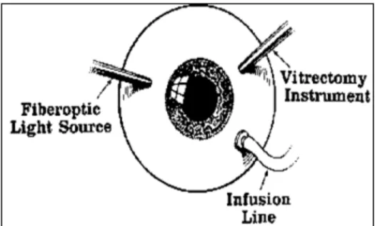
The procedure can be done under either general or topical anesthesia. For vitreous aspiration the one-port technique can be used. However, in eyes with uveitis, where there can be coexistent media opacities or intraocular inflammation, the three-port technique is preferable (Figure 1).
Figure 1: A Standard three-port PPV
Balanced salt solution (BSS) infusion is used to control the intraocular pressure during the procedure. Control of the pressure can minimize the risk of severe intraoperative hypotony as well as choroidal detachments and expulsive hemorrhage, all of which are more common in eyes with uveitis. However in diagnostic vitrectomy, the initial vitreous specimen is obtained undiluted (before the infusion port is opened); approximately 1.0 ml of vitreous is obtained directly from the vitrectomy cutter hand piece, with a cutting rate at 1200/minute, through an in-line stopcock and tubing attached to a syringe. The infusion line is subsequently opened, and a standard, total vitrectomy is performed. Effort is taken to remove as much of the vitreous body as possible by harvesting material from all areas of the fundus including with scleral indentation.
Both the undiluted vitreous specimen and the vitreous washings from the subsequent total vitrectomy should be delivered immediately for analysis. Methods of vitreous analysis The harvested vitreous can be analyzed in various ways. These include: microbiological and polymerase chain reaction (PCR) analysis, cytopathological analysis, flow-cytometry and immunohistochemistry methods as well as antibody and cytokine determination methods. More tests become available as new laboratory testing techniques develop. It is neither cost-effective nor feasible to perform all the spectrum of analysis in each and every case. The
decision, which tests to order in each case, is based on the preoperative clinical examination and the results of the non-invasive work-up.
Microbiological analysis of the vitreous
In cases of infectious uveitis, the responsible organism may be identified with the use of appropriate stains and cultures of the ocular fluids.
The smears and stains are very useful for rapid initial diagnosis of endophthalmitis but their role is limited. Gram stain is positive in 66% of culture proven cases. 4,7 However, positive smears can help the clinician choose the appropriate antibiotic for the organism before the results from the cultures are available.
Cultures can be performed in both diluted and undiluted vitreous. Undiluted samples can be used directly for cultures or smears. Vitreous washings are initially passed through Millipore filters. During filtration, microorganisms or any cellular elements concentrate on the filter surface. The filter is then cut under sterile conditions and used for culture. Bacterial cultures should be kept 5 -14 days, if the presence of a slowly growing anaerobic bacterium (i.e. Propionibacterium acnes) is suspected. 7
The sensitivity of vitreous cultures has been estimated to be 50% and is much higher than that of aqueous. 8-9 Processing both diluted and undiluted vitreous increases the sensitivity of vitreous cultures to 57.4%. 10
Polymerase chain reaction (PCR) analysis of the vitreous
PCR is a technique that is used to amplify the number of copies of a specific region of DNA in order to produce enough DNA to be adequately tested. To perform PCR one needs a source of DNA (i.e. DNA extracted from the vitreous specimen) and some knowledge of the sequence to be amplified. This sequence may be a gene in the patient¬s DNA (i.e. the t(14;18) locus in patients with intraocular lymphoma), or a gene from a suspected pathogen (i.e. herpes simplex virus). The initial sample containing the target DNA is mixed with the appropriate primers, DNA polymerase, nucleotide triphosphates and buffered salts. The primers allow annealing to the target DNA. The DNA polymerase is activated and template-directed DNA replication of the target sequence occurs by increasing the temperature. The original and newly synthesized strains of DNA denature and separate as the temperature elevates further. The temperature is again lowered; allowing a set of primers to anneal to the target DNA and the cycle is repeated. Because both strands are copied during PCR, there is an exponential increase of the number of copies of the initial gene. Suppose there is only one copy of the wanted gene before the cycling starts; after one cycle there will be 2 copies, after two cycles, there will be 4 copies, three cycles will result in 8 copies and so on. The products of PCR can be detected in one of several ways. The most common is gel electrophoresis. PCR analysis of the vitreous is indicated in cases with suspected infectious or malignant uveitis.
PCR analysis for infectious uveitis
Among the most powerful uses of PCR in medical practice is the detection of foreign organisms. The list of the organisms that are associated with ophthalmic disease and for which specific primers are available is long (Figure 2).
Figure 2: Infectious agents detected by PCR
As more specific primers are designed, the number of organisms that can be detected by PCR increases. Currently, the primary use of PCR is detection of viral pathogens particularly the herpes family of viruses.
PCR analysis for intraocular malignancy
The BCL2 gene is a proto-oncogene located on chromosome 18.
Translocation of this gene is the fundamental event in many hematological malignancies including non-Hodgkin lymphoma. In the latter instance a t(14;18) translocation brings the BCL2 gene in proximity to the Ig heavy-chain promoter that is located on chromosome 14. This results in BCL2 gene over expression.
The Ig heavy-chain rearrangement can be detected by PCR in ocular specimens of patients with intraocular-CNS lymphoma. 11
PCR can be performed on very small amounts of tissue by using DNA - specific primers and it can be completed in less than 24 hours. The sensitivity and specificity of this test is very high and outweighs that of cultures. 12
PCR appears to have the ideal characteristics for a diagnostic test; however some limitations are worth noting. High sensitivity and specificity may cause significant diagnostic pitfalls. False positives may result from contamination of the sample in the laboratory or from latent viral DNA that is incorporated in the host genome13.The need to perform a separate reaction for each suspected pathogen makes this procedure time consuming and expensive. These impediments in return decrease the spectrum of possible infectious etiologies that could be evaluated in a given
sample. However, the recent advancements in PCR and particularly the introduction of multiplex nested primers (panbacterial, panviral, and panfungal) prior to individual speciation, may improve the diagnostic yield of the procedure. 14-15
Cytopathological analysis of the vitreous
Cytopathology plays an important role in the diagnosis of ocular inflammatory disorders and masquerade syndromes. Sophisticated cytological techniques such as membrane filters, air-dried smear-slide preparations, cytocentrifugations, and transmission electron microscopy have facilitated the analysis of vitreous specimens. However, because cells are distorted after collection and processing access to a truly expert cytologist, preferably ocular pathologist is essential for correct interpretation of the results.
Cytopathological examination of the vitreous can be diagnostic in cases with intraocular
malignancy; it can provide valuable information in cases of infectious, traumatic and autoimmune uveitis (Figure 3). Cytopathological criteria for the diagnosis of intraocular lymphoma include irregular nuclear outline, coarse chromatin pattern and prominent nucleoli of the lymphocytes. 16
Flow cytometry and immunohistochemistry of the vitreous
Both methods detect cell or tissue bound antigens. Immunohistochemistry uses fluorescence- labeled (immunofluorescence) or enzyme-labeled antibodies (immunoperoxidase). Flow cytometry uses fluorescence activated -cell sorters.
These methods are an adjunct to cytopathology as they allow further classification of cell types according to physiological and biochemical properties. For example, T lymphocytes may be detected on the cytological examination, but it is immunohistochemistry, or flow cytometry that will provide information as to whether these lymphocytes represent helper - inducer (CD4+) or
In cases of intraocular lymphoma a monoclonal population of B-lymphocytes can be detected by immunohistochemistry. The amount of T-lymphocyte activation, which correlates with the uveitis activity, can also be measured by measuring the soluble IL-2 receptor levels. 17-18
Antibody determination in the vitreous
Antibodies detected in the vitreous are more specific that in the serum as they suggest topical disease activity. Specific vitreous Ab levels can be measured by enzyme-linked immunosorbent assay (ELISA), which should be performed on undiluted vitreous specimens. Current methods allow detection of Toxoplasma gondii, Toxocara canis, HSV, VZV, CMV and EBV. 19
The disruption of the blood-retina barrier in uveitis may result in antibodies from the serum entering the vitreous. To eliminate the possibility of false positive results, the levels of antibody in both the vitreous and serum are measured. The Goldmann-Witmer antibody coefficient (AC), which relates vitreous to serum antibodies is subsequently used to determine local antibody production:
Goldmann-Witmer AC = [specific vitreous Ig/ total vitreous Ig] / [specific serum Ig/ total serum Ig] A coefficient >3.0 is considered diagnostic. 20
Cytokine analysis the vitreous
Cytokines are small soluble proteins that enhance differentiation, proliferation, migration and activity of T-cells, monocyte and macrophages.
The levels of cytokines in undiluted vitreous can be measured by ELISA. Currently detectable cytokines include: Il-1, -2, -3, -4, -6, -10 and -12, INF-gamma and TNF-a. Measurement of the levels of intravitreal cytokines helps differentiate between inflammation and malignancy. Il-10 is a growth and differentiation factor for B-lymphocytes and it has been implicated in the pathogenesis of lymphomas and leukemias. Increased vitreal levels of IL-10 are suggestive of intraocular lymphoma. 21 Interleukin-1, -2 and -6 correlate with uveitis or infection. 22
In summary, diagnostic vitrectomy is a safe procedure that should be considered sooner than later in cases that constitute diagnostic dilemmas. Various methods are available for vitreous analysis. Key elements for the success of the procedure are: total vitrectomy, rapid delivery of the vitreous specimens for analysis and availability of an expert pathologist. In cases whereas the first PPV is inconclusive, repeat vitrectomies may help make the diagnosis.
REFERENCES
1. Rodriquez A, Calogne M, Pedroza-Seres M et al. Referral patterns of uveitis in a tertiary eye care center. Arch Ophthalmol 1996;114:593-599
2. Ausburger J. Invasive diagnostic techniques for uveitis and simulating conditions. Trans Am Ophthalmol Soc 1990;88:89-104
3. Park SS, D¬Amico DJ, Foster CS. The role of invasive diagnostic testing in inflammatory eye diseases. Int Ophthalmol Clin 1994;34:229-38
4. Forster R, Abbott R, Gelender H. Management of infectious endophthalmitis. Ophthalmology 1980; 87:313-319
5. Verbraeken H. Diagnostic vitrectomy and chronic uveitis. Graefes Arch Clin Exp Ophthalmol 1996;234 (S):2-7
6. Bovey EH, Herbort CP. Vitrectomy in the management of uveitis. Ocul Immunol Inflamm. 2000;8(4):285-91
7. Savitri S, Subhadra J, Muralidhar V et al. Sensitivity and predictability of vitreous cytology, biopsy and membrane filter culture in endophthalmitis. Retina 1996;16: 525-529
8. Forster RK. Endophthalmitis: doagnostic cultures and visual results. Arch Ophthalmol 1974;92:387-392
9. Allansmith MR, Skaggs C, Kimura SJ. Anterior chamber paracentisis. Diagnostic value in postoperative endophthalmitis. Arch Ophthalmol 1970;84:745-748
10. Donahue SP, Kowalski RP, Jewart BH, Friberg TR Vitreous cultures in suspected endophthalmitis. Biopsy or vitrectomy? Ophthalmology. 1993; 100(11):1597-8
11. Harris NL, Stein H, Coupland S, at al. New approaches to lymphoma diagnosis. Hematology (Am. Soc Hematol Educ Program).2001: 194-220
12. Lohman CP, Linde HJ, Reischl U. Improved detection of microorganisms by polymerase chain reaction in delayed endophthalmitis after cataract surgery. Ophthalmology 2000;107:1047-1051
13. van Gelder RN. Applications of the polymerase chain reaction to diagnosis of ophthalmic disease. Surv Ophthalmol. 2001; 46(3): 248-58.
14. van Gelder RN. Frontiers of polymerase chain reaction diagnostics for uveitis. Ocul Immunol Inflamm. 2001;9(2):67-73.
15. Dabil H, Boley M, Schmitz T, Van Gelder R. Validation of a diagnostic multiplex polymerase chain reaction assay for infectious posterior uveitis. Arch Ophthalmol 2001;119:1315-1322 16. Green WR. Diagnostic cytopathology of ocular fluid specimens. Ophthalmology 1984;91:726-749
17. Chevez-Barrios P. Immunohistochemistry in ophthalmic pathologic diagnosis. Adv Clin Ophthalmol 1994;1:141-178
18. Davis JL, Solomon D, Nussenblatt RB et al. Immunohistochemical staining of vitreous cells. Indications, techniques and results. Ophthalmology 1992;99:250-256
19. Baarsma GS, Luyendijk L, Kijlstra A, de Vries J, Peperkamp E, Mertens DA, van Meurs JC. Analysis of local antibody production in the vitreous humor of patients with severe uveitis. Am J Ophthalmol. 1991;112(2): 147-50.
20. Witmer R. Clinical implications of aqueous humor studies in uveitis. Am J Ophthalmol 1978;86:39-44
21. Whitcup SM, Stark-Vancs V, Wittes RE et al. Association of interleukin 10 in the vitreous and cerebrospinal fluid and primary central nervous system lymphoma. Arch Ophthalmol. 1997 Sep;115(9):1157-60
22. de Boer JH, Verhagen C, Bruinenberg M, et al. Serologic and polymerase chain reaction analysis of intraocular fluids in the diagnosis of infectious uveitis. Am J Ophthalmol. 1996 Jun;121(6):650-8.
Vitreous biopsy in the diagnosis of uveitis.
Thekla Papadaki M.D.1. Vitreous biopsy is indicated in cases of :
A. Chronic uveitis resistant to anti-inflammatory medications B. Uveitis with atypical clinical manifestation
C. Acute, sight-threatening uveitis D. All of the above
2. The technique of vitreous biopsy determines the diagnostic yield of the procedure. A. True
B. False
3. Vitreous biopsy by vitreous aspiration is safer than by pars plana vitrectomy. A. True
B. False
4. For cultures, the vitreous specimen should be obtained: A. Diluted
B. Undiluted C. Both
5. For PCR, the vitreous specimen should be obtained: A. Diluted
E. Undiluted F. Both
A. Diluted G. Undiluted H. Both
7. The following test(s) can be diagnostic of intraocular lymphoma A. PCR
B. Cytopathological analysis C. Flow-cytometry
D. All of the above
8. The results of vitreous antibody determination are interpretable only when the test is performed in both the vitreous and serum.
A. True B. False
9. Only viral pathogens can be detected by PCR analysis of the vitreous. A. True
B. False
10. PCR analysis of the vitreous:
A. is more sensitive than cultures for identification of infectious agents B. has a high rate of false positives
C. can be performed rapidly (within 24 hours), on a small amount of tissue. D. all of the above
Answer key: 1D, 2B, 3B, 4C, 5B, 6A, 7D, 8A, 9B, 10D

