I
NTRODUCCTIONThe effective adhesion of restorative composi-te resins to mineralized dental tissues has been the subject of research for the latest 40 years, regar-ding permanent teeth. Since 1955, when Buonoco-re2 achieved significant bond strength between an
Avaliação do tempo de condicionamento ácido na resistência da união de dois
sistemas de união em dentes decíduos
Regina Maria PUPPIN-RONTANI
Professor of Pediatric Dentistry Department – Dental School of Piracicaba – UNICAMP
Angela Scarparo CALDO-TEIXEIRA
Pediatric Dentistry Specialist – Master in Dental Materials – Dental School of Piracicaba – UNICAMP, Student in Pediatric Dentistry – Federal University of Santa Catarina – UFSC
Mário Alexandre Coelho SINHORETI Lourenço CORRER SOBRINHO
Professor of Dental Materials Department – Dental School of Piracicaba – UNICAMP
A
BSTRACTPurpose: to evaluate the effect of the etching time and adhesive systems on the shear bond strength in primary teeth. Methodology: 48 sound primary teeth (USP teeth bank) were used. They were longitudinally sectioned (mesio-distal direction) and embedded in epoxy resin, leaving the buccal or lingual surface externally. The specimens were ground flat until dentin. They were randomly divided into 3 groups according to the etching time (7, 15 and 20 s) and the adhesive system used (Scotchbond Multipurpose Plus SBMP and Prime & Bond 2.1 -PB): G1 - 7s + SBMP; G2 - 15 s + SBMP; G3 - 20 s + SBMP; G4 - 7 s + PB; G5 - 15 s + PB; G6 - 20 s + PB. Next the teeth were restored (Z100), on the dentin surface and stored in distilled water (37oC, for 72h). Then, the
specimens were submitted to the SBS test in an Instron machine (0.5 mm/min). The fracture sites were analyzed in a Stereomicroscopic and SEM. The values for the SBS test were submitted to ANOVA and Tukey tests (p<0.05). Results: the SBS was higher for the 7s time groups. However, no statistical difference was observed when SB was used. The PB system showed higher values of SBS at 7 and 15s (p<0.05). The adhesive failure (86.5%) was more frequent. Conclusion: 1 - The interaction material versus time showed that for the SBMP the performance of the system did not vary due to the etching time, while for the PB the best results were found for the lower times of acid etching; 2 - The analysis of fractured sites showed that the most frequently failure type found was the adhesive type (86.5%); 3 - The larger the etching time the lower the shear bond strength values for the PB adhesive system.
U
NITERMSEtching time, acid etching, dental; primary teeth; adhesive systems
Relevância clínica: considerando-se dentes decíduos é possível alcançar bons resultados de resistência da união em menor tempo de
condicionamen-to ácido.
acrylic resin and the dental enamel after the acid etching of its surface using 85% phosphoric acid, the restorative dentistry concepts have been under modifications. Thus, the industry began to value the esthetic adhesive materials, which save heal-thy dental tissue in instead of using metals, which sacrifice sound dental structure.
Due to the success achieved by the use of the enamel etching, the dentin etching has also being studied. The acid etching of the dentin removes the smear layer, which is compound of debris and bac-teria, produced by carious tissue removal when sharp instruments are used. This procedure provi-des disclosing and enlarging of the dental tubules, creating porosities in the inter-tubular area in whi-ch the dentin matrix is exposed7,21. Studies indica-te that the removal or alindica-teration of the smear layer increases the effectiveness in the adhesion betwe-en composite resin and the tooth10. Gwinnett5 and Olmez et al.19, reported that the excessive removal of the smear layer would lead to a collapse of the collagen zone weakening the bonding between the composite resin and the tooth.
One factor that hinders the adhesion to the den-tin is it inherent humidity. In order to minimize the effects of this humidity, adhesive systems with hydrophilic characteristics were developed. Incre-ase in the bond strength values was achieved when dentin was intentionally moistened before the pla-cement of these adhesive systems4,14.
The dental products available are indicated for simultaneous use in primary as well as permanent teeth. However, considering the adhesive process, changes in the substrate can determine decrease in the bond strength, increase in the microleakage, and consequently, affecting the restoration longevity8. Primary teeth show peculiar characteristics due to its function in the oral cavity. The life cycle of those teeth is much shorter than their permanent successors. In comparison to the permanent ones, they are less mineralized, and according to some authors, they show different tubular density and per-meability11.
In association to those characteristics, primary dentin is a dynamic tissue that undergoes alterati-ons in its function with aging and external stimu-lus. As the substrate is different from the perma-nent teeth, related to the adhesion, this process should suffer adaptations in order to guarantee the physiological characteristics of the primary teeth18. Etched primary teeth tend to have the dentin surface demineralized faster than permanent ones, and consequently, they can exhibit thicker hybrid layer, which can decrease the bond strength betwe-en the bonding agbetwe-ent and the dbetwe-entin18. In addition, organic acids tend to be more efficient in the adhe-sion to the dentin structure; while the inorganic and more concentrated, acids tend to provide a deep
demineralization, leaving debris on the contact sur-face that can interfere with the adhesive process21. Considering the different morphological cha-racteristics of the primary teeth when compared to the permanent ones6,24 and previous reports11, such characteristics would influence the action of the adhesive system used1,9,12,16,17. The use of adhesive systems should be evaluated regarding the time and type of etching agents25.
The aim of this study was to evaluate the shear bond strength of the adhesive system in primary dentin, concerning different etching times.
M
ATERIALS ANDM
ETHODSForty-eight sound primary molars donated by the teeth bank of the São Paulo University were used. The teeth were stored in a 2.5% glutaraldehy-de solution, until they were processed. The roots were sectioned at the cement-enamel junction (CEJ) and discarded, and the crowns were longitu-dinally sectioned mesio-distally in a saw machine (ISOMET 1000 - Buehler UK Ltd). Each tooth re-sulted into two specimens, which were embebbed in epoxy resin, inside plastic cylinders (P.V.C), with 20mm of external diameter and 20mm of height, with the buccal or lingual surface turned externally and projected 2mm above the border of the P.V.C. cylinders. The specimens were randomly divided into three groups according to the etching time (7, 15 or 20 seconds) and the adhesive systems tested, being G1 7s + SBMP; G2 15s + SBMP; G3 20s + SBMP; G4 – 7s + PB; G5 – 15s + PB; G6 -20 s + PB.
The specimens were positioned individually in the central area of a metallic round base, measu-ring 20.5mm of internal diameter X 75mm of ex-ternal diameter, for 29mm in height and 500g wei-ght. The insertion of the sample was made until the superior border of the P.V.C. cylinder was pa-rallel to the surface of the metallic base, with the teeth face projected above the borders, maintained in that position by means of a knob inserted in one of the faces of the metallic base. The specimens were flattened in a horizontal machine (Minimet 1000, Buehler UK Ltd) with a sandpapers sequen-ce from grit 240 to 600, under water cooling, using a metallic support, until next an area of 5mm in diameter was obtained at the dentin surface of all the samples. Next, the surfaces were examined through a Stereomicroscope (Model XLT30 - New
Optical Systems) with 25X magnification in order to verify any enamel spot remained on the surface. Adhesive procedures
After the preparation of the dentin surfaces, an adhesive tape (Contact®) with a central hole of 3mm in diameter was bonded to the dentin surface, in order to define the adhesion area. The bonding pro-cedure was accomplished following the manufac-tures’ instructions, except the etching time.
After light curing, a split mold with a central perforation in 3mm of diameter and 5mm in height was positioned on the established area of the sam-ples. Then, the set was taken to a metallic holder to facilitate the insertion of the Z100 composite resin, A2 shade. The composite resin was inserted in 1mm thickness layers, and light cured, with a blue light Elipar TriLight. - (ESPE America Co), for 40 seconds and which the light intensity was measured in a radiometer (470 mW/cm2), before each restorative procedure.
Next, the sample was carefully removed from the metallic holder and the split mold was separa-ted using a surgical blade to avoid inducing tensi-ons at the adhesion areas during its removal.
The specimens were stored in distilled water, for 72 hours, at 37 ± 1oC and relative humidity of 100%.
The shear bond strength was accomplished in an Instron Testing Machine (model 4411) in a 0.5-mm/min crosshead speed. The specimens were horizontally placed in a metallic glove, with 20.5mm of internal diameter and 20mm height, fastened to the upper holder of the machine of uni-versal testing. In the lower holder the extremities of stainless steel strip (5mm width x 10cm leng-th) were fastened, forming a loop that involved the composite cylinder bonded to the dentin sur-face.
The shear bond strength data were analyzed by ANOVA and Tukey tests, at the 95% confidence level.
Analysis of the fractured sites
The samples were examined in a Stereomicros-cope at 25X magnification in order to observe the failure sites, and they were classified as cohesive (composite resin or dentin), adhesive or mix failu-re.
SEM Analysis of the dentin/resin-bonding inter-face
The bonding interfaces were observed with SEM in order to illustrate the resin/dentin-bonding inter-face. Three primary molars were prepared for each group described previously. The teeth were ground flat by occlusal surface until reach the dentin surfa-ce. Then, the dentin surfaces were treated similar to those described for the experimental groups and they were longitudinally sectioned, in the mesio-distal direction into three sections. The sections were fixed with 2.5% glutaraldehyde in sodium ca-codylate solution (pH=7.2) for 1 hour. After, they were washed in 0.2M sodium cacodylate buffer for 1 hour, which was changed for three times. The sec-tions were immersed in distilled water for one hour and then, dehydrated in a graded series of ethanol (25-100%). The sections were etched with 50% phosphoric acid for three seconds, and ultrasoni-cally washed for 25 minutes. Next, they were im-mersed in 1% sodium hypochlorite solution for five minutes, and ultrasonically washed for 25 min.
The sections were dried in hexamethyldisila-zane (HMDS) for teen minutes, and left untouched for 12 hours in a room temperature. Next, they were placed on aluminum stubs and sputter-coated by gold/palladium for SEM examination.
R
ESULTSShear bond strength
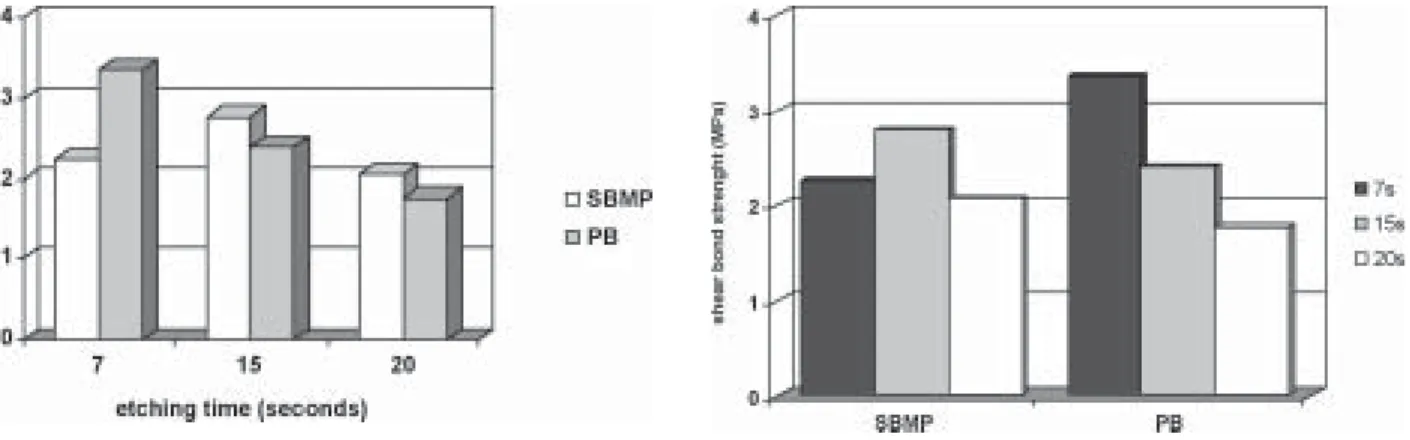
The results of the shear bond strength test are displayed in Table1 and Figures 1-2.
The seven sec etching time showed the highest values of SBS (p<0.05). There was no statistically significant difference between the adhesive syste-ms studied. However, the interaction of adhesive system and etching time was significant and it was observed that the sevens etching time showed the highest SBS values when PB adhesive system was used.
Analysis of failure sites
The failure sites showed to all etching times and for both adhesive systems adhesive failure. Ho-wever, the SBMP showed the higher percentual va-lues of cohesive failure (18%) than PB (6%) as sho-wed at Table 2.
Table 1 - Shear bond strength values (MPa) SBMP PB TOTAL 7s 2.25 a A 3.36 b A 2.81 a 15s 2.79 a A 2.41 a B 2.60 a b 20s 2.08 a A 1.77 a B 1.92 b Total 2.37 a 2.51 a
* Different lower case letters on lines mean values with statistical significant difference (p<0.05). ** Different upper case letters on columns mean values with statistical significant difference (p<0.05).
Table 2 - Type of failures found on the fractured sites for the analyzed samples
PB SBMP
Etching times Adhesive Cohesive Adhesive Cohesive
7 s 93.75% 6.25% 81.25% 18.75%
15 s 93.55% 6.45% 81.45% 18.55%
20 s 100% 0% 98.5% 1.5%
FIGURE 1 – Shear bond strength values (MPa) for the all etching times used in the study.
FIGURE 2 - Shear bond strength values (MPa) for all adhesive sys-tems used in the study.
D
ISCUSSIONIn this study significant statistical difference was found in the interaction between the etching time and adhesive systems (p=0.02) indicating that the success of the adhesive technique does not de-pend exclusively on the material employed, but also on its interaction with the substratum. On the other hand, this presents intrinsic characteristics that should be considered in the bonding process. The conditioning of the dentin substrate is an attempt of improving the effectiveness of the adhesive res-torative materials when replacing the dental struc-ture lost by the decay or trauma8. As the dentin is a dynamic substrate that suffers alterations with time, and from external stimuli, the adhesive process fin-ds some barriers, which hinder the bonding pro-cess26.
Researches have been accomplished in order to verify the effectiveness of the etching acid in the modification of the substratum. The aim for using etching acids is to enable the penetration of resi-nous monomers inside the dentin; so resistant struc-tures to bonding can be produced between restora-tive material and dental structures5, 15.
The modifications in the substrate are directly related to the concentration and application time of etching agents, which consequently will influ-ence in the values of adhesion25 depending on their effectiveness. However, the relationship between bonding strength values and clinical performance of the restoration is not well known, yet.
In permanent teeth, a shorter conditioning time than that for primary ones can lead to insignificant alterations in the structure of the substrate, and the-refore to a decrease in the bond strength values. However, in primary teeth, which the amount of minerals seems to be smaller, and dentin tubules are less wide, the etching time may determine more intense alterations in extension that would decrea-se the bond strength values17.
The highest shear bond strength values were obtained for seven seconds (2.81 MPa), however, these values were not statistically different from the values obtained for 15 seconds (2.60 MPa), suggesting that a shorter exposure time of the subs-tratum to the etching acid did not negatively influ-ence the results. The significant interaction betwe-en adhesive system and etching time suggests that different adhesive systems may have different re-action according to alterations in the etching time.
The increase in etching time reduced the values of the SBS for the PB, though not interfering in the action of the SBMP system.
It could be observed that the larger the etching time the smaller the shear bond strength values; the results showed 1.92 MPa when 20 seconds of etching time was used.
These findings suggest that excessive demine-ralization, due to a longer etching time, could form deeply etched zones where bonding agents perhaps were not able to diffuse, and consequently produ-ced weaker bonding areas due to the formation of a demineralized area not filled out by restorative material3,8,13,16-17,22.
It is important to point out that, the values ob-tained for primary teeth were much smaller than the ones found for permanent ones, as reported in the literature18, as well as for the nominal values of mechanical test that has demonstrated discrepan-cies depending on the methodology used27. In this study, the shear bond strength was accomplished using a steel strip whose width was similar to the thickness of the resin cylinder of the specimen. Based on mechanical laws this type of test provi-des smaller nominal values28 because the load is distributed on the structure of the specimen, which would cause a sliding of the cylinder in relation to the bonding surface, what is not verified when a plain or round knife was used23. The specimen fai-led before the shear effort using a knife, due to the flexuring moment caused by the steel strip on the surface of the resin cylinder, resulting in smaller forces in the bonding surface, producing a real shear effort in the area23.
Considering the adhesive system, in all etching times tested, no statistically significant difference was observed between SBMP and PB, 2.37 MPa and 2.51 MPa, respectively.
However, it was observed that for the etching time seven seconds, PB system showed higher nu-merical values for shear bond strength (3.36 MPa) than for SBMP system (2.25 MPa). The numerical difference presented should be attributed to the di-fferent compositions of the adhesive systems. The PB system, has acetone as solvent, and possesses 37% phosphoric acid as etching agent, while the SBMP system, has the same acid the 35%, howe-ver has water as solvent.
These differences concerning the solvent can suggest that after the etching acid for 7s and rin-sing of the dentin surface, the PB was more
effec-tive concerning the intrinsic dentin humidity, fa-voring a larger interlocking of the bond agent with the substratum, resulting in larger values of SBS.
There was not statistically significant differen-ce between the adhesive systems For the etching times (15 and 20s). This corroborates the suggesti-ons given above for the findings in the times of 7s. This way, a longer etching time (15 and 20s), in dentin of primary teeth, would produce larger amount of demineralized dentin. Consequently, af-ter the rinsing of the surface, a greaaf-ter amount of residual water is found, demonstrating that SBMP exhibited more homogeneous action among the different demineralization levels (Table 1), while PB, due to its composition, seems to be more vul-nerable to the amount of residual water, exactly for interacting in a more effective way with the intrin-sic dentin humidity.
These findings can be emphasized when the obtained patterns of failures are observed after SBS test (Table 2), because both systems presented adhe-sive failures. However, the PB presented predomi-nantly a larger frequency of adhesive failures (95.76%) and smaller of cohesive failures (12.9%). Concerning the performance of the materials studied, the interface material/dentin was observed using photomicrographs and it was found that the amount of demineralization produced by the etching acid of the dentin is related to the amount
of minerals and quality of their distribution on the surface. Thus, possibly a fast demineralization pro-cess is developed in primary teeth due to smaller mineralization of its dentin surface, leading to lar-ge demineralization zones as demonstrated by some authors who found a thicker hybrid layer18,19,20.
It was observed that both adhesive systems and all etching times studied showed the presence of resin tags in all the samples, presenting an interfa-ce with uniform adaptation, suggesting an appro-priate adhesiveness, in the morphological point of view (Figures 3-8).
Specifically, for the seven seconds of etching time, the same characteristics at the resin/dentin interface were observed for both adhesive syste-ms, suggesting that the reduction of the etching time should be applied in primary teeth (Figures 3 and 6), not interfering in the adaptation of the material on the dentin surface.
However, clinical and in vitro microleakage stu-dies should be accomplished to evaluate and to substantiate the in vitro results of the bond streng-th and analysis of streng-the interface, assuring streng-the use of the adhesive process as an efficient restorative pro-cedure, with a different protocol to primary teeth. It could be concluded that the material versus time interaction of acid etching showed that larger bond strength values can be obtained when PB is used for a short period of time in primary teeth,
FIGURE 3 – SEM photomicrograph illustrating the resdentin in-terface reached in G1. Note: C (composite resin), HL (hybrid layer), D (dentin) and RT (resin tags).
FIGURE 4 – SEM photomicrograph illustrating the resdentin in-terface reached in G2. Note: C (composite resin), A (bond agent), HL (hybrid layer), D (dentin) and RT (resin tags).
FIGURE 6 – SEM photomicrograph illustrating the resdentin in-terface reached G4. Note: C (composite resin), A (bond agent), HL (hybrid layer), D (dentin) and RT (resin tags).
FIGURE 5 – SEM photomicrograph illustrating the resdentin in-terface reached in G3. Note: C (composite resin), A (bond agent), HL (hybrid layer), D (dentin) and RT (resin tags).
FIGURE 8 – SEM photomicrograph illustrating the resdentin in-terface reached in G6. Note: C (composite resin), A (bond agent), HL (hybrid layer), D (dentin) and RT (resin tags).
FIGURE 7 – SEM photomicrograph illustrating the resdentin in-terface reached in G5. Note: C (composite resin), A (bond agent), HL (hybrid layer), D (dentin) and RT (resin tags).
although, the clinical implication is unknown whe-ther larger averages of SBS would be beneficial or not to the longevity of the restoration.
Thus, it must be emphasized that the smaller the time of work the smaller the chances of
con-tamination of the dentin and, therefore, the smal-ler the microleakage levels that would be found, mainly in a child’s dental treatment, in which the time of work is a decisive factor for the child beha-vior control.
C
ONCLUSIONSAccording to the results, it can be concluded that:
1. The interaction material versus time showed that for the SBMP the performance of the system did not vary due to the etching time, while for the PB the best results were found for the lower times of acid etching;
2. The analysis of fractured sites showed that the most frequently failure type found was the adhesive type (86.5%);
3. The larger the etching time the lower the she-ar bond strength values for the PB adhesive system.
A
CKNOWLEDGMENTS We thank to:FAPESP #99/07551-3 for supported this rese-arch;
To Professor Dr. José Carlos P. Imparato, Head of Bank of teeth of the São Paulo University – Den-tal School; Prof. Dr. Elliot W. Kitajima, Head of NAP/MEPA – ESALQ/USP.
R
ESUMOObjetivo: avaliar o efeito do tempo de condicionamento ácido e sistemas de união na resistência ao
cisalhamen-to (RUC) em dentes decíduos. Mecisalhamen-todologia: 48 molares decíduos, hígidos, doados pelo Banco de dentes da USP, foram seccionados longitudinalmente (mésio-distal) e embutidos em resina epóxica, deixando as superfíci-es V ou L expostas. As amostras foram lixadas até a obtenção de uma superfície plana em dentina e distribuídas em 3 grupos de acordo com o tempo de condicionamento ácido (7, 15 ou 20 s) e sistemas de união (Scotchbond Multipurpose Plus -SBMP e Prime & Bond 2.1-PB): G1 - 7 s + SBMP; G2 - 15 s + SBMP; G3 - 20 s + SBMP; G4 - 7 s + PB; G5 - 15 s + PB; G6 - 20 s + PB. Confeccionou-se restaurações com compósito Z100, sendo armaze-nados em água destilada a 37oC, por 72h. Os corpos-de-prova foram submetidos ao ensaio de RUC (Instron - 0,5 mm/min). Os sítios de fratura foram analisados em Microscópio Estereoscópico e MEV e os resultados subme-tidos à análise estatística ANOVA e Teste de Tukey (p<0,05). Resultados: os maiores valores de RUC foram obtidos por G1 e G4. Não houve diferença estatística entre G1 e G2, enquanto o G4 apresentou maiores valores em relação aos G1 e G2 (p<0,05). A falha adesiva foi a mais freqüente (86,5%). Conclusões: 1 – A interação material*tempo de condicionamento demonstrou que para o SBMP o desempenho do sistema não diferiu em relação do tempo de condicionamento ácido, enquanto que para o PB os melhores resultados foram observados para os menores tempos de condicionamento ácido; 2 – A análise dos sítios de fratura demonstrou que a falha mais freqüentemente observada foi a do tipo adesiva (86,5%); 3 – Quando maior o tempo de condicionamento ácido, menor os valores de resistência da união para o sistema adesivo PB.
U
NITERMOSAtaque ácido dentário, condicionamento; condicionamento ácido; dente decíduo, sistemas de união
R
EFERENCES1. Araújo FB. Adesão à dentina de dentes decíduos: a micromorfo-logia da dentina condicionada e da interface resina/dentina e sua relação com a resistência ao cisalhamento. São Paulo; 1993. [Tese de Doutorado – Faculdade de Odontologia – Universidade de São Paulo].
2. Buonocore MG. A Simple method of increasing the adhesion of acrylic filling materials to enamel surfaces. J Dent Res. 1955 Dec; 34(6): 849-53.
3. Eick JD, Robinson SJ, Chapell RP, Cobb CM, Spencer P. The dentinal surface: its influence on dentinal adhesion. Part I. Quin-tessence Int. 1991; 22: 967-77.
4. Eick JD, Robinson SJ, Chapell RP, Cobb CM, Spencer P. The dentinal surface: its influence on dentinal adhesion. Part II. Quin-tessence Int. 1992 Jan; 23: 43-51.
5. Gwinnett AJ. Quantitative contribution of resin infiltration/hy-bridization to dentin bonding. Am J Dent. 1993 Feb; 6:7-9. 6. Gwinnett AJ. Altered tissue contribution to interfacial bond
streng-th wistreng-th acid conditioned dentin. Am J Dent 1994 Oct; 7: 243-6. 7. Gwinnett AJ. Adesivos dentais. In: Baratieri LN, Monteiro
Juni-or S, Andrada, MAC, Vieira, LCC, Cardoso AC, Ritter AV. Esté-tica: restaurações adesivas diretas em dentes anteriores fratura-dos. São Paulo: Santos; 1998. p.57-74.
8. Heymann HO, Bayne SC. Current concepts in dentin bonding: focusing on dentinal adhesion factors. J Am Dent Assoc. 1993 May; 124: 27-35.
9. Hosoya Y, Nishiguchi M, Kashiwabara Y, Horiuchi A, Goto G. Comparison of two dentin adhesives to primary vs. permanent bovine dentin. J Clin Pediatr Dent 1997; 22(1): 69-76. 10. Johnson GH, Powell LV, Gordon GE. Dentin bonding systems: a
review of current products and techniques. J Am Dent Assoc 1991 July; 22: 34-41.
11. Lakomaa EL, Rytomaa I. Mineral composition of enamel and dentin of primary and permanent teeth in Finland. Scand J Dent Res 1977 Jan/Feb; 85: 89-95.
12. Marshall Jr GW, Marshall SJ, Kinney JH, Balooch M. The dentin substrate: structure and properties related to bonding. J Dent 1997 June; 25: 441-58.
13. Matos AB, Palma RG, Saraceni CHC, Matson E. Effects of acid etching on dentin surface: SEM morphological study. Braz Dent J 1997; 8(1): 35-41.
14. Miears JR, Charlton DG, Hermesch CB. Effect of dentin moisture and storage time on resin bonding. Am J Dent 1995 Apr; 8: 80-2. 15. Nakabayashi N, Pashley DH. Hibridização dos tecidos den-tais duros. São Paulo: Quintessence; 2000.
16. Nakabayashi N, Saimi Y. Bonding to intact dentin. J Dent Res 1996 Sept; 75(9): 1706-15.
17. Nör JE, Feigal RJ, Dennison JB, Edwards CA. Dentin bonding: SEM comparison of the resin-dentin interface in primary and per-manent teeth. J Dent Res 1996 June; 75(6): 1396-403.
18. Nör JE, Feigal RJ, Dennison JB, Edwards CA. Dentin bonding: SEM comparison of the dentin surface in primary and permanent teeth. Pediatr Dent 1997 May/June; 19(4): 246-52.
19. Olmez A, Oztas N, Basak F, Erdal S. Comparison of the resin-dentin interface in primary and permanent teeth. J Clin Pediatr Dent 1998; 22(4): 293-8.
20. Pioch T, Stotz S, Buff E, Duschner H, Staehle HJ. Influence of different etching times on hybrid layer formation and tensile bond strength. Am J Dent 1998 Oct; 11: 202-6.
21. Puppin-Rontani RM, Caetano E, Garcia-Godoy F, De Goes MF. Effect of antimicrobial agents on the morphology of primary den-tin. J Clin Pediatr Dent. 2001; 25(2): 137-41.
22. Sano H, Takatsu T, Ciucchi B, Horner JA, Matthews WG, Pash-ley DH. Nanoleakage: leakage within the hybrid layer. Oper Dent 1995; 20(1): 18-25.
23. Sinhoreti MAC et al. Influence of loading types on the shear strength of the dentin-resin interface bonding. J Mater Sci 2001; 12: 39-44.
24. Ten Cate AR. Histologia bucal: desenvolvimento, estrutura e fun-ção. Rio de Janeiro: Guanabara Koogan; 1988.
25. Triolo Jr PT, Swift Jr EJ, Mudgil A, Levine A. Effects of etching time on enamel bond strengths. Am J Dent 1993 Dec; 6: 302-4. 26. Van Meerbeek B, Braem M, Lambrechts P, Vanherle G.
Morpho-logical characterization of the interface between resin and scle-rotic dentine. J Dent Res 1994 June; 22: 141-6.
27. Van Noort R, Cardew GE, Howard IC, Noroozi S. The effect of local interfacial geometry on the measurement of the tensile bond strength to dentin. J Dent Res 1991; 70(5): 889-93.
28. Versluis A, Tantbirojn D, Douglas WH. Why do shear bond tests pull out dentin? J Dent Res 1997; 76(6): 1298-307.
Recebido em: 26/03/04 Aprovado em: 30/06/04 Regina Maria Puppin-Rontani Department of Pediatric Dentistry – Piracicaba Dental School – UNICAMP CEP: 13414-018 – Av. Limeira, 901 Piracicaba – SP tel: 55-019-3412 5286 fax: 55-019-3412 5218 [email protected]