From monoclonal gammopathy
of undetermined significance
to symptomatic multiple myeloma:
genetic heterogeneity on all levels
B.A. Walker, G.J. MorganMolecular Haematology, Haemato-Oncology Research Unit, Division of Molecular Pathology, The Institute of Cancer Research, London, UK E-mail: brian.balker@icr.ac.uk I Simposio Conjunto EHA - SAH HEMATOLOGÍA, Vol.17 Número Extraordinario XXI CONGRESO Octubre 2013 Abstract
Multiple myeloma (MM) is preceded by pre-malig-nant disease phases of monoclonal gammopathy of undetermined significance (MGUS) and smoldering myeloma (SMM). The genetic abnormalities found in MM comprise of intrachromosomal transloca-tions, largely involving the IGH locus, copy number abnormalities, somatic mutations and changes in DNA and histone methylation. Many of these ge-netic lesions are also present in MGUS and SMM but do not result in the clinical symptoms associated with MM. Here we discuss the common abnormali-ties in these disease phases along with the impact of intraclonal heterogeneity on the future of myeloma biology and treatment.
Learning goals
At the conclusion of this activity, participants should be able to:
- describe the common genetic abnormalities in multiple myeloma;
- know the common somatic mutations in myelo-ma and targeted therapy options;
- understand the complex subclonal genetic archi-tecture of myeloma.
Introduction
Multiple myeloma is a genetically complex isease that is becoming more common in today’s aging population. Myeloma belongs to a group of related paraproteinemias that are characterized by an ab-normal clonal plasma cell infiltration in the bone marrow.1,2 A number of distinct clinical phases of myeloma can be recognized, including monoclonal gammopathy of undetermined significance (MGUS) and asymptomatic or smoldering multiple myeloma (SMM). Both these phases lack the clinical features of myeloma but share some of the genetic features of a myeloma clone.3 By contrast, symptomatic multi-ple myeloma (MM) is defined by clinical symptoms
and evidence of organ damage.
A characteristic feature of myeloma cells is the re-quirement for an intimate relationship with the bone marrow microenvironment, where plasma cells are nurtured in specialized niches that maintain their survival long term.4-8 However, during the progres-sion of the disease, clonal cells develop the ability to proliferate at sites outside of the bone marrow, man-ifesting as extra-medullary myeloma (EMM) and plasma cell leukemia (PCL).9 These cells constitute the end stages in the multistep transformation pro-cess from normal to malignant plasma cells. Here we will review the genetics and techniques used to study the events in the process of transformation from MGUS through SMM, MM and finally to PCL. These include the classically studied translocations and hyperdiploidy, copy number abnormalities and, finally, how genome sequencing strategies are identi-fying new potential targets in somatic mutations and how these can be used to determine the evolutionary course of disease progression.
Translocations
Chromosomal translocations arise when DNA dou-ble strand breaks at different sites in the genome are brought together and aberrantly rejoined.10
They are common in tumors of the lymphoid lineage because of the ‘off target’effects of the normal physi-ological mechanismsmediating DNA rearrangement at theimmunoglobulin (IGH) locus. Translocation-sinto the IGH locus predominantly occur eitherdur-ing recombination activation gene (RAG) complex-mediated V(D)J rearrangement, such as in mantle cell lymphoma (t(11;14)),11 or during class switch re-combination (CSR). Inmyeloma, the primary trans-locations are thought to be generated via abnormal CSR events mediated by activation-induced cytidine deaminase (AID).12 This concept has been devel-oped and is based on the location of the transloca-tion breakpoints determined in myeloma cell lines and a few primary samples.
Added to this, the myeloma clone is derived from a mature plasma cell that has undergone somatic hy-permutation in the germinal center13 and does not express the RAG complex.
In myeloma, primary aberrant rearrangementsinto the IGH locus are present in up to 40% of cases.14,15 There are five main translocation partner
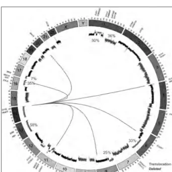
chromo-somes including the t(4;14) (11%), t(6;14) (2%), t(11;14) (15%), t(14;16) (3%) and t(14;20) (1.5%) which result in the overexpression of MMSET and FGFR3, CCND3, CCND1, MAF and MAFB, respec-tively, and are thought to confer a selective advan-tage to the clone (Figure 1).16 Although the trans-locations over-express very different genes, they have in common downstream deregulation of cyclin D genes, which have been grouped together under the Translocation/Cyclin D (TC) classification.17 In its simplest form, this classification defines groups of myeloma samples based on their expression of CCND1 (t(11;14)), CCND2 (t(4;14),t(14;16) and t(14;20)), and CCND3 (t(6;14)). However, the trans-locations themselves are not sufficient to cause pro-gression to myeloma. Evidence for this comes from analysis of MGUS, SMM and MM samples in which translocations are detected, but not at the same fre-quency.18 For example, the t(14;20) is present in 5% of MGUS samples but only 1.5% of MM samples, and conversely, the t(4;14) is present in 3% of MGUS but rises to 11% in MM samples.
Figure 1: The common genetic abnormalities in myeloma. The circos plot shows chromosomes arranged around the outside in a clockwise direction. The internal track shows the common copy number changes with deletions (red) and gains (blue) shown with their frequencies in myelo-ma. Translocations are indicated by lines across the center between loci. The genes of interest are shown around the outside of the circle and are color-coded according to the legend.
The conclusions drawn from these data are that some translocations, such as the t(14;20), can be stable in MGUS patients for long periods of time resulting in higher frequencies present in MGUS, whereas the t(4;14) progresses to MM faster, resulting in a lower frequency in MGUS patients.
Copy number changes
In addition to translocations, copy number abnor-malities are common in myeloma (Figure 1). These abnormalities have been studied by many techniques from karyotyping and fluorescence in situ hybridiza-tion (FISH) through to SNP-based mapping arrays, and more recently, exome sequencing. The most prevalent copy number abnormality is the presence of hyperdiploidy, through trisomy of chromosomes 3, 5, 7, 9, 11, 15, 19 and 21, and like the transloca-tions is considered a primary event.
Hyperdiploidy is present in approximately 50% of myeloma samples and is almost mutually exclusive with IGH translocations, where both translocations and hyperdiploidy occur in only 9% of samples. The most commonly gained chromosomes are 9, 15 and 19 but the genetic mechanism of gain and pathogenic advantage still remain elusive. Hyperdiploid patients tend to have a better prognostic outcome than those with IGH translocations. The myeloma genome is rife with additional copy number abnormalities, with almost all chromosomes being affected across samples, indicating genomic instability in myeloma. Aside from the trisomies related to hyperdiploidy, the most common chromosomal abnormalities are del(1p) (30%), 1q+ (36%), del(6q) (33%), del(8p) (25%), 11q+ (24%), del(13q) (58%), del(16q) (35%) and del(17p) (7%).19 In some of these chromosomes, the genes of interest have been identified but in oth-ers they remain elusive. For example, on 1p FAM46C, CDKN2C and FAF1 have been identified as poten-tial targets,19-21 on 16q CYLD and WWOX are targets of interstitial deletions,22,23 on 1q CKS1B, ANP32E, BCL9 and PDZK1 have all attracted interest,19,24,25 and on 17p TP53 is the clear gene of interest.19,26,27 However, for many of the chromosomal abnormali-ties (6q, 8p) there is no clear target gene. These last two regions have not been so well studied, in part because they currently have no prognostic value.
Cytogenetic risk stratification
Cytogenetics has been used to determine which genetic lesions have an impact on overall and pro-gression-free survival. Concerning the translocation groups, t(4;14), t(14;16) and t(14;20) are considered to be high risk genetic events resulting in a decreased overall survival.28
However, much of the high risk nature of the t(4;14) can be overcome by treatment with bortezomib.29 t(11;14) and (6;14) are considered standard risk groups, as is hyperdiploidy.
Many of the copy number abnormalities do have a prognostic value in several datasets. In the UK MRC Myeloma IX trial, we have shown del(1p), 1q+ and del(17p) all have an independent statistically sig-nificant impact in overall survival.19,26 This has been confirmed in other datasets with several different treatment contexts.30-33 Together with t(4;14), these cytogenetic markers have been used to identify pa-tients with high-risk myeloma, which could be man-aged differently to standard risk patients. One analy-sis has also determined that the poor prognostic effect of high-risk genetics (t(4;14), t(14;16), t(14;20) or del(17p)) can be ameliorated by the presence of trisomies.34 Bortezomib administration can also improve outcome in patients with del(17p) when administered before and after autologous stem cell transplantation.35
The accumulation of adverse markers has a profound effect on the overall survival of a patient. Many of the adverse lesions co-segregate, so the chance of a pa-tient having more than one abnormality is increased, for example 72% of patients with an IGH translo-cation also have 1q+. By integrating these known adverse lesions it is possible to more accurately es-timate the overall survival of a patient where those without any adverse markers (OS=60.6 months) do better than those with one (OS=41.9 months), two (OS=23.4 months) or three (OS=9.1 months) ad-verse markers.36
Somatic mutations
The most recent developments in myeloma ge-netics revolve around genome and exome sequenc-ing of samples, allowsequenc-ing the identification of somatic mutations and structural variations. This has been exemplified by the initial publication of the land-scape of mutations in myeloma through sequencing
of 38 myeloma samples.37 The number of non-syn-onymous (NS) somatic mutations found in myelo-ma is around 30-35.37,38 This number is higher than some other hematologic malignancies such as hairy cell leukemia (NS-mutations = 5),39 acute myeloid leukemia (NS-mutations = 8)40 but much lower than solid tumors such as lung cancer (NS-mutations = 540).41 This level of mutation indicates that myeloma is more complex than most hematologic malignan-cies. The main finding of this initial screen is that there is no unifying mutation in myeloma. In some other hematologic malignancies, a common muta-tion in most or all samples has been discovered and is thought to be the primary driver mutation. For example, in hairy cell leukemia, the BRAF V600E mutation is found in all samples,39 and in Walden-ströms macroglobulinemia, the MYD88 L265P mu-tation is found in 91% of samples.42 In myeloma, the most frequent mutations were found in NRAS (23%) and KRAS (26%), followed by FAM46C (13%, pre-viously identified as deleted and mutated)19,20 and TP53 (8%). The NRAS and KRAS mutations, with the addition of BRAF mutations (4%), indicates the ERK pathway is critical in at least 53% of myeloma patients and points to a treatment strategy that has so far not been harnessed. ERK pathway mutations are not new to myeloma, but the whole genome strategies have identified some novel mutations not previously identified by other means. These include DIS3 (mutated in 10%) on chromosome 13, a highly conserved RNA nuclease, which is also deleted in 58% of samples. The function of this mutation is not understood, but may be involved in regulation of the available pool of mRNAs available for trans-lation.43 However, the number of myeloma samples sequenced to date is small and the true landscape of somatic mutations is yet to be realized. As the num-ber of samples sequenced increases, it will be possi-ble to identify groups of genes with related functions or pathways that can be used as therapeutic targets. For example, DNA and histone methylation are im-portant biological processes in myeloma which is characterized by overexpression of MMSET, a his-tone methyltransferase, in t(4;14) myeloma, and mutations in other methyltransferases, such as EZH2 and MLL3, can also be present. Additionally, histone lysine demethylases such as KDM6A (also known as UTX) can be deleted or mutated in my-eloma, 44 making histone methylation a common
and attractive target for drug therapy. The discovery of BRAF mutations in 4% of myeloma patients has also brought the possibility of targeted therapy to the forefront of myeloma treatment in the clinic. BRAF is part of the MAP kinase pathway, which is acti-vated by RAS through phosphorylation and results in the subsequent activation of the MEK/MAPK/ ERK signaling cascade, resulting in proliferation and survival.45 The BRAFV600E mutation is present in 50%-60% of all melanomas and results in constitu-tive activation of BRAF, bypassing the requirement for RAS, activating the MEK/MAPK/ERK cascade, and culminating in cell proliferation and malignant conversion.46 The drug vemurafenib is a competi-tive seleccompeti-tive inhibitor of BRAFV600E which is ap-proved for use in melanoma and results in relative reduction of 63% in risk for death compared to other treatments.47 Vemurafenib, therefore, represents a potential targeted therapy for patients harboring a BRAFV600E mutation and clinical trials are under-way in myeloma to determine its efficacy.
Intraclonal heterogeneity
Like many malignancies, myeloma cells are not uni-form within a patient. A great deal of genetic varia-tion exists within the populavaria-tion of tumor cells, and it is this variation that allows the cancer to persist and diversify. The genetic events within a cancer cell consist of ‘driver’ and ‘passenger’ lesions, where drivers confer a selective advantage to the progeny. The acquisition of these lesions allows for the rapid evolution of a clone in a Darwinian fashion.
Selection pressures are exerted on the tumor cells al-lowing the outgrowth of any favorable trait. These selection pressures may give a growth advantage to a cell, confer a better interaction with the bone mar-row microenvironment, or even allow independence from the bone marrow resulting in a plasma cell leu-kemia or an extramedullary tumor.
Aside from this, mutations gained in subpopula-tions of cells may confer drug resistance, allowing the eventual repopulation of the tumor in a drug resistant state. Although myeloma is considered to be a clonal disease, due to the presence of one V(D) J rearrangement and a monoclonal secreted immu-noglobulin, at a genetic level the cells are far from clonal. IGH translocations and hyperdiploidy are ac-cepted as being primary events in myeloma patho-genesis; however, the rate at which other
abnormali-ties are accrued has been less well studied. Studies utilizing FISH were the first to investigate the rela-tionship of abnormalities within a sample by using probes to a translocation and a copy number abnor-mality and comparing the frequencies. When com-paring a translocation with del(13q) it was found that the majority of cells carry the translocation (as expected given it is a primary event) but the pro-portion of cells with del(13q) can vary dramatically from patient to patient, but is always lower than the frequency of the translocation.48 It can be inferred from these data that the copy number abnormalities occur subsequent to the translocation. By analyz-ing the disease at different time points it becomes clear that the frequency of any given abnormality increases through MGUS and SMM towards MM in a population of individuals. This has been shown for del(13q), del(17p) and 1q+where the proportion of myeloma patients with an abnormality increases as the disease progresses.18,49 However, such an analysis can be even more informative if sequential samples from the same patient are used, particularly when they are taken at different stages of disease (for ex-ample SMM and MM). Several papers have been published analyzing such patients by FISH and SNP-based mapping array.49,50 The overarching theme of these papers is that the frequency of abnormalities increases within a tumor sample as the disease pro-gresses, but they are generally always present at low levels in the preceding stage of disease. For example, in a patient there may be 29% of cells with del (17p) when the patient is diagnosed with high risk-SMM and this may increase to 86% when they present with symptomatic MM.51 The genetic landscape of these tumors gets more interesting as the technologies used get more advanced. Using genome sequencing technologies it is possible to estimate the proportion of cells in a tumor mass with any somatic mutation found. This has been achieved in many cancers,52,53 including myeloma.38,54 Taking the RAS pathway mutations as an example, it has been shown that these activating oncogenic driver mutations are not necessarily present in the dominant clone. That is, they can be present only in a subset of the cells in the tumor.38 This is true for NRAS, KRAS and BRAF mutations, indicating that although they are known oncogenic drivers they are not necessarily present early on in the disease and can be acquired as the tumor evolves.
Using information on the subclonal nature of mul-tiple mutations or copy number abnormalities it is possible to piece together the history of a tumor, determining which genetic events occurred first or occurred together.52,55 This can also be done at the single cell level using FISH withmultiple probes per cell, or at a nucleotide level using singlecell sorting and genotyping assays.38,55 These techniques clearly indicate a complex substructure of branched evolu-tion in tumor development. Other studies have fo-cused on the genetic evolution of myeloma follow-ing treatment.54 Analysis of tumor DNA collected at multiple time points during a patient’s treatment can illustrate the genetic diversity within a myeloma tumor and the effect that treatment has on the dy-namics of the sub-clones present. By studying seven time points from diagnosis, remission, four relapse phases and progression to plasma cell leukemia the different subclones present can be seen using arrays, gaining and losing dominance in the myeloma pop-ulation as the patient undergoes different treatment regimens. Ultimately, the clone that was dominant as the disease progresses to PCL was barely detectable at diagnosis. Given that myeloma exists as multiple foci of lytic lesions throughout the bone marrow, it remains to be determined how these subpopulations of cells relate to one another, whether they evolve independently, and whether they can be treated as a whole.
Conclusions
Myeloma is a genetically complex malignancy in which translocations involving the IGH locus and hyperdiploidy are primary events. These events are followed by an accrual of additional lesions through MGUS and SMM before transforming to MM. These additional lesions include, but are not limited to, chromosomal gains and losses, somatic mutations and DNA methylation changes. It is clear that there is a subclonal genetic structure within the myeloma cell population where copy number and somatic mutations are gained or lost over time, resulting in a mixed population of cells capable of exploiting any selective advantages laid upon them. This intraclonal heterogeneity may prove to be an extra obstacle in the fight towards curing myeloma, but through using therapies towards key genetic mechanisms it should prove possible to selectively target mutated clones.
References
1. Greipp PR, San MJ, Durie BG, et al. International staging system for multiple myeloma. J Clin Oncol. 2005;23(15):3412-20.
2. Kyle RA, Gertz MA, Witzig TE, et al. Review of 1027 patients with newly diagnosed multiple myeloma. Mayo Clin Proc. 2003;78(1):21-33.
3. Kyle RA, Remstein ED, Therneau TM, et al. Clini-cal course and prognosis of smoldering (asymp-tomatic) multiple myeloma. New Eng J Med. 2007;356(25):2582-90.
4. Akiyama M, Hideshima T, Hayashi T, et al. Cytokines modulate telomerase activity in a human multiple myeloma cell line. Cancer Res. 2002;62(13):3876-82. 5. Chauhan D, Li G, Hideshima T, et al. Blockade of
ubiquitinconjugating enzyme CDC34 enhances anti-myeloma activity of Bortezomib/Proteasome inhibitor PS-341. Oncogene. 004;23(20):3597-602. 6. Damiano JS, Cress AE, Hazlehurst LA, Shtil AA,
Dalton WS. Cell adhesion mediated drug resis-tance (CAM-DR): role of integrins and resisresis-tance to apoptosis in human myeloma cell lines. Blood. 1999;93(5):1658-67.
7. Hideshima T, Catley L, Yasui H, et al. Perifosine, an oral bioactive novel alkylphospholipid, inhib-its Akt and induces in vitro and in vivo cytotox-icity in human multiple myeloma cells. Blood. 2006;107(10):4053-62.
8. Mitsiades CS, Mitsiades NS, Munshi NC, Richard-son PG, AnderRichard-son KC. The role of the bone mi-croenvironment in the pathophysiology and thera-peutic management of multiple myeloma: interplay of growth factors, their receptors and stromal inter-actions. Eur J Cancer. 2006;42(11):1564-73. 9. Palumbo A, Anderson K. Multiple myeloma. N Engl
J Med. 2011;364(11):1046-60.
10. Richardson C, Jasin M. Frequent chromosomal translocations induced by DNA double-strand breaks. Nature. 2000;405(6787):697-700.
11. Jares P, Colomer D, Campo E. Molecular patho-genesis of mantle cell lymphoma. J Clin Invest. 2012;122(10):3416-23.
12. Gonzalez D, van der Burg M, Garcia-Sanz R, et al. Immunoglobulin gene rearrangements and the pathogenesis of multiple myeloma. Blood. 2007;110(9):3112-21.
13. Bakkus MH, Heirman C, van Riet I, van Camp B, Thielemans K. Evidence that multiple myeloma Ig heavy chain VDJ genes contain somatic muta-tions but show no intraclonal variation. Blood. 1992;80(9):2326-35.
14. Bergsagel PL, Chesi M, Nardini E, Brents LA, Kirby SL, Kuehl WM. Promiscuous translocations into immunoglobulin heavy chain switch regions in multiple myeloma. Proc Natl Acad Sci USA. 1996;93(24):13931-6.
15. Pratt G, Fenton JA, Proffitt JA, et al. True spectrum of 14q32 translocations in multiple myeloma. Br J Haematol. 1998;103(4):1209-10.
16. Morgan GJ, Walker BA, Davies FE. The genetic ar-chitecture of multiple myeloma. Nat Rev Cancer. 2012;12(5):335-48.
17. Bergsagel PL, Kuehl WM. Molecular pathogenesis and a consequent classification of multiple myelo-ma. J Clin Oncol. 2005;23(26):6333-8.
18. Ross FM, Chiecchio L, Dagrada G, et al. The t(14;20) is a poor prognostic factor in myeloma but is asso-ciated with long-term stable disease in monoclonal gammopathies of undetermined ignificance. Hae-matologica. 2010;95(7):1221-5.
19. Walker BA, Leone PE, Chiecchio L, et al. A compen-dium of myeloma-associated chromosomal copy number abnormalities and their prognostic value. Blood. 2010;116(15):e56-65.
20. Boyd KD, Ross FM, Walker BA, et al. Mapping of chromosome 1p deletions in myeloma identifies FAM46C at 1p12 and CDKN2C at 1p32.3 as being genes in regions associated with adverse survival. Clin Cancer Res. 2011;17(24):7776-84.
21. Leone PE, Walker BA, Jenner MW, et al. Deletions of
CDKN2C in multiple myeloma: biological and clinical implications. Clin Cancer Res. 2008;14(19):6033-41.
22. Jenner MW, Leone PE, Walker BA, et al. Gene map-ping and expression analysis of 16q loss of heterozy-gosity identifies WWOX and CYLD as being im-portant in determining clinical outcome in multiple myeloma. Blood. 2007;110(9):3291-300.
23. Keats JJ, Fonseca R, Chesi M, et al. Promiscu-ous mutations activate the noncanonical NF-kap-paB pathway in multiple myeloma. Cancer Cell. 2007;12(2):131-44.
24. Shaughnessy J. Amplification and overexpression of CKS1B at chromosome band 1q21 is associated with reduced levels of p27Kip1 and an aggressive clinical course in multiple myeloma. Hematology. 2005;10(1 Suppl):117-26.
25. Le Baccon P, Leroux D, Dascalescu C, et al. Novel evidence of a role for chromosome 1 pericentric heterochromatin in the pathogenesis of B-cell lym-phoma and multiple myeloma. Gene Chromosome Canc. 2001;32(3):250-64.
26. Boyd KD, Ross FM, Tapper WJ, et al. The clinical impact and molecular biology of del(17p) in mul-tiple myeloma treated with conventional or thal-idomide-based therapy. Gene Chromosome Can. 2011;50(10):765-74.
27. Chng WJ, Price-Troska T, Gonzalez-Paz N, et al. Clinical significance of TP53 mutation in myeloma. Leukemia. 2007;21(3):582-4.
28. Fonseca R, Bergsagel PL, Drach J, et al. International Myeloma Working Group molecular classification of multiple myeloma: spotlight review. Leukemia. 2009;23(12):2210-21.
29. Avet-Loiseau H, Leleu X, Roussel M, et al. Bort-ezomib plus dexamethasone induction improves outcome of patients with t(4;14) myeloma but not outcome of patients with del(17p). J Clin Oncol. 2010;28(30):4630-4.
30. Fonseca R, Van Wier SA, Chng WJ, et al. Prognostic value of chromosome 1q21 gain by fluorescent in situ hybridization and increase CKS1B expression in myeloma. Leukemia. 2006;20(11):2034-40. 31. Hanamura I, Stewart JP, Huang Y, et al. Frequent
gain of chromosome band 1q21 in plasma-cell dy-scrasias detected by fluorescence in situ hybridiza-tion: incidence increases from MGUS to relapsed myeloma and is related to prognosis and disease progression following tandem stem-cell transplan-tation. Blood. 2006;108(5):1724-32.
32. Shaughnessy JD Jr, Haessler J, van Rhee F, et al. Test-ing standard and genetic parameters in 220 patients with multiple myeloma with complete data sets: superiority of molecular genetics. Br J Haematol. 2007;137(6):530-6.
33. Avet-Loiseau H, Attal M, Moreau P, et al. Genetic abnormalities and survival in multiple myeloma: the experience of the Intergroupe Francophone du Myelome. Blood. 2007;109(8):3489-95.
34. Kumar S, Fonseca R, Ketterling RP, et al. Triso-mies in multiple myeloma: impact on survival in patients with high-risk cytogenetics. Blood. 2012;119(9):2100-5.
35. Neben K, Lokhorst HM, Jauch A, et al. Adminis-tration of bortezomib before and after autologous stem-cell transplantation improves outcome in mul-tiple myeloma patients with deletion 17p. Blood. 2012;119(4):940-8.
36. Boyd KD, Ross FM, Chiecchio L, et al. A novel prog-nostic model in myeloma based on co-segregating adverse FISH lesions and the ISS: analysis of pa-tients treated in the MRC Myeloma IX trial. Leuke-mia. 2012;26(2):349-55.
37. Chapman MA, Lawrence MS, Keats JJ, et al. Initial genome sequencing and analysis of multiple myelo-ma. Nature. 2011;471(7339):467-72.
38. Walker BA, Wardell CP, Melchor L, et al. Intraclonal heterogeneity and distinct molecular mechanisms characterize the development of t(4;14) and t(11;14) myeloma. Blood. 2012;120(5):1077-86.
39. Tiacci E, Trifonov V, Schiavoni G, et al. BRAF mutations in hairy-cell leukemia. N Engl J Med. 2011;364(24):2305-15. 40. Ley TJ, Mardis ER, Ding L, et al. DNA sequencing of a cytogenetically nor-mal acute myeloid leukaemia genome. Nature. 2008;456(7218):66-72.
41. Lee W, Jiang Z, Liu J, et al. The mutation spectrum revealed by paired genome sequences from a lung cancer patient. Nature. 2010;465(7297):473-7. 42. Treon SP, Xu L, Yang G, et al. MYD88 L265P
somat-ic mutation in Waldenstrom’s macroglobulinemia. N Engl J Med. 2012;367(9):826-33.
43. Ibrahim H, Wilusz J, Wilusz CJ. RNA recognition by 3’-to-5’ exonucleases: the substrate perspective. Biochim Biophys Acta. 2008;1779(4):256-65. 44. van Haaften G, Dalgliesh GL, Davies H, et al. Somatic
mutations of the histone H3K27 demethylase gene UTX in human cancer. Nat Genet. 2009;41:521-3. 45. Satyamoorthy K, Li G, Gerrero MR, et al.
Consti-tutive mitogen-activated protein kinase activation in melanoma is mediated by both BRAF mutations and autocrine growth factor stimulation. Cancer Res. 2003;63(4):756-9.
46. White RM. The natural history of malignancies un-der conditions of BRAF inhibitor stimulation. Ex-pert Opin Investig Drugs. 2011;20(1):135-6.
47. Ravnan MC, Matalka MS. Vemurafenib in patients with BRAF V600E mutation-positive advanced melanoma. Clin Ther. 2012;34(7):1474-86.
48. Chiecchio L, Dagrada GP, Ibrahim AH, et al. Tim-ing of acquisition of deletion 13 in plasma cell dy-scrasias is dependent on genetic context. Haemato-logica. 2009;94(12):1708-13.
49. Lopez-Corral L, Sarasquete ME, Bea S, et al. SNP-based mapping arrays reveal high genomic com-plexity in monoclonal gammopathies, from MGUS to myeloma status. Leukemia. 2012;26(12):2521-9. 50. Lopez-Corral L, Gutierrez NC, Vidriales MB, et al.
The progression from MGUS to smoldering my-eloma and eventually to multiple mymy-eloma involves a clonal expansion of genetically abnormal plasma cells. Clin Cancer Res. 2011;17(7):1692-700.
51. Lopez-Corral L, Mateos MV, Corchete LA, et al. Genomic analysis of high-risk smoldering multiple myeloma. Haematologica. 2012;97(9):1439-43. 52. Nik-Zainal S, Van Loo P, Wedge DC, et al. The life
history of 21 breast cancers. Cell. 2012;149(5):994-1007.
53. Yates LR, Campbell PJ. Evolution of the cancer ge-nome. Nat Rev Genet. 2012;13(11):795-806. 54. Keats JJ, Chesi M, Egan JB, et al. Clonal competition
with alternating dominance in multiple myeloma. Blood. 2012;120(5):1067-76.
55. Anderson K, Lutz C, van Delft FW, et al. Genetic variegation of clonal architecture and propagating cells in leukaemia. Nature. 2011;469(7330):356-61.