Comparison of Oral Microbiome in Siblings
Using Next Generation Sequencing
Hyo-Seol Lee
The Graduate School
Yonsei University
Comparison of Oral Microbiome in Siblings
Using Next Generation Sequencing
Directed by Professor Jae-Ho Lee
A Doctoral Dissertation
Submitted to the Department of Dental Science
and the Graduate School of Yonsei University
in partial fulfillment of requirements
for the degree of Doctor of Philosophy in Dental Science
Hyo-Seol Lee
감사의 글
이 논문의 작성을 끝으로 모든 박사과정을 마치게 되었습니다. 지금 의 제가 있을 수 있도록 도와주신 모든 분과 하나님께 감사를 드립니다. 우선, 연세대학교 소아치과의 모든 교수님께 감사를 드립니다. 성실함 과 인자함을 보여주시는 이종갑, 양정강 명예교수님, 뵙기만 해도 의지를 느끼고, 배울 수 있는 손흥규 교수님, 석사논문을 잘 이끌어주셨던 최병 재 교수님, 논문과 삶에 대한 조언을 해주셨던 최형준 교수님, 어떤 일에 도 응원을 해주셨던 김성오 교수님, 물심양면 도와주신 송제선 교수님께 진심으로 감사드립니다. 그리고, 박사과정 지도교수님이자, 장애인치과학 으로 이끌어주신 이제호 교수님께 아낌없는 감사의 마음 전합니다. 도움 을 주기보다 받기만 한 것 같아 미안한 소아치과 의국원들, 강정민, 김재 은, 전혜림, 조성현, 김수현, 이혜원, 정연욱, 주기훈, 박민경, 신민경, 신 영섭, 이명연 선생님 고맙습니다. 임상에서 같이 일하며서 많은 도움을 주신 김성은, 윤민희, 정유주 위생사님 고맙습니다. 도와주신 전미정, 정 재은 연구원, 이다선 위생사 비서 임경은씨에게도 감사합니다. 연세대학교 치과병원의 모든 교수님들께 감사합니다. 병원 내에서 인사만 해주셔도 제겐 큰 힘이 되었습니다. 항상 따뜻하게 보살펴주시는 담임반 유형석 교수님께 감사의 마음을 전합니다. 구강내과 최종훈, 김성택, 안형준, 권정승 교수님 감사합니다. 보철과 심준성 교수님, 치 주과 채중규, 조규성, 이중석 교수님, 보존과 이승종, 정일영, 신유석 교 수님, 방사선과 박창서 교수님, 교정과 이기준, 차정열 교수님, 구강외 과의 김형준, 강정완, 정휘동 교수님 감사했습니다. 통합진료과의 김기 덕, 박원서, 방난심 교수님 감사합니다. 병리학 교실의 김현실 교수님 감사합니다. 선생님은 제 멘토입니다. 치의학교육실의 김주아 교수님도 감사합니다. 구강 생물학교실의 허경석 교수님 감사합니다. 책 더 많이 읽겠습니다. 약리학교실의 서정택교수님 감사합니다. 또한, 제게 물심양 면으로 도와주신 예방치과의 김백일 교수님, 정회인, 이은송 선생님께 깊은 감사의 말씀 전합니다. 그리고, 이 논문이 나오는 데 결정적인 도 움을 주신 이지현 교수님께 감사드리며, 앞날에 무한한 축복이 있기를 기원합니다. 제가 힘들고, 목표가 보이지 않을 때 가야 할 길을 보여주시는 대한 장애인치과학회의 학회장님, 이사님들께 모두 감사합니다. 이긍호 교수 님, 나성식 원장님, 백승호 교수님, 김광철 교수님, 김영재 교수님, 이재 천 원장님, 김선미 교수님, 장주혜 교수님, 서광석 교수님, 현홍근 교수 님, 최재영 원장님, 최충호 교수님, 김지연 교수님, 정태성 교수님과 민 여진 사회복지사님과 함께 할 수 있어서 영광이었습니다. 그 과정에서
제가 배운 많은 것들이 제가 끈기있게 생활할 수 있는 원동력이 되었습 니다. 감사합니다. 또한, 수련을 마친 후 3년간 근무했던 서울특별시 장 애인치과병원의 모든 분께도 감사드립니다. 김혜정 선생님, 김미경 선 생님, 김인선 선생님, 이은영 선생님, 남선회 선생님, 황지영 선생님과 김민선 사회복지사님에게 깊은 감사를 드립니다. 그리고, 논문으로 지칠 때 위로해준 FM 93.9 CBS 라디오, podcast 아워리스트와 이적씨, 이승열씨, 서태지씨, 故 신해철씨에게 깊은 감사 를 드립니다. 중, 고등학교 때부터 지금까지 좋은 음악 들려주셔서 감 사합니다. 논문작업에 많은 도움을 주시고, 생각이 막힐 때 돌파구가 되어주는 많은 책을 빌려주신 의학도서관의 사서분들께도 감사의 마음 을 전합니다. 교회 영유아부와 구름반, 무지개반 아이들 부모님께 감사 합니다. 또한, 초,중,고등학교의 좋은 친구들 고맙습니다. 마지막으로 저의 모든 것이자 살아가는 의미인 가족들에게 감사의 말씀을 전합니다. 아버지, 어머니, 큰언니, 큰형부, 예원, 예윤, 작은언니, 작은형부, 소희, 소정과 시아버지, 시어머니, 형님, 아주버님, 다은이, 평 소에 바쁘다고 소홀하고, 표현하지 못해서 죄송합니다. 정말 사랑하고 감사합니다. 생각만해도 마음이 따뜻해지는 나의 작은 영웅 김재원군과 큰 영웅 김정인씨에게 이 논문을 바칩니다. 사랑합니다.
i
Contents
Contents ... i
List of Tables and Figures ... iii
Abstract ... iv
I. Introduction ... 1
II. Materials and Methods ... 6
1. Ethics statement ... 6
2. Data collecting ... 6
3. Sample preparation ... 8
4. PCR amplification and pyrosequencing ... 9
5. Pyrosequencing data analysis ... 12
III. Results ... 15
1. Demographics and clinical information ... 15
2. Taxonomic identification and operational taxonomical unit (OUT) diversity ... 17
3. Comparison of the composition of the microbiome between caries and caries-free group ... 20
4. Correlation of the siblings ... 26
ii
V. Conclusion ... 35 VI. References ... 36 Abstract (in Korean) ... 48
iii
List of Table and Figures
Figure 1. Flow chart of experimental design ... 7 Figure 2. Variable region of 16S rRNA gene and PCR amplicon ... 11 Figure 3. Data analysis procedure ... 14 Table 1. Demographic and clinical information of the participants ... 16 Table 2. Taxonomic identification ... 18 Figure 4. Comparison of the richness and diversity of samples at 3%
distance using TDC-TBC method ... 19 Figure 5. The relative abundance of oral microbiome by Box-and-
whiskers plot ... 21 Figure 6. The relative abundance of oral microbiome ... 24 Figure 7. Mean pairwise weighted UniFrac distance for subsets of
samples ... 27 Table 3. Oral microbiome in healthy and caries status in previous
iv
Abstract
Comparison of Oral Microbiome in Siblings
Using Next Generation Sequencing
Hyo-Seol Lee
Department of Dentistry
The Graduate School, Yonsei University
(Directed by Professor Jae-Ho Lee)
The purpose of this study was to identify the oral microbiome in siblings with or without dental caries using next generation sequencing to verify the ecological changes in health and caries state and to identify the horizontal transmission. To investigate the composition of microbiome, 14 young siblings including 7 in caries group and 7 in caries-free group (average of 4.3 years) were enrolled in 7
v
families. Supragingival plaques were collected from cervico-buccal area of posterior teeth. All samples were analyzed by pyrosequencing based on 16S rRNA gene V1-V4 hypervariable regions. At the phylum level of caries-group, Firmicutes increased and Proteobacteria, Bacteroidetes, Fusobacteria, and Actinobacteria decreased. At the genus level of caries-group, Streptococcus increased and Haemophilus, Capnocytophage, and Leptotrichia decreased. At species level of caries group, Veillonella dispar, Veillonella tobetsuensis, Veillonella rodentium, Actinomyces viscosus, and Streptococcus sanguinis increased and Streptococcus mitis, Capnocytophage sputiger, Capnocytophage leadbeltt and AF366267 decreased. Similarity between the siblings was evident in UniFrac distance (p<0.05). In conclusion, these results showed the ecological change of oral microbiome in caries, the same as the previous researches. In addition, it appeared that the horizontal transmission contributed to the colonization of oral microbiome in siblings. We suggested that the strategy of caries prevention and management have to be considered in more holistic and personalized approach.
Keywords: Oral microbiome, Next generation sequencing, Pyrosequencing, Dental caries, Sibling, Ecological plaque theory, Horizontal transmission
Comparison of Oral Microbiome in Siblings with or
without Dental Caries Using Next Generation Sequencing
Hyo-Seol Lee
Department of Dentistry
The Graduate School, Yonsei University
(Directed by Professor Jae-Ho Lee)
I.
INTRODUCTION
Since Miller (Miller, 1890) announced the Chemoparasitic theory in 1890, there were two conflicting opinions about the role of bacteria in dental caries. Non-specific plaque theory was that the dental caries were caused by indigenous microorganism in plaque and treated by its elimination (Theilade, 1986). In contrast, specific plaque theory was that the dental caries caused by a single
specific pathogen, and treated by elimination of pathogen or leaving non-pathogenic plaque (Loesche, 1976). Prevention was focused on inhibiting the transmission or development of vaccination (Emilson and Krasse, 1985). In the early and middle 1900s, lactobacillus was indicated to be a certain bacteria (Green et al., 1957; Howe, 1917). It was appeared only in advanced caries lesion and the amount was generally less (Glass, 1952). In 1960, Fitzgeral and Keys experimented the successful generation of dental caries by transmission of mutans streptococci in the hamster (Fitzgerald and Keyes, 1960). Since, through a number of studies, the virulence of Streptococcus mutans (S. mutans), such as acidogenegic and aciduric potential, tooth-adhesion property, intra- and extra- cellular polysaccharide production – have been discovered (Holbrook, 1993; Krasse, 1988; Masuda et al., 1979; Tanzer, 1989; Thenisch et al., 2006). However, the amount of S. mutans and the dental caries showed correlation in population not in the individual level (Lang et al., 1987; Sullivan et al., 1989). In addition, it was found in experiments that other bacteria were involved in occurrence of early dental caries (de Soet et al., 2000; van Houte et al., 1996; Van Houte et al., 1991; van Ruyven et al., 2000)
Ecological plaque theory reconciles these two theory (Marsh, 1994). Commensal or residual oral microorganisms maintain the health status by adapting to the environmental changes. But if the pathological changes were persisted, microorganisms cause pathological conditions. In the development of dental caries, dental plaque in the healthy oral environment was dominant with
non-mutans streptococci (MS) and actinomyces, balancing reminralization and demineralization (dynamic stability state) (Takahashi and Nyvad, 2008). However, along with the pathological changes, such as highfrequent diets, reduction of salivary flow, acid environment (acidogenic state), dominant bacteria were changing to acidogenic and aciduric bacteria, such as S. mutans and lactobacillus (aciduric state) (Takahashi and Nyvad, 2011). As a result, the accumulated acid in dental plaque enhances demineralization and visual lesion, named dental caries was exposed in oral cavity.
The recent understanding of the human microbiome by the development of molecular biology was the background of ecological theory. A microbiome was “the ecological community of commensal, symbiotic, and pathogenic microorganisms that literally share our body space” (Group et al., 2009). Human microbiome plays an important role in metabolic processes, homeostasis, nutrition, protection against deleterious infections, and even heredity (Do et al., 2013; Human Microbiome Project, 2012; Wade, 2013). In addition, it was associated with the human disease, such as allergy, rhinitis, atopy, obesty, immune disease, enteritis and heart disease (Aagaard et al., 2013).
Dental plaque was considered as a related biofilm, composed of a tooth-related microbiome embedded within a self-produced matrix of extracellular polymeric substance (Seneviratne et al., 2011; Tanzer, 1989; Wade, 2013). Oral microbiome was naturally transmitted from mothers, siblings, and environment and selected to reside by the oral environment (Berkowitz and Jordan, 1975;
Caufield et al., 1988; Kulkarni et al., 1989). Acquisition in oral cavity starts with birth and soon the streptococcus species including S. salivarius, S. mitis and S. oralis increase as „pioneer group‟ (Hegde and Munshi, 1998; Nyvad and Fejerskov, 1987). Nutrition, micro-environment such as pH and redox potential and adhesions on tooth surface made by the pioneer group make other microorganisms survive in dental plaque and increase the diversity of microbiome (microbial succession) (ten Cate and Zaura, 2012). Finally, a stable climax community resides but, it could be changed by its environmental changes (Kononen et al., 1999; Li et al., 2003; Marsh and Devine, 2011).
The molecular biology was essential to reveal the microbiome, because previous cultural method can identify only 50% of oral microorganisms (Nyvad et al., 2013; Wade, 2011). The molecular biology of microbiome was based on 16S rRNA which was 1000pb in length, easy to read and to distinguish, common in all microorganism (Tanner et al., 2011). Many methods using 16S rRNA, such as denaturing gradient gel electrophoresis (Ling et al., 2010), polymerase chain reaction (PCR) (Marchant et al., 2001; Zhang et al., 2012), 16s rRNA gene microarray (Tanner et al., 2011; Tanner et al., 2012), DNA-DNA checkerboard hybridization (Becker et al., 2002; Lima et al., 2011), fluorescent in situ hybridization (Dige et al., 2014), and DNA sequencing(Gross et al., 2010; Munson et al., 2004) have been conducted in oral microbiology.
However, only DNA sequencing can identify ‘de novo’ species (Nyvad et al., 2013). DNA sequencing was the process of determining the precise order of
nucleotides within a DNA molecule and has been used in applied fields such as diagnostic, biotechnology, virology and biological systematics (Pettersson et al., 2009). The 1st generation DNA sequencing, known as Sanger sequencing, was exact but time-consuming and expensive (Zaura, 2012). The 2nd generation DNA sequencing, so called Next generation sequencing (NGS), was possible to read more and economically faster (Xu and Gunsolley, 2014). There were several kinds of NGS, but pyrosequencing was the most appropriate method in microbiology due to the longest read length. Therefore, pyrosequencing has been used in oral microbiology and researches about dental caries, such as the healthy state (Keijser et al., 2008; Zaura, 2012; Zaura et al., 2009), white spot lesions (S. H. Kim et al., 2012), various caries states (Jiang et al., 2014; Tanner et al., 2012), and impact of pH (Kianoush et al., 2014). In addition, the heritability of oral microbiome was researched in twins (Corby et al., 2007; Stahringer et al., 2012). According to these researches, holistic change of microbiome in dental caries including S. mutans has been revealed. However, there was no research in sibling with or without dental caries.
The purpose of this research was to identify the oral microbiome in siblings with or without dental caries using NGS. We expect to verify the ecological changes in health and caries state and to the horizontal transmission.
II.
Materials and Methods
1. Ethics statement
Approval from the Institutional Review Board of the Dental Hospital, Yonsei University (#2-2014-0028) was obtained for this research, and written consents were obtained from the parents of all participants.
2. Data collecting
We presented the schematic flow chart of experimental design in Fig. 1. Participants were recruited from the department of pediatric dentistry, Dental Hospital, Yonsei University. Inclusion criteria were i) children under 12 years with primary or mixed dentition and ii) siblings with 5 more caries experienced teeth and without caries. Exclusion criteria were i) children visiting dental clinic for orthodontic treatment or dental trauma and ii) having antibiotics within last a month.
All caries surfaces were scored by DMFT system in participants with caries. For collecting dental plaque of participants, a sterile cotton swab was rubbed across the cervico-buccal area of the plaque 2-3 times under pressure in each child. Each plaque sample was obtained by pooling from multiple teeth. Samples were placed in a sterile 1.5ml microcentrifuge tube and frozen for storage.
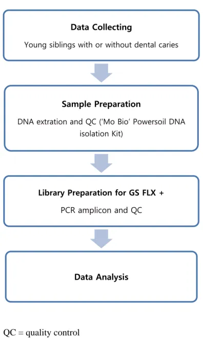
QC = quality control
Figure 1. Flow chart of experimental design
Data Collecting
Young siblings with or without dental caries
Sample Preparation
DNA extration and QC (‘Mo Bio’ Powersoil DNA isolation Kit)
Library Preparation for GS FLX +
PCR amplicon and QC
3. Sample preparation
DNA was extracted using PowerSoil® DNA Isolation Kit (MO BIO Laboratories, Carlsbad, CA, USA) according to the manufacturer‟s instructions. Briefly, samples were put in the PowerBead Tubes and homogenized with solution C1 for 10 minutes. Homogenized supernatants were transferred to 2 ml collection tubes and centrifuged with solution C2C5 four times. DNA was eluted with deionized water and stored at -20°C. DNA concentration and purity were quantified using a NanoDrop Spectrophotometer (ND-1000, Thermo Scientific, Waltham, MA, USA) by measuring absorbance at 260 and 280 nm.
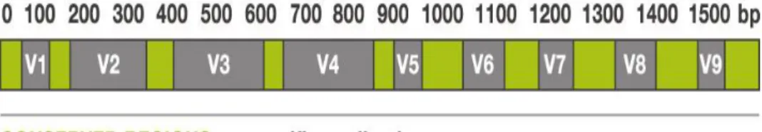
4. PCR amplification and pyrosequencing
PCR amplification was performed using primers targeting from V1 to V4 regions of the 16S rRNA gene with extracted DNA (Fig. 2 (A)). For bacterial amplification, barcoded primers of 27F (5‟-
CCTATCCCCTGTGTGCCTTGGCAGTC-TCAG-AC-AGAGTTTGATCMTGGCTCAG-3‟; underlining sequence indicates the
target region primer) and 800R
(5‟-CCATCTCATCCCTGCGTGTCTCCGAC-TCAG-X-AC-
GAGTTTGATCMTGGCTCAG-3‟; „X‟ indicates the unique barcode for each subject) was designed (Fig. 2 (B)). The amplifications were carried out under the following conditions: initial denaturation at 95 ºC for 5 min, followed by 30 cycles of denaturation at 95 ºC for 30 sec, primer annealing at 55 ºC for 30 sec, and extension at 72 ºC for 30 sec, with a final elongation at 72 ºC for 5 min. The PCR product was confirmed by using 2% agarose gel electrophoresis and visualized under a Gel Doc system (BioRad, Hercules, CA, USA). The amplified products were purified with the QIAquick PCR purification kit (Qiagen, Valencia, CA, USA). Equal concentrations of purified products were pooled together and removed short fragments (non-target products) with Ampure beads kit (Agencourt Bioscience, MA, USA). The quality and product size were assessed on a Bioanalyzer 2100 (Agilent, Palo Alto, CA, USA) using a DNA 7500 chip. Mixed amplicons were conducted emlusion PCR, and
then deposited on Picotiter plates. The sequencing was carried out at Chunlab, Inc. (Seoul, Korea), with GS FLX Plus system (Roche, Branford, CT, USA) according to the manufacturer‟s instructions.
(A)
(B)
PCR ; polymerase chain reaction
Figure 2. Variable region of 16S rRNA gene and PCR amplicon; (A) GS FLX plus uses the V1 – V4 and 27F-800R as primer; (B) Production of amplicon library
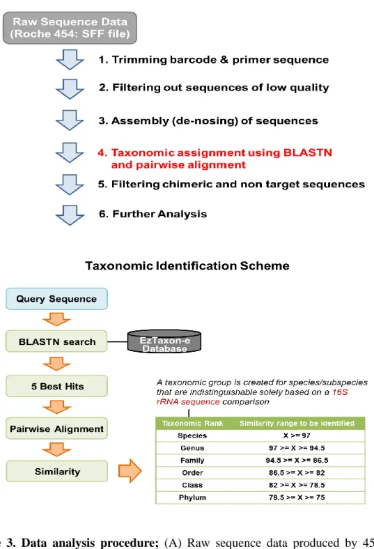
5. Pyrosequencing data analysis
The basic analysis was conducted according to the previous descriptions in other studies (Fig. 3) (Chun et al., 2010; Hur et al., 2011; B. S. Kim et al., 2012). Obtained reads from the different samples were sorted by unique barcodes of each PCR product. The sequences of the barcode, linker, and primers were removed from the original sequencing reads. Any reads containing two or more ambiguous nucleotides, low quality score (average score < 25), or reads shorter than 300bp, were discarded. Potential chimera sequences were detected by the bellerophone method, which was comparing the BLASTN search results between forward half and reverse half sequences (Huber et al., 2004). After removing chimera sequences, the taxonomic classification of each read was assigned against the EzTaxon-e database (http://eztaxon-e.ezbiocloud.net) (O. S. Kim et al., 2012), which contains 16S rRNA gene sequence of type strains that have valid published names and representative species level phylotypes of either cultured or uncultured entries in the GenBank database with complete hierarchical taxonomic classification from the phylum to the species. The richness and diversity of samples were determined by Chao1 estimation and Shannon diversity index at the 3% distance. Random subsampling was conducted to equalize read size of samples for comparing different read sizes among samples. The overall phylogenetic distance between communities was estimated using the Fast UniFrac
(Hamady et al., 2010) and visualized using principal coordinate analysis (PCoA). To compare OTUs between samples, shared OTUs were obtained with the XOR analysis of CLcommunity program (Chunlab Inc., Seoul, Korea).
(A)
(B)
Figure 3. Data analysis procedure; (A) Raw sequence data produced by 454 pyrosequencing was processed in 6 steps. (B) Detailed explanation of the taxonomic identification ((A)-4).
III.
RESULTS
1. Demographics and clinical information
7 children with caries and 1 children without caries in 7 families were participated in this research (Table 1). Mean age was 4.3 years, ranging from 1 to 10. The gender ratio was almost 1:1 (8 males and 6 females). In caries group, average DMFT index was 7.3, ranging from 5 to 15. Especially, # 51 showed the highest DMFT index, 15.
Table 1. Demographic and clinical information of the participants
Caries group Caries-free group Family group ID Age (Year) Gender DMFT ID Age (Year) Gender 1 11 1 M 6 12 4 F 2 21 4 M 5 22 2 M 3 31 5 M 6 32 3 M 4 41 4 F 5 43 6 F 5 51 3 M 15 52 2 M 6 61 6 F 6 62 3 F 7 71 10 F 8 72 7 M
Gender ; M=male, F=Female
2. Taxonomic identification and operational taxonomical unit (OTU) diversity
PCR amplification and subsequent pyrosequencing of the 16S rRNA gene hypervariable region V1-V4 of 15 samples resulted in 624,630 raw reads (Table 2). 391,325 were used for analysis after filtering. Operation taxonomical unit (OTU) diversity was an operational definition of a species or a group of species often used only when DNA sequence data were available. OTUs were defined at the 3% divergence (97% similarity) threshold using the average neighbor clustering algorithm. OTUs for all children were average 523. OTUs for the children without caries (average 562) were more than OTUs for the children with caries (average 479), but found no significant difference by independent t-test (α=0.05).
The richness and diversity of the samples were determined by Shanon diversity index at the 3% distance using taxonomy-dependent clustering - taxonomy-based clustering (TDC-TBC) method (Fig. 4). The alpha-diversity (Shannon) index of the caries group was lower than caries-free group. It demonstrated that the caries group displayed lower bacterial diversity. There was significant difference between the caries and caries-free group by Wilcoxon signed-rank test (p=0.022).
Table 2. Taxonomic identification Group ID Number of raw reads Number of final reads Number of OTU Caries 11 43,755 24,475 466 21 63,625 46,354 185 31 43,101 22,552 460 41 35,815 19,669 649 51 41,936 25,175 397 61 43,994 23,667 554 71 42,811 25,073 645 Average 45,214 26,709 482 Caries-free 12 46,346 26,448 597 22 51,476 36,767 203 32 41,503 22,366 526 42 38,726 21,400 755 52 29,112 16,591 389 62 40,570 20,858 750 72 54,760 30,959 872 Average 43,213 25,056 585 OTU = operational taxonomic units
(A)
(B)
Figure 4. Comparison of the richness and diversity of samples at 3% distance using TDC-TBC (taxonomy-dependent clustering - taxonomy-based clustering) method; In alpha-diversty (Shannon) index, the caries group showed lower bacterial diversity, but there was significant difference by Wilcoxon signed-rank test (p=0.022).
3. Comparison of the composition of the microbiome between caries and caries-free group
(1) Comparison of all samples at phylum, genus, and species level
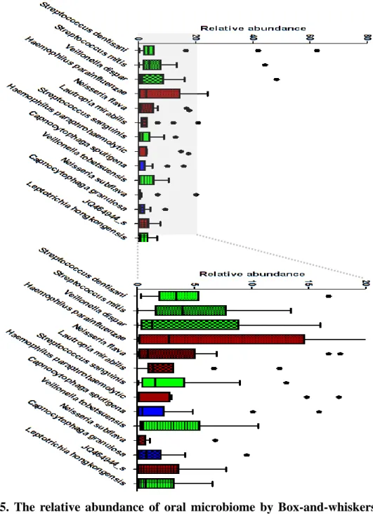
At phylum level, five major bacterial phyla, Firmicutes, Proteobacteria, Bacteroidetes, Fusobacteria and Actinobacteria, were identified (over 5% of all microbiome) (Fig. 5 (A)). In caries group, Firmicutes was increasing and Proteobacteria, Bacteroidetes, Fusobacteria, and Actinobacteria were decreasing.
At genus level, 7 major bacterial genera, Streptococcus, Veillonella, Neisseria, Haemophilus, Capnocytophaga, Leptotrichia and Actinomyces, were identified (over 5% of all microbiome) (Fig. 5 (B)). In caries group,
Streptococcus and Neisseria were increasing and Haemophilus,
Capnocytophage, and Leptotrichia were decreasing.
At species level, 14 major bacterial species, Streptococcus dentisani, S. mitis, S. sanguinis, Veillonella dispar, V. tobetsuensis, Haemophilus parainfluenzae, H. paraphrohaemolytic, Neiseria flava, N. subflava, Lautropia mirabills, Capnocytophaga sputigena, C. granulosa, Leptotrichia hongkongensis, and JQ464944, were identified (over 5% of all microbiome) (Fig. 5 (C)). In caries group, Veillonella dispar, Veillonella tobetsuensis, Veillonella rodentium, Actinomyces viscosus, and Streptococcus sanguinis were increasing and Streptococcus mitis, Capnocytophage sputiger, Capnocytophage leadbeltt and AF366267 were decreasing.
(A)
(C)
Figure 5. The relative abundance of oral microbiome by Box-and-whiskers plot; (A) Relative abundance of bacterial phyla were identified (over 5% of all microbiome). (B) At genus level, 7 major bacterial genus were identified (over 5% of all microbiome). (C) At species level, 14 major bacterial species were identified (over 5% of all microbiome).
(2) Comparison in individuals at phylum, genus, and species level
At phylum level, individuals showed various composition of microbiom. But, Firmicutes increased in the caries group of 5 families (samples # 11,21 41,51,71) (Fig. 6 (A)).
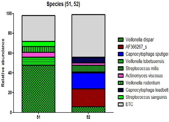
At genus level, Streptococcus increased in the caries group of 5 families (samples # 11,21,31,41,71) (Fig. 6 (B)). Also, Veillonella increased in the caries group of 3 families (samples # 11,41,51), especially # 51 showing the most DMFT index among all samples.
We compared samples # 51 and 52, becuase # 51 have the most DMFT index and it might reveal the notable ecological changes in dental caries in child (Fig. 6 (C)). At species level, 14 major bacterial species, Streptococcus dentisani, S. mitis, S. sanguinis, Veillonella dispar, V. tobetsuensis, In the sample # 51, Veillonella dispar, Veillonella tobetsuensis, Veillonella rodentium, Actinomyces viscosus, and Streptococcus sanguinis increased
and Streptococcus mitis, Capnocytophage sputiger, Capnocytophage
(A)
(C)
Figure 6. The relative abundance of oral microbiome; (A) Relative abundance of bacterial phyla were identified (over 5% of all microbiome). (B) At genus level, 7 major bacterial genera were identified (over 5% of all microbiome). (C) At species level, 9 major bacterial species were compared in sample # 51 which showed the most DMFT index and # 52 (over 5% of all microbiome).
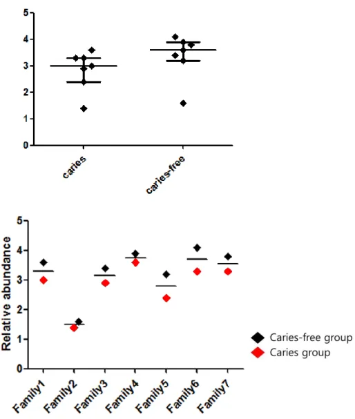
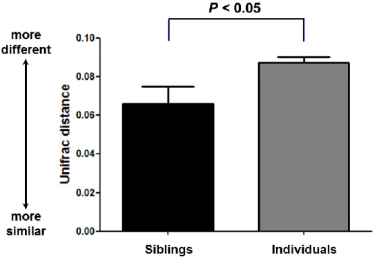
4. Correlation of the siblings
Comparison of the sharing of microbiome of siblings permited an assessment of the horizontal transmission. The metric used for comparison was the weighted UniFrac distance (Lozupone et al., 2006; Lozupone and Knight, 2005), a widely used qualitative (presence/absence) community comparison measure based on phylogenetic information. UniFrac values range from 0 (identical communities) to 1 (maximum difference). We compared siblings to unrelated individuals and there was a statistical difference by a two-tailed t-test with unequal variance (p<0.05) (Fig. 7).
Figure 7. Mean pairwise weighted UniFrac distance for subsets of samples; Mean pairwise weighted UniFrac distance reflecting siblings and individuals. Means were shown ± SEM, and p-values were calculated by a two-tailed t-test with unequal variance.
IV.
DISCUSSION
For this study of the oral microbiome, cross-sectional samples from 15 young siblings with or without dental caries were collected. Microbiome analysis was conducted using 16S rRNA gene cloning and pyrosequencing, and bacteria were identified using an oral 16S rRNA database. This open-ended approach allowed a holistic approach at the microbiome associated with caries and health in children and the horizontal transmission.
Participants in this study were children under 12 years with primary or mixed dentition. This period was the most vulnerable time to dental caries in life and the early established microbiome might influence the entire oral health (Kohler and Andreen, 2010; Kohler et al., 2003). In addition, the microbiome was changing with ages, but it was quiet stable without external stimulation before the pubescent (Crielaard et al., 2011). Dental plaques were collected in the cervico-buccal area of posterior teeth in all samples. In spite of the site-specific characteristics (Bowden et al., 1975; Van Houte and Green, 1974), we expected to identify the basic commensal or residual microbiome in the same location.
In this research, oral microbiome in carie and caries-free groups showed several similarities with the previous researches. At first, the number of OUT was lower in caries group. Several previous researches showed the diversity decreased with increasing caries within subject (Arif et al., 2008; Gross et al., 2012; Gross et al., 2010; Jiang et al., 2011). The reason was supposed that acid-senditive species
were eliminated in acidic environment produced by acidogenic specied and only a few aciduric species survived. This loss of diversity might damage the resilience of bacterial community in oral disease.
The change of microbiome in caries and caries-free group was similar with the previous researches, particularly in phylum level (Table 3). Oral microbiomes in health and caries status in previous researches were listed in Table 4. At the phylum level of caries-group, Firmicutes increased and Proteobacteria, Bacteroidetes, Fusobacteria, and Actinobacteria decreased. At the previous researches, Proteobacteria, Actinobacteria and Bacteroidetes were all health associated (Gross et al., 2010; Stahringer et al., 2012). The reason was the acidogenic and aciduric species, such as S. mutan, S. sobrinus, non-MS, and Lactobacillus, were included in Firmicutes.
At the genus level of caries-group, Streptococcus and Neisseria increased and Haemophilus, Capnocytophage, and Leptotrichia decreased. At species level of caries group, Veillonella dispar, Veillonella tobetsuensis, Veillonella rodentium, Actinomyces viscosus, and Streptococcus sanguinis increased and Streptococcus mitis, Capnocytophage sputiger, Capnocytophage leadbeltt and AF366267 decreased.
Table 3. Oral microbiome in healthy and caries status in previous researches (Corby et al., 2007; Crielaard et al., 2011; Gross et al., 2012; Jiang et al., 2014; Tanner et al., 2011; Tanner et al., 2012)
Healthy Phylum 5 Major : Bacteroidetes, Fusobacteria, Actinobacteria, Firmicutes, Proteobacteria
Genus Capnocytophage, Fusobacterium, Porphyromonas, Leptotrichia, Selenomonas, Abiotrophia, Comamonas,
Tannerella, Eikennella, Paludibacter, Treponeme,
Actinobaculum, Stenotrophomonas, Aestuariimicrobium, Peptococcus
Species Streptococcus parasanquinis, S. mitis/oralis, S. sanguinis, S. cristatus, S. salivarius, streptococcus sp.
Gemella. morbillorum, G. haemolysans, Actinomyces defective, Porphyromonas catoniae, Neisseria flavescens Caries Phylum Increasing: Firmicutes
Decreasing: Bacteroidetes, Actinobacteria, Proteobacteria Genus Streptococcus, Veillonella, Cryptobacterium,
Lactobacillus, Megasphaera, Olsenella, Scardovia,
Shuttleworthia, Cryptobacterium
Species Streptococcus mutans, , S. salivarius, S. sobrinus, S.
parasanquinis, Scardovia wiggsiae, S. exigua,
Veillonella granulicatella, Lactobacillus sp., Prevotella sp., Atopobium sp., Olsenella sp., Actinomyces sp.
Especially, Veillonella was abundant in sample # 51 showing the highest DMFT index. The genus of Veillonella has regularly been found in the oral cavity. These genera have been distinguished from each other based on phenotype, but they cannot be reliably differentiated based on 16S rRNA sequence. Veillonella rely soley on lactate and other organic acids as an evergy source (Rogosa, 1964). Veillonella was found to be significantly associated with caries in children in previous molecular studies as well (Aas et al., 2008; Gross et al., 2012; Tanner et al., 2011). Veillonella was highly correlated with the total of all known acid producing species. This was not surprising given its reliance on lactate as its nutrient source, and has potential clinical utility since Veillonella levels may serve as a sensitive biologic indicator and early warning of acid production. Gross et al. suggested that Veillonella, but not S. mutans or other acid-producing species, was the microbial risk factors, predicting future caries (Gross et al., 2012).
In contrast, the correlation of dental caries and well-known cariogenic species, such as S. mutans, S. sobrinus, and Lactobicillus was insignificant. S. mutans was higly acidogenic and aciduric, and considerable clinical and laboratory data implicates this species as the primary pathogen in human dental caries. S. sobrinus was closely related to S. mutans, and these species were often referred to collectively as the mutans streptococci. S. sobrinus was associated with caries in the current study, and appeared to be the primary pathogen in some subjects. Lactobacilli were rarely detected, and when present were at very low levels in this cohort of young children with the earliest stages of caries. Lactobacilli have
shown a robust association with more advanced caries in many studies and in older children. The reason was supposed that the location of sample collecting was cervico-buccal surface, not the caries site. If we collected at the caries progressing site, the more acidogenic and aciduric microbiome would be revealed than this result due to the site specific colonization.
We identified horizontal transmission in siblings in this research. In previous researches, intrafamilial transmission of S. mutans including horizontal transmission (Domejean et al., 2010; Hoshino et al., 2012) and vertical transmission (Caufield and Walker, 1989; Mannaa et al., 2012) was approved. However, the result of this study and the recent researches (Stahringer et al., 2012) by NGS approved the transmission of the whole oral microbiome. Even, Corby et al.(Corby et al., 2007) used the term „Heritability‟ and suggested that the relative abundance of oral microbial species was in part determined by the host genome. Heritability was the proportion of observed differences on a trait among individuals of a population that were due to genetic differences and factors including genetics, environment and random chance can all contribute to the variation between individuals in their observable characteristics (Raj and van Oudenaarden, 2008). In the same vein, the human microbiome was considered as human second genome (Grice and Segre, 2012). This heritability could be used in personalized dental medicine.
Personalized dental medicine was personalized medicine in dentistry, which is the medical model that proposes the customization of healthcare using molecular
analysis – with medical decisions, practices, and/or products being tailored to individual patient. In this model, diagnostic testing is often employed for selecting appropriate and optimal therapies based on the context of a patient`s genetic content (Eng et al., 2012). In fact, the genetic approach to the dental caries is difficult because it is multifactorial disease. But, Werneck et al. (Werneck et al., 2011) represent the genetic risk factors controlling caries susceptibility. In addition, Li et al (Li et al., 2007) revealed the genetic profiling of the oral microbiome and suggested that the diversity and complexity of the microbiome were significantly less in caries children. Host factors such as tooth and salivary factors and dietary factors were influenced by genes in some ways. (Brook, 2009; Kim et al., 2005; Poulter et al., 2013; Vitorino et al., 2005; Wendell et al., 2010; Wright et al., 2011). These genes could be the sources of personalized dental medicine for dental caries.
We also think about the prevention and management of dental caries in more holistic and ecologic aspect. The targets of specific plaque theory, which was believed in world-wide, were the blockage of transmission and the vaccination, but the transmission of microbiome including S. mutans, s. sobrinus, and Lactobacillus was natural from family or environment. We should try to reduce the number of transmitted pathologic microbiome by the improvement of hygiene in family and environment and to make the oral environment not suitable for the selective colonization of the pathologic microbiome. In addition, the acquisition timing was not limited in the „window of infectivity‟, all the time from the birth
was critical until the stable and healthy climax community was built. The timing of the climax community and the influencing factor was needed to be researched further. Also, the oral hygiene care of pregnant women has to be emphasized far more for reducing the cariogenic microbiome in empirically and theoretically.
The one of the limitations of this research was a small number of participants. In addition, the duplication or triplication of sampling would make more accurate research. And, the collected amount of sample # 21 and 22 were so small that a couple of species were dominant. Larger scale and precise research is needed for overcoming this limitation.
V.
CONCLUSIONS
In conclusion, these results showed the ecological change of oral microbiome in caries, such as the reducing of the diversity. In addition, it appeared that the horizontal transmission contributed to the colonization of oral microbiome in siblings. We suggested that the strategy of caries prevention and management have to be considered in more holistic and personalized approach.
VI.
REFERENCES
Aagaard K, Petrosino J, Keitel W, Watson M, Katancik J, Garcia N, et al. 2013. "The Human Microbiome Project strategy for comprehensive sampling of the human microbiome and why it matters." FASEB J. 27(3):1012-1022. Aas JA, Griffen AL, Dardis SR, Lee AM, Olsen I, Dewhirst FE, et al. 2008.
"Bacteria of dental caries in primary and permanent teeth in children and young adults." J Clin Microbiol. 46(4):1407-1417.
Arif N, Sheehy EC, Do T, Beighton D. 2008. "Diversity of Veillonella spp. from sound and carious sites in children." J Dent Res. 87(3):278-282.
Becker MR, Paster BJ, Leys EJ, Moeschberger ML, Kenyon SG, Galvin JL, et al. 2002. "Molecular analysis of bacterial species associated with childhood caries." J Clin Microbiol. 40(3):1001-1009.
Berkowitz RJ, Jordan HV. 1975. "Similarity of bacteriocins of Streptococcus mutans from mother and infant." Arch Oral Biol. 20(11):725-730.
Bowden GH, Hardie JM, Slack GL. 1975. "Microbial variations in approximal dental plaque." Caries Res. 9(4):253-277.
Brook AH. 2009. "Multilevel complex interactions between genetic, epigenetic and environmental factors in the aetiology of anomalies of dental development." Arch Oral Biol. 54 Suppl 1:S3-17.
Caufield PW, Ratanapridakul K, Allen DN, Cutter GR. 1988. "Plasmid-containing strains of Streptococcus mutans cluster within family and racial cohorts: implications for natural transmission." Infect Immun. 56(12):3216-3220.
Caufield PW, Walker TM. 1989. "Genetic diversity within Streptococcus mutans evident from chromosomal DNA restriction fragment polymorphisms." J Clin Microbiol. 27(2):274-278.
Chun J, Kim KY, Lee JH, Choi Y. 2010. "The analysis of oral microbial communities of wild-type and toll-like receptor 2-deficient mice using a 454 GS FLX Titanium pyrosequencer." BMC Microbiol. 10:101.
Corby PM, Bretz WA, Hart TC, Schork NJ, Wessel J, Lyons-Weiler J, et al. 2007. "Heritability of oral microbial species in caries-active and caries-free twins." Twin Res Hum Genet. 10(6):821-828.
Crielaard W, Zaura E, Schuller AA, Huse SM, Montijn RC, Keijser BJ. 2011. "Exploring the oral microbiota of children at various developmental stages of their dentition in the relation to their oral health." BMC Med Genomics. 4:22. de Soet JJ, Nyvad B, Kilian M. 2000. "Strain-related acid production by oral
streptococci." Caries Res. 34(6):486-490.
Dige I, Gronkjaer L, Nyvad B. 2014. "Molecular Studies of the Structural Ecology of Natural Occlusal Caries." Caries Res. 48(5):451-460.
Do T, Devine D, Marsh PD. 2013. "Oral biofilms: molecular analysis, challenges, and future prospects in dental diagnostics." Clin Cosmet Investig Dent. 5:11-19.
Domejean S, Zhan L, DenBesten PK, Stamper J, Boyce WT, Featherstone JD. 2010. "Horizontal transmission of mutans streptococci in children." J Dent Res. 89(1):51-55.
Emilson CG, Krasse B. 1985. "Support for and implications of the specific plaque hypothesis." Scand J Dent Res. 93(2):96-104.
Eng G, Chen A, Vess T, Ginsburg GS. 2012. "Genome technologies and personalized dental medicine." Oral Dis. 18(3):223-235.
Fitzgerald RJ, Keyes PH. 1960. "Demonstration of the etiologic role of streptococci in experimental caries in the hamster." J Am Dent Assoc. 61:9-19.
Glass RL. 1952. "The lack of relationship between salivary lactobacillus counts and dental caries activity." Oral Surg Oral Med Oral Pathol. 5(2):210-213. Green GE, Dodd MC, Inverso HS. 1957. "Comparative microflora of developing dental plaques in caries-immune and susceptible individuals." J Dent Res. 36(3):331-337.
Grice EA, Segre JA. 2012. "The human microbiome: our second genome." Annu Rev Genomics Hum Genet. 13:151-170.
Gross EL, Beall CJ, Kutsch SR, Firestone ND, Leys EJ, Griffen AL. 2012. "Beyond Streptococcus mutans: dental caries onset linked to multiple species by 16S rRNA community analysis." PLoS One. 7(10):e47722. Gross EL, Leys EJ, Gasparovich SR, Firestone ND, Schwartzbaum JA, Janies DA,
et al. 2010. "Bacterial 16S sequence analysis of severe caries in young permanent teeth." J Clin Microbiol. 48(11):4121-4128.
Group NHW, Peterson J, Garges S, Giovanni M, McInnes P, Wang L, et al. 2009. "The NIH Human Microbiome Project." Genome Res. 19(12):2317-2323.
Hamady M, Lozupone C, Knight R. 2010. "Fast UniFrac: facilitating high -throughput phylogenetic analyses of microbial communities including analysis of pyrosequencing and PhyloChip data." ISME J. 4(1):17-27.
Hegde S, Munshi AK. 1998. "Influence of the maternal vaginal microbiota on the oral microbiota of the newborn." J Clin Pediatr Dent. 22(4):317-321. Holbrook WP. 1993. "Dental caries and cariogenic factors in pre-school urban
Icelandic children." Caries Res. 27(5):431-437.
Hoshino T, Fujiwara T, Kawabata S. 2012. "Evolution of cariogenic character in Streptococcus mutans: horizontal transmission of glycosyl hydrolase family 70 genes." Sci Rep. 2:518.
Howe PR. 1917. "A Study of the Microorganisms of Dental Caries." J Med Res. 36(3):481-492 485.
Huber T, Faulkner G, Hugenholtz P. 2004. "Bellerophon: a program to detect chimeric sequences in multiple sequence alignments." Bioinformatics. 20(14):2317-2319.
Human Microbiome Project C. 2012. "Structure, function and diversity of the healthy human microbiome." Nature. 486(7402):207-214.
Hur M, Kim Y, Song HR, Kim JM, Choi YI, Yi H. 2011. "Effect of genetically modified poplars on soil microbial communities during the phytoremediation of waste mine tailings." Appl Environ Microbiol. 77(21):7611-7619.
Jiang W, Jiang Y, Li C, Liang J. 2011. "Investigation of supragingival plaque microbiota in different caries status of Chinese preschool children by denaturing gradient gel electrophoresis." Microb Ecol. 61(2):342-352. Jiang W, Ling Z, Lin X, Chen Y, Zhang J, Yu J, et al. 2014. "Pyrosequencing
analysis of oral microbiota shifting in various caries states in childhood." Microb Ecol. 67(4):962-969.
Keijser BJ, Zaura E, Huse SM, van der Vossen JM, Schuren FH, Montijn RC, et al. 2008. "Pyrosequencing analysis of the oral microflora of healthy adults." J Dent Res. 87(11):1016-1020.
Kianoush N, Adler CJ, Nguyen KA, Browne GV, Simonian M, Hunter N. 2014. "Bacterial profile of dentine caries and the impact of pH on bacterial population diversity." PLoS One. 9(3):e92940.
Kim BS, Kim JN, Yoon SH, Chun J, Cerniglia CE. 2012. "Impact of enrofloxacin on the human intestinal microbiota revealed by comparative molecular analysis." Anaerobe. 18(3):310-320.
Kim JW, Hu JC, Lee JI, Moon SK, Kim YJ, Jang KT, et al. 2005. "Mutational hot spot in the DSPP gene causing dentinogenesis imperfecta type II." Hum Genet. 116(3):186-191.
Kim OS, Cho YJ, Lee K, Yoon SH, Kim M, Na H, et al. 2012. "Introducing EzTaxon-e: a prokaryotic 16S rRNA gene sequence database with phylotypes that represent uncultured species." Int J Syst Evol Microbiol. 62(Pt 3):716-721.
Kim SH, Choi DS, Jang I, Cha BK, Jost-Brinkmann PG, Song JS. 2012. "Microbiologic changes in subgingival plaque before and during the early period of orthodontic treatment." Angle Orthod. 82(2):254-260.
Kohler B, Andreen I. 2010. "Mutans streptococci and caries prevalence in children after early maternal caries prevention: a follow-up at eleven and fifteen years of age." Caries Res. 44(5):453-458.
Kohler B, Lundberg AB, Birkhed D, Papapanou PN. 2003. "Longitudinal study of intrafamilial mutans streptococci ribotypes." Eur J Oral Sci. 111(5):383-389.
Kononen E, Kanervo A, Takala A, Asikainen S, Jousimies-Somer H. 1999. "Establishment of oral anaerobes during the first year of life." J Dent Res. 78(10):1634-1639.
Krasse B. 1988. "Biological factors as indicators of future caries." Int Dent J. 38(4):219-225.
Kulkarni GV, Chan KH, Sandham HJ. 1989. "An investigation into the use of restriction endonuclease analysis for the study of transmission of mutans streptococci." J Dent Res. 68(7):1155-1161.
Lang NP, Hotz PR, Gusberti FA, Joss A. 1987. "Longitudinal clinical and microbiological study on the relationship between infection with Streptococcus mutans and the development of caries in humans." Oral Microbiol Immunol. 2(1):39-47.
Li Y, Dasanayake AP, Caufield PW, Elliott RR, Butts JT, 3rd. 2003. "Characterization of maternal mutans streptococci transmission in an African American population." Dent Clin North Am. 47(1):87-101.
Li Y, Ge Y, Saxena D, Caufield PW. 2007. "Genetic profiling of the oral microbiota associated with severe early-childhood caries." J Clin Microbiol. 45(1):81-87.
Lima KC, Coelho LT, Pinheiro IV, Rocas IN, Siqueira JF, Jr. 2011. "Microbiota of dentinal caries as assessed by reverse-capture checkerboard analysis." Caries Res. 45(1):21-30.
Ling Z, Kong J, Jia P, Wei C, Wang Y, Pan Z, et al. 2010. "Analysis of oral microbiota in children with dental caries by PCR-DGGE and barcoded pyrosequencing." Microb Ecol. 60(3):677-690.
Loesche WJ. 1976. "Chemotherapy of dental plaque infections." Oral Sci Rev. 9:65-107.
Lozupone C, Hamady M, Knight R. 2006. "UniFrac--an online tool for comparing microbial community diversity in a phylogenetic context." BMC Bioinformatics. 7:371.
Lozupone C, Knight R. 2005. "UniFrac: a new phylogenetic method for comparing microbial communities." Appl Environ Microbiol. 71(12):8228-8235.
Mannaa A, Carlen A, Dahlen G, Lingstrom P. 2012. "Intra-familial comparison of supragingival dental plaque microflora using the checkerboard DNA-DNA hybridisation technique." Arch Oral Biol. 57(12):1644-1650.
Marchant S, Brailsford SR, Twomey AC, Roberts GJ, Beighton D. 2001. "The predominant microflora of nursing caries lesions." Caries Res. 35(6):397-406. Marsh PD. 1994. "Microbial ecology of dental plaque and its significance in
health and disease." Adv Dent Res. 8(2):263-271.
Marsh PD, Devine DA. 2011. "How is the development of dental biofilms influenced by the host?" J Clin Periodontol. 38 Suppl 11:28-35.
Masuda N, Tsutsumi N, Sobue S, Hamada S. 1979. "Longitudinal survey of the distribution of various serotypes of Streptococcus mutans in infants." J Clin Microbiol. 10(4):497-502.
Munson MA, Banerjee A, Watson TF, Wade WG. 2004. "Molecular analysis of the microflora associated with dental caries." J Clin Microbiol. 42(7):3023-3029.
Nyvad B, Crielaard W, Mira A, Takahashi N, Beighton D. 2013. "Dental caries from a molecular microbiological perspective." Caries Res. 47(2):89-102. Nyvad B, Fejerskov O. 1987. "Scanning electron microscopy of early microbial
colonization of human enamel and root surfaces in vivo." Scand J Dent Res. 95(4):287-296.
Pettersson E, Lundeberg J, Ahmadian A. 2009. "Generations of sequencing technologies." Genomics. 93(2):105-111.
Poulter JA, Brookes SJ, Shore RC, Smith CE, Abi Farraj L, Kirkham J, et al. 2013. "A missense mutation in ITGB6 causes pitted hypomineralized amelogenesis imperfecta." Hum Mol Genet.
Raj A, van Oudenaarden A. 2008. "Nature, nurture, or chance: stochastic gene expression and its consequences." Cell. 135(2):216-226.
Rogosa M. 1964. "The Genus Veillonella. I. General Cultural, Ecological, and Biochemical Considerations." J Bacteriol. 87:162-170.
Seneviratne CJ, Zhang CF, Samaranayake LP. 2011. "Dental plaque biofilm in oral health and disease." Chin J Dent Res. 14(2):87-94.
Stahringer SS, Clemente JC, Corley RP, Hewitt J, Knights D, Walters WA, et al. 2012. "Nurture trumps nature in a longitudinal survey of salivary bacterial communities in twins from early adolescence to early adulthood." Genome Res. 22(11):2146-2152.
Sullivan A, Granath L, Widenheim J. 1989. "Correlation between child caries incidence and S. mutans/lactobacilli in saliva after correction for confounding factors." Community Dent Oral Epidemiol. 17(5):240-244. Takahashi N, Nyvad B. 2008. "Caries ecology revisited: microbial dynamics and
the caries process." Caries Res. 42(6):409-418.
Takahashi N, Nyvad B. 2011. "The role of bacteria in the caries process: ecological perspectives." J Dent Res. 90(3):294-303.
Tanner AC, Kent RL, Jr., Holgerson PL, Hughes CV, Loo CY, Kanasi E, et al. 2011. "Microbiota of severe early childhood caries before and after therapy." J Dent Res. 90(11):1298-1305.
Tanner AC, Sonis AL, Lif Holgerson P, Starr JR, Nunez Y, Kressirer CA, et al. 2012. "White-spot lesions and gingivitis microbiotas in orthodontic
patients." J Dent Res. 91(9):853-858.
Tanzer JM. 1989. "On changing the cariogenic chemistry of coronal plaque." J Dent Res. 68 (Spec Iss):1576-1587.
ten Cate JM, Zaura E. 2012. "The numerous microbial species in oral biofilms: how could antibacterial therapy be effective?" Adv Dent Res. 24(2):108-111.
Theilade E. 1986. "The non-specific theory in microbial etiology of inflammatory periodontal diseases." J Clin Periodontol. 13(10):905-911.
Thenisch NL, Bachmann LM, Imfeld T, Leisebach Minder T, Steurer J. 2006. "Are mutans streptococci detected in preschool children a reliable predictive factor for dental caries risk? A systematic review." Caries Res. 40(5):366-374.
Van Houte J, Green DB. 1974. "Relationship between the concentration of bacteria in saliva and the colonization of teeth in humans." Infect Immun. 9(4):624-630.
van Houte J, Lopman J, Kent R. 1996. "The final pH of bacteria comprising the predominant flora on sound and carious human root and enamel surfaces." J Dent Res. 75(4):1008-1014.
Van Houte J, Sansone C, Joshipura K, Kent R. 1991. "Mutans streptococci and non-mutans streptococci acidogenic at low pH, and in vitro acidogenic potential of dental plaque in two different areas of the human dentition." J Dent Res. 70(12):1503-1507.
van Ruyven FO, Lingstrom P, van Houte J, Kent R. 2000. "Relationship among mutans streptococci, "low-pH" bacteria, and lodophilic polysaccharide-producing bacteria in dental plaque and early enamel caries in humans." J Dent Res. 79(2):778-784.
Vitorino R, Lobo MJ, Duarte JR, Ferrer-Correia AJ, Domingues PM, Amado FM. 2005. "The role of salivary peptides in dental caries." Biomed Chromatogr. 19(3):214-222.
Wade WG. 2011. "Has the use of molecular methods for the characterization of the human oral microbiome changed our understanding of the role of bacteria in the pathogenesis of periodontal disease?" J Clin Periodontol. 38 Suppl 11:7-16.
Wade WG. 2013. "The oral microbiome in health and disease." Pharmacol Res. 69(1):137-143.
Wendell S, Wang X, Brown M, Cooper ME, DeSensi RS, Weyant RJ, et al. 2010. "Taste genes associated with dental caries." J Dent Res. 89(11):1198-1202. Werneck RI, Lazaro FP, Cobat A, Grant AV, Xavier MB, Abel L, et al. 2011. "A major gene effect controls resistance to caries." J Dent Res. 90(6):735-739. Wright JT, Torain M, Long K, Seow K, Crawford P, Aldred MJ, et al. 2011. "Amelogenesis imperfecta: genotype-phenotype studies in 71 families." Cells Tissues Organs. 194(2-4):279-283.
Xu P, Gunsolley J. 2014. "Application of metagenomics in understanding oral health and disease." Virulence. 5(3):424-432.
Zaura E. 2012. "Next-generation sequencing approaches to understanding the oral microbiome." Adv Dent Res. 24(2):81-85.
Zaura E, Keijser BJ, Huse SM, Crielaard W. 2009. "Defining the healthy "core microbiome" of oral microbial communities." BMC Microbiol. 9:259. Zhang XH, Zhou Y, Zhi QH, Tao Y, Lin HC. 2012. "Genetic polymorphisms of
the sortase A gene and early childhood caries in two-year-old children." Arch Oral Biol. 57(7):948-953.
국문요약
차세대시퀀싱을 이용한
형제간의 구강마이크로바이옴의 비교연구
연세대학교 대학원 치의학과 이 효 설 지도교수 : 이제호 본 연구의 목적은 치아우식증의 유무가 다른 형제에서의 구강미생물균을 차 세대시퀀싱으로 동정하여 비교하는 것으로 건강한 상태와 치아우식증에서의 생태학적 변화를 조사하고 수평적 전이를 확인하는 것이다. 구강미생물의 조 성을 파악하기 위해, 7 가족에서 14명의 어린이(평균 4.6 세)가 참여하였고, 7 명은 치아우식증이 있고 7명은 치아우식증이 없었다. 구치부 협측치경부에서 치은연상치태를 채취하였다. 시료를 16S rRNA 유전자의 V1-V4 부위에 대해 파이로시퀀싱(pyrosequencing)으로 분석하였다. 개개인은 자신의 형제와 더 비슷한 결과를 보였다. 문(門, Phylum) 영역에서는 Firmicutes이 증가하였고,다. 속(屬, Genus) 영역에서는 Streptococcus가 증가하고, Haemophilus와
Capnocytophage, Leptotrichia가 감소하였다. 종(種, Species) 영역에서는
Veillonella dispar와 Veillonella tobetsuensis, Veillonella rodentium,
Actinomyces viscosus가 증가하고 Streptococcus mitis와 Capnocytophage
sputiger, Capnocytophage leadbeltt, AF366267가 감소하였다. 형제간의 유
사성은 UniFrac distance로 유의성있게 나타났다 (p<0.05). 결론적으로 본 연 구의 결과는 이전의 연구와 유사하게 치아우식증 발생에 있어 구강미생물의 생태학적 변화를 보였다. 또한, 수평적 전이가 형제간의 구강미생물의 집락에 영향을 주는 것으로 나타났다. 치아우식증의 예방과 관리 전략을 수립할 때 보다 더 전체적인 접근이 필요하다. 핵심되는 말: 구강미생물, 차세대시퀀싱, 파이로시퀀싱, 치아우식증, 형제, 생 태학적 치태가설, 수평적 전이