DEVELOPED AND ENDORSED BY:
American Association of Medical Dosimetrists (AAMD) American Association of Physicists in Medicine (AAPM) American Board of Radiology (ABR)
American Brachytherapy Society (ABS) American College of Radiology (ACR)
American College of Radiation Oncology (ACRO) American Radium Society (ARS)
American Society for Radiation Oncology (ASTRO) American Society of Radiologic Technologists (ASRT)
Association of Freestanding Radiation Oncology Centers (AFROC) Society of Chairmen of Academic Radiation Oncology Programs (SCAROP)
Society for Radiation Oncology Administrators (SROA)
T A R G E T I N G C A N C E R C A R E
Safety
is no
accident
A F R A M E W O R K F O R
Q U A L I T Y R A D I AT I O N
O N C O L O G Y A N D C A R E
S P O N S O R E D B YSafety
is no
accident
A F R A M E W O R K F O R
Q U A L I T Y R A D I AT I O N
O N C O L O G Y A N D C A R E
SPONSORED BY:
American Society for Radiation Oncology (ASTRO)
DEVELOPED AND ENDORSED BY:
American Association of Medical Dosimetrists (AAMD) American Association of Physicists in Medicine (AAPM) American Board of Radiology (ABR)
American Brachytherapy Society (ABS) American College of Radiology (ACR)
American College of Radiation Oncology (ACRO) American Radium Society (ARS)
American Society for Radiation Oncology (ASTRO) American Society of Radiologic Technologists (ASRT)
Association of Freestanding Radiation Oncology Centers (AFROC)
Society of Chairmen of Academic Radiation Oncology Programs (SCAROP) Society for Radiation Oncology Administrators (SROA)
i i S A F E T Y I S N O A C C I D E N T | 2 0 1 2
SAFETY IS NO ACCIDENT: A Framework for Quality Radiation Oncology and Care
Technologic advances and systemic changes in health care delivery mean that the fi eld of radiation oncology and its processes of care are in continuous evolution. Th ese changes must be refl ected in this book and so a mechanism for timely review and revision is necessary. Th e radiation oncology intersociety meeting is held biennially, bringing together the participating societies to discuss issues of importance to the fi eld. As planning begins for each intersociety meeting, this safety document will be reviewed to assess whether a signifi cant update is needed. If so, the update will become the subject of the next intersociety meeting.
Th e content in this publication is current as of the publication date. Th e information and opinions provided in the book are based on current evidence and consensus in the radiation oncology community. However, no such guide can be all-inclusive, and, especially given the evolving environment in which we practice, the recommendations and information provided in the book are subject to change and are intended to be updated over time.
Th is book is made available to ASTRO and endorsing organization members and to the public for educational and infor-mational purposes only. Any commercial use of any content in this book without the prior written consent of ASTRO is strictly prohibited. Th e information in the book presents scientifi c, health and safety information and may to some extent refl ect ASTRO and the endorsing organizations’ understanding of the consensus scientifi c or medical opinion. ASTRO and the endorsing organizations regard any consideration of the information in the book to be voluntary. All radiation oncology medical practice management and patient care decisions, including but not limited to treatment planning and implementation; equipment selection, maintenance and calibration; staffi ng and quality assurance activities, are exclusively the responsibility of duly licensed physicians and other practitioners. Th e ultimate determination regarding the practices utilized by each provider must be made by the provider, considering any local, state or federal laws and certifi ca-tion and/or accreditaca-tion standards that apply to the provider’s practice, the applicable policies, rules and regulaca-tions of third-party payors, their own institution’s policies, procedures, and safety and quality initiatives, and their independent medical judgment.
Th e information and opinions contained in the book are provided on an “as-is” basis; users of the information and opinions provided by the book assume all responsibility and risk for any and all use. Neither ASTRO, nor any endorsing organization, gives any warranty, express or implied, as to the accuracy, reliability, utility or completeness of the information or opinions provided in the book or provided in response to user inquiry. Neither ASTRO, nor any endorsing organization, nor any ASTRO or endorsing organization’s offi cers, directors, agents, employees, committee members or other representatives, shall have any liability for any claim, whether founded or unfounded, of any kind whatsoever, including but not limited to any claim for costs and legal fees, arising from the use of these opinions.
Introduction 1
Chapter 1: The Process of Care in Radiation Oncology
3
1.1
Patient
Evaluation
3
1.2
Preparing
for
Treatment
4
1.3
Radiation
Treatment
Delivery
8
1.4
Radiation
Treatment
Management
9
1.5 Follow-up Evaluation and Care
9
Chapter 2: The Radiation Oncology Team
11
2.1 Roles and Responsibilities
11
2.2
Qualifi cations and Training
11
2.3 Continuing Education and Maintenance of Certifi cation
13
2.4
Staffi
ng Requirements
14
Chapter 3: Safety
19
3.1
Th
e Need for a Culture of Safety
19
3.2 Leadership and Empowering Others
19
3.3 Evolving Roles and Responsibilities of Each Team Member
20
3.4 Examples of Tools/Initiatives to Facilitate Safety and the Safety Culture
20
3.5 Ingraining Safety into Everyday Practice
24
3.6 Collaborations Between Users and Vendors
25
3.7 Involving Th
ose Beyond Radiation Oncology
26
Chapter 4: Management and Assurance of Quality in Radiation Oncology
29
4.1 Quality Requirements of Radiation Oncology Programs
29
4.2 Patient-Related Quality Management
38
Appendix I: Acronym Glossary
51
i v S A F E T Y I S N O A C C I D E N T | 2 0 1 2
WRITING CHAIRMEN
Anthony L. Zietman, MD, Massachusetts General Hospital Jatinder R. Palta, PhD, University of Florida/Davis Cancer Center Michael L. Steinberg, MD, UCLA Center for Health Sciences
AUTHORS
Albert L. Blumberg, MD, Greater Baltimore Medical Center (ACR)
R. Alan Burns, BS, RT, Mercy St. John’s Hospital, O’Reilly Cancer Center (SROA) Susan W. Cagle, MS, CMD, Piedmont Hospital, Atlanta (AAMD)
Laura A. Dawson, MD, Princess Margaret Hospital (ASTRO)
Vanna Marie Dest, MSN, APRN, BC, AOCN, Hospital of Saint Raphael (ASTRO) Th eodore L. DeWeese, MD, Johns Hopkins University (SCAROP)
Benedick Fraass, PhD, University of Michigan (ASTRO)
James M. Galvin, DSc, Th omas Jeff erson University Hospital (ASTRO) Arve W. Gillette, MD, Texas Oncology (ACRO)
Bruce G. Haff ty, MD, UMDNJ Robert Wood Johnson Medical School (ABR)
Sandra E. Hayden, MA, RT(T), Th e University of Texas MD Anderson Cancer Center (ASRT) Peter A. S. Johnstone, MD, Indiana University School of Medicine (ACR, ARS)
Lisa Ann Kachnic, MD, Boston University (ABR)
Th eresa Kwiatkowski, BS, RT(T), CMD, Lipson Cancer Center (AAMD) Lawrence Marks, MD, University of North Carolina (ASTRO) Karen McGraner, Insight Oncology, Inc. (SROA)
Michael D. Mills, PhD, University of Louisville (AAPM)
Christopher J. Moore, BS, CMD, Virtua Memorial Hospital (AAMD) Bhudatt R. Paliwal, PhD, University of Wisconsin (ASTRO) Daniel Pavord, MS, Vassar Brothers Medical Center (AAPM) David J. Rice, MD, 21st Century Oncology (AFROC) Mark J. Rivard, PhD, Tufts Medical Center (ABS)
Seth A. Rosenthal, MD, Radiological Associates of Sacramento (ACR) Christopher F. Serago, PhD, Mayo Clinic (AAPM)
Giles C. Toole III, MS, Tallahassee Memorial Hospital (SROA) Prabhakar Tripuraneni, MD, Scripps Clinic (ASTRO)
Akila N. Viswanathan, MD, MPH, Brigham and Women’s Hospital/Dana-Farber Cancer Center (ABS) Paul Wallner, DO, 21st Century Oncology (ABR)
David E.Wazer, MD, Tufts Medical Center (ACRO)
uring the later part of the twentieth century, the “Blue Book” had a unique importance in defi ning the shape of a modern radiation oncology department. It set standards regarding personnel, equipment and quality assurance and has been an invaluable guide for department chairs and practice leaders. Twenty years have elapsed since the last edition was published and during that time the world of radiation oncology has changed beyond measure. Th ese two decades have seen an unprecedented expansion in the technological tools at our disposal with clear benefi ts to our patients. At the same time, however, the “Great Expansion” has added the challenge of deep complexity to our planning and treatment delivery. Th ese decades have also been associated with a vigorous awareness of safety in medicine generally and radiation oncology in particular. Th is movement is pushing the practice of medicine toward integrated teamwork and eff ective, simple, quality assurance procedures. Th e safe delivery of radiation therapy was never a simple matter and is now exceedingly
complex. Th is new document is designed to address the specifi c requirements of a contemporary radiation oncology facility in terms of structure, personnel and technical process in order to ensure a safe environment for the delivery of radiation therapy. It was developed through collaboration between all of the major societies in the fi eld representing physicians, medical physicists, radiation therapists, medical dosimetrists, nurses and administrators. It explicitly sets a high bar below which no radiation oncology facility should operate, and it foresees that the bar will be raised further in the years ahead. Th is book is unapologetic in its strong stance because, as the title states, safety is no accident. It comes from well-run facilities with good processes operating harmoniously within their capabilities. We recognize that some with smaller facilities may fi nd the standards set here hard to achieve but we do not believe that they are impossible. We recognize that, in a declining economy, these high bars may prove a challenge but we believe this interdisciplinary document will help facility leaders advocate on behalf of patients from a position of strength. Th e authors wish this book to be a living manifesto of the specialty’s dedication to patient safety and, after initial publication, will place it on the web with regular updating to follow.
Anthony L. Zietman, MD Jatinder R. Palta, PhD Michael L. Steinberg, MD
2011-2012 “Safety Is No Accident” Writing Chairmen
Introduction
D
2 S A F E T Y I S N O A C C I D E N T | 2 0 1 2
he “process of care” in radiation oncology refers to a conceptual framework for guaranteeing the appropriateness, quality and safety of all patients treated with radiation for therapy. Each of the aspects of the process of care in radiation oncology requires knowledge and training in the natural history of cancer, certain benign disease processes, radiobiology, medical physics and radiation safety that can only be achieved by Board certifi cation in radiation oncology (or equivalent training), to synthesize and integrate the necessary knowledge base to safely and completely render care. Th is high level of training and Board certifi cation applies as a recommendation for all of the specialists on the radia-tion oncology team. Th e medical therapeutic application of ionizing radiation is irreversible, may cause signifi cant morbidity and is potentially lethal. Use of ionizing radiation in medical treatment, therefore, requires direct or personal physician management, as the leader of the radiation oncology team, as well as input from various other essential coworkers.
Th e radiation oncology process of care can be separated into the following fi ve operational categories. • Patient Evaluation
• Preparing for Treatment o Clinical Treatment Planning o Th erapeutic Simulation o Dosimetric Treatment Planning
o Pretreatment Quality Assurance and Plan Verifi cation
• Radiation Treatment Delivery • Radiation Treatment Management • Follow-up Evaluation and Care
Leader:James Galvin
Albert L. Blumberg Kevin Camphausen Sandra E. Hayden Michael Kuettel Mark J. Rivard Seth A. Rosenthal
Giles C. Toole III
The Process of Care In
Radiation Oncology
CHAPTER 1
A course of radiation therapy is a function of the in-dividual patient situation, composed of a series of distinct activities of varying complexity. All components of care involve intense cognitive medical evaluation, interpreta-tion, management and decision-making by the radiation oncologist and other members of the clinical team. Each time a component of care is completed and reported, it should be appropriately documented in the patient record.
Th e clinical team, led by the radiation oncologist, provides the medical services associated with the process of care. Other team members involved in the patient’s plan-ning and treatment regimen include the medical physicist, medical dosimetrist, radiation therapist and nursing staff . Many of the procedures within each phase of care will be carried to completion before the patient’s care is taken to the next phase. Others will occur and recur during the course of treatment, and they are by necessity repeated during treatment due to patient tolerance, changes in tumor size, need for boost fi elds or port size changes, protection of normal tissue or as required by other clinical circumstances (that is, certain procedures may need to occur multiple times during the treatment course).
1.1.0 PATIENT EVALUATION
Patient evaluation is a service provided by a physician at the request of another physician, the patient, or an appropriate source, and is intended to recommend care for a specifi c condition or problem, including further work-up, or to recommend treatment. Th e radiation oncologist, as part of this process, will review the pertinent history, patient complaints, physical fi ndings, imaging studies, pathology and lab fi ndings. If treatment is recommended and accepted, this patient visit, or a return visit, should
T
4 S A F E T Y I S N O A C C I D E N T | 2 0 1 2
also be used for patient counseling, informed consent, coordinating care and making recommendations about other aspects of oncologic management or staging.
Th e evaluation with the radiation oncologist will often be followed by discussions with other members of the multidisciplinary care team, as indicated. Th is will include a review of details regarding pathology, disease extent based on radiographic imaging and other procedures, and potential sequencing of treatment modalities either used or planned, including surgery, chemotherapy, hormonal therapy or molecular targeted therapy. Full details of the patient evaluation are beyond the scope of this safety document.
1.2.0 PREPARING FOR TREATMENT
1.2.1 Clinical Treatment Planning
Clinical treatment planning is a comprehensive, cogni-tive eff ort performed by the radiation oncologist for each patient undergoing radiation treatment. Th e radiation oncologist is responsible for understanding the natural history of the patient’s disease process, conceptualization of the extent of the disease relative to the adjacent normal anatomical structure, integration of the patient’s overall medical condition and associated comorbidities. Knowl-edge of the integration of chemotherapeutic and surgical treatment modalities with radiation therapy is essential. An understanding of the integration of the various radiation treatment modalities is an essential part of this phase in the process of care.
Clinical treatment planning for either external beam radiation therapy (EBRT) or brachytherapy is an impor-tant step in preparing for radiation oncology treatment. Th is planning includes several components: determining the disease-bearing areas based on the imaging studies described above and pathology information; identifying the type (brachytherapy, photon beam, particle beam, etc.) and method of radiation treatment delivery (intensity modulated radiation therapy [IMRT], intensity modulated proton therapy [IMPT], three-dimensional conformal radiation therapy [3-D CRT], two-dimensional conformal radiation therapy [2-D CRT], low-dose-rate [LDR] or high-dose-rate [HDR] brachytherapy, etc.); specifying areas to be treated; and specifying dose and dose fractionation. In developing the clinical treatment plan, the radiation oncologist may use information obtained from the patient’s initial clinical evaluation, as well as the additional tests, studies and procedures described above that are necessary to complete treatment planning. Studies ordered as part of clinical treatment planning may or may not be associated with studies necessary for staging the cancer, and may be
needed to obtain specifi c information to accomplish the clinical treatment plan. In this regard, the radiation on-cologist must consider toxicities and tolerances associated with defi nitive radiation therapy or combined-modality therapy. Review is needed of imaging studies and lab tests to determine treatment volume and critical structures, commonly referred to as organs at risk (OARs), in close proximity to the treatment area or more distant and receiving a dose of radiation that needs monitoring.
For either EBRT or brachytherapy, clinical treatment planning results in a complete, formally documented and approved directive. Details including total desired dose to all targets and OARs, fractionation, treatment modality, energy, time constraints and all other aspects of the radia-tion prescripradia-tion are recorded in a written or electronic format and must be provided by the radiation oncologist prior to the start of treatment planning. In some cases, this prescription can require modifi cation based on the results of the treatment planning process.
1.2.2 Therapeutic Simulation, Fabrication of Treatment Devices and Preplanning Imaging Simulation is the process by which the geometry of the treatment device in juxtaposition to the patient is simulated for the purpose of developing an accurate and reproducible treatment delivery plan. For this purpose, it is necessary initially to acquire radiographic images of the patient in the preferred treatment position. Selecting a comfortable and appropriate patient position for treatment is an important part of the simulation process. Th e selected position should consider the location of the target and anticipated orientation of the treatment beams. Appropri-ate immobilization devices provide comfort, support and reproducibility.
In some cases the exact treatment position cannot be duplicated for some imaging procedures that are not under direct control of the radiation oncology team; clinical considerations should be made to compensate for such diff erences.
Some computed tomography (CT)-Simulator devices include the ability to register imaging datasets. However, most image registration is still performed manually with rigid datasets. Treatment planning systems can also provide the software for this capability. Using the software included with the treatment planning system shifts this part of the process to the treatment planning phase of the overall care process.
1.2.2.1 Team Interaction
Th e radiation oncology team, under the leadership of the radiation oncologist, works to deliver irradiation safely and reproducibly. Most radiation treatments use standard
operating procedures (SOPs) describing the treatment approach. Th ese SOPs are considered to be an essential component of any radiation oncology department. In those situations where the patient presents with target and critical sensitive structure geometric relationships that are not easily handled using available SOPs, the involve-ment of the radiation therapy team is necessary. Team interactions that include the radiation therapists, medical dosimetrists, medical physicists and representation from the departmental nursing staff can prove helpful in specifi c situations. Th e purpose of these meetings is to carefully consider how treatments might be tailored to a particular patient’s situation.
1.2.2.2 Fabrication of Immobilization Treatment Devices
Immobilization of the patient in a comfortable position for treatment might involve the construction or selection of certain treatment devices, facilitating accurate treatment delivery. Th is step must take into account the potential treatment planning considerations so that the immobi-lization aids do not restrict the treatment techniques. A personalized approach is required here, taking into consid-eration each patient’s unique anatomy, at times requiring special accommodations appropriate for the individual case-specifi c concerns.
1.2.2.3 Therapeutic Simulation for EBRT
Simulation is the process of determining critical informa-tion about the patient’s geometry, to permit safe and repro-ducible treatments on a megavoltage machine. Simulation for external beam radiation treatment is imaging based. Most simulation procedures have now shifted away from the direct use of the treatment beam to using X-rays in the diagnostic range of energies. In general, this part of the overall process of care reveals the relationship between the position of the target, or targets, and the surrounding critical structures. It is helpful here to think of the simula-tion step as the imaging needed for input to the treatment planning process. Th ese images can be obtained in a large number of ways. Modern conventional simulators, like the CT-Simulator, can include the ability to produce volumetric data in addition to 2-D images. Intravenous contrast should be used during simulation, as indicated, to improve enhancement within both target and normal tissues/structures.
Preparing for EBRT treatment can also depend on other imaging modalities that are directly or indirectly introduced in the simulation process. In some cases, extra time and eff ort are required to directly incorporate the information available from other imaging modalities. For example, magnetic resonance (MR) and/or positron
emission tomography (PET) are now available, and treat-ment planning systems that include image registration capabilities allow combining of information from other imaging modalities with the standard CT dataset obtained during simulation in appropriate situations. It is now pos-sible to produce image datasets that quantify the motion of structures and targets due to respiration, cardiac motion and physiologic changes in the body. Th ese four-sional (4-D) datasets include time as the fourth dimen-sion and are used for motion management techniques like respiratory tracking or gating. Ultrasound imaging has a role in both EBRT and brachytherapy. Ultrasound comes into play as a preplanning imaging technique and can also be used as an image guided radiation therapy (IGRT) tech-nique during the treatment delivery and verifi cation steps of the care process.
1.2.2.4 Therapeutic Simulation for Brachytherapy
For certain brachytherapy procedures, preparing for treat-ment is similar to the procedure described above for EBRT. Th e simulation portion for this treatment modality is also imaging based, and can involve either planar X-rays or CT scans. Other imaging modalities may be important for some brachytherapy procedures and these studies can be obtained as part of the preplanning imaging process.
1.2.2.5 Treatment Planning for Radiation Therapy Using Unsealed Sources
For clinical situations where therapy using unencapsulated radionuclides is indicated, a distinct treatment planning process is necessary due to its multidisciplinary execution. Th e process can involve calculations of the anticipated dose distribution to the target organ or tumor(s) based on knowledge of the patient’s vascular anatomy or biologi-cal imaging, such as nuclear medicine scans. Th is process should include multidisciplinary evaluation of the patient and consideration of clinical indications and radiation safety precautions. Th e American College of Radiology (ACR)/American Society for Radiation Oncology (ASTRO) Practice Guidelines regarding the Performance of Th erapy with Unsealed Radiopharmaceutical Sources and NRC Guidelines discuss these issues in greater detail[1].
1.2.3 Dosimetric Treatment Planning
Th e computer-aided integration of the patient’s unique anatomy, the desired radiation dose distribution to the tumor and normal tissues inside the patient, and the tech-nical specifi cations of the treatment delivery device yield a work product referred to as the dosimetric treatment plan. Th e plan is a programmed set of instructions for the linear accelerator or brachytherapy device whereby a combination of external beams or internal source
position-6 S A F E T Y I S N O A C C I D E N T | 2 0 1 2
ing will administer the intended dose of radiation to the target volume while minimizing the exposure of normal tissues. Accordingly, before the medical dosimetrist begins the treatment planning process, the radiation oncologist needs to defi ne the target volumes and dose limiting organs and structures on the diagnostic images obtained during simulation.
Th e skills of the appropriately trained and credentialed medical dosimetrist and medical physicist relate to the effi cient and eff ective use of the complex treatment plan-ning system hardware and software. Th ese individuals must also understand the clinical aspects of radiation oncology in order to interact with the radiation oncologist during the planning process. Th e role of the medical physicist is to guarantee proper functioning of the hardware and software used for the planning process, consult with the radiation oncologist and medical dosimetrist, check the accuracy of the selected treatment plan and perform measurements and other checks aimed at assuring accurate information exchange between radiation therapy devices and delivery of the treatment plan.
At various steps in the treatment planning process, the radiation oncologist is presented with one or more treatment plans for evaluation. Th e plans are evaluable by a combination of graphic visual representations of the radiation dose distribution inside the patient and quantita-tive statistics describing the dose to the tumor and normal tissue of interest. Th e radiation oncologist must then decide whether to accept or reject a given plan. Typically, this process is iterative and requires multiple revisions and adjustments to the initial plan in order to achieve a dose distribution that is both clinically acceptable and techni-cally feasible. Th e radiation oncologist is responsible for selecting and formally approving the plan ultimately selected for treatment.
1.2.4 Pretreatment Quality Assurance (QA) and Plan Verifi cation
Th e QA steps taken after treatment planning is completed and before the start of treatment are critical for guarantee-ing patient safety. An important initial step is an indepen-dent calculation of the machine output setting (monitor units) for external beam radiation therapy or radioactive source dwell times for brachytherapy. Th is recalculation may be accomplished as a manual point dose verifi cation in the center of the treatment volume based on printed tables relating the eff ective fi eld size to the administered dose at given depths from the surface. Alternatively, this can be performed in a computer-assisted manner, whereby data from the patient’s planning images are entered into a separate software program along with parameters describ-ing the prescription dose to the tumor and beam or source
arrangements. In either case, the key result is confi rma-tion of linear accelerator output settings or brachytherapy dwell times that reduce the risk of error related to an input mistake in the initial treatment planning software opera-tion. If an independent calculation method is not available, then an appropriate measurement technique should be used. Th e radiation onologist ensures that a pretreatment quality assurance program is in place and followed for every patient.
In the past, treatment verifi cation consisted of fi eld aperture imaging using radiographic fi lm. Th ese images are referred to as portal images or port fi lms. Th ese images are now frequently obtained using electronic portal imaging devices (EPIDs). With the introduction of IMRT, imaging of individual apertures is no longer practical. However, the traditional method of verifying the plan isocenter position using orthogonal imaging is often used for both 3-D CRT and IMRT. For IMRT, this important QA technique is not considered to be completely suffi cient to guarantee patient safety. In addition to this isocenter check procedure, patient-specifi c QA measurements are also required for IMRT and other complex delivery techniques that use in-verse treatment planning. In terms of clearly organizing the diff erent steps in the process of care for radiation oncology, a blurring of the separation between the verifi cation step described in this subsection and the treatment delivery step described in section 1.3.0 occurs on the fi rst day of treat-ment and whenever the treattreat-ment plan is changed. While patient-specifi c QA measurements are obtained prior to the start of treatment, dosimeters are sometimes also placed on the patient as a verifi cation of correct dose delivery. Th e information gathered on the fi rst day of treatment, if within acceptable limits, allows the treatment to continue for all fractions using the same treatment plan.
IGRT equipment is now available for checking the patient setup on the treatment table immediately prior to treatment delivery and then adjusting the patient position as needed to localize the target volume precisely within the volume that receives the prescription dose. Th is equipment can be used to verify the patient setup daily and
can supplement port fi lm information. IGRT has the advantage that it sometimes provides volumetric imaging capabilities. Th e extra setup accuracy provided by IGRT can allow for the use of treatment plans that reduce the volume of normal tissue around the tumor receiving a high dose of radiation, since there is less uncertainty in the target volume location. Th is process goes well beyond the simple plan verifi cation process discussed further in the treatment delivery section.
For either portal imaging or isocenter verifi cation imaging (using volumetric or planar images), it is necessary to have a reference image for comparison. Th is information
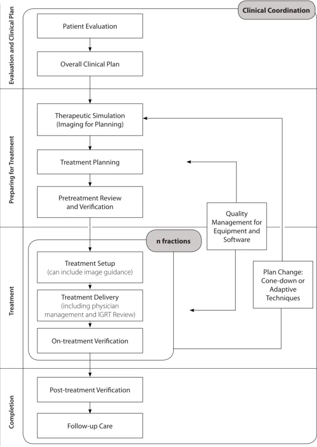
Figure 1.1. Process of Care for External Beam Radiation Therapy Patient Evaluation Quality Management for Equipment and Software Plan Change: Cone-down or Adaptive Techniques Overall Clinical Plan
Therapeutic Simulation (Imaging for Planning)
Treatment Planning
Pretreatment Review and Verifi cation
Treatment Setup (can include image guidance)
Treatment Delivery (including physician management and IGRT Review)
On-treatment Verifi cation
Post-treatment Verifi cation
Follow-up Care n fractions Clinical Coordination C ompletion T rea tment P reparing f or T rea tment E v alua
8 S A F E T Y I S N O A C C I D E N T | 2 0 1 2
1.3.3 Calibration Procedures, Ongoing Equipment QA and Preventive Maintenance
Th e initial commissioning, ongoing performance evalu-ation and periodic calibrevalu-ation of radievalu-ation treatment delivery devices are important tasks that are vital to the safe administration of radiation therapy. In general, it is the medical physicist who is primarily responsible for the device evaluations necessary for compliance with applicable state and federal regulations concerning radiation treat-ment delivery technology. Th e American Association of Physicists in Medicine (AAPM) has published extensive guidelines on the conduct of these duties and regularly updates its educational materials when new technologies enter into standard clinical practice. Th e radiation oncolo-gist, medical physicist and other members of the radiation therapy team should maintain a clear channel of commu-nication on this issue of treatment device performance so that any possible sign of impending machine malfunction is quickly recognized and diagnosed, and any necessary corrective or reparative action is taken prior to use of the machine to deliver a clinical treatment to a patient. is obtained from the imaging that is performed during the
therapeutic simulation step in the process.
Th e QA process must include other steps that are aimed at checking the accuracy of both the dose calcula-tions and the data used for treatment through the complete chain of systems (e.g., CT-Simulator to treatment planning to record and verify to accelerator control computer).
Another important step in the QA part of the process is the performance of secondary monitor unit calculations to check the primary calculation used to treat the patient.
1.3.0 RADIATION TREATMENT DELIVERY
1.3.1 External Beam Radiation Therapy
With treatment plan and treatment portal verifi cation complete, the patient is ready for treatment. Th e initial step in this part of the process is patient setup on the treatment table using several diff erent techniques, such as simple skin marks or a room laser system that localizes the treatment unit isocenter in space. Alternatively, the IGRT system may be used on each day of treatment.
Radiation treatment delivery includes various meth-ods, modalities and complexities of radiation therapy. Th e physician is responsible for verifi cation and documentation of the accuracy of treatment delivery as related to the initial treatment planning and setup procedure.
IGRT may be performed to ensure accurate targeting of precise radiation beams where certain needs of dose and organs at risk (OARs) tolerance exist. IGRT corrects for the positioning errors encountered when an internal target can move from day to day and can be reliably identifi ed. Th e physician is responsible for the supervision and review of these images and shifts in order to ensure the therapy delivered conforms to the original clinical and dosimetric plans. Similarly, management of organ motion during treatment delivery, when indicated, is the responsibility of the treating physician (Figure 1.1, see page 7).
Th e overall clinical plan can involve selection of chemotherapy, surgery, EBRT, brachytherapy or a combi-nation of modalities. Adaptive techniques can involve a modifi cation to the initial treatment plan to adjust for an observed change.
1.3.2 Brachytherapy
Brachytherapy involves the temporary or permanent placement of radioactive material inside or immediately adjacent to a tumor-bearing region. One example is perma-nent seed implants for prostate cancer, either as defi nitive therapy for early stage disease or as a boost treatment following external beam treatment for intermediate- or high-risk disease (Figure 1.2).
Figure 1.2.
Process of Care for Brachytherapy Patient Evaluation
Follow-up Evaluation and Care Clinical Treatment Planning
Imaging for Planning Treatment Planning
Pretreatment Review and Verifi cation
Treatment Delivery (implant, applicator, seeds, other) Quality Management for equipment and software Review dosimetry and adjust plan
1.4.0 RADIATION TREATMENT
MANAGEMENT
Radiation treatment management encompasses the radiation oncologist’s overall management of the course of treatment and care for the patient as well as checks and approvals provided by other members of the radiation therapy team that are necessary at various points in the process. For the radiation oncologist, radiation treatment management requires and includes a minimum of one examination of the patient by the physician for medical evaluation and management. Th e professional services furnished during treatment management may include: • Review of portal images
• Review of dosimetry, dose delivery and treatment parameters
• Review of patient treatment setup
• Patient evaluation visit (described in section 1.1.0) Not all of these parameters of treatment management are required for all patients for each week of management (except for the patient evaluation visit) because the clinical course of care may diff er due to variation in treatment modality and individual patient requirements. For exam-ple, use of port fi lms may vary based on certain technical characteristics (i.e., electron beams) and modifi cation of dose delivery can vary based on individual patient needs, depending on the patient’s tolerance of therapy or variation in tumor response. Examinations and evaluations may be required more often than weekly.
It should be emphasized that weekly treatment management requires the integration of multiple medical and technical factors, which may be required on any day through the treatment course. While nurses and nonphysi-cian providers can eff ectively participate in the manage-ment of patients receiving radiation therapy, typically by helping to manage side eff ects associated with the treatment (Table 2.1, see page 12), their eff orts do not
represent the comprehensive eff ort of management for which the radiation oncologist is solely responsible. Additionally, regardless of whether a nurse or nonphysician provider evaluates the patient, the proper quality care for a patient receiving radiation therapy involves a personal evaluation by the radiation oncologist at least once for every fi ve treatments given, and this evaluation should be documented in the patient’s record.
1.5.0 FOLLOW-UP EVALUATION AND CARE
Continued follow-up evaluation and care of patients who have completed irradiation is necessary to manage acute and chronic morbidity resulting from treatment, as well as to monitor the patient for tumor relapse. Such follow-up is preferably provided through in-person examinations by the radiation oncologist and/or nonphysician provider, or when this is not feasible, by electronic communications and/or patient reports. Th e radiation oncologist should consult with the other members of the radiation therapy team when unexpected morbidity is observed or reported for the purpose of trying to identify measures that might reduce the risk of toxicity for future patients.
Th e ultimate goal for radiation treatment is to achieve the best possible outcome for the patient. Th is result depends on a number of factors. Th e training of the vari-ous members on the radiation therapy team is a major consideration. Board certifi cation is one useful measure of competency of the team members. After receiving this important credential, the members of the team should actively pursue continuing education as required by the certifying Board.
Creating an error-free environment is an essential part of any radiation oncology department. Th is can be accom-plished by understanding and properly implementing all steps in the process of care as described here.
CHAPTER REFERENCES
[1] Henkin RE, Del Rowe JD, Grigsby PW, et al. ACR-ASTRO practice guideline for the performance of therapy with unsealed radiopharmaceutical sources. Clin Nucl Med 2011;36(8):e72-80.
1 0 S A F E T Y I S N O A C C I D E N T | 2 0 1 2
2.1.0 ROLES AND RESPONSIBILITIES
Th e radiation oncology team ensures every patient under-going radiation treatment receives the appropriate level of medical, emotional and psychological care before, during and after treatment, through a collaborative multidisci-plinary approach.
Th e primary radiation oncology team consists of, but is not limited to, radiation oncologists, medical physi-cists, medical dosimetrists, oncology nurses and radiation therapists. On-site or by consultation, services provided by nonphysician providers can include, but are not limited to, nurse practitioners, clinical nurse specialists, advanced practice nurses and physician assistants, dentists, clinical social workers, psychologists/psychiatrists, nutritionists, speech/swallowing therapists, physical therapists, occupa-tional therapists, genetic counselors, integrative medicine specialists and pastoral care providers. Th ese services are available to the interdisciplinary team to meet the complex needs of patients.
Th e process of care in radiation oncology involves close collaboration between a team of qualifi ed individuals. Th e attending radiation oncologist has ultimate and fi nal responsibility, as well as accountability for all aspects of patient care.
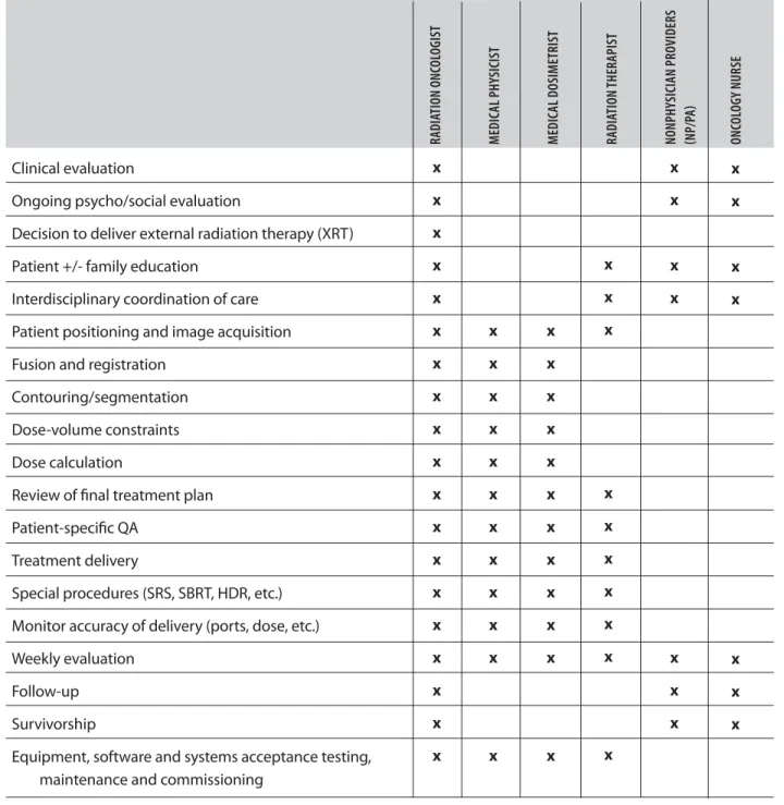
While Table 2.1 (see page 12) does not specifi cally defi ne individual roles within the radiation oncology team, it is an attempt to clarify those roles and relative responsi-bilities. Th e scope of practice of each team member should be based on the criteria established by their professional organization and local jurisdiction. Each facility must have policies and procedures defi ning the roles of these team members.
Leader:Prabhakar Tripuraneni
Theresa Kwiatkowski Michael D. Mills Bruce G. Haff ty Daniel Pavord Daniel Low Bhudatt R. Paliwal Albert L. Blumberg Paul Wallner Vanna Marie Dest
The Radiation Oncology Team
CHAPTER 2
2.2.0 QUALIFICATIONS AND TRAINING
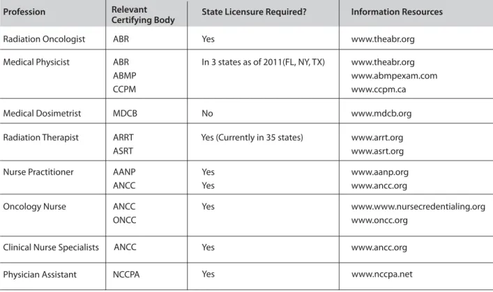
Board certifi cation is the primary consideration for estab-lishing proper qualifi cations and training for a professional working in radiation oncology. Th e relevant professional societies establish the eligibility requirements to sit for a board exam, including education, training and clinical residency requirements. In addition, where applicable, professionals must meet requirements for obtaining a state license, as shown in Table 2.2 (see page 13).
Each facility should have a policy regarding orienta-tion, competency, credentialing and periodic evaluations of all team members.
2.2.1 Medical Director
Th e medical director is a radiation oncologist who is responsible for oversight of the facility, in addition to establishing policies and procedures.
2.2.2 Radiation Oncologist
Th e radiation oncologist has American Board of Radiology (ABR) certifi cation in Radiation Oncology, Th erapeutic Radiology or equivalent certifi cation. Additional processes of certifi cation as defi ned by ABR are published at: www.theabr.org.
2.2.3 Nonphysician Providers (Physician Extenders) Nonphysician providers include, but are not limited to, nurse practitioners, clinical nurse specialists, advanced practice nurses and physician assistants. Th e roles, quali-fi cations, licensure requirements and maintenance of credentials for these individuals should be determined by their professional organizations, scope of practice, rules and
1 2 S A F E T Y I S N O A C C I D E N T | 2 0 1 2
regulations of individual institutions and licensure regula-tions within individual jurisdicregula-tions (American Academy of Nurse Practitioners [AANP], www.aanp.org;
American Nurses Credentialing Center [ANCC], www.nursecredentialing.org; National Commission on Certifi cation of Physician Assistants [NCCPA],
www.nccpa.net; American Academy of Physician Assistants [AAPA], www.aapa.org).
2.2.4 Medical Physicist
Medical physicists should be certifi ed in accordance with the appropriate qualifi cation for the designation of Qualifi ed Medical Physicist (as published at www.aapm.org), Th erapeutic Medical Physicist (as pub-lished at www.theabr.org) or equivalent certifi cation.
Table 2.1. Roles and Responsibilities of the Radiation Oncology Team
RADIA TION ONC OL OGIST MEDIC AL PHY SICIST MEDIC AL DOSIMETRIST RADIA TION THERAPIST NONPHY SICIAN PROVIDERS NP /P A ONC OL OGY NURSE Clinical evaluation
Ongoing psycho/social evaluation
Decision to deliver external radiation therapy (XRT) Patient +/- family education
Interdisciplinary coordination of care Patient positioning and image acquisition Fusion and registration
Contouring/segmentation Dose-volume constraints Dose calculation
Review of fi nal treatment plan Patient-specifi c QA
Treatment delivery
Special procedures (SRS, SBRT, HDR, etc.) Monitor accuracy of delivery (ports, dose, etc.) Weekly evaluation
Follow-up Survivorship
Equipment, software and systems acceptance testing, maintenance and commissioning
x x x x x x x x x x x x x x x x x x x x x x x x x x x x x x x x x x x x x x x x x x x x x x x x x x x x x x x x x x x x x x x x x x x
2.2.5 Medical Dosimetrist
A medical dosimetrist is competent to practice under the supervision of a qualifi ed physician and qualifi ed medi-cal physicist. An individual is considered competent to practice in medical dosimetry if that individual is eligible or certifi ed in accordance with the appropriate qualifi ca-tion for the designaca-tion of Qualifi ed Medical Dosimetrist through the Medical Dosimetrist Certifi cation Board (MDCB) at www.mdcb.org.
2.2.6 Radiation Therapist
A qualifi ed radiation therapist is considered competent to practice in radiation therapy if he or she is eligible or certi-fi ed in accordance with the appropriate qualicerti-fi cation for the designation of Radiation Th erapist, published by the American Registry of Radiologic Technologists (ARRT) at www.arrt.org and the American Society of Radiologic Technologists (ASRT) at www.asrt.org.
Table 2.2. Certifi cation and Licensure Requirements
Profession State Licensure Required? Information Resources
Radiation Oncologist ABR Yes www.theabr.org
Medical Physicist ABR In 3 states as of 2011(FL, NY, TX) www.theabr.org ABMP www.abmpexam.com
CCPM www.ccpm.ca
Medical Dosimetrist MDCB No www.mdcb.org
Radiation Therapist ARRT Yes (Currently in 35 states) www.arrt.org
ASRT www.asrt.org
Nurse Practitioner AANP Yes www.aanp.org
ANCC Yes www.ancc.org
Oncology Nurse ANCC Yes www.www.nursecredentialing.org
ONCC www.oncc.org
Clinical Nurse Specialists Yes www.ancc.org
Physician Assistant NCCPA
2.2.7 Radiation Oncology Nurse
A qualifi ed oncology or radiation oncology nurse has oncology certifi cation, in addition to basic educational preparation to function as a registered professional nurse, as determined by the individual jurisdiction. Oncology certifi cation can be obtained through the Oncology Nurs-ing Certifi cation Corporation (ONCC, www.oncc.org), American Nurses Credentialing Center (ANCC, www. nursecredentialing.org), or National Association of Clinical Nurse Specialists (NACNS, www.nacns.org).
2.3.0 CONTINUING EDUCATION AND
MAINTENANCE OF CERTIFICATION
Th e applications, technologies and methodologies of radia-tion oncology continue to expand and develop. Lifelong learning is vital to ensure incorporation of new knowledge into clinical practice, therefore, each member of the interdisciplinary team should participate in available Continuing Medical Education (CME) and, where appli-cable, Maintenance of Certifi cation (MOC) programs. Yes www.nccpa.net
Relevant
Certifying Body
1 4 Safe t y iS no accident | 2012 Safe t y iS no accident | 2012 15 ChAPTeR APPendIx:
IlluSTRATIve SAfeTy STAffInG MOdel
In the current environment, radiation oncology as a profes-sion is providing more complex special procedures. The above guidelines reflect the combined input from the sur-veys performed by several professional organizations (ACR, ASTRO, AAMD, AAPM and the ABR studies) during the last decade. Additional personnel will be required for research, education and administration. For a progressive clinic, the above recommendations may be insufficient to accurately estimate the medical physics and dosimetry FTE effort required to provide all special patient procedures and services.
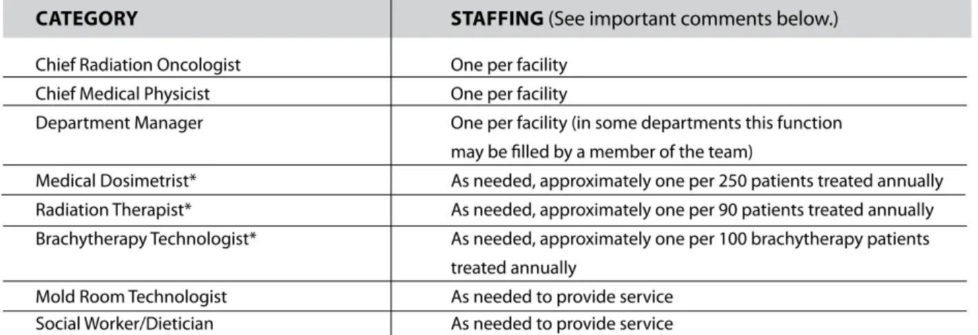
Table 2.3 Minimum Personnel Requirements for Clinical Radiation Therapy
CATEGORy STAFFING (See important comments below.)
chief radiation oncologist one per facility chief medical physicist one per facility
department manager one per facility (in some departments this function may be filled by a member of the team)
medical dosimetrist* as needed, approximately one per 250 patients treated annually radiation therapist* as needed, approximately one per 90 patients treated annually brachytherapy technologist* as needed, approximately one per 100 brachytherapy patients
treated annually
mold room technologist as needed to provide service Social worker/dietician as needed to provide service
2.4.0 STAffInG ReQuIReMenTS
The staffing needs of each facility are unique and vary based greatly upon the patient mix, as well as on the type and complexity of the services offered. Patient load, number of machines and satellites/affiliated centers also influence the need to allocate management manpower and full-time employees (FTEs) (Table 2.3), as well as teaching responsibilities and vacation time. As such, it is impossible, in the current era, to prescribe hard staffing levels.
The radiation oncology facility should have a qualified radiation oncologist on-call 24 hours a day, seven days a week, to address patient needs and/or emergency treat-ments. An adequate number of other members of the radiation oncology team should be available to deliver urgent treatments in off-hours. Otherwise, the facility must have arrangements for referral of emergency patients for timely treatments.
* this number may be higher or lower depending upon the complexity of patients treated by an individual physician or by the complexity of technology.
**it is recommended that a minimum of two qualified individuals be present for any routine external beam patient treatment.
Rela tiv e FTE F a c tor Requir ed FTE Requir ed T otal FTE Equipment, Sour ces and S ystems S e rvic es -– # of Units or Lic enses* No . of sy st ems* Ph y sicist Dosimetrist Ph y sicist Dosimetrist Ph y sicist Dosimetrist Multi ener gy ac celerat ors 0.25 0.05 Single ener gy ac celerat ors 0.08 0.01 Tomotherap y, C yberK nif e , G ammaK nif e 0.3 0.03 C obalt Units , IMR T, P A CS, EMR & C o nt ouring 0.08 0.03 Or tho v
oltage and super
fi cial units 0.02 0.01 M anual brach ytherap y ; LDR S eed I mplants 0.2 0.03 HDR brach ytherap y 0.2 0.02 Simulat o r, C T -Simulat o r, PE T, MRI F usion 0.05 0.02 C omput er planning sy st em (per 10 w o rkstations) 0.05 0.02 HDR planning sy st em 0.2 0.01 Subtotal No. P atien t Proc edures Annual # of P a tients under going P roc edur es** No . of pa tients** Ex te rnal Beam R T with 3D planning 0.0003 0.003 Ex te rnal Beam R T with c o n v entional planning 0.0002 0.002 S ealed sour ce Brach ytherap y (LDR & HDR) 0.008 0.003 Unsealed sour ce therap y 0.008 0.005 IMR T, IGR T, SRS, TBI, SBR T 0.008 0.005 Subtotal Nonclinical - Estimated T otal FTE Eff or t Estima ted Total (Ph y
s & Dosim) FTE Eff
or t*** FTE Eff or t*** Education & T raining (FTE) 0.667 0.333 Generation of I n te rnal Repor ts (FTE) 0.667 0.333 C ommitt ees & M eetings; I n c. R a d . Saf e ty (FTE) 0.667 0.333 A dministration and M anagement (FTE) 0.667 0.333 Subtotal T otal
A sample w
o
rksheet f
or calcula
ting medical ph
y
sics and dosimetr
y staffi
ng in r
adia
tion onc
ology:
ap y units , imaging systems , w orkstations , support systems and technologies in each c
a
tegor
y (
column 3).
go each of the follo
wing planning and tr
eatment deliv er pr oc edur es; c ount
each new patient one time (
column 3).
al ph
ysicist and medic
al dosimetrist estimated FTE eff
or
t in each of the follo
wing c
a
tegories
. S
ee C
omponent FTE table for t
ypic
al FTE (
1 6 S A F E T Y I S N O A C C I D E N T | 2 0 1 2 Rela tiv e FTE F a c tor Requir ed FTE Requir ed T otal FTE Equipment, Sour ces and S ystems S e rvic es -– # of Units or Lic enses* No . of sy st ems* Ph y sicist Dosimetrist Ph y sicist Dosimetrist Ph y sicist Dosimetrist Multi ener gy ac celerat ors 4 0.25 0.05 1 0.2 Single ener gy ac celerat ors 0 0.08 0.01 0 0 Tomotherap y, C yberK nif e , G ammaK nif e 1 0.3 0.03 0.3 0.03 C obalt Units , IMR T, P A CS, EMR & C o nt ouring 0 0.08 0.03 0 0 Or tho v
oltage and super
fi cial units 0 0.02 0.01 0 0 M anual brach ytherap y ; LDR S eed I mplants 1 0.2 0.03 0.2 0.03 HDR brach ytherap y 1 0.2 0.02 0.2 0.02 Simulat o r, C T -Simulat o r, PE T, MRI F usion 1 0.05 0.02 0.05 0.02 C omput er planning sy st em (per 10 w o rkstations) 1 0.05 0.02 0.05 0.02 HDR planning sy st em 1 0.2 0.01 0.2 0.01 Subtotal 2.00 .033 No. P atien t Proc edures Annual # of P a tients under going P roc edur es** No . of pa tients** Ex te rnal Beam R T with 3D planning 500 0.0003 0.003 0.15 1.5 Ex te rnal Beam R T with c o n v entional planning 200 0.0002 0.002 0.04 0.4 S ealed sour ce Brach ytherap y (LDR & HDR) 100 0.008 0.003 0.8 0.3 Unsealed sour ce therap y 2 5 0.008 0.005 0.2 0.125 IMR T, IGR T, SRS, TBI, SBR T 400 0.008 0.005 3.2 2 Subtotal 4.39 4.33 Nonclinical - Estimated T otal FTE Eff or t Estima ted Total (Ph y
s & Dosim) FTE Eff
or t*** FTE Eff or t*** Education & T raining (FTE) 0.1 0.667 0.333 0.0667 0.00333 Generation of I n te rnal Repor ts (FTE) 0.1 0.667 0.333 0.0667 0.00333 C ommitt ees & M eetings; I n c. R a d . Saf e ty (FTE) 0.1 0.667 0.333 0.0667 0.00333 A dministration and M anagement (FTE) 0.5 0.667 0.333 0.0667 0.00333 Subtotal 0.53 0.27 T otal 6.92 4.92
Multiply the entries in c
olumn 3 b
y
the Ph
y
sicist FTE fac
tor (c
olumn 4) and the Dosimetrist FTE fac
tor (c
olumn 5);
repor
t these in c
olumns 6 and 7. Sum and t
otal in c
olumns 8 and 9. Example belo
w : Another r esour ce f
or calculating radiation onc
ology staffi ng is: Battista JJ et al . M edical ph y sics staffi ng for radia-tion onc ology : a decade of experienc e in Ontario , C anada. J A ppl Clin Med Ph ys . 2012;13(1):3704.
1 8 S A F E T Y I S N O A C C I D E N T | 2 0 1 2
Leader: Lawrence B. Marks
Leader Designee: Daniel Pavord
R. Alan Burns Laura A. Dawson Lisa A. Kachnic Peter A. S. Johnstone Christopher J. Moore Christopher F. Serago
Safety
CHAPTER 3
3.1.0 THE NEED FOR A CULTURE OF
SAFETY
Modern radiation therapy is complex and rapidly evolving. Th e safe delivery of radiation therapy requires the concert-ed and coordinatconcert-ed eff orts of many individuals with variconcert-ed responsibilities. Further, safety and effi ciency go hand in hand. Ineffi cient systems lead to staff frustration, rushing and sometimes cutting corners, thus, all team members need to work together to create a safe and effi cient clinical environment and workfl ow.
Th e need for effi ciency is heightened by the increas-ing demands beincreas-ing placed on all members of the radiation oncology team. Changes in the levels of reimbursement for some clinical activities, global changes in the national healthcare system (e.g., structural, fi nancial) and increas-ing levels of administrative burden (e.g., documentation requirements) require physicians to search for improved levels of effi ciency. Th is is essential in order to provide staff with necessary time to perform critical safety-related activities.
Th e rapidly-evolving nature of radiation oncology requires that processes and workfl ows be continually reassessed. Each member of the team needs to accept that optimal approaches are not static, but will necessarily change to accommodate the evolving practice. Long-held traditional approaches will need to be challenged and possibly modifi ed.
People may be hesitant to change, often for good reasons. Good clinical practices usually evolve over years if not decades, so change should be carefully implemented. It is critical that a culture that appropriately manages change exists, ensuring change facilitates safety and quality.
Furthermore, all team members must be open to having any member of the team (whether in leadership positions or not) raise concerns about safety as well as suggesting and considering change. Indeed, it is often the frontline staff that are more likely to understand the limitations of cur-rent procedures and suggest improvements. Th us, an ideal open environment with a safety-minded culture only exists where staff are permitted and encouraged to suggest and lead change to improve safety, quality and effi ciency.
3.2.0 LEADERSHIP AND EMPOWERING
OTHERS
Physicians and medical physicists comprise the primary leadership roles within a radiation oncology clinical site. Th ey must empower all members of their team to be active participants in improving clinical processes. Th is is true from a practical perspective, as one person cannot pos-sibly understand all aspects of the complex fi eld. Further, such empowerment is a meaningful way to provide team members with a feeling of responsibility, thereby increasing job satisfaction, raising expectations and enhancing perfor-mance. Staff should know that they have a meaningful and benefi cial impact in the work environment.
In the radiation oncology clinic, these professionals are ultimately responsible for creating a culture of safety. Soci-ety has entrusted physicians and medical physicists as the guardians of both the individual and societal health care structure. With this trust, they are empowered to operate as advocates for safety-related initiatives. Leadership needs to make all staff feel comfortable to raise concerns about safety without fear of reprimand or reprisal.
2 0 S A F E T Y I S N O A C C I D E N T | 2 0 1 2
3.3.0 EVOLVING ROLES AND
RESPONSIBILITIES OF EACH TEAM
MEMBER
Th e fi eld of radiation oncology is ever-evolving, and as such, there are rapid changes in the roles and responsibili-ties of each team member. Table 3.1 (see page 21) sum-marizes some of these changes and associated challenges. Entries are meant as examples, as this is not an exhaustive list.
3.4.0 EXAMPLES OF TOOLS/INITIATIVES
TO FACILITATE SAFETY, AND THE SAFETY
CULTURE
3.4.1 Staffi ng/Schedules
Staffi ng levels need to be adjusted to refl ect the workload, particularly in physics, dosimetry and treatment, where the demands have markedly increased (e.g., patient-specifi c QA for IMRT). Schedules should be realistic to avoid/ minimize hurrying through a given task and risking error. An excessive workload can lead to errors. Conversely, light workloads can also be a problem since a certain level is needed to maintain “situational awareness” [1, 2].
3.4.2 Communication/Facilities
Systems that facilitate clear, unambiguous and effi cient communication between all team members are critical. Th is is particularly true between physicians, medical dosimetrists, medical physicists and radiation therapists, given the large number of hand-off s and interdepen-dent tasks that routinely occur during the planning and treatment-implementation processes. Well-defi ned charting procedures, either paper or preferably electronic, are criti-cal. In planning the layout of a department, one might centrally locate dosimetry, and/or establish dedicated time for physicians and medical dosimetrists to work together, thereby facilitating the iterative “directive-segment-com-putation-review-repeat” cycle. Th is is a particular challenge when physicians and planners rotate between facilities. Enhanced tools are needed to enable effi cient and accurate communication/transfer of complex 3-D data between centers. A well-defi ned communication pathway between workers will reduce the need for ad hoc/variable solutions and provide for messages being sent, received and verifi ed.
3.4.3 Workfl ow/Effi ciency
Clinical practice is complex, often mired in administrative and historically-derived procedures. Effi ciency impacts quality and safety. Harried workers are more prone to error, therefore eliminating nonessential tasks increases time available for critical tasks. Lean approaches (adapted from the Toyota Production System)[3] have been adopted
by many to streamline clinical workfl ow and alter the work environment. Some have implemented rapid improvement events (Kaizens [4]) where participating representative
members of involved groups create process maps for particular tasks. Value-added steps are identifi ed, with wasteful steps and unnecessary stressors being eliminated, and a more streamlined, unambiguous, standardized process emerges. Having stakeholders meet to discuss and defi ne their work builds teamwork and mutual respect, while fostering an environment in which staff know that they can positively impact their work.
3.4.4 Standardization
Standardization is widely recognized as a means to reduce errors and confusion. Th is might be particularly useful in group practices where radiation therapists, medical dosimetrists and medical physicists interact with numerous physicians, each having their own preferred methods. Having too many diverse approaches can lead to con-fusion. It is helpful if providers can agree on standard approaches to common diseases using reference or guide sheets to avoid confusion among planning staff . Standard treatment practices and QA mechanisms, as well as associ-ated policies and procedures, should be vetted through a review committee and required for every technique or site, with regular updates, as needed. Th ese should be posted with easy access for all who may need to refer to them. 3.4.5 Hierarchy of Eff ectiveness
Diff erent methods used to aff ect behaviors have variable expectations for success [5]. Reliance on policies and
training is the usual but least eff ective approach. In a large database of errors from the State of New York, “failure to follow policies/procedures” was implicated as a contrib-uting factor in 84 percent of events, versus “inadequate policies/procedures” in 16 percent of events. Whenever possible, it is best to “hardwire” the systems for success using simplifi cation, standardization, automation and forced functions to create workfl ows and systems that support human work. Checklists and time-outs are eff ective [6, 7] especially if:
• Th ey are focused on the task at hand; • Th e user believes in their utility; and
Table 3.1. Examples of Safety-Related Roles and Challenges – Radiation Oncology Staff
Team Member Traditional Role Evolving Role Challenges
Physician Medical Physicist Medical Dosimetrist Radiation Therapist Nurse Administrator IT Specialist All Clinical Staff • Patient care
• Supervises RT (e.g., sets dose/ volume criteria, approves plan and treatment images, manages toxicity)
• Assure the safe and eff ective delivery of radiation as prescribed
• Treatment planning • Plan and TPS QA
• Provide safe and eff ective delivery of radiation as prescribed • Daily equipment and new
patient treatment QA • Assist with patient care/
education • Manage toxicity
• Oversight of regulatory compliance
• Desktop support
• Proper patient identifi cation • Peer review
• Relinquish some autonomy to other personnel
• Engaging others in safety mission
• Role shift to increase emphasis on safety-related work
• Education in advanced process analysis tools for patient safety
• Adequate instruction in anatomy
• Proper utilization of emerging imaging/segmentation tools
• Safe and proper use of additional imaging and treat-ment delivery systems
• Adequate instruction in evolving technologies • Knowledge of evolving chemotherapy agents • Resource allocation • Resources • Space • Vendor interoperability • Identifi cation/discussion of near-misses • Continuous education • Increased reliance on EMR • Adequate instruction with
software/technological advances
• Dedicating time for safety initiatives
• Minimizing distractions • Team leader for patient safety
• Coordination with multidisci-plinary team
• Continuous education (e.g., image evaluation/segmenta-tion, new software/technology) • Incorporating technological
innovations to improve patient/ staff safety
• Assess safety of treatment processes, (e.g., with statistic processes, failure mode analysis, fault trees, etc.)
• Image cataloging/manipulation (e.g., fusion/registration/ segmentation) • Assist in IMRT/IGRT/equipment QA • Assessment of 2-D/3-D images to make decisions concerning patient treatment/ motion/ alignment
• Patient pain
• Assist in multidisciplinary coordination
• Support patient safety program
• Connectivity • Failure mode analysis • Data archiving/recovery • QA/Quality Improvement (QI) • Increased documentation in
EMR
• Evolving peer review • Compliance with evolving
regulatory requirements Nonphysician
Providers
• Assist physician with patient care
• Coordination with multidisci-plinary team
• Legal or regulatory restrictions
2 2 S A F E T Y I S N O A C C I D E N T | 2 0 1 2
“Knowledge in the fi eld” (automatic computer/machine functions and checklists) is more likely to improve human performance than is “knowledge in the head” (memory).
3.4.6 Human Factors Engineering [5, 8]
Human-machine interactions are ubiquitous. Human factors engineering aims to defi ne processes, interfaces and machinery that facilitate correct usage. For example, the forcing function of an automated teller machine can require withdrawal of the bankcard before money is dispensed. Similarly, placing console control buttons that perform particular functions in a consistent loca-tion enables users to more reliably operate equipment in a predictable and correct manner. Safety is improved with workspaces that are designed to reduce noise, interruptions and visual clutter. Improving lighting, temperature and desk height are additional factors proven to aff ect performance.
In the radiation oncology fi eld, complicated computer screen layouts, keyboard functions and treatment consoles are a few examples of the hundreds of human-machine interfaces that are navigated daily. Th ese require increasing mental eff ort as they become more complicated or lack standardization. Many are well designed, but there is ample room for improvement. For example, within individual products, shortcut keyboard commands should be consis-tent whenever possible. Standardization of nomenclature, monitor layouts and shortcuts across diff erent vendors are examples of enhancements that might also be helpful. 3.4.7 Incorporating QA Tools/Functionality Into Software
Often, QA is not incorporated into the planning or record and verify delivery systems. For example, user-confi gurable checklists and time-outs are not an option. Although potentially valuable, such embedded checklists still require the user to verify that checklist items are appropriately addressed rather than being automatic. Some embedded automatic QA functions would be useful, such as: • For a new plan, the system searches its directory
archive for patients with the same name to identify inadvertent retreatment.
• For common diagnoses, the planning system com-pares the proposed target volumes and associated dose parameters to a library of user-specifi ed “expected” parameters and issues predefi ned alerts.
• Normal tissue dose-volume parameters are compared to user-specifi ed constraints.
• Automatic highlighting of under-dosed target, or normal tissue hot-spots.
• Beams and plans are named automatically to refl ect the treatment planner, date, etc.
• Common nomenclature of target volumes, organs at risk and plans to facilitate review of plans and identifi -cation of outliers.
Some of these functions may already exist. At least one manufacturer is “training” their planning system to identify discrepancies between pending plans and their library of “similar plans” [9].
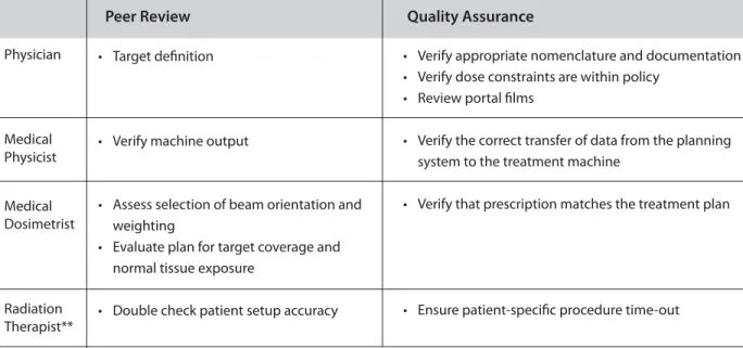
3.4.8 Peer and Interdisciplinary Review Peer review is an essential part of the safe delivery of radiation. Prospective peer review is critical, especially for new technologies such as IMRT and IGRT [10, 11]. Once
treatment has been initiated, the threshold for making a meaningful change in image segmentation or motion-management strategy is relatively high because it may result in time-consuming replanning and QA. Physician-to-physician peer review is useful, and review of target delineation and image segmentation prior to planning deserves more standardization. Peer review is also con-ducted as part of the chart rounds process. See Chapter 4, sections 4.1.5 and 4.1.6, in this document for the specifi cs regarding the components of this process.
Peer review is clearly important for other team mem-bers as well. As an example, medical dosimetrists can check each other’s work (e.g., choice of beam selection/weight-ing). A distinction is often made between quality assurance and peer review (Table 3.2, see page 23). Quality assur-ance is often taken to relate to objective/quantitative “right versus wrong” actions (e.g., was the correct plan sent from the planning system to the treatment machine? Is the ma-chine beam output correct?), that can readily lead to major clinical events that aff ect one or many patients. Peer review is often used to refer to somewhat more subjective items (e.g., target defi nition or dose selection) that are perhaps less likely to lead to major clinical events, and not aff ect a large number of patients. Th ese interactions tradition-ally occur roughly as physics-, planning- or therapy-based versus physician-based. However, this distinction can be readily blurred. For example, should there be a double check for things such as machine QA? (e.g., there may be two people to confi rm the machine output). Similarly, a physician can make gross right or wrong type errors in target delineation (e.g., mislabeling the left atrium as a sub-carinal lymph node) or misinterpreting published da