Wavelet-enhanced convolutional neural network:
a new idea in a deep learning paradigm
https://doi.org/10.1515/bmt-2017-0178
Received October 13, 2017; accepted February 19, 2018
Abstract
Purpose:
Manual brain tumor segmentation is a
chal-lenging task that requires the use of machine learning
techniques. One of the machine learning techniques
that has been given much attention is the convolutional
neural network (CNN). The performance of the CNN can
be enhanced by combining other data analysis tools such
as wavelet transform.
Materials and methods:
In this study, one of the famous
implementations of CNN, a fully convolutional network
(FCN), was used in brain tumor segmentation and its
architecture was enhanced by wavelet transform. In this
combination, a wavelet transform was used as a
com-plementary and enhancing tool for CNN in brain tumor
segmentation.
Results:
Comparing the performance of basic FCN
archi-tecture against the wavelet-enhanced form revealed a
remarkable superiority of enhanced architecture in brain
tumor segmentation tasks.
Conclusion:
Using mathematical functions and
enhanc-ing tools such as wavelet transform and other
math-ematical functions can improve the performance of CNN
in any image processing task such as segmentation and
classification.
Keywords:
brain tumor; convolutional neural network;
segmentation; wavelet transform.
Introduction
Brain tumor is one of the main causes of death in all age
groups. According to reports by the National Brain Tumor
Foundation (NBTF) and American Brain Tumor
Associa-tions (ABTA), research in developed countries indicates
that the number of people with this disorder has
drasti-cally increased during the past decade [1–6].
One of the routine tests in brain tumor diagnosis is
the use of magnetic resonance (MR) imaging [7] which is
capable of creating the optimal contrast of soft tissues
[8]. Because of its high resolution and non-ionizing
nature, it is widely used in brain studies [9–14]. There
are many challenges in the manual segmentation of
brain tumors [12, 15–18], as one of the important steps in
the processing of brain MR images [19], including
differ-ences in the size, shape, texture and intensity of tumors
in the MR images. In fact, these challenges increase the
manual segmentation error and lead to disagreement
among experts [20]. Besides that, the large number of
brain scans increases the time required for the analysis
of the MR images [21]. All of the mentioned problems
turn brain tumor segmentation into a complex and
time-consuming process and cause misdiagnosis or delay in
decision-making [22, 23]. The presence of these
prob-lems illustrates the necessity of using machine learning
techniques for automatic brain tumor segmentation [20,
24, 25].
In recent years, the use of deep learning techniques
has increased in the image processing as well as in the
medical image processing applications [26–29]. Among
techniques introduced for automatic brain tumor
segmen-tation [2, 26, 27, 30–39], the portion of deep learning-based
techniques has rapidly increased [40] and several
exam-ples of these techniques’ application have been recently
proposed for brain tumor segmentation [30–33, 40–53].
As a matter of fact, the best technique for exploiting the
benefits of multidimensional spatial data such as image,
*Corresponding author: Mohamadreza Hajiabadi, Brain and Spinal Cord Injury Research Center, Neuroscience Institute, and Iranian International Neuroscience Institute, Shariati Hospital, Tehran University of Medical Sciences, Tehran, Iran,
E-mail: [email protected]
Behrouz Alizadeh Savareh: Student Research Committee, School of Allied Medical Sciences, Shahid Beheshti University of Medical Sciences, Tehran, Iran
Hassan Emami: Faculty of Allied Medical Sciences, Shahid Beheshti University of Medical Sciences, Tehran, Iran
Seyed Majid Azimi: Chair of Remote Sensing Technology, Technical University of Munich, Munich, Germany
Mahyar Ghafoori: Department of Radiology, Hazrat Rasoul Akram Hospital, School of Medicine, Iran University of Medical Sciences, Tehran, Iran
sound and time series is a convolutional neural network
(CNN) [34]. The CNN performance can be enhanced if it is
combined with other data analysis tools. One of the most
useful tools in signal and image analysis is the wavelet
transform which is considered as a good candidate for the
CNN enhancement. In the following sections, the idea of
combining the CNN and wavelet transform is explained
and then the application of this combination in brain
tumor segmentation is investigated.
Related works
Evolution of brain tumor segmentation techniques
represents a move toward achieving an automatic and
accurate segmentation where three levels of algorithms
were developed to achieve these goals [1, 2, 10, 14, 36–39,
54–58].
In the first generation, heuristic ideas were employed
by algorithms such as the threshold level [59], area growth
[60] and edge detections [10]. Simplicity of the
implemen-tation was the main feature of these algorithms; however,
the big challenge was posed when they were faced with
situations different from the training setting.
Techniques in the second generation were based
on the probabilistic and optimization methods such as
artificial neural networks [61], Bayesian models [62],
fuzzy clustering [63] and support vector machines [64].
In addition, techniques like Gaussian mixture models,
linear and non-linear dynamic systems, conditional
random fields, maximum entropy (MaxEnt) models,
logistic regression, kernel regression and extreme
learn-ing machines were in this generation too. This group
of techniques was effective in solving simple or
well-constrained problems [65–68], but their low modeling
capacity caused some problems while dealing with
complex real-world situations [69].
There are techniques in the third generation which
seek to achieve the desired result using the higher levels
of knowledge such as tacit knowledge, rules and models
extracted directly or indirectly from data.
The significant examples of this generation’s
tech-niques include Atlas-based segmentation [70] and
deep learning-based methods [40] which fascinatingly
modeled the human brain information processing system.
Although various versions of deep learning techniques
have been proposed for image segmentation, the most
successful technique in brain tumor segmentation is the
CNN [30–33, 40–53].
Method
Idea description
The deep learning hypothesis is based on the fact that, in order to achieve a high level of representation, a hierarchy of initial and mid-dle representations is required [71–74].
The CNN structure is based on the multilayered perceptron (MLP) [75–77], and its function is similar to the time delayed neural networks which share the intra-network weights in order to reduce the computations [78]. Employing operations like convolution, sam-pling and linear correction unit, the CNN is able to directly extract features from raw data [79]. In fact, the use of the convolutional operation gives CNN spatial flexibility and the use of the sampling results in higher levels of representation. The learning process occurs in the CNN using intermediate representations (known as the feature map) [80], exactly the same as the hierarchical learning in biological brains [81]. The feature maps pass through layers, so the hierarchy of learning occurs and the information becomes more meaningful by going through the layers [82, 83]. This unique ability has led the CNN to succeed in most of the image processing applications [35].
Along with the CNN properties in the hierarchical learning, a wavelet transform has interesting features that make it a candidate to enhance the CNN. The main functionality of the wavelet transform in image processing is the ability to decompose images into different scales with different levels of details [84–86]. During the decomposi-tion, the separation of the information at different levels is repeated on the remainder part of the previous layer [79]. Therefore, it can be deduced that CNN and wavelet transform have different approaches toward image details in that CNN creates a high-level representation by upward combinations of low-level representations such as pixels, lines and object elements, whereas wavelet transform decomposes the image into its elements at different levels [75]. The difference between the viewpoints of these two data analysis tools is the basis for an idea according to which wavelet transform can be considered as an enhancing tool for the learning algorithm of CNN. In fact, according to the very idea, the compressed forms of the input image, as the result of various levels of wavelet decomposition, can be injected into the different layers of the CNN architecture and enhance its performance.
The application of this idea in brain tumor segmentation
In this section, the application of the introduced idea in brain tumor segmentation is investigated and its details are described in the form of the implementation.Data: The Brain Tumor Segmentation (BRATS) dataset provided by the Medical Image Computing and Computer Assisted Intervention Society was used in this study. There were 220 sample MR images of patients with a high-grade glioma, and 54 samples of patients with a low-grade glioma with 155 axial scans stored in three-dimensional (3D) (mha) format for each patient. Each scan included a collection of four types of images: T1, T2, T1 with contrast (T1C) and Flair with a corresponding manually segmented (ground truth) image [76]. The provided manual segmentation was in the five-class mode: back-ground, necrosis, edema, enhancing tumor and non-enhancing
tumor. But, in terms of neurosurgery application, the existence of binary classification (tumor and non-tumor) is preferable for neu-rosurgeons as they are the main beneficiaries of the segmentation. Therefore, the model introduced in this study was trained based on two-class segmentation as shown in equation 1.
1, Seg class is in (Necrosis, Enhancing Tumor and NonEnhancing Tumor) NewSeg 0, Seg class is in (Edema and Background) = (1) Equation 1: New segmentation criteria on BRATS dataset images.
Figure 1 shows examples of BRATS data with ground truth seg-mentation.
Implementation: In this study, a python-based implementation [87], based on [88] and adapted from vgg-net, was used for pixel-wise semantic segmentation. In order to implement the introduced idea of combination, we used the TensorFlow framework which has a great potential in designing and testing the deep learning models. In this framework, the data model is composed of multidimensional arrays, named tensors, with the operational model in graph form [77]. As the most successful model of CNN in image segmentation, a fully convolutional network (FCN) was selected to implement the wavelet-enhanced fully convolutional network (WFCN) model in brain tumor segmentation. The FCN consists of convolution, deconvolution and max-pooling layers for the image segmentation task [78]. In this regard, new paths were defined for wavelet injection. Employing Pywt as the main library of wavelet transform implementation in python, the first order of Daubechies wavelet family (db1) was used for wavelet injections [89]. Regarding the proven success of Daubechies in signal decomposition and identification of image edges, db1 was selected
as the mother wavelet function, the simplest form of this family with lower computation and less wavelet filter bank coefficients [90]. Computation and hardware: To increase the generalizability and to avoid overfitting in the introduced models, the data was augmented with a 180° rotation and increased to 84,940 images. Then, it was randomly divided into two groups: 90% for training and 10% for test-ing. In order to evaluate the performance of WFCN, four tests were run. Each time one of the four wavelet injection paths was turned on and the remaining ones were turned off. The introduced architec-tures’ training of 100 epochs, on Linux server CentOS release 6.8 with 128 GB shared main memory and GeForce GTX 980Ti (6 GB memory) gpu, takes about 18 h.
Results
The basic design for brain tumor segmentation was
gener-ated with a modification to the first layer of the vgg-based
FCN, where the three-channel input was replaced by the
four-channel input.
Figure 2 shows the basic form of segmentation with
a sequence of convolution and sampling to feature
com-pression and a reverse process to reconstruct the
seg-mented image.
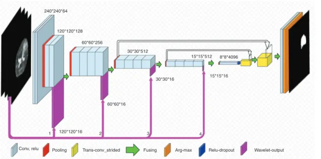
As mentioned before, the main idea behind the WFCN
is to enhance the FCN using wavelet transform injections
as shown in Figure 3. Given the FCN architecture, four
paths were proposed for the injection. Each path transfers
the compressed form of the image to an appropriate
posi-tion, in terms of size, in the basic FCN architecture.
As shown in Figure 3, using four levels of the wavelet
transform on input images resulted in the compression of
the images to H/2*W/2, H/4*W/4, H/8*W/8 and H/16*W/16
pixel sizes.
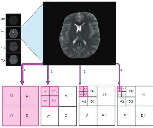
According to Figure 4, for each input channel with
240
×
240-pixel size, the wavelet compression was
accom-plished by four (approximate, horizontal, vertical and
diagonal) compressed forms. On the other hand, through
four input channels, 16 compressed images were formed
as T1
A, T1C
A, T2
A, Flair
A, T1
H, T1C
H, T2
H, Flair
H, T1
V, T1C
V, T2
V,
Flair
V, T1
D, T1C
D, T2
Dand Flair
D(Figure 4).
Whenever one of the paths (1–4) is activated, the
injected images get concatenated with the feature maps
extracted by the basic FCN architecture layers. The
concatenation provides more features (FCN’s feature
map
+
wavelet injection), which can be used in the
subse-quent layers and consesubse-quently in creating a higher-level
representation. Table 1 illustrates the results of testing
dif-ferent WFCN architectures in brain tumor segmentation.
Conv, relu 240 120 60 30*30*512 15*15*512 8*8*4096 60 256 120 128 240 64
Pooling Trans-conv_strided Fusing Arg-max Relu-drop out
Figure 2: Original FCN for semantic segmentation.
Conv, relu 1 120*120*16 60*60*16 30*30*16 30*30*512 60*60*256 120*120*128 240*240*64 15*15*16 15*15*512 8*8*4096 2 3 4
Pooling Trans-conv_strided Fusing Arg-max Relu-dropout Wavelet-output
Dice, the most important benchmark for analyzing the
performance of segmentation techniques, was used in the
initial assessment of the architectures.
According to Table 1, the best performance belongs to
WFCN1 in which the first path was activated and a
one-step wavelet compression with H/2*W/2 size was injected
into the architecture.
In order to analyze the WFCN1 performance in more
detail, a set of evaluation parameters was used as shown
in equations 2–5.
2 |
|
Dice=
| |+| |
S G
S G
∗ ∩
(2)
Equation 2: Dice in brain tumor segmentation.
In the above equation,
S
is equal to the region
seg-mented by the algorithm and
G
is equal to the reference
segmentation region (ground truth).
+
=
Accuracy
All pixels
Tp Tn
(3)
Equation 3: Accuracy in brain tumor segmentation.
In the above equation,
Tp
denotes the number of
tumor pixels which are correctly identified by the
tech-nique as tumor, and
Tn
refers to the number of non-tumor
pixels that are correctly identified as non-tumor by the
algorithm.
=
+
Sensitivity
Tp
Tp Fn
(4)
Equation 4: Sensitivity in brain tumor segmentation.
=
+
Specificity
Tn
Tn Fp
(5)
Equation 5: Specificity in brain tumor segmentation.
Fn
denotes the number of pixels that are actually
tumor but misclassified by the technique as non-tumor.
On the other hand,
Fp
refers to the number of pixels that
are not actually tumor, but they are classified as tumor.
A detailed evaluation of the WFCN1 is summarized in
Table 2.
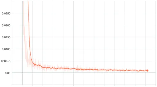
Figure 5 shows the WFCN1 network entropy reduction
diagram for training.
Also Figure 6 shows segmented samples as WFCN1
outputs.
T2 1 2 A1 H1 H1 V2 D2 V1 D1 H3 A4 H2 V3 D3 V2 D2 H1 V1 D1 H2 H1 D1 V1 D1 A2 A3 V3 D3 H3 H2 V2 D2 V1 3 4 T1C Flair T1Figure 4: Wavelet compression.
Table 1: Test result of different architectures.
Architecture Result (dice)
Basic FCN architecture (Figure 2) 77.9%
WFCN1 (1st level injection – Figure 3) 91.8% WFCN2 (2nd level injection – Figure 3) 91.4% WFCN3 (3rd level injection – Figure 3) 91.3% WFCN4 (4th level injection – Figure 3) 91.3%
Discussion
Surveying the related studies show a large number of
CNN usage in brain tumor segmentation [30–33, 40–53].
The average segmentation dice in these studies is about
84% and the standard deviation of them is around 5.5%.
Figure 7 demonstrates the performance comparison
between the superior ones from the surveyed studies
(yellow columns) and our method (blue column).
In the present study, a total of 20 layers of convolution
and deconvolution for brain tumor segmentation were
used for modeling of the network. The comparison of the
surveyed studies shows that the number of convolution
layers in the study by Chang was nine, while this number
in the study by Casamitjana et al. was 20. On the other
hand, Chen et al. used 25 convolutional layers with four
deconvolutional layers, which was the most number of
layers in modeling. Besides that, Yi et al. used five
convo-lutional layers and Kasamitjana et al. used a 14-layer
con-volutional model. There is a coincidence in the number
of layers used in the modeling between the present study
and the study by Casamitjana et al., as both studies are
based on the vgg_net19.
Convolutional layers are considered as
feature-extractors, because they look for a special pattern as
the kernel in the image. Using more convolution layers
implies the use of more levels of abstraction on the image
analysis and leads to more modeling power in solving
complex problems. Using more layers of abstraction
is desired; however, increasing the number of layers in
the designed model will increase the demand for more
computational power. Deeper models (with more layers)
require more memory usage and more power of
process-ing. It is of great importance in the modeling to create a
balance between the number of layers used in the model
and the hardware capabilities.
– The WFCN dice was 91.8%. This is a better
perfor-mance against all previous studies where the best of
them was reported in [30] by 91.59% dice.
– This superiority can be explained according to the
spe-cific reasons and be examined from various aspects:
– The first reason is the difference in the number
of target classes in segmentation. In fact,
previ-ous studies worked with five classes, while in
the present study the segmentation was based
on two classes. Although the use of five classes
in segmentation clearly has some advantages, it
should be considered that as the neurological
sur-geons are the ones who make the most benefit of
the segmentation of brain tumor images, binary
segmentation would be more helpful for them in
tumor resection. It is due to the fact that they are
practically seeking to analyze tumor MR images
in binary format (tumors and non-tumors).
– The second and more important factor for the
superiority of the proposed method compared
0.0250 0.0200 0.0150 0.0100 5.000e–3 0.00 0.000 20.00 k 40.00 k 60.00 k 80.00 k 100.0 k 120.0 k 140.0 k 160.0 k 180.0 k 200.0 k 220.0 k 240.0 k
Figure 5: WFCN1 entropy reduction. Table 2: WFCN1 evaluation.
Parameter Value
Dice 0.918
Pixel accuracy 0.99
Mean pixel accuracy 0.96
Sensitivity 0.93
with the similar studies is the idea presented in
this study, the combination of the wavelet and the
CNN. The important thing in this combination, as
mentioned earlier, is that no part of the basic FCN
network has been eliminated here, and in fact its
innovation is the definition and use of new paths
that did not exist before. New paths add features
to the structure which are derived from the
wave-let transform and are different from those
pro-duced by the FCN layers itself.
Figure 6: Brain tumor segmentation’s result for WFCN1, from top to bottom: Flair, T1, T1C, T2, manual segmentation by National Institute of Health and WFCN1 output.
0.00%
Our method (WFCN) Casamitjana Chen Yi Kamnitsas Dice 91.80% 91.59% 89% 89% 89% 87% Chang 5.00% 10.00% 15.00% 20.00% 25.00% 30.00% 35.00% 40.00% 45.00% 50.00% 55.00% 60.00% 65.00% 70.00% 75.00% 80.00% 85.00% 90.00% 95.00% 100.00%
This creates a variety of features that help the network to
figure out the problem space better (the shape of brain
tumors and their structural features) and increase the
accu-racy of the network. Therefore, it can be said that the
com-position is the main factor of the segmentation result. This
claim can be easily verified by comparing the performance
of WFCN with the raw FCN, where the segmentation dice for
raw FCN cannot exceed over 78% for the same data.
There is a negative point in addition to the advantages
of using a wavelet transform as a complementary part
to the CNN. In fact, this combination increases the CNN
computational burden. Although the amount of
computa-tional burden imposed by using the wavelet transform is
trivial compared to the overall computation, this increase
in the computational burden should be managed as much
as possible. In this regard, there are some ways to reduce
this computational burden:
One of the ways to reduce the computational burden
is the use of low-computational wavelet functions, such as
db1, which is used in this study as the simplest member of
the Daubechies wavelet family. Of course, wavelet
selec-tion should be done with causelec-tion, as sometimes there may
be an equilibrium between the computational burden of
using a particular type of wavelet function and its ability
in image decomposition and feature extraction. In the
present case, the nature of the problem (like brain tumor
segmentation) and, also, the time complexity can affect
the selection of the desired wavelet function.
Another way to reduce the computational burden is
to store the wavelet compressed images on the hard disk
and reuse them in training epochs. So, in the first epoch
of CNN training, the network input images are once
com-pressed by the wavelet transform and will be used in the
remaining epochs. In this study, due to the limited shared
hard disk space on the server, there was no way to store
wavelet compressed images. But in other cases where
hard disk space is sufficient enough, it is possible to speed
up the computation by storing the compressed images.
– Comparing WFCN performance with the surveyed
studies leads to an interesting point. The network
introduced in [30] as the most accurate CNN in brain
tumor segmentation uses a dual-path architecture to
combine various levels of detail into the network
lay-ers in order to build more complex representations.
A fair similar idea is employed in WFCN architecture
when new paths are defined for wavelet injection, in
addition to the routine convolutional path in FCN. In
fact, what succeeds in both architectures is the use of
combining components through a variety of paths to
construct higher-level concepts. But, in general, there
is a significant difference between these two studies
in terms of network paths and their combinations in
network architecture.
– The main functionality of wavelet transform is data
decomposition with different scales and levels of
details that lead to the increasing success of this
tech-nique in the analysis of signal data such as images.
That is why the wavelet transform, known as a
multi-level analysis tool, is capable of compressing images
with various details. In fact, by wavelet transform,
the details of the input image are deleted in different
levels. This is exactly what a CNN needs, as the main
idea behind the CNN is based on the information
com-pression into higher-level concepts by eliminating
unnecessary details. Therefore, by passing
informa-tion through the CNN layers, more details are removed
from the input and a more compressed feature map is
achieved. So, the combination of these two techniques
can lead to a fantastic result as proven in this study.
– There is also another interesting point with WFCN
architecture, where the performance of the WFCN1
is better than the other levels of injections (WFCN2,
WFCN3 and WFCN4). The small differences between
the injection levels depend on some parameters like
the type of the application which is expected to be
per-formed by the network (segmentation, classification
and
…
), the type of the function used as the mother
wavelet Daubechies, Haar, Coiflets and
…
) and the
basic architecture used in the implementation.
As shown in this study, wavelet injections, in the first
layers of the network, were somewhat more effective than
its injection into the subsequent layers. This fact seems to
be due to the complexity of the network analysis in the
subsequent layers, as passing the feature maps through
the network layers makes this analysis far more complex
than the wavelet transformation, and, therefore, the
wavelet injection in the first layers helps to make the CNN
work better. The application of the mentioned point in
the present study and other similar studies is that future
studies can be developed in which the initial layers can be
based on wavelet injections and more complex analyses
can be used in the middle and final layers.
Conclusion
Although the deep learning paradigm emphasizes on
automatic feature extraction and avoids feature
engineer-ing processes, the use of other mathematical functions, in
data analysis, as an enhancing tool in the CNN
architec-ture can improve their performance in image processing
ing tools can be a turning point in the design of new CNN
architectures in the future. Particularly, by increasing
the computational power in the modern hardware, the
advancement of deep learning optimization algorithms
can also be facilitated by the development of the new
com-bination ideas. So, in the future, new ideas can be used in
order to enhance deep learning using other mathematical
functions and be considered as topics for future studies.
Acknowledgments:
The authors are grateful to School of
Computer Science, Institute for Research in Fundamental
Science (IPM), Tehran, Iran, for professional technical
assistance.
Author Statement
Conflict of interest:
Authors state no conflict of interest.
References
[1] Charutha S, Jayashree MJ. An efficient brain tumor detection by integrating modified texture based region growing and cellular automata edge detection. In: Control, Instrumentation, Com-munication and Computational Technologies (ICCICCT), 2014 International Conference on, USA; 2014.
[2] Idrissi N, Ajmi FE. A hybrid segmentation approach for brain tumor extraction and detection. In: Multimedia Computing and Systems (ICMCS), 2014 International Conference on, Morocco; 2014.
[3] Chandra S, Bhat R, Singh H. A PSO based method for detection of brain tumors from MRI. In: Nature & Biologically Inspired Computing, 2009. NaBIC 2009. World Congress on, India; 2009. [4] Logeswari T, Karnan M. An improved implementation of brain
tumor detection using soft computing. In: Communication Software and Networks, 2010. ICCSN ’10. Second International Conference on, Singapore; 2010.
[5] Cancer Council Australlia. Adult gliomas: a guide for patients, their families and carers. Sydney, Australia: Cancer Council Australia/Clinical Oncology Society of Australia; 2011. [6] Damodharan S, Raghavan D. Combining tissue segmentation
and neural network for brain tumor detection. Int Arab J Inf Technol 2015;12:42–52.
[7] American Brain Tumor Association. About brain tumors: a primer for patients and caregivers. Chicago, IL, USA: American Brain Tumor Association; 2015.
[8] Bauer S. Medical Image Analysis and Image-based Modeling for Brain Tumor Studies. Switzerland: ETH Zürich; 2013. [9] Wein W, Brunke S, Khamene A, Callstrom MR, Navab N.
Auto-matic CT-ultrasound registration for diagnostic imaging and image-guided intervention. Med Image Anal 2008;12:577–85. [10] Maiti I, Chakraborty M. A new method for brain tumor
segmen-tation based on watershed and edge detection algorithms in HSV colour model. In: Computing and Communication Systems (NCCCS), 2012 National Conference on, India; 2012.
In: Instrumentation and Measurement, Sensor Network and Automation (IMSNA), 2013 2nd International Symposium on, Canada; 2013.
[12] Wang G, Wang D. Segmentation of brain MRI image with GVF snake model. In: Pervasive Computing Signal Processing and Applications (PCSPA), 2010 First International Conference on, China; 2010.
[13] Ming-Ni W, Chia-Chen L, Chin-Chen C. Brain tumor detection using color-based K-means clustering segmentation. In: Intel-ligent Information Hiding and Multimedia Signal Processing, 2007. IIHMSP 2007. Third International Conference on, Taiwan; 2007.
[14] Jude Hemanth D, Selvanth D, Anitha J. Effective fuzzy cluster-ing algorithm for abnormal MR brain image segmentation. In: IEEE International Advance Computing Conference (IACC 2009), Patiala, India; 2009;6:609–14.
[15] Wang Y, Ma S. Automatic detection and segmentation of brain tumor using fuzzy classification and deformable models. In: Biomedical Engineering and Informatics (BMEI), 2011 4th Inter-national Conference on, China; 2011.
[16] Heiss WD, Raab P, Lanfermann H. Multimodality assessment of brain tumors and tumor recurrence. J Nucl Med
2011;52:1585–600.
[17] Moon N, Bullitt E, Van Leemput K, Gerig G. Automatic brain and tumor segmentation. In: International Conference on Medical Image Computing and Computer-Assisted Intervention. Berlin, Heidelberg: Springer; 2002:372–9.
[18] Bara S, Maia HE, Hammouch A, Aboutajdine D. A robust approach for the detection of brain tumors by variational b-spline level-set method and brain extraction. In: Multimedia Computing and Systems (ICMCS), 2014 International Confer-ence on, Morocco; 2014.
[19] Costin H. Recent trends in medical image processing. Comput Sci 2014;22:65.
[20] Prastawa M, Bullitt E, Ho S, Gerig G. A brain tumor segmenta-tion framework based on outlier detecsegmenta-tion. Med Image Anal 2004;8:275–83.
[21] El-Melegy MT, Mokhtar HM. Tumor segmentation in brain MRI using a fuzzy approach with class center priors. EURASIP J Image Video Process 2014;2014:1–14.
[22] Amsaveni V, Singh NA. Detection of brain tumor using neural network. In: Computing, Communications and Networking Technologies (ICCCNT), 2013 Fourth International Conference on, India; 2013.
[23] Schmidt M, Levner I, Greiner R, Murtha A, Bistritz A. Segment-ing brain tumors usSegment-ing alignment-based features. In: Machine Learning and Applications, 2005. Proceedings. Fourth Interna-tional Conference on, USA; 2005.
[24] Prajapati SJ, Jadhav KR. Brain tumor detection by various image segmentation techniques with introduction to non negative matrix factorization. Brain 2015;4:600–3.
[25] Al-Ashwal RH, Supriyanto E, Anati N. Digital processing for com-puted tomography images: brain tumor extraction and histogram analysis. In: Math Comput Contemp Sci. 14th International Conference on Mathematical Methods and Computational Tech-niques in Electrical Engineering (MMACTEE13), Singapore; 2012. [26] Kang J, Lu C, Cai M, Zhang W-Q, Liu J. Neuron sparseness
vocabulary speech recognition. In: 2015 IEEE International Con-ference on Acoustics, Speech and Signal Processing (ICASSP). IEEE, Australia; 2015.
[27] Kleć M, Koržinek D. Pre-trained deep neural network using sparse autoencoders and scattering wavelet transform for musical genre recognition. Comput Sci 2015;16:133–44. [28] Oyallon E, Mallat S, Sifre L. Generic deep networks with
wave-let scattering. arXiv preprint arXiv:1312.5940; 2013.
[29] Hassairi S, Ejbali R, Zaied M. Supervised image classification using deep convolutional wavelets network. In: Tools with Artificial Intelligence (ICTAI), 2015 IEEE 27th International Con-ference on. IEEE, Italy; 2015.
[30] Casamitjana A, Puch S, Aduriz A, Sayrol E, Vilaplana V. 3D convolutional networks for brain tumor segmentation. In: Pro-ceedings of the MICCAI Challenge on Multimodal Brain Tumor Image Segmentation (BRATS), Greece; 2016:65–8.
[31] Rao V, Shari Sarabi M, Jaiswal A. Brain tumor segmentation with deep learning. In: MICCAI Multimodal Brain Tumor Segmentation Challenge (BraTS); 2015:56–9.
[32] Lyksborg M, Puonti O, Agn M, Larsen R. An ensemble of 2D convolutional neural networks for tumor segmentation. In: Scan-dinavian Conference on Image Analysis. Berlin: Springer; 2015. [33] Yi D, Zhou M, Chen Z, Gevaert O. 3-D convolutional neural
networks for glioblastoma segmentation. arXiv preprint arXiv:1611.04534; 2016.
[34] Vo DM, Le TH. Deep generic features and SVM for facial expres-sion recognition. In: Information and Computer Science (NICS), 2016 3rd National Foundation for Science and Technology Development Conference on. Vietnam: IEEE; 2016.
[35] Zhou X, Zhang Y, Bai X, Zhu J, Zhu L, Quian X. Product image search with deep attribute mining and re-ranking. In: Pacific Rim Conference on Multimedia. China: Springer; 2016. [36] Withey D, Koles Z. A review of medical image
segmenta-tion: methods and available software. Int J Bioelectromagn 2008;10:125–48.
[37] Sachin N, Khairnar V. Brain tumor detection based on symme-try information. Int J Eng Res Appl 2013;3:430–2.
[38] Constantin AA, Bajcsy BR, Nelson CS. Unsupervised segmenta-tion of brain tissue in multivariate MRI. In: Biomedical Imaging: From Nano to Macro, 2010 IEEE International Symposium on, Netherlands; 2010.
[39] Kobashi S, Matsui M, Inoue N, Kondo K, Sawada T, Hata Y. Adaptive brain tissue classification with fuzzy spatial modeling in 3T IR-FSPGR MR images. In: Automation Congress, 2006. WAC ’06. World; 2006.
[40] Pereira S, Pinto A, Alves V, Silva CA. Brain tumor segmentation using convolutional neural networks in MRI images. IEEE Trans Med Imaging 2016;35:1240–51.
[41] Havaei M, Davy A, Warde-Farley D, Biard A, Courville A, Bengio Y, et al. Brain tumor segmentation with deep neural networks. Med Image Anal 2017;35:18–31.
[42] Zikic D, Ioannou Y, Criminisi A, Brown M. Segmentation of brain tumor tissues with convolutional neural networks. In: Proceed-ings MICCAI-BRATS; 2014:36–9.
[43] Agn M, Puonti O, Law I, af Rosenschöld PM, van Leemput K. Brain tumor segmentation by a generative model with a prior on tumor shape. In: Proceeding of the Multimodal Brain Tumor Image Segmentation Challenge; 2015:1–4.
[44] Dvorak P, Menze B. Structured prediction with convolutional neural networks for multimodal brain tumor segmentation. In:
Proceeding of the Multimodal Brain Tumor Image Segmenta-tion Challenge; 2015:13–24.
[45] Rewari R. Automatic tumor segmentation from MRI scans. Available from: http://cs231n.stanford.edu/reports/2016/ pdfs/328_Report.pdf.
[46] Zhao X, Wu Y, Song G, Li Z, Zhang Y, Fan Y. A deep learning model integrating FCNNs and CRFs for brain tumor segmenta-tion. arXiv preprint arXiv:1702.04528; 2017.
[47] Pan Y, Huang W, Lin Z, Zhu W, Zhou J, Wong J, et al. Brain tumor grading based on neural networks and convolutional neural networks. In: 2015 37th Annual International Conference of the IEEE Engineering in Medicine and Biology Society (EMBC). Italy: IEEE; 2015.
[48] Chen H, Dou C, Yu L, Heng P-A. VoxResNet: deep voxelwise residual networks for volumetric brain segmentation. arXiv preprint arXiv:1608.05895; 2016.
[49] Lun T, Hsu W. Brain tumor segmentation using deep convolu-tional neural network. In: Proceedings of BRATS-MICCAI; 2016. [50] Kamnitsas K, Ferrante E, Parisot S, Ledig C, Nori AV, Criminisi
A, et al. DeepMedic on Brain Tumor Segmentation. Athens, Greece Proc. BRATS-MICCAI; 2016.
[51] Zhao L, Jia K. Deep feature learning with discrimination mecha-nism for brain tumor segmentation and diagnosis. In: 2015 International Conference on Intelligent Information Hiding and Multimedia Signal Processing (IIH-MSP). Australia: IEEE; 2015. [52] Chang PD. Fully convolutional deep residual neural networks
for brain tumor segmentation. In: International Workshop on Brainlesion: Glioma, Multiple Sclerosis, Stroke and Traumatic Brain Injuries. Cham: Springer; 2016:108–18.
[53] Ghafoorian M, Karssemeijer N, Heskes T, van Uden I, Sanchez C, Litjens G, et al. Location sensitive deep convolutional neural networks for segmentation of white matter hyperintensities. arXiv preprint arXiv:1610.04834; 2016.
[54] Yu C-P, Ruppert G, Collins R, Nguyen D, Falcao A, Liu Y. 3D blob based brain tumor detection and segmentation in MR images. In: Biomedical Imaging (ISBI), 2014 IEEE 11th International Symposium on, China; 2014.
[55] Karnan M, Selvanayaki K. Improved implementation of brain MR image segmentation using meta heuristic algorithms. In: Computational Intelligence and Computing Research (ICCIC), 2010 IEEE International Conference on, India; 2010. [56] Vijay J, Subhashini J. An efficient brain tumor detection
methodology using K-means clustering algoriftnn. In: Com-munications and Signal Processing (ICCSP), 2013 International Conference on, India; 2013.
[57] Fazli S, Nadirkhanlou P. A novel method for automatic segmen-tation of brain tumors in MRI images. arxiv; 2013.
[58] Kumar M, Mehta KK. A texture based tumor detection and automatic segmentation using seeded region growing method. Int J Comp Tech Appl 2011;2.
[59] Mustaqeem A, Javed A, Fatima T. An efficient brain tumor detection algorithm using watershed & thresholding based segmentation. Int J Image Graph Signal Process 2012;4:34. [60] Cuadra MB, Pollo C, Bardera A, Cuisenaire O, Villemure JG,
Thiran JP. Atlas-based segmentation of pathological MR brain images using a model of lesion growth. IEEE Trans Med Imag-ing 2004;23:1301–4.
[61] Shen S, Sandham W, Granat M, Sterr A. MRI fuzzy segmentation of brain tissue using neighborhood attraction with neural- network optimization. IEEE Trans Inf Technol Biomed 2005;9:459–67.
2008;27:629–40.
[63] Hall LO, Bensaid AM, Clarke LP, Velthuizen RP, Silbiger MS, Bezdek JC. A comparison of neural network and fuzzy clustering techniques in segmenting magnetic resonance images of the brain. IEEE Trans Neural Netw 1992;3:672–82.
[64] Bauer S, Nolte L-P, Reyes M. Fully automatic segmentation of brain tumor images using support vector machine classification in combination with hierarchical conditional random field regularization. In: Medical Image Computing and Computer-Assisted Intervention – MICCAI, vol 11; 2011:354–61.
[65] Aslan Ö, Cheng H, Zhang X, Schuurmans D. Convex two-layer modeling. In: Advances in Neural Information Processing Systems; 2013. Available from: https://papers.nips.cc/ paper/4867-convex-two-layer-modeling.pdf.
[66] Cho Y, Saul LK. Kernel methods for deep learning. In: Advances in Neural Information Processing Systems; 2009.
[67] Deng L, Tur G, He X, Hakkani-Tur D. Use of kernel deep convex networks and end-to-end learning for spoken language under-standing. In: Spoken Language Technology Workshop (SLT), 2012 IEEE. USA: IEEE; 2012.
[68] Vinyals O, Jia Y, Deng L, Darrell T. Learning with recursive perceptual representations. In: Advances in Neural Information Processing Systems, USA; 2012.
[69] Huertas-Company M, Gravet R, Cabrera-Vives G, Pérez-González PG, Kartaltepe JS, Barro G, et al. A catalog of visual-like morphologies in the 5 CANDELS fields using deep-learning. arXiv preprint arXiv:1509.05429; 2015.
[70] Bauer S, Seiler C, Bardyn T, Buechler P, Reyes M. Atlas-based segmentation of brain tumor images using a Markov random field-based tumor growth model and non-rigid registration. In: Engineering in Medicine and Biology Society (EMBC), 2010 Annual International Conference of the IEEE. Argentina: IEEE; 2010.
[71] Hinton GE, Osindero S, Teh Y-W. A fast learning algorithm for deep belief nets. Neural Comput 2006;18:1527–54.
[72] Mohamed A-R, Hinton GE. Phone recognition using restricted Boltzmann machines. In: ICASSP; 2010.
[73] Le QV, Zou WY, Yeung SY, Ng AY. Learning hierarchical invariant spatio-temporal features for action recognition with independent subspace analysis. In: Computer Vision and Pat-tern Recognition (CVPR), 2011 IEEE Conference on. USA: IEEE; 2011.
[74] Vincent P, Larochelle H, Lajoie I, Bengio Y, Manzagol P-A. Stacked denoising autoencoders: learning useful
Process 2014;62:4114–28.
[76] MICCAI. About MICCAI. 2016 [cited 2016]. Available from: http://www.miccai.org/organization.
[77] Misale C, Drocco M, Aldinucci M, Tremblay G. A comparison of big data frameworks on a layered dataflow model. Parallel Process Lett 2017;27:1740003.
[78] Zhu W, Xie X. Adversarial deep structural networks for mammo-graphic mass segmentation. arXiv preprint arXiv:1612.05970; 2016.
[79] Singh B, Singh J. Classification of brain MRI in wavelet domain. Int J Electron Comput Sci Eng 2011:2277–1956.
[80] Cogswell M. Understanding Representations and Reducing their Redundancy in Deep Networks. Blacksburg, VA: Virginia Polytechnic Institute and State University; 2016.
[81] Chen J, Kang X, Liu Y, Wang ZJ. Median filtering forensics based on convolutional neural networks. IEEE Signal Process Lett 2015;22:1849–53.
[82] Arel I, Rose DC, Karnowski TP. Deep machine learning – a new frontier in artificial intelligence research [research frontier]. IEEE Comput Intell Mag 2010;5:13–8.
[83] LeCun Y, Bengio Y, Hinton G. Deep learning. Nature 2015;521:436–44.
[84] Boussion N, Hatt M, Lamare F, Bizais Y, Turzo A, Cheze-Le Rest C, et al. A multiresolution image based approach for correction of partial volume effects in emission tomography. Phys Med Biol 2006;51:1857.
[85] Sun J, Yao M, Xu B, Bel P. Fabric wrinkle characterization and classification using modified wavelet coefficients and support-vector-machine classifiers. Text Res J 2011;81:902–13. [86] Srivastava R. Research Developments in Computer Vision and
Image Processing: Methodologies and Applications: Method-ologies and Applications. Iran: IGI Global; 2013.
[87] Shekkizhar S. Tensorflow Implementation of Fully Convolu-tional Networks for Semantic Segmentation. 2016 [cited 2016]. Available from: https://github.com/shekkizh/FCN.tensorflow. [88] Long J, Shelhamer E, Darrell T. Fully convolutional networks for
semantic segmentation. In: Proceedings of the IEEE Conference on Computer Vision and Pattern Recognition, USA; 2015. [89] Pu R, Gong P. Wavelet transform applied to EO-1 hyperspectral
data for forest LAI and crown closure mapping. Remote Sens Environ 2004;91:212–24.
[90] Satiyan M, Hariharan M, Nagarajan R. Comparison of perfor-mance using Daubechies wavelet family for facial expression recognition. In: Signal Processing and its Applications (CSPA), 2010 6th International Colloquium on. Malaysia: IEEE; 2010.