General Protocol for RK-031-30
Ghrelin (Human) - RIA Kit
(range: 10-1280 pg/ml)
introduction
contents:
This kit is designed to measure a specific peptide and its related peptides by a competitive radioimmunoassay. It is intended for in vitro protocols only. The antiserum used for this assay was raised against a synthetic form of the peptide.
1. RIA buffer, 50ml (4x concentrate)
2. Standard peptide, 12.8 µg (lyophilized powder) 3. Rabbit antiserum specific for the peptide, 13 ml
(lyophilized powder)
4. 125I-peptide, 1.5 µCi (lyophilized powder)
5. Goat Anti-Rabbit IgG Serum (GAR), 13 ml (lyophilized
powder)
6. Normal Rabbit Serum (NRS), 13ml (lyophilized powder) 7. Positive Control (lyophilized powder)
8. Instructions, 1 booklet
Materials for extraction are not included.
If needed, extraction procedure for plasma is provided.
Note: Phoenix Pharmaceuticals, Inc. guarantees that its products conform to the information contained in this publication. The purchaser must determine the suitability of the product for their particular needs and establish optimum sample concentration.
storage
general information
assay conditions
This kit contains reagents sufficient for 125 RIA tubes. 125I-peptide
expires in approximately 6 weeks. Store at -20°C upon receipt. We strongly recommend that this kit be used as soon as possible upon receiving it. All solutions should be used on the same day as rehy -dration.
The assay is based upon the competition of 125I-peptide and peptide
(standard or unknown) binding to the limited quantity of antibod -ies specific for peptide in each reaction mixture. As the quantity of standard or unknown sample in the reaction increases, the amount of 125I-peptide able to bind to the antibody is decreased. By measur
-ing the amount of 125I-peptide bound as a function of the
concentra-tion of peptide (in standard reacconcentra-tion mixtures), it is possible to con -struct a “standard curve” from which the concentration of peptide in the unknown sample can be determined. The assay requires two overnight incubations, so plan accordingly.
Plasma, serum, culture media, tissue homogenate, CSF, urine or any biological fluid can be assay as long as the level of sample is high enough for the sensitivity of the kit to detect.
Blood Collection: See page 10.
Plasma Extraction: Extraction is strongly recommended but not required. It is up to the discretion of the paper reviewers. See page 10.
Tissue Extraction Method: Visit www.phoenixpeptide.com and click on the link, “Sample Preparation”, for more information.
generalprocedure forutilization ofthe riakit: 1. Dilute the RIA buffer (4X concentrate) with 150ml of dis
-tilled water. This buffer will be used to reconstitute all of the other compounds in this kit and should be used for dilution of samples if needed.
2. Reconstitute the standard peptide with 1ml of RIA buffer. Mix well and store on ice.
Note: Before adding buffer, carefully examine the eppendorf tube containing the standard. During shipping, part or all of the lyophilized standard may have come loose from the bottom of the tube causing it to stick to the cap or walls of the tube. Gently tap or centrifuge the tube to dislodge powder from the cap or walls. Carefully open the tube and add buffer.
After adding the RIA buffer, vortex for approximately 2 minutes until ALL the peptide powder is completely dissolved. For hydrophobic and hard-to-dissolve peptides, longer vortexing may be required.
3. Reconstitute the rabbit anti-peptide serum with 13ml of RIA buffer, mix well and store on ice.
Note: The remaining reagents are not required at this time and should be stored in their lyophilized state until needed.
4. Reconstitute the Positive Controls with 1ml of RIA buffer. Mix well and store on ice.
5. Reconstitute samples with RIA buffer (we cannot ensure suc -cess with other buffers since they have not been tested).
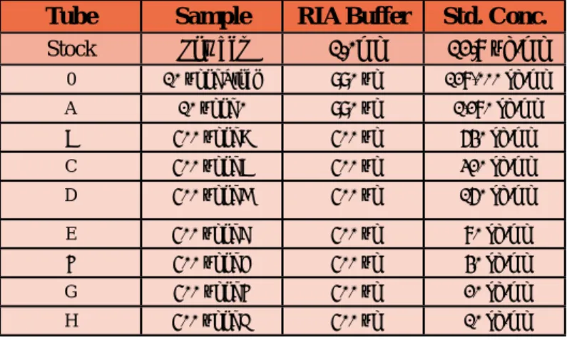
6. Prepare dilutions of the standard as below:
Tube Sample RIA Buffer Std. Conc.
Stock Powder 1.0ml 12.8 µg/ml 0 10 µl of Stock 990 µl 128,000 pg/ml A 10 µl of 0 990 µl 1,280 pg/ml B 500 µl of A 500 µl 640 pg/ml C 500 µl of B 500 µl 320 pg/ml D 500 µl of C 500 µl 160 pg/ml E 500 µl of D 500 µl 80 pg/ml F 500 µl of E 500 µl 40 pg/ml G 500 µl of F 500 µl 20 pg/ml H 500 µl of G 500 µl 10 pg/ml Table 1: Standard Dilutions
7. Set up initial RIA reactions (see diagram on page 5) in 12 x 75 mm polystyrene tubes.
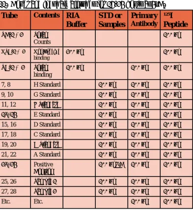
a) Number tubes TC-1, TC-2, NSB-1, NSB-2, TB-1, TB-2 and #7 - #22 for the standards.
b) Number tubes #23, #24 for the positive controls. c) Number tubes #25 up to #125 for the unkown samples. d) Pipette 200 µl of RIA buffer into each NSB tube. e) Pipette 100 µl of RIA buffer into each TB tube. f) Pipette 100 µl of standards H through A into duplicate
tubes #7-#22.
Note: Reverse the order of preparation so that the concentration increases as the number of the tube increases. For example: Pipette 100 µl of standard H into tubes #7 & #8.
g) Pipette 100 µl of positive control in tubes #23 & #24. h) Pipette 100 µl of unknown sample into duplicate tubes:
i) Pipette 100 µl of primary antibody (rabbit anti-peptide serum) into all tubes EXCEPT TC AND NSB TUBES. j) Vortex the contents of each tube.
k) Cover and incubate all tubes for 16-24 hours at 4°C.
8. Reconstitute the 125I-peptide with 13ml of RIA buffer and mix
well to make tracer solution. Please check the concentration of this tracer solution and adjust it with RIA buffer until the concentration (total activity) is 8,000-10,000 cpm/100 µl. 9. Add 100 µl of the tracer solution to each tube.
10. Vortex the contents in each tube.
11. Cover and incubate all tubes for 16-24 hours at 4°C.
Tube Contents RIA
Buffer STD or Samples Primary Antibody 125I Peptide TC-1 &2 Total Counts 100 µl NSB-1 & 2 Non-specific binding 200 µl 100 µl TB-1 & 2 Total binding 100 µl 100 µl 100 µl 7, 8 H Standard 100 µl 100 µl 100 µl 9, 10 G Standard 100 µl 100 µl 100 µl 11, 12 F Standard 100 µl 100 µl 100 µl 13, 14 E Standard 100 µl 100 µl 100 µl 15, 16 D Standard 100 µl 100 µl 100 µl 17, 18 C Standard 100 µl 100 µl 100 µl 19, 20 B Standard 100 µl 100 µl 100 µl 21, 22 A Standard 100 µl 100 µl 100 µl 23, 24 Positive Control 100 µl P.C 100 µl 100 µl 25, 26 Sample 1 100 µl 100 µl 100 µl 27, 28 Sample 2 100 µl 100 µl 100 µl Etc. Etc. 100 µl 100 µl
12. Reconstitute the Goat Anti-Rabbit IgG serum (GAR) with 13ml of RIA buffer
13. Reconstitute the Normal Rabbit Serum(NRS) with 13ml of RIA buffer.
14. Add 100 µl of GAR to each tube except the TC tubes. 15. Add 100 µl of NRS to each tube except the TC tubes. 16. Vortex the contents of each tube. Incubate all tubes at room temperature for 90 minutes
17. Add 500 µl of RIA buffer to each tube except the TC tubes and vortex.
18. Centrifuge all tubes (except the TC tubes) at 3,000 rpm (approx. 1700 x g) for 20 minutes at 4°C.
19. Carefully aspirate ALL the supernatant (without touching
the pellet) immediately following centrifugation (do not
decant as the pellet might be lost or excess liquid could be left). DO NOT ASPIRATE THE TC TUBES.
Note: For best results, the supernatant should be immediately aspirated after centrifugation. If the pellet sits for more than 15-30 minutes, it may become detached and make aspiration difficult. Do not aspirate any solids.
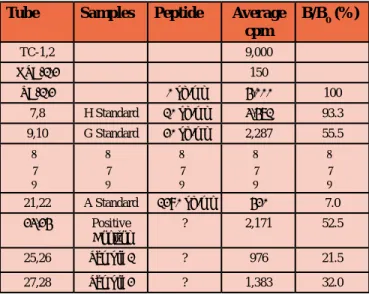
calculations:
1. Using cpm, calculate the average NSB and label this as NSB. 2. Using cpm, calculate the average TB and label this as TB. 3. To find B0 use the following equation: B0 = TB-NSB
4. To determine the B/B0 (%) for paired standards and unknown samples use the following calculation:
a) Example for standard H:
B/B0 (%) = (Avg. cpm Std. H) - (NSB) B0
b) Standards G through A (tubes #9-#22), Positive Controls (tubes #23 & #24) and the unknown samples (tubes #25 up to #125) are handled as shown above for standard H.
5. Examples of tabulated data:
Tube Samples Peptide Average cpm B/B0 (%) TC-1,2 9,000 NSB-1,2 150 TB-1,2 0 pg/ml 4,000 100 7,8 H Standard 10 pg/ml 3,471 93.3 9,10 G Standard 20 pg/ml 2,287 55.5 ▲ │ ▼ ▲ │ ▼ ▲ │ ▼ ▲ │ ▼ ▲ │ ▼ 21,22 A Standard 1280 pg/ml 420 7.0 23,24 Positive Control ? 2,171 52.5 25,26 Sample 1 ? 976 21.5 27,28 Sample 2 ? 1,383 32.0 x 100%
Total Count (Total activity) (cpm/100µl) = 9,000 cpm
NSB = 150 cpm TB = 4,000 cpm
B0 = 4,000 cpm - 150 cpm = 3850 cpm
6. On semi log graph paper, plot B/B0 (%) (in decimal scale )
versus the standard peptide concentrations (in log scale). a) Label the concentrations of standard H through A
(10-1280 pg/ml) on the X-axis (log scale).
b) Label B/B0 (%) (0 to 100%) on the Y-axis (decimal scale)
c) Plot B/B0 (%) for each standard concentration directly
above its X-axis designation. d) Draw the “Best-Fit” curve.
7. Determination of the concentration of peptide in unknown samples.
a) Using B/B0 (%) calculated for each unknown sample,
read across the graph to the point of intersection with the “Best-Fit” curve.
b) The corresponding X-axis coordinate is equivalent to the concentration of peptide (pg/ml) in the assayed sample. c) To calculate the amount of peptide in the original
sample, multiply the concentration of the assayed sample by any dilution factor used to prepare the sample. 8. Conversion of units:
Summary of aSSay Protocol
Add sample or standard and antibody ▼
Vortex and incubate 16-24 hours at 4ºC ▼
Add 125I-peptide
▼
Vortex and incubate for 16-24 hours at 4ºC ▼
Add GAR and NRS ▼
Vortex and incubate at room temperature for 90 minutes ▼
Add RIA buffer ▼
Vortex and centrifuge for 20 minutes at 1,700 x g ▼
Aspirate off the supernatant (except TC tubes) ▼
Count assay tubes ▼
suggested methodfor theextraction of peptides
from plasma
Blood Withdrawal:
Collect blood samples into Lavender Vacutainer tubes (Cat. No. VT-6450) which contain EDTA. Each tube can collect 7ml of blood/tube. Gently rock the Lavender Vacutainer tubes several times immediately after collection of blood for anti-coagulation. Transfer the blood from the Lavender Vacutainer tubes to centri -fuge tubes containing aprotinin (Cat. No. RK-APRO) (0.6 TIU/ ml of blood) and gently rock several times to inhibit the activity of proteinases. Centrifuge the blood at 1600 x g for 15 minutes at 4ºC and collect the plasma. Plasma kept at -70ºC is stable for up to one month.
Elution Solvents:
1. Buffer A (Cat. No. RK-BA-1) 2. Buffer B (Cat. No. RK-BB-1)
Extraction of Peptides from Plasma:
1. Acidify the plasma with an equal amount of buffer A. For example, if you are using 1ml of plasma, add 1ml of buffer A. Mix and centrifuge at 6,000 to 17,000 x g for 20 minutes at 4ºC. Keep the supernatant.
2. Equilibrate the SEP-COLUMN containing 200mg of C18 (Cat. No. RK-SEPCOL-1) by washing with buffer B (1ml, once) followed by buffer A (3ml, 3 times)
Note: From steps 3-5, no pressure should be applied to the column.
3. Load the acidified plasma solution onto the pre-treated C-18 SEP-COLUMN.
4. Slowly wash the column with buffer A (3ml, twice) and discard the wash.
5. Elute the peptide slowly with buffer B (3ml, once) and collect eluant into a polystyrene tube.
6. Evaporate eluant to dryness in a centrifugal concentrator or by a suitable substitute method
7. Dissolve the residue in RIA buffer for radioimmunoassay as follows: For normal subjects, dissolve in 250µl of RIA buffer for a two-tube assay. Aliquot 100µl into each tube (50µl is left over). Please note, at this point the sample has been concentrated by 4. If each tube is found to contain 100pg/ml of the peptide, then the total level of peptide in plasma = 100pg/ml ÷ 4 = 25pg/ml (where the concentration factor is 4). If upon assay the peptide value exceeds or does not fall in the range of detection, dilute or concentrate the sample
tips forextraction of plasma:
When using SEP-COLUMN for the first time, use the enclosed bulb to apply pressure to the column after addition of 1ml of buffer B to facilitate flow. From steps 3-5, no pressure should be applied. Ensure there is a constant flow for all solutions during the extrac -tion procedure. Do not allow air bubbles to enter the C-18 matrix for optimal sample processing and recovery.
Drying Sample After Extraction:
A combination of centrifugal concentrator (i.e Speedvac) and a lyophilizer (freeze-dryer) produces the best results for drying the sample after extraction. First, use a Speedvac to dry sample for approximately 15 minutes to remove the organic layer. Then snap-freeze the remaining sample, and freeze-dry overnight using a lyophilizer. This two-step procedure produces a more consistent fluffy powder that is easier to rehydrate than a sample dried only with a centrifugal concentrator. However, if a centrifugal concen -trator is not accessible, freeze-drying overnight using a lyophilizer will be sufficient.
references:
1. Berson, S.A. and Yalow, R.S. Kinetics of reaction between insulin and insulin binding antibody. J. Clin. Invest 36:873, (1957).
2. Patrono, C. and Peskar, B.A., (eds) Radioimmunoassay in ba -sic and clinical pharmacology. Heidelberg, Springer-Verlag, (1987).
3. Reuter, A., Vrindts-Gevaerts, Y., Meuleman-Gathy, R., Joris, J., Chretien, M. and Franchimont, P. A Radioimmunoassay for Beta-Endorphins. (BETA-END) and (BETA-LPH) in Plasma. Horm Res 25:236, (1987).
4. Dwenger, A. Radioimmunoassay: An Overview, J.Clin. Bio -chem. 22: 883, (1984)
5. Wang, Y.N., Chou J., Chang, D., Chang, J.K., Avila, C. and Romero, R. Endothelin-1 in Human Plasma and Amniotic Fluid. In Endothelin-Derived Contracting Factors, edited by G. Rubanyi and P. Vanchoutte, Karger, Basel, pg. 143, (1990).
CAUTION: SOME REAGENTS IN THIS KIT CONTAIN SODIUM
AZIDE WHICH MAY REACT WITH LEAD AND COPPER PLUMB
-ING TO FORM EXPLOSIVE METAL AZIDES. FLUSH WITH LARGE VOLUMES OF WASTE DURING DISPOSAL.
instructions for possession, handling and use of radioactive material
This radioactive material shall only be received, acquired, pos -sessed and used by physicians and veterinarians in clinical labo -ratories or hospitals for in vitro laboratory tests. Its use should not involve internal or external administration of the material and ra -diation therefrom to human beings or animals. Its receipt, acquisi -tion, possession, use and transfer are subject to the regulations and general license requirements of the U.S. Nuclear Regulatory Com -mission or of a State with which the Com-mission has entered into an agreement for the exercise of regulatory authority.
Precautions in Handling Radioactive Material:
The user should store the by-product material, until used, in the original shipping container or in a container providing equivalent radiation protection. There should be no drinking, eating or smok -ing while radioactive material is be-ing handled. Hands should be covered with gloves during, and thoroughly washed after the han -dling of radioactive material. When han-dling radioactive material do not pipette by mouth. Spills must be quickly and thoroughly cleaned up. Surfaces involves should be washed with an alkali de -tergent (alconox or the equivalent). Persons under 18 should not be permitted to handle radioactive material or enter radioactive areas.
Disposal:
Used radioactive test solutions must be disposed of by flushing down a laboratory sink drain with copious amounts of water. Ra -dioactive waste should be disposed of in compliance with Federal, State, and Local Government regulations.
THIS PACKAGE CONFORMS TO THE CONDITIONS AND LIMITA
-TIONS SPECIFIED IN 49 CFR173.421 FOR EXCEPTED RADIOAC
for research only not for use in diagnostic
procedures
USA
Phoenix PhArmAceUticAlS, inc.
330 Beach Rd. Burlingame, California 94010 Tel: 650-558-8898, 800-988-1205 Fax: 640-558-1686 [email protected] www.phoenixpeptide.com eUroPe Phoenix eUroPe Gmbh Viktoriastrasse 3-5. D-76133 Karlsruhe Germany Tel: +49 (721) 16 11 950 Fax: +49 (721) 16 11 952 [email protected]