R E V I E W
Tissue Cells Feel and Respond to the
Stiffness of Their Substrate
Dennis E. Discher,1* Paul Janmey,1
Yu-li Wang2 Normal tissue cells are generally not viable when suspended in a fluid and are
therefore said to be anchorage dependent. Such cells must adhere to a solid, but a solid can be as rigid as glass or softer than a baby’s skin. The behavior of some cells on soft materials is characteristic of important phenotypes; for example, cell growth on soft agar gels is used to identify cancer cells. However, an understanding of how tissue cells—including fibroblasts, myocytes, neurons, and other cell types—sense matrix stiffness is just emerging with quantitative studies of cells adhering to gels (or to other cells) with which elasticity can be tuned to approximate that of tissues. Key roles in molecular pathways are played by adhesion complexes and the actin-myosin cytoskeleton, whose contractile forces are transmitted through transcellular structures. The feedback of local matrix stiffness on cell state likely has important implications for development, differentiation, disease, and regeneration.
Anchorage dependence refers to a cell_s need for adhesion to a solid. Most tissue cells are simply not viable upon dissociation and sus-pension in a fluid, even if soluble proteins are added to engage cell adhesion moleculesEe.g., integrin-binding RGD peptide (1, 2)^. Fluids are clearly distinct from solids in that fluids will flow when stressed, whereas solids have the ability to resist sustained pushing and pull-ing. In most soft tissues—skin, muscle, brain, etc.—adherent cells plus extracellular matrix contribute together to establish a relatively elastic microenvironment. At the macro scale, elasticity is evident in a solid tissue_s ability to recover its shape within seconds after mild pok-ing and pinchpok-ing, or even after sustained com-pression, such as sitting.
At the cellular scale, normal tissue cells probe elasticity as they anchor and pull on their surroundings. Such processes are de-pendent in part on myosin-based contractility and transcellular adhesions—centered on in-tegrins, cadherins, and perhaps other adhesion molecules—to transmit forces to substrates. A normal tissue cell not only applies forces but also, as reviewed here, responds through cytoskeleton organization (and other cellular processes) to the resistance that the cell senses, regardless of whether the resistance derives from normal tissue matrix, synthetic substrate, or even an adjacent cell. Furthermore, physical properties of tissues can change in diseaseEas imaged now by magnetic resonance imaging (MRI) or ultrasound elastography (3–5)^, and cellular responsiveness to matrix solidity can
likewise change, as illustrated by the growth of cancer cells on soft agarEe.g., (6)^.
Contractile forces in cells are generated by cross-bridging interactions of actin and myosin filaments. For adherent cells, some of these forces are transmitted to the substrate (referred to as traction forces) and cause wrinkles or strains when the substrate consists of a thin film or a soft gel (7–12) (Fig. 1A). The cell, in
turn, is shown to respond to the resistance of the substrate, by adjusting its adhesions, cyto-skeleton, and overall state. Although con-siderable attention has been directed at the responsiveness of individual cells to external forces (outsideYin) that range from fluid flow to direct stretching and local twisting (13), we are now beginning to understand that cellular responses to cell-exerted forces involve a feedback loop of insideYoutsideYin that couples to the elasticity of the extracellular microenvironment. An analogy to muscle building is perhaps useful: A bicep is not built by passive flexing; the muscle must do active work against a load. Moreover, a load of 1 kg clearly feels different from a load of 2 kg. Similar sensitivity, growth, and remodeling principles seem to apply to most anchored cells.
On ligand-coated gels of varied stiffness, epithelial cells and fibroblasts (14) were the first cells reported to detect and respond
dis-1School of Engineering and Applied Science and Cell
and Molecular Biology Graduate Group, University of Pennsylvania, Philadelphia, PA 19104–6315, USA.
2Departments of Physiology and Cell Biology,
Uni-versity of Massachusetts, Worcester, MA 01655, USA. *To whom correspondence should be addressed. E-mail: [email protected]
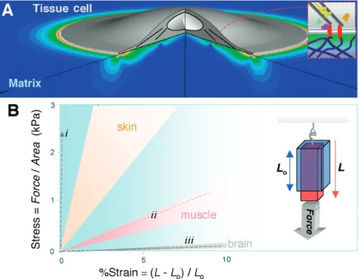
Fig. 1.Substrate strain and tissue stiffness. (A) Strain distribution computed in a soft matrix beneath a cell. The circular cell has a uniform and sustained contractile prestress from the edge to near the nucleus (81). (B) Stress versus strain illustrated for several soft tissues extended by a force (per cross-sectional area). The range of slopes for these soft tissues subjected to a small strain gives the range of Young’s elastic modulus,E, for each tissue (24, 28, 30). Measurements are typically made on time scales of seconds to minutes and are in SI units of Pascal (Pa). The dashed lines (- - -) are those for (i) PLA, a common tissue-engineering polymer (89); (ii) artery-derived acellularized matrix (90); and (iii) matrigel (42). PE CI A L
S
EC TI ONtinctly to soft versus stiff substrates. Although molecular pathways are still only partially known, muscle cells, neurons, and many other tissue cells have since been shown to sense substrate stiffness (15–17). Unlike cells on soft gels or in tissues, cells cultured on tissue-culture plastic or glass coverslips are attached (often via adsorbed matrix protein) to essen-tially rigid materials. The question therefore arises: Do cells perceive and respond to the rigidity of these conventional materials in ways that contrast with their behavior in much more compliant tissues, gels, or sub-layers of cells? The increasingly clear, af-firmative answer to this question appears important in its impact not just on standard cell culture but also, perhaps, in understand-ing disease processes, morphogenesis, and tissue-repair strategies.
Soft Tissue Benchmarks
Cells adhere to solid substrates that range in stiffness from soft to rigid and that also vary in topography and thickness (e.g., basement mem-brane). Regardless of geometry, the intrinsic resistance of a solid to a stress is measured by the solid’s elastic modulusE, which is most simply obtained by applying a force—such as hanging a weight—to a section of tissue or other material and then measuring the relative change in length or strain (Fig. 1B, inset). Another common method to obtainE involves controlled poking by macro- and micro-indenters, including atomic force microscopes (AFMs) (18,19). Many tissues and biomaterials exhibit a relatively linear stress versus strain relation up to small strains of about 10 to 20%. The slopeEof stress versus strain is relatively constant at the small strains exerted by cells (20), although stiffening (increased E) at higher strains is the norm (21, 22). Nonetheless, microscopic views of both natural and synthetic matrices [e.g., collagen fibrils and polymer-based mimetics (23)] suggest that there are many subtleties to tissue mechanics, particularly concerning the length and time scales of greatest relevance to cell sensing. Sample preparation or state is another obvious issue; for example, elastic moduli of whole brain in macroscopic mea-surements can vary by a factor of 2 or more, depending on specifics of preparation, tissue perfusion, etc. (24). In addition, with cells as well as tissues, many probing methods involve high-frequency stressing (25), whereas relevant time scales for cell-exerted strains seem likely to range from seconds to hours, motivating long time studies of cell rheology [recent cell me-chanics references (26, 27)]. Regardless, com-parisons of three diverse tissues that contain a number of different cell types show that brain tissue is softer than muscle (28,29), and muscle is softer than skin (30) (Fig. 1B). Although mapping soft tissue micro-elasticities at a resolution typical in histology seems impor-tant, the implication here is that there are
dis-tinct elastic microenvironments for epithelial cells and fibroblasts in skin, for myotubes in fiber bundles, and for neurons in brain.
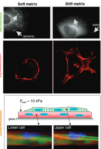
Correlations have long been made between increased cell adhesion and increased cell contractility [e.g., (31)], but it now seems clear that tactile sensing of substrate stiffness feeds back on adhesion and cytoskeleton, as well as on net contractile forces, for many cell types. Seminal studies on epithelial cells and fibroblasts exploited inert polyacrylamide gels with a thin coating of covalently attached collagen (14). This adhesive ligand allows the cells to attach and—by controlling the extent of polymer cross-linking in the gels—E can be adjusted over several orders of magnitude, from extremely soft to stiff. Images of adhe-sion proteins such as vinculin are revealing (Fig. 2, top): Soft, lightly cross-linked gels (EÈ1 kPa) show diffuse and dynamic adhesion complexes. In contrast, stiff, highly cross-linked gels (E È30 to 100 kPa) show cells with stable focal adhesions, typical of those seen in cells attached to
glass. Similarly, rigidifi-cation of cell-derived three-dimensional (3D) matrices alters 3D-matrix adhesions, because the adhesions are replaced by large, nonfibrillar fo-cal adhesions similar to those found on fixed 2D substrates of fibronectin (32). Consistent with a role for signaling in stiff-ness sensing, tyrosine phosphorylation on mul-tiple proteins (including paxillin) appears broad-ly enhanced in cells on stiffer gel substrates (14), whereas pharmacologi-cally induced, nonspecif-ic hyperphosphorylation drives focal adhesion for-mation on soft materials. Inhibition of actomyosin contractions, in contrast, largely eliminates promi-nent focal adhesions, whereas stimulation of contractility drives in-tegrin aggregation into adhesions (33). Ad-ditionally, although mi-crotubules have been proposed to act as ‘‘struts’’ in cells and thus limit wrinkling of thin films by cells (34), quantifica-tion of their contribu-tions to cells on gels shows that they provide only a minor fraction of
the resistance (14%) to contractile tensions; most of a cell’s tension is thus resisted by matrix (35).
Traction stresses (t, force per area) exerted by fibroblasts on gels were the first to be mapped by embedding fluorescent microbeads near the gel surface and then imaging bead displacements before and after cell detachment (10,20). Although larger tractions are exerted on stiffer gels, typical tractions ofbtÀ È1 kPa exceed by orders of magnitude the viscous fluid drag on any cell crawling in culture. In addition, mean cell tractions equate to mean gel strains that differ very little (eout0bt/EÀ ; 3 to 4%) between gels that differ by twofold inE. This suggests thateoutis sensed by cells as a tactile set-point, perhaps analogous to other physiological set-points such as extra-cellular ion concentrations or optimal growth factor concentrations. Furthermore, if matrix strain is approximately constant, then cells on soft gels need be less contractile than on stiff gels, and if they are less contractile, then
Fig. 2. Substrate stiffness influences adhesion structures and dy-namics (14), cytoskeleton assembly and cell spreading (17, 42), and differentiation processes such as striation of myotubes (28). (Top) The arrows point to dynamic adhesions on soft gels and static, focal adhesions on stiff gels. [Adapted from (14)] (Middle) The actin cyto-skeleton. (Bottom) A cell-on-cell layering in which the lower layer is attached first to glass so that the upper layer, which fuses from myoblasts that are added later, perceives a soft, cellular substrate.
S
PECIALS
EC TI ONtheir adhesions need not be as strong. This is consistent with a reduced adhesion strength as measured by reduced forces to peel cells from soft gels versus glass (28). This is also consistent with more dynamic adhesions on soft substrates (Fig. 2, top). Fluorescence imaging also shows increasingly organized F-actin and stress fibers on increasingly stiff substrates in fibroblasts (Fig. 2, middle). Neu-rons, in contrast, appear to apply very little stress to their substrate, because they can only deform very soft gels (36). Neurons also branch more on softer substrates (37), perhaps because the cytoskeleton is more pliable, if less structured.
Differentiation and a Cell-on-Cell Hypothesis
Cytoskeletal organization in muscle cells also depends on substrate stiffness and reveals an optimal substrate stiffness for striation of actomyosin (28, 38)—the contractile element of the myotube. On very soft gels that are micropatterned with collagen strips so as to generate well-separated myotubes, actomyosin appears diffuse after weeks in culture. On very stiff gels, as well as on glass micropatterns, stress fibers and strong focal adhesions pre-dominate, suggesting a state of isometric con-traction. Notably, however, on gels with an
elasticity that approximates that of relaxed muscle bundles (EÈ10 kPa), a large fraction of myotubes in culture exhibit definitive actomyosin striations. Actomyosin striation is even more prominent when cells are cultured on top of a first layer of muscle cells (Fig. 2). The lower myotubes attach strongly to glass and form abundant stress fibers, whereas the upper myotubes differentiate to the more physiological, striated state. Although cell-cell contact may provide additional signals, the elasticityEof the myotubes, as measured by atomic force microscopy, is in the same range as that of gels optimal for differentiation and—importantly—in the same range as that of normal muscle tissue.
Cell-cell contact appears to induce similar cell-on-gel effects for systems other than muscle. Astrocytes growing on glass, for ex-ample, appear to provide a soft cell ‘‘stroma’’ adequate for neuronal branching that is simi-lar to gels having brainlikeE (39). Cell-cell contact may have a similar effect when cells are grown at a high density. When endo-thelial cells are confluent, the cells have indistinguishable morphologies on soft versus stiff substrates (40), whereas cells attached only to an underlying stiff surface differ in their spreading and cytoskeletal organization (Fig. 2). Related results are also emerging
with epithelial cells and fibroblasts, as well as cardiomyocytes that show a tendency to ag-gregate and form cell-cell contacts in pref-erence to contact with soft gels (41). Such studies may set the stage for a better un-derstanding of mechanosensitivity in cell-cell interactions during embryogenic and tissue regeneration processes.
Materials ranging from fibrin gels and microfabricated pillars to layer-by-layer poly-mer assemblies (41–45) all suggest a similar trend of more organized cytoskeleton and larger, more stable adhesions with increasing Eas outlined here, despite likely differences in adhesive ligand density and long-time elasticity. However, the responses appear to be specific to anchorage-dependent and/or relatively contractile cells. Highly motile amoe-boid cells such as human neutrophils are perfectly viable in blood (a fluid) and do not appear to be sensitive to substrate stiffness; neutrophils spread on soft gels just as much as they do on stiff gels and glass, whether activated or not (46–48). Although additional study is needed and could prove ligand dependent, the initial contrast with cells derived from solid tissue highlights the compelling need for insights into molecular pathways of stiffness sensing in relation to anchorage dependence and contractility. Variation with cell type implies an active, regulated response, rather than a universal need of cells to exert traction forces on a stiff matrix. Differences no doubt depend in part on expression and engagement of adhesion molecules. Integrins reportedly undergo adhesion-modulating conforma-tional changes in response to force (49), and they also appear to be down-regulated on soft gels [e.g.,a5-integrin (40)]. However, over-expression of a5-integrin does not override the limited spreading of cells on soft gels, whereas overexpression of actin drives cyto-skeletal assembly and strongly promotes spreading (17).
Nonlinear Response to Compliance Signals and Molecular Effectors Myosin inhibitors—including a potent non-muscle myosin II inhibitor, blebbistatin (50)—have provided key evidence for the crit-ical role of contractility in substrate sensing (14,38). Important roles are also reported for integrating activator proteins of the Ras superfamily, especially Rho subfamily mem-bers that are broadly known to regulate the cytoskeleton, cell growth, and transcription. In cells such as fibroblasts, it is well established that Rho-stimulated contractility drives stress fiber and focal adhesion formation and that up-regulation of a smooth muscle actin correlates with contractility on rigid substrates (33, 51). Rac1 is another Rho family protein that when activated in macrophages, promotes engulfment of antibody-bearing soft beads, which otherwise are not engulfed (48). RhoA, Fig. 3.Substrate stiffness influences contractility, motility, and spreading. (A) Interplay of physical and
biochemical signals in the feedback of matrix stiffness on contractility and cell signaling as extended from (91). (B) Cells exert less tension on softer, collagen-coated gels but crawl faster (20), causing an accumulation of cells toward the stiff end of a soft-to-stiff gradient gel (54). Curves are schematic. [Image adapted from (54)] (C) Spread area,a, of smooth muscle cell versus ligand density and matrix stiffness, based on measurements fitted by a thermodynamic model (17). Similar nonlinear responses are also seen for adhesions, cytoskeleton organization, tractions exerted on the substrate, and other cellular processes.
PE CI A L
S
EC TI ONin contrast, has no observable effect in these measurements. Current views of signaling pathways, especially various physical signals (Fig. 3A), clearly implicate Rac in cell mo-tility (versus contracmo-tility)—indeed, myosin inhibition activates Rac (52). The involvement of contractile-effector proteins in sensing im-plies that cell crawling is also likely to be sensitive to substrate stiffness (Fig. 3B), as demonstrated in studies of the ‘‘cell on gel’’ effect with epithelial cells (14), fibroblasts (20), and smooth muscle cells (53,54). With the latter cell type, crawling speed appears maximal at an intermediate stiffness. The re-sult is reminiscent of a bell-shaped curve of crawling speed versus the concentration of adhesive ligand (55), which has been mathe-matically modeled as a shift in the balance between mediated traction and ligand-mediated anchorage (56). Additionally, smooth muscle cells on gels are slowed by inhibition of Rho kinase, suggesting that RhoA activity contributes to the tensions needed to detach any established adhesions at the rear of a motile cell (a process not needed in engulf-ment) (57). The dependence of cell crawling speed and direction on substrate stiffness, particularly gradients in stiffness, is now re-ferred to as ‘‘durotaxis’’ (20).
Molecular mechanisms involved in cellu-lar responses to matrix stiffness are still open to investigation, but it seems important to consider close relationships (or not) be-tween ‘‘insideYoutsideYin’’ pathways and ‘‘outsideYin’’ pathways (Fig. 3A). Adhe-sions on stiff materials are multifaceted mech-anosensors [for a review, see, e.g., (5)], and, on the one hand, contractility does appear to regulate the formation and dynamics of adhesion structures (14). Indeed, myosin II has a well-established role on rigid substrates in adhesion and cytoskeletal organization (33), as well as spreading (58) and cell tension (13). On the other hand, applying external forces to cells (outsideYin) leads to growth of focal adhesions on rigid materials, with or without myosin contractile forces (59). None-theless, insideYoutside activity can trigger outsideYin pathways such as the opening
of stress-activated channels (60), with induc-tion of calcium transients and activainduc-tion of calmodulin and myosin II.
Additional work from the outsideYin perspective has shown that stretching well-spread cells leads to deactivation of Rac (for G30 min) without affecting Rho activity (52). Stretching can also create new cytoskele-tal binding sites for activator and adapter proteins (61) and thus alter the balance be-tween protrusion and contractility. The mech-anism may involve conformational changes to uncover scaffold binding sites or other activities; for example, one key focal adhe-sion protein, talin, must unfold for vinculin binding (62–64), and although the unfolding forces are not yet clear, similar helical bundle cytoskeletal proteins unfold at forces that just a few myosin molecules can generate. On the other hand, fluid shearing of endothelial cells activates Rho and also increases cell traction forces (65), but how such stimulation— transient or sustained—depends on myosin ac-tivity and compares with substrate-mediated pulling forces or substrate compliance effects remains unknown.
The effects and effectors of contractility can be transient as well as nonlinear, but are nonetheless essential to clarify. The tempo-rary deactivation of Rac with stretch may have to be integrated over time to understand its place in signaling (66), and although myosin II activity is crucial for stiffness sensing, on rigid substrates it only delays the earliest phase of cell spreading (by È2 hours), ap-parently through stiffening of the cell cortex (67). Overstimulation of myosin, like over-stimulation of most motors, is also likely to slow and eventually stall cell migration. The effect may be related to the formation of less dynamic myosin assemblies on progressively stiffer substrates, fostering larger, more stable focal adhesions. Reconstitution experiments with mixtures of actin, myosin, adenosine 5¶ -triphosphate (ATP), and cross-linkers might lend important insight into motor-driven self-assembly processes.
Varied responses to mechanical signals at the cellular and molecular scales are
increas-ingly in need of multivariate analyses. More data are needed to define coupled responses to substrate stiffness, contractile state, ligand density, and activator levels, as well as ef-fects such as growth factor stimulation. A number of studies have already revealed nonlinear response maps, as illustrated by the spread area of cells on gels (Fig. 3C). Modeling efforts to date have been thermo-dynamic (17,68), kinetic (56), and—for cell-cell interactions—purely mechanical (69), but all generally yield nontrivial responses, saturable or even bell-shaped inEand other inputs. A major challenge in all such mod-eling is to clarify the principal enigma: how contractile traction forces exerted by a cell tend to increase with stiffness of the cell’s substrate.
Do Cells Feel Their Way in Organogenesis?
Cell type–dependent increases in contractility with increasing substrate stiffness may offer partial answers to some long-standing ques-tions of cell-cell organization. Random mix-tures of two cell types often lead to shell-core cell aggregates (Fig. 4), as first observed when heart cells segregated into the interior of a mass of retinal cells after 1 day in culture (70). Individual cell clusters form by ‘‘pulling’’ away from each other (71). Such observations are now being used to manipulate aggregate morphologies through printing of cell masses into gels as toroids and other shapes (72). Such phenomena have been explained by a ‘‘differential adhesion hypothesis’’ in which different cell types bear different numbers and types of adhesion proteins (e.g., cadherins), giving rise to an effective surface tension,g, at interfaces with cell layers (73). Although pos-sible contributions of cytoskeleton and cell tension have not yet been reported, studies of zebrafish embryos (74) have shown that (i) disruption of actin filaments dissociates cells entirely, even though cadherins remain at the cell surface; and (ii) the effect is potentiated by at least one drug that inhibits actomyosin contractility.
Quantitative estimates ofgfor the spherical aggregates of cadherin-expressing cells (73) exceed the rate-dependent cohesive strength of lipid bilayers [as low as 2 to 3 mN/m (75)] and suggest adhesion energies per cadherin that are orders of magnitude larger than would be expected of individual cadherin bonds. Such largegvalues could be due to the cytoskele-ton or even contractility (becauseg/btÀ ;1 to 10mm is a stress fiber length scale), especially because there is growing evidence of common RhoGTPase-cytoskeleton signaling among integrin- and cadherin-mediated adhesion (76–79). A major role for contractility in cell sorting was speculated long ago (80), but results reviewed here make it clear that contractile state can be strongly influenced by Fig. 4.Sorting of two cell types into a 3D shell-core aggregate (È300mm in diameter) in which low
expressers of N-cadherin (labeled in red) surround high expressers of N-cadherin (labeled in green) (73). Scanning electron micrograph of a typical spheroid’s surface shows well-spread cells. [Adapted from (73) with permission from Elsevier. Image courtesy of G. Forgacs, University of Missouri]
S
PECIALS
EC TI ONthe stiffness of the anchoring substrate. Heart cells pulling on equally stiff heart cells can generate a positive and steady feedback on their cytoskeleton that may not occur when these cells pull on other tissue cell types. Cell aggregation of less differentiated cells such as some stem cells that assemble into ‘‘embryoid bodies’’ has yet to be studied with myosin inhibitors or related methods, but the principles may extend to stem cell differentiation, partic-ularly because at least some stem cells express nonmuscle myosin II at levels similar to those of myoblasts (81).
Added Facets and Prospects
Mechanobiology is a broad field. Emphasized here is the recent recognition that most tissue cells not only adhere to but also pull on their microenvironment and thereby respond to its stiffness in ways that relate to tissue elasticity. Many emerging topics are not dealt with ad-equately in this brief review of substrate stiffness effects. These include in vitro models of fibrotic stiffening and related disease pro-cesses (82,83); perturbed secretion and uptake (84,85); 2D versus 3D responses (32,86); de-formations of fibronectin and other matrix molecules (87); structure formation such as capillary development (15,88); deeper aspects of cell differentiation such as with stem cells (81); the relative sensitivity and contractility of some cells relative to others; and broader effects of matrix elasticity, as well as fluidity (i.e., matrix rheology), on cells in tissue de-velopment, remodeling, and regeneration. For the cell biologist, this review may suggest the need for a better understanding of mechano-chemical pathways and the benefit of more biologically relevant elastic substrates than rigid coverslips and polystyrene for in vitro studies. For the applied biologist or bio-engineer, modified strategies for tissue repair and cell scaffolding may emerge, such as the development of fibrous scaffolds for cell seeding (23), where careful attention can be given to fiber flexibility. All of these topics seem likely to add to our rapidly growing recognition that tissue cells feel and respond to the mechanics of their substrate in many contexts.
References and Notes
1. E. Ruoslahti, M. D. Pierschbacher,Science238, 491 (1987).
2. H. L. Hadden, C. A. Henke,Am. J. Respir. Crit. Care Med.162, 1553 (2000).
3. For example, classic palpation self-exam reveals that tumor stiffening can be quantitated by both MRI (4) and ultrasound elastography (5).
4. A. L. McKnightet al.,AJR Am. J. Roentgenol.178, 1411 (2002).
5. J. Bercoff et al., Ultrasound Med. Biol. 29, 1387 (2003).
6. D. R. Welchet al.,Cancer Res.60, 1552 (2000). 7. A. K. Harris, P. Wild, D. Stopak, Science208, 177
(1980).
8. Since (7), methods for measuring traction force have become more quantitative by embedding beads in
either films (9) or gels (10) or by using deformable patterns (11) or posts (12).
9. T. Oliver, M. Dembo, K. Jacobson,J. Cell Biol.145, 589 (1999).
10. W. A. Marganski, M. Dembo, Y. Wang,Methods Enzymol. 361, 197 (2003).
11. N. Q. Balabanet al.,Nat. Cell Biol.3, 466 (2001). 12. J. L. Tanet al.,Proc. Natl. Acad. Sci. U.S.A.100, 1484
(2003).
13. F. J. Alenghat, D. E. Ingber, Sci. STKE 119, pe6 (2002).
14. (Fibroblasts and epithelial cells) R. J. Pelham, Y. Wang. Proc. Natl. Acad. Sci. U.S.A. 94, 13661 (1997) with Erratum95, 12070a (1998).
15. (Endothelial cells) C. F. Deroanne, C. M. Lapiere, B. V. Nusgens.,Cardiovasc. Res.49, 647 (2001). 16. (Transformed cells) H.B. Wang, M. Dembo, Y. Wang.,
Am. J. Physiol. Cell Physiol.279, C1345 (2000). 17. (Smooth muscle cells) A. Engleret al. Biophys. J.86,
617 (2004).
18. R. E. Mahaffy, C. K. Shih, F. C. MacKintosh, J. Kas, Phys. Rev. Lett.85, 880 (2000).
19. The Hertz model for a spherical probe of radiusR indenting a thick slab givesEÈf/d3/2R1/2where the
forcefindents to a depthd. The shear modulus,G, of an isotropic, incompressible material isG,E/3, reflecting the fact that shearing and tensile exten-sion are related by a rotation of frame.
20. C. M. Lo, H. B. Wang, M. Dembo, Y. Wang,Biophys. J. 79, 144 (2000).
21. C. Storm, J. J. Pastore, F. C. MacKintosh, T. C. Lubensky, P. A. Janmey,Nature435, 191 (2005).
22. Y. C. Fung,A First Course in Continuum Mechanics: For Physical and Biological Engineers and Scien-tists (Prentice Hall, Englewood Cliffs, NJ, ed. 3, 1994).
23. M. M. Stevens, J. George,Science310, 1135 (2005). 24. A. Gefen, S. S. Margulies, J. Biomech. 37, 1339
(2004).
25. S. Huet al.,Am. J. Physiol. Cell Physiol.287, C1184 (2004).
26. G. Bao, S. Suresh,Nat. Mater.2, 715 (2003). 27. F. Wottawah et al., Phys. Rev. Lett. 94, 098103
(2005).
28. (Skeletal Muscle Cells) A. J. Engleret al.,J. Cell Biol. 166, 877 (2004) .
29. Y. Yoshikawa, T. Yasuike, A. Yagi, T. Yamada,Biochem. Biophys. Res. Commun.256, 13 (1999).
30. S. Diridollou et al., Skin Res. Technol. 6, 214 (2000).
31. W. M. Leader, D. Stopak, A. K. Harris,J. Cell Sci.64, 1 (1983).
32. E. Cukierman, R. Pankov, D. R. Stevens, K. M. Yamada, Science294, 1708 (2001).
33. M. Chrzanowska-Wodnicka, K. Burridge,J. Cell Biol. 133, 1403 (1996).
34. O. J. Pletjushkinaet al.,Cell Motil. Cytoskeleton48, 235 (2001).
35. N. Wanget al.,Proc. Natl. Acad. Sci. U.S.A.98, 7765 (2001).
36. P. C. Bridgman, S. Dave, C. F. Asnes, A. N. Tullio, R. S. Adelstein,J. Neurosci.21, 6159 (2001).
37. (Neurons) L. A. Flanagan, Y. E. Ju, B. Marg, M. Osterfield, P. A. Janmey,Neuroreport13, 2411 (2002). 38. M. A. Griffin, S. Sen, H. L. Sweeney, D. E. Discher,
J. Cell Sci.117, 5855 (2004).
39. P. C. Georges, P. A. Janmey, in preparation. 40. T. Yeung et al., Cell Motil. Cytoskeleton 60, 24
(2005).
41. Y. Wang, in preparation.
42. P. C. Georges, P. A. Janmey,J. Appl. Physiol.98, 1547 (2005).
43. G. P. Raeber, M. P. Lutolf, J. A. Hubbell,Biophys. J.89, 1374 (2005).
44. A. Saez, A. Buguin, P. Silberzan, B. Ladoux,Biophys J., in press .
45. A. Engler, L. Richert, J. Wong, C. Picart, D. E. Discher, Surf. Sci.570, 142 (2004).
46. Phagocytosis of rigid yeast particles by neutro-phils involves a large contractile stress (È1 kPa) and implicates myosin (47), consistent with stiffness sensitivity of phagocytosis by macro-phages (48).
47. E. A. Evans, A. Leung, D. Zhelev,J. Cell Biol. 122, 1295 (1993).
48. (Macrophages) K. A. Beningo, Y. Wang,J. Cell Sci. 115, 849 (2002).
49. M. Jin, I. Andricioaei, T. A. Springer, Structure (Cambridge)12, 2137 (2004).
50. A. F. Straightet al.,Science299, 1743 (2003). 51. B. Hinz, G. Celetta, J. J. Tomasek, G. Gabbiani, C.
Chaponnier,Mol. Biol. Cell12, 2730 (2001). 52. A. Katsumiet al.,J. Cell Biol.158, 153 (2002). 53. (Smooth muscle cells) S. R. Peyton, A. J. Putnam,J. Cell.
Physiol.204, 198 (2005).
54. N. Zaari, P. Rajagopalan, S. K. Kim, A. J. Engler, J. Y. Wong,Adv. Mater.16, 2133 (2004).
55. S. L. Goodman, G. Risse, K. von der Mark,J. Cell Biol. 109, 799 (1989).
56. M. H. Zaman, R. D. Kamm, P. Matsudaira, D. A. Lauffenburger,Biophys. J.89, 1389 (2005). 57. P. Y. Jay, P. A. Pham, S. A. Wong, E. L. Elson,J. Cell
Sci.108, 387 (1995).
58. D. Rivelineet al.,J. Cell Biol.153, 1175 (2001). 59. L. P. Cramer, T. J. Mitchison,J. Cell Biol.131, 179
(1995).
60. A. Doyle, W. Marganski, J. Lee,J. Cell Sci.117, 2203 (2004).
61. M. Tamada, M. P. Sheetz, Y. Sawada,Dev. Cell7, 709 (2004).
62. I. Fillinghamet al., Structure (Cambridge) 13, 65 (2005).
63. Recent unfolding of spectrin helical bundles at force levels that myosins can generate are described in (64).
64. R. Lawet al.,Biophys. J.85, 3286 (2003). 65. Y. T. Shiuet al.,Biophys. J.86, 2558 (2004). 66. A. R. Asthagiri, C. A. Reinhart, A. F. Horwitz, D. A.
Lauffenburger,J. Cell Sci.113, 4499 (2000). 67. T. Wakatsuki, R. B. Wysolmerski, E. L. Elson,J. Cell
Sci.116, 1617 (2003).
68. A. Nicolas, B. Geiger, S. A. Safran,Proc. Natl. Acad. Sci. U.S.A.101, 12520 (2004).
69. I. B. Bischofs, U. S. Schwarz,Proc. Natl. Acad. Sci. U.S.A.100, 9274 (2003).
70. M. S. Steinberg,Science137, 762 (1962). 71. J. P. Trinkaus, J. P. Lentz,Dev. Biol.89, 115 (1964). 72. K. Jakab, A. Neagu, V. Mironov, R. R. Markwald, G. Forgacs, Proc. Natl. Acad. Sci. U.S.A. 101, 2864 (2004).
73. R. A. Foty, M. S. Steinberg,Dev. Biol.278, 255 (2005). 74. S. E. Zalik, E. Lewandowski, Z. Kam, B. Geiger,Biochem.
Cell Biol.77, 527 (1999).
75. E. A. Evans, V. Heinrich, F. Ludwig, W. Rawicz, Biophys. J.85, 2342 (2003).
76. N. K. Noren, W. T. Arthur, K. Burridge,J. Biol. Chem. 278, 13615 (2003).
77. W. T. Arthur, N. K. Noren, K. Burridge,Biol. Res.35, 239 (2002).
78. In vitro studies of cadherin-coated surfaces show no cytoskeleton assembly and no response to contract-ile inhibitors (79).
79. H. Delanoe-Ayari, R. Al Kurdi, M. Vallade, D. Gulino-Debrac, D. Riveline,Proc. Natl. Acad. Sci. U.S.A.101, 2229 (2004).
80. A. K. Harris,J. Theor. Biol.61, 267 (1976). 81. A. Engler, S. Sen, H. L. Sweeney, D. E. Discher, in
preparation.
82. R. G. Wells,J. Clin. Gastroenterol.39, S158 (2005). 83. M. A. Griffinet al.,J. Cell Sci.118, 1405 (2005). 84. E. J. Semler, P. A. Lancin, A. Dasgupta, P. V. Moghe,
Biotechnol. Bioeng.89, 296 (2005). 85. H. J. Konget al.,Nat. Mater.4, 460 (2005). 86. K. A. Beningo, M. Dembo, Y. Wang,Proc. Natl. Acad.
Sci. U.S.A.101, 18024 (2004).
87. G. Baneyx, L. Baugh, V. Vogel,Proc. Natl. Acad. Sci. U.S.A.99, 5139 (2002).
88. A. L. Sieminski, R. P. Hebbel, K. J. Gooch,Exp. Cell Res.297, 574 (2004).
89. Y. Shikinami, M. Okuno, Biomaterials 24, 3161 (2003).
90. P. F. Gratzer, J. P. Santerre, J. M. Lee,Biomaterials.25, 2081 (2004).
91. K. Rottner, A. Hall, J. V. Small,Curr. Biol.9, 640 (1999).
92. Support was provided by NIH, NSF-PECASE (D.E.D.), and Penn’s NSF-MRSEC.
10.1126/science.1116995 PE CI A L