10
Pain in Joint Diseases
B. A. Michel and P. Greminger10
10.1
Inflammatory Rheumatic Joint
Disorders
338
Rheumatoid Arthritis 338
Felty Syndrome 339
Adult Still Disease 339
Sjögren Syndrome 339
Juvenile Chronic Arthritis 340 Spondylarthropathies 341 Ankylosing Spondylitis
(Bekhterev Disease) 341 Psoriatic Arthritis 342 Reactive Arthritis (Reiter syndrome) 343
Rheumatic Fever 343 Arthropathies Associated with Enterocolitis 343 Behçet Disease 344 SAPHO Syndrome 344 Undifferentiated Spondylarthropathy 344 Arthropathies Associated with
Metabolic Diseases 345 Arthritis Urica (Gout) 345 Chondrocalcinosis (Pseudogout) 345 Diffuse Idiopathic Skeletal Hyperostosis
(DISH) 346 Ochronosis (Alkaptonuria) 347 Primary Amyloidosis 347 Hemochromatosis 347 Wilson Disease 348 Other Arthropathies 348 Hematologic Disorders 348 Arthrititis Associated with Neoplasms 348 Arthropathies in Endocrine Disorders 348 Arthropathies in Neurologic Disorders 348 Cartilage Disorders 348
10.2
Degenerative Joint Disorders
349
Osteoarthritis 349
Degenerative Disease of the Spine (Ostearthritis of the Intervertebral
Joints, Spondylosis Deformans) 350
10.3
Soft Tissue Rheumatism
352
Fibromyalgia 352
Periarthropathies 352 Periarthropathia Humeroscapularis 353 Other Localized Periarthropathies 353
General Differential Considerations in Joint Pain
The symptom “joint pain” has to be clinically substan-tiated. In not all cases does the joint itself represent the origin of the disorder. Affections of soft tissue may also give rise to joint symptoms. Joint disorders usually are as-sociated with the following symptoms:
앫 swelling (sometimes with effusion) 앫 warmth
앫 pain upon pressure 앫 functional deficit.
Acute monoarthritis needs immediate evaluation: an in-fectious disorder must be considered. Other forms of acute joint inflammation include arthritis in gout and pseudogout (calcium pyrophosphate arthropathy). These affections are often associated with severe redness of the skin and tenderness. Other joint disorders usually take a chronic course from the very beginning, such as rheumatoid arthritis, connective tissue diseases, and osteoarthritis. Joint disorders of the spondylarthropathy group usually show intermittent acute to chronic courses.
10.1
Inflammatory Rheumatic Joint Disorders
Rheumatoid Arthritis
Epidemiology.Rheumatoid arthritis is the most frequent inflammatory rheumatic joint disease. Women are af-fected about three times more often than men. Clinical Findings.Symmetric distribution of the joint dis-orderis characteristic. In the early phase of the disease hand, metacarpophalangeal and interphalangeal joints (Fig. 10.1), as well as knee and metatarsophalangeal joints, are affected. Involvement of the distal inter-phalangeal joints is rare and points to differential diag-noses, such as psoriatic arthritis or reactive arthritis. Usual symptoms of rheumatoid arthritis include joint pain and swelling, often associated with severe and long-standing morning stiffness, as well as loss of strength, particularly in the hands. Fatigue and general malaise, and at times slightly elevated temperatures, are often the first indications of theongoing disease process.
Without effective medication, rheumatoid arthritis is characterized by functional deficit due to progressive joint destruction. Late stages are characterized by de-formities, rheumatic nodules, as well as postinflam-matory changes of bones, joints, and soft tissue.
In later stages of the diseaseextra-articular manifesta-tionsmay occur. This includes pleuropericarditis, rheu-matic nodules, eye involvement, and more rarely, vasculi-tis with sensorimotor disturbances or amyloidosis. Diagnosis.Already in the early course of the disease radi-ologic changesmay be detected in the hands and feet. In the early stage changes include periarticular soft tissue swelling and demineralization of the periarticular bones. In later stages they include joint space narrowing along with erosions and subluxations (Fig. 10.2). It is rare for ankylosis to be a feature. Involvement of the cervical
Fig. 10.1 Rheumatoid arthritis with joint swelling and moderate ulnar deviation of the fingers.
Table 10.1 Sjögren syndrome
Sicca complex and connective tissue disease Xerophthalmia Rheumatoid arthritis Xerostomia Systemic sclerosis
Systemic lupus erythematosus Periarteritis nodosa
Dermatomyositis
spine however is frequent. Rheumatoid arthritis may lead to spondylarthritis, instability, or rarely ankylosis, but also to destruction of the atlantal dentate ligaments through inflammatory pannus resulting in atlantoaxial subluxation or even compression of the spinal cord.
Laboratory examination often reveals elevated ery-throcyte sedimentation rate (ESR) and C-reactive pro-tein (CRP), normochromic and normocytic anemia, thrombocytosis, and low serum iron. Rheumatoid fac-tors tend to be positive at later stages.
The diagnosis of rheumatoid arthritis is based on the history, typical clinical findings (pattern of joint in-volvement), as well as radiographic and laboratory findings.
Differential Diagnosis.In the differential diagnosis the following disorders have to be considered:
➤ connective tissue diseases (particularly systemic lupus erythematosus and systemic sclerosis)
➤ polymyalgia rheumatica (patients over 60 years of age)
➤ Parvovirus-induced arthritis (usually self-healing)
➤ osteoarthritis of the fingers.
Rarely, difficulties in the delineation of reactive arthritis are encountered. Most often this disorder shows asym-metric oligoarticular joint involvement along with en-thesiopathies.
Felty Syndrome
Felty syndrome is a rare, systemic manifestation of
rheumatoid arthritis. Its hallmarks are hepato-splenomegaly, leukopenia, and frequently treatment resistant skin ulceration of the lower extremities. The majority of patients show rheumatoid nodules, lymph-adenopathy, high titer rheumatoid factors, as well as antinuclear antibodies. A genetic disposition is charac-terized by the association with HLA-DR4, which is en-countered in over 90 % of patients.
Adult Still Disease
Clinical Findings.Still disease is arare form of rheumatoid arthritis. Men and women less than 40 years of age are equally effected. Acute episodes of this inflammatory disorder are characterized by high fever spikes (usually over 39 °C). Arthralgias or even oligoarthritis (especially wrists), phalangitis, weight loss, and transient, salmon-colored exanthema of the trunk and the proximal ex-tremities are characteristic manifestations.
Further findings include hepatosplenomegaly, lym-phadenopathy, elevated ESR, marked leukocytosis, and very high values for serum ferritin. Rheumatoid factors and antinuclear antibodies are negative.
Fig. 10.2 Rheumatoid arthritis: considerable erosions, joint space narrowing, and periarticular osteopenia.
Differential Diagnosis.Other causes of fevers such as in-fections and inflammatory intestinal disorders, includ-ing Crohn disease and colitis ulcerosa, have to be con-sidered in a differential diagnosis.
Sjögren Syndrome
Definition and Epidemiology. Sjögren syndrome is characterized by inflammatory involvement of tear, salivary, and, mucosal glands (in the intestinal and the pulmonary airways) resulting insicca symptoms. The syn-drome may evolve as a primary synsyn-drome or as an accom-panying disorder in association with rheumatoid arthritis or another connective tissue disease (Tab. 10.1). Women over 50 years of age constitute more than 90 % of patients.
Fig. 10.3 Dry, cracked tongue in Sjögren syndrome. 79-year-old woman.
Fig. 10.4 Swelling of both parotid glands in Sjögren syn-컄 drome. 50-year-old woman.
Clinical Findings. Dryness leads to the characteristic manifestations, which are:
➤ xerophthalmia (keratoconjunctivitis sicca with sen-sation of foreign bodies, burning, and redness)
➤ xerostomia (Fig. 10.3) with impaired swallowing, hoarseness, coughing, and development of severe caries.
Characteristic are recurrent, symmetrical, painful, en-larged salivary glands, particularly the parotid glands (Fig. 10.4). Further symptoms include fatigue, fever, arthritis similar to rheumatoid or pillar arthralgias, lymphadenopathy, vasculitic ulcerations, particularly of the legs, as well as neuropathies. Renal involvement is more rare (interstitial nephritis, tubular acidosis). Tran-sitions to malignant lymphomas may occur.
Diagnosis.ESR is often highly elevated. Further typical findings include hypergammaglobulinemia, rheuma-toid factors, and anti-SS-A (Ro), as well as anti-SS-B (La) antibodies.
Differential Diagnosis. Treatment with antidepressants (dry mouth), sarcoidosis, and HIV infection have to be considered in the differential diagnosis. The diagnosis of Sjögren syndrome is strongly supported with the posi-tive Schirmer test (measurement of tear flow), as well as biopsy of the lips.
Juvenile Chronic Arthritis
Classification. Juvenile chronic arthritis (JCA) is classified according to its presentation at the onset of disease. There are three forms:
➤ Thesystemic form(Still disease) is characterized by episodes of fever with salmon-colored rash, hepato-splenomegaly, and lymphadenopathy. Often arthritic complaints will follow.
➤In thepolyarticular form, distal finger joints are often involved, in contrast to rheumatoid arthritis in adults. In this form, rheumatoid factor usually is negative.
➤ The oligoarticular form shows two special aspects: under the age of four years destructive iridocyclitis may occur; children with positive antinuclear anti-bodies should undergo regular ophthalmologic ex-amination since iridocyclitis may be asymptomatic. Oligoarthritis after the age of eight years usually dis-plays features similar to ankylosing spondylitis. Differential Diagnosis.The following disorders have to be considered: acute rheumatic fever, bacterial polyar-thritis, tuberculosis, and sarcoidosis. In the differential diagnosis puncture or synovial biopsy of the involved joint are helpful.
Table 10.2 Frequency of HLA-B27 and sacroiliitis in spondylar-thropathies HLA-B27 (%) Sacroiliitis (%) Ankylosing spondylitis 95 100 Reactive arthritis (Reiter
syndrome) 70 30 Psoriatic arthropathy 50 20 Enterocolitic arthropathy 50 20 SAPHO syndrome 40 30 Undifferentiated spondyl-arthropathy 50 20
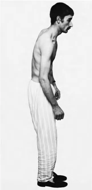
Fig. 10.5 Typical positioning of a patient with Bekhterev dis-ease.
Spondylarthropathies
Common Features.Tab. 10.2lists all disorders within the family of spondylarthropathies. These inflammatory, mostly chronic, musculoskeletal diseases share clinical, radiologic, histopathologic, and genetic features:
➤ peripheral, usually oligoarticular arthritis involve-ment of the spine and sacroiliacal joints
➤ involvement of tendons and tendon insertions (en-thesiopathy)
➤ extra-articular features (eyes, skin, mucosa, more rarely heart and lungs)
➤ familiar aggregation and association with HLA-B27. Often these diseases are calledseronegativedue to the fact that rheumatoid factors and autoantibodies are usually not found.
Ankylosing Spondylitis
(Bekhterev Disease)
Clinical Findings.Ankylosing spondylitis represents the classical spondylarthropathy. This chronic inflam-matory disorder affects sacroiliacal, costovertebral, and facet joints leading to progressive ankylosis. Hips and shoulders are most commonly affected whereas other joints are less often involved. Systemic manifestations are rare (uveitis anterior, aortitis with aortic insuffi-ciency, pulmonary fibrosis).
Men are affected more often and more severely than women. First symptoms usually develop between the ages of 20−40. Typical symptoms include low back and buttock pain at night, radiating towards the dorsal aspects of the knees. Motion is followed by decreased pain. Stressing the sacroiliacal joints is painful due to in-flammation (Mennel or Patrick sign). Axial involvement often leads to early ankylosis with typical deformities:
hyperkyphosis of the thoracic spine, flattening of the lower spine (Fig. 10.5). Plantar and achilles enthe-siopathies are frequent origins of heel pain. Hip involve-ment leads to a tendency towards contracture.
Diagnosis.Although elevated ESR is typical, it may also be normal. Typical changes of the sacroliacal joints in radiographs show narrowing of the joint space along with sclerosing and erosive features (Fig. 10.6), finally leading to ankylosis. Involvement of the spine is charac-terized by ossification of the ligaments (Fig. 10.7).
Fig. 10.6 Sacroiliac joints in Bekhterev disease.
Fig. 10.7 Ossified ligaments along the spine in Bekhterev dis-컄 ease.
Fig. 10.8 Psoriatic arthritis
Hallmarks for diagnosis are the classical history of pain, clinical features, and radiographic examina-tion. Determination of HLA-B27 usually is not neces-sary.
Psoriatic Arthritis
Clinical Findings. The typical pattern of joint involve-ment in psoriatic arthritis is either characterized by ar-thritis of all finger joints (DIP, PIP, and MCP joints) re-sulting in a so-called dactylitis or sausage finger, as well as a transverse involvement of the DIP joints. In contrast to rheumatoid arthritis, the joint swelling is rather tight and the skin often shows a red livid coloration. Arthritis may appear after skin involvement, which is especially the case in children. Radiographically, the hallmark findings consist in both ankylosing ossification and de-structive processes at the same time. Given this typical feature, diagnosis is often possible without skin involve-ment (Fig. 10.8). Nails may show so-called oil spots or onycholysis, particularly when distal joints of fingers or toes are affected. The typical course of psoriatic arthritis is characterized by episodes of highly acute disease and long-standing remissions. Involvement of the spine and the sacroiliac joints (mostly asymmetrical) is less frequent, as compared to ankylosing spondylitis.
10
Pain in Joint Diseases
Differential Diagnosis.Similarities exist with rheuma-toid arthritis (symmetrical pattern of joint involvement without affecting the DIP joints), osteoarthritis of the fingers (joint involvement preferentially of DIP and PIP joints), reactive arthritis (manifestation after intestinal or urogenital infection), as well as crystal arthropathies (joint fluid examination).
Reactive Arthritis (Reiter syndrome)
Definition.Reactive arthritis in its full form is called Re-iter syndrome. The syndrome is characterized by ar-thritis, urear-thritis, and conjunctivitis, occasionally as-sociated with mucocutaneous lesions. Men between 20 and 40 years of age are most often affected.
Causes. Reactive arthritis affects women and men equally after intestinal infections. After urogenital infec-tions most patients are men. Triggering microorganisms includes Salmonella, Shigella, Campylobacter, Yersinia,
Brucella, as well asChlamydiaandUreaplasma. In con-trast to septic arthritis, these microorganisms can not be cultivated from joint tissue or fluid.
Clinical Findings. The first symptoms of a reactive ar-thritis may evolve only a few weeks after intestinal or urogenital infection. Along with fatigue and occasional fever, several manifestations may occur. Usually, acute asymmetrical oligoarthritis of larger joints of the lower extremities is the first manifestation. Other manifesta-tions include involvement of a single finger or toe with livid skin coloration (so-called dactylitis or sausage fin-ger), spondylarthropathy with lower back pain in the early morning and stiffness of the spine, frequently along with asymmetric involvement of the sacroiliac joints and inflammatory changes in tendon sheaths, tendons, and ligaments. Extra-articular symptoms are found in the skin and mucosa (keratoderma blennor-rhagicum of palms and soles, erythema nodosa, oral ulceration), eyes (often conjunctivitis), and urogenital tract (sterile urethritis, balanitis and cystitis), as well as in the gastrointestinal tract (enteritis). Only very oc-casionally, nail changes may occur similar to the ones encountered in psoriasis.
Diagnosis.The culture of the feces, as well as PCR exami-nation of the serum may detect the enteral infectious agents, as well asChlamydiain the urine in the early stages of the disease. Determination of HLA-B27 does not really contribute to confirming the diagnosis, al-though 70 % of the cases are positive. In the differential diagnosis gonococcal urethritis with septic arthritis should be considered.
Rheumatic Fever
The typical example of a reactive arthritis is rheumatic fever. Currently, this disorder is very rarely encountered in Europe. After infection with beta-hemolytic strepto-cocci group A, fever and polyarthritis, predominantly of the larger joints, carditis, and later chorea minor, tran-sient erythema anulare marginatum of the trunk and thighs, as well as subcutaneous rheumatic nodules, may evolve.
Anti-streptolysin titers should always be considered in association with the history and clinical findings, since this titer is not specific for rheumatic fever.
Arthropathies Associated
with Enterocolitis
Colitis ulcerosa and Crohn disease may be associated with inflammatory changes of the spine and peripheral joints in about 10 %−20 % of patients. More rare causes of enterocolitic arthropathies are Whipple disease, ar-thritis after gastrointestinal bypass operation, as well as gluten-induced enteropathy.
Clinical Findings.Incolitis ulcerosathe arthropathy most often evolves after the enteric symptoms. In contrast, joint involvement is not really a primary manifestation inCrohn disease, although inflammatory changes in the gastrointestinal tract may be detected endoscopically in the early stages.
Axial involvement as well as sacroiliitis may precede intestinal symptoms by years. Clinical and radiographic findings often can not be distinguished from classical ankylosing spondylitis. This activity in the peripheral joints often reflects the general intestinal inflammatory activity. Spinal inflammation however appears to take place independently. In contrast to ankylosing spondyl-itis, arthritis in enterocolitis disorders lasts only from a few days to a few weeks and may change location frequently. Sacroiliitis associated with gastrointestinal diseases often remains asymptomatic and is frequently detected by chance in radiographic examinations.
Systemic manifestationsinclude anterior uveitis (up to 10 %, most often along with axial involvement), pain-ful stomatitis ulcerosa, erythema nodosa, and pyoderma gangrenosum.
In Whipple disease arthralgias or transient nonde-structive arthritis of small and larger joints may precede abdominal manifestations by years. Involvement of the spine and sacroiliac joints, as well as spondylarthritis is rare. Men between 40 and 60 years of age should be ex-amined forTropheryma whippleiif any arthritic condi-tion of unclear origin occurs. The leading clinical symp-toms for Whipple disease are abdominal discomfort with diarrhea and weight loss, slightly elevated
temperatures, lymphadenopathy, uveitis, and more rarely eye muscle paresis and encephalopathy.
Behçet Disease
Behçet disease belongs to the vasculitides. Most patients are originally from the eastern Mediterranean regions and the Orient. The main rheumatological com-plaints include chronic synovitis of larger and smaller joints. The diagnosis is based on two main symptoms plus one minor symptom.
Clinical Findings. Main symptoms are:
➤mucosal ulcerations in the mouth and/or intestinal tract
➤genital ulcerations (vulva, penis, scrotum)
➤ocular manifestations (uveitis anterior, hypopyon, retinal vasculitis).
Minor symptoms are:
➤skin disease (erythema nodosa, folliculitis)
➤arthritis (predominantly knee or ankle joints)
➤neurologic symptoms (meningitis, involvement of cranial nerves)
➤vessel disease (venous thrombosis, arterial aneu-rysms).
SAPHO Syndrome
Definition.SAPHO describes the most frequent manife-stations of the syndrome: synovitis, acne, pustulosis,
hyperostosis, and osteomyelitis. Men and women are equally affected and the disorder may evolve in patients of any age.
Fig. 10.9 SAPHO syndrome with swelling of the clavicular-costosternal area on the right side.
Clinical Findings.A hallmark finding of this disorder of unclear origin is an often asymmetrical, painful swelling in the clavicular-costosternal area (Fig. 10.9). The fol-lowing features are frequent:
➤sternoclavicular hyperostosis
➤palmoplantar pustulosis
➤inflammatory axial involvement
➤peripheral oligoarthritis, particularly of large joints. Manifestations often evolve sequentially; skin and bone symptoms may be found many years apart. Low back pain, axial stiffness, and painful joint swellings are, as with other spondylarthropathies, typical. Palmoplantar pustulosis is characterized by well delineated vesicles or pustules, as well as superficial scaling of the palms and soles. Differentiation from psoriatic skin disease is often not possible. As a complication of clavicular-costal hy-perostosis, thrombosis of the subclavian vein or the su-perior vena cava may occur as a consequence of com-pression.
Diagnosis.Imaging of the strong increase in activity in bones and joints is best done by skeletal scintigraphy. At times, bone changes may simulate infectious osteomy-elitis or tumors in radiographs. Such features therefore need more intensive examination.
Undifferentiated Spondylarthropathy
In about 30 % of all patients with spondylarthropathies no specific diagnosis is possible. Many manifestations of spondylarthropathies may occur without fulfilling clear criteria for a specific disease, such as isolated per-itendinitis of the achilles tendon or severe low back pain in the early morning in a young man. Such, mostly early, forms of spondylarthopathies are classified as undiffer-entiated.
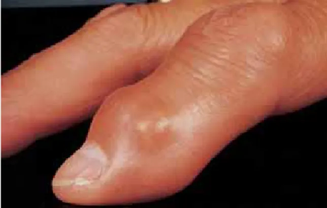
Fig. 10.10 Chronic tophaceous gout of the index finger.
Arthropathies Associated with Metabolic Diseases
Arthritis Urica (Gout)
Epidemiology and Causes.Primary goutin men occurs about 10 times more often than in women. In men, gout develops most frequently between the ages of 40 and 50 years, but in women usually only after the age of 60 years. Triggering factors include rich meals with high purine intake and/or alcohol consumption.
Clinical Findings.Classical acute goutis always very pain-ful, it occurs during the night, and patients may not be able to walk on the inflamed joints. Typically, the in-volved joint is red, swollen, and extremely painful. Slightly raised temperatures, elevated ESR, and leucocy-tosis are the rule. Without treatment the acute attack will subside in about a week, leaving the involved joint slightly painful for longer periods. The first metatar-sophalangeal joint is most often involved, however, gout may occur in any joint. In such cases the differential di-agnosis of acute septic arthritis or reactive arthritis, as well as psoriatic arthritis has to be considered. Acute onset in rheumatoid arthritis is also possible. In elderly people, pseudogout (chondrocalcinosis) has to be differ-entiated from gout.
Inlater stages of the diseasedeposition of urate oc-curs in tendons, bursa, and joints, which is known as chronic tophaceous gout (Fig. 10.10). Tophi are located most often in the neighborhood of the involved joints, but may be found at the external ear as well (Fig. 10.11). Diagnosis. In radiographs, well-delineated erosions at the end of bones are characteristic for gout (Fig. 10.12). The diagnosis of gout is supported by increased serum uric acid levels and proven by the positive finding of uric acid crystals in the joint fluid.
Pathogenesis. The cause of primary gout is multifac-torial. About 20 % of cases show enzyme defects leading to the overproduction of uric acid. In all other patients uric acid is not excreted sufficiently, probably based on an epithelial insufficiency.
Acute attacks of gout with normal or only slightly in-creased serum uric acid may occur. This is especially true when uricosuric agents have been taken before the attack.
In elderly patients pseudogout (see Chondrocalcinosis, below) always has to be considered. Serum uric acid levels do not parallel clinical symptoms. However, most patients with increased serum uric acid of over 600μmol/L will sooner or later be symptomatic.
Complications.The most important complication of gout is the so-calledgout kidney. Impairment of the kidney is a consequence of hyperuricemia and increased excre-tion of uric acid. On histological examinaexcre-tion, inflam-matory interstitial infiltrates as a consequence of deposition with uric acid are found along with an even-tual pyelonephritis caused by lithiasis, as well as vascu-lar changes (nephrosclerosis). Many patients with gout develop hypertension. Therefore it may be difficult to differentiate between secondary consequences of hy-pertension or gout when interpreting renal insuffi-ciency. Gout nephropathy itself may lead to hyperten-sion.
Hyperuricemia and gout are often associated with diabetes mellitus, hyperlipoproteinemia, and hyperten-sion; therefore gout is considered a risk indicator. Whether gout alone or only in combination with other risk factors may lead to coronary sclerosis is controver-sial.
Secondary Gout.Features of gout may evolve in all dis-eases associated with increased cell death (e. g., myelo-and lymphoproliferative diseases) or by any treatment leading to decreased excretion of uric acid, such as diuret-ics. In addition, secondary hyperuricemia may be found in ketosis (fasting, decompensated diabetes mellitus, fat-rich meals), acromegaly, hypo- and hyperparathyroid-ism, CO intoxication, lead intoxication, myxedema, and with the intravenous application of fructose.
Chondrocalcinosis (Pseudogout)
Deposition of calcium pyrophosphate crystals may lead to joint inflammation. The acute attack may not be differentiated clinically from a gout joint attack. Most often larger joints are involved. Differentiation of crys-tals is possible by the examination of joint fluid. Radio-graphically, typical calcifications may be found in
meniscae and superficial layers of the joint cartilage (Fig. 10.13).
Chondrocalcinosis has a preference for middle aged and elderly persons, but may occur at any age. Often chondrocalcinosis will be found as an accompanying disorder in damaged joints (osteoarthritis, posttrau-matic arthritis) or in metabolic diseases
(hyperparathy-Fig. 10.11 Auricular tophus. 72-year-old man. Fig. 10.12 Cystic defects in the bone in a patient with gout.
Fig. 10.13 Chondrocalcinosis with calcifications of meniscus and cartilage.
roidism, hematochromatosis, Wilson disease, gout, ochronosis).
Axial involvement is rare and may show calcifica-tions of the intervertebral disks. The course of axial dis-ease is clinically silent with the diagnosis most often re-sulting from a chance finding in a radiograph of the vertebrae.
Diffuse Idiopathic Skeletal Hyperostosis
(DISH)
Axial hyperostosis is often detected in radiographs by chance. This disorder is characterized by overshooting ossifications with osseous bridge building between single vertebrae without narrowing of the interverte-bral spaces. It is based on ossification of the longitudinal ligaments of the spine. The right side of the thoracic spine is preferentially involved, however, ossifications may develop at any site including peripheral joints.
Usually hyperostosis is followed by clinical ankylosis, whereas occurrence of pain is rare. In the shoulder area, severe symptoms may be due to hyperostosis at the in-ferior aspect of the acromion leading to irritation of the subacromial area (impingement). In the pathogenesis,
10
Pain in Joint Diseases
Fig. 10.14 Dark discoloration of the external ear in ochronosis.
Fig. 10.15 Bands of calcifi-cation of the intervertebral disks in ochronosis.
metabolic disturbances are thought to be the possible cause of the hyperostosis, since metabolic disorders are associated with this disorder, such as impaired glucose tolerance, hyperlipidemia, and hyperuricemia.
Ochronosis (Alkaptonuria)
Definition and Pathogenesis.Ochronosis is a congenital disorder of metabolism characterized by a deficiency of homogentisic acid oxidase leading to an incomplete me-tabolism of phenylalanine. This may all take a symptom-free course for years while it is only recognizable in the urine: homogentisic acid is excreted at higher levels and when oxidized the urine will change color to a dark blue.
Clinical Findings.Black spots may evolve in many areas and are often recognized as first symptoms of the dis-ease. Homogentisic acid is deposited in cartilage, ten-dons, and sclerae leading to a darker brown to black dis-coloration which is called ochronosis (Fig. 10.14). Only many years later does cartilage destruction occur. In this stage, changes in the spine, ossifications of tendon in-sertion, especially at the pelvis and hips, leading to osteoarthritis of the hips and knees, as well as the el-bows occurs. The changes of the spine include severe sclerosis of the horizontal plates of the vertebrae as well as osteophytoselike changes along with severe degeneration of the intervertebral disks. The radio-graphic hallmarks at the spine are horizontal calcifica-tions of the disks (Fig. 10.15).
Primary Amyloidosis
Primary amyloidosis may give rise to amyloid deposits in synovial tissue and hyaline cartilage resulting in pain, stiffness, swelling, and occasionally impaired mobility of the involved joints. In the differential diagnosis, rheu-matoid arthritis and other arthritis, which in turn may lead to secondary amyloidosis, must be considered.
Hemochromatosis
The first symptoms of the arthropathy in hemochroma-tosis occur in the inflammatory stage of the disease. In about 20 % of all patients, joint symptoms are the first sign of the disease. In later stages of disease about 90 % of all patients will have some joint symptoms.
The most characteristic feature of the arthropathy is the involvement of the MCP joints II and III, most often in a symmetrical pattern. In active disease, peri-articular tissue swelling with redness and warmth may be found. Cysts, loss of cartilage, and larger osteophytes towards the radial aspects of the finger
are typical radiologic changes. The laboratory diagnos-tic investigations are described in Chapter 25 (includ-ing find(includ-ings in liver biopsy and determination of mu-tations in theHFE gene).
Other Arthropathies
Hematologic Disorders
Severe joint destruction may be found incoagulopathies. Hemolytic anemias (thalassemia, sickle cell disease), acute leukemia, and malignant lymphoma may be as-sociated with arthritides as well (see Chapters 13, 14, and 15).
Arthrititis Associated with Neoplasms
Hypertrophic Osteoarthropathy. The hallmark disorder of paraneoplastic arthritides is the so-called hyper-trophic osteoarthropathy often preceding the mani-festation of a tumor by a long period of time. This dis-order is characterized by:
➤clubbed fingers and hourglass nails
➤arthralgias and arthritides of hands, elbows, ankles, knees, and MCP joints
➤periostial proliferations in radiographs in the area of the diaphyses of long bones
➤neurovegetative symptoms (hyperhydrosis, hyper-thermia, peripheral vasodilatation)
➤possibly gynecomastia.
The full picture of hyperthrophic osteoarthropathy is most often found in bronchial carcinoma. However, it may be found in many intrathoracic and extrathoracic disorders as well (see Chapter 3).
Arthropathies in Endocrine Disorders
Endocrine disorders such as acromegaly, hyperparathy-roidism, and hyperthyroidism/hypothyroidism may be associated with arthropathies. Long-standing cortisone intake may be followed by osteonecrosis of the femur head. Such complications may evolve in systemic lupus erythematosus and progressive sclerosis as well.
Arthropathies in Neurologic Disorders
The so-calledneuropathic joint disordersresult in an im-pressive diffuse destruction of the joints, which usually is pain-free. Such changes may occur with disturbances of deep and superficial sensitivity, whereas repeated microtraumas and overuse of joint tissue may lead to extensive destruction of the joints. Similar joint dis-orders may be observed intabes dorsalisand in syringo-myelia.
About 10 % of patients who have diabetic poly-neuropathywill develop neuropathic arthropathy with special preference for tarsal and MTP joints, and more rarely of the finger joints.
Cartilage Disorders
Polychondritis (relapsing polychondritis) belongs to the connective tissue diseases and is characterized by in-flammation and partial destruction of the cartilage, especially of the nose, ears, trachea, and larynx. Asym-metrical arthropathy of large and small joints is charac-teristic. The eyes may be also affected (episcleritis, uveitis). Further findings may include alterations of the heart valves (aortic insufficiency) or renal involvement. This rare disease may develop as a primary disease or in association with systemic lupus erythematosus, rheu-matoid arthritis, or multiple myeloma.
Osteochondritis dissecansis a disorder based on me-chanical traumatic damage of the superficial joint car-tilage which may lead to arthropathy. Most often in-volved are the knee and hip, more rarely the elbow.
10
Pain in Joint Diseases
Wilson Disease
Diffuse osteoporosis is most often found in Wilson dis-ease (hepatolenticular degeneration). Degenerative changes, particularly in the knee joints, periarticular classification, as well as osteochondritis dissecans may be observed.