Abstract
Objective:The purpose of this study was to determine thyroid hormone levels of amniotic fluid and correlate with gestasional ages.
Methods:125 pregnant women underwent amniocentesis procedure for prenatal diagnosis were inclueded in study between May 2004 and May 2005. Thyroid hormone levels were analyzed with using Roche E170 Modular analytics (Hitachi, Japan) sys-tem. Statistical analyses were performed by using One-Way Anova test. A value of p<0.05 was considered statistically signifi-cant.
Results:The mean age of patients was 34.5 ± 5.6 (21-40) The mean gestational age of patients who underwent amniocente-sis was 17.88 ± 1.58 (16-20) Karyotype analyamniocente-sis of all patients was normal. Amniotic fluid levels of total and free T4 increased progressively with gestasional age (p< 0.001). Although total T3 , free T3 and TSH levels did not increase with gestasional age (p>0.05).
Conclusion: The levels of thyroxine (T4) hormone in amniotic fluid was higher than T3 and TSH hormones. The need of thyrox-ine (T4) hormone increased with gestasional age.
Keywords:Thyroid hormone, amniotic fluid, gestational age.
Ikinci trimester amniotik s›v› tiroid hormon düzeyleri ile gestasyonel hafta aras›ndaki iliflki
Amaç:Amniosentez uygulanan gebelerin amnion mayilerindeki tiroid hormon düzeylerini ölçmek ve bu de¤erlerin gestasyonel hafta ile olan iliflkisini de¤erlendirmek.
Yöntem:Çal›flmaya, May›s 2004 ile May›s 2005 tarihleri aras›nda prenatal tan› amac›yla, klini¤imizde amniosentez yap›lan top-lam 125 gebe dahil edildi. Tiroid hormon analizleri, 1ml amnion s›v›s›nda, Roche E170 Modular analytics (Hitachi, Japan) cihaz› ile, Roche marka ticari kit kullan›larak gerçeklefltirildi. Veriler, istatistiksel olarak One-Way Anova testi kullan›larak karfl›laflt›r›ld›, p<0.05 de¤eri istatistiksel anlaml› olarak kabul edildi.
Bulgular:Çal›flmaya al›nan hastalar›n ortalama yafl›, 34.5 ± 5.6 (21-40) y›l idi. Amniosentez uygulanan olgular›n ortalama gebe-lik haftas›, 17.88 ± 1.58 (16-20) olarak bulundu. Tüm hastalarn amniosentez sonucu normal karyotip olarak tesbit edildi. Olgu-lar›n amnion s›v›s›ndaki total T4ve serbest tiroksin (f T4) seviyeleri, gestasyonel hafta art›kça progresif olarak art›fl gösterdi (p< 0.001). Buna karfl›l›k total T4, serbest triiodotronin (f T4) ve TSH seviyeleri gestasyonel hafta ile birlikte progresif olarak art›fl gös-termedi (p>0.05).
Sonuç:Amnion s›v›s›ndaki tiroksin (Tv) hormonun miktar›n›n, T4ve TSH’a göre daha fazla oldu¤unu bulduk ve bu hormona olan ihtiyac›n gebelik haftas› artt›kça belirginleflti¤ini düflünüyoruz.
Anahtar Sözcükler:Tiroid hormon, amnion s›v›s›, gestasyonel hafta.
The Correlation of Thyroid Hormone Levels
and Gestasional Weeks in
Amniotic Fluid at Second Trimester
Ahmet Kale1, Nurten Akdeniz1, Ebru Kale2, Ahmet Yal›nkaya1, Murat Yayla11Department of Gynecology and Obstetrics, Department of Biochemistry, Faculty of Medicine, Dicle University, Diyarbak›r 2Clinics of Gynecology and Obstetrics, Haseki Hospital, Istanbul
Correspondence: Dr. Ahmet Kale, Dicle Üniversitesi T›p Fakültesi, Kad›n Hastal›klar› ve Do¤um Anabilim Dal›, 21280 Diyarbak›r e-mail: [email protected]
Introduction
Thyroid hormones that accelerate tissue growth synthesis through DNA and RNA synthesis are nec-essary for normal growth and development. Thyroid hormones are responsible for dendritic and axonal development, synaptogenesis, neu-ronal migration, myelinization, and cerebral
differ-entiation in fetal period.1
Thyroid follicles and T4synthesis was detected
in 10 week-fetus. As from 10th week, a growth in fetal serum TSH, T4 and Thyroid binding globulin
levels was detected. Total and free T4level
reach-es the adolreach-escent levels in 36th week. On the
fourth hour following the birth, serum T3 and T4
hormones reach the peak level. Since iodotironin deiodinaz activity was high in placenta,
trans-forming T3and T4into the inactive metabolites T3
and T4, TSH, T3and T4trespass to the fetus at low
level.3
The purpose of this study was to determine thyroid hormone levels of amniotic fluid and cor-relate with gestational ages.
Methods
125 pregnant women underwent amniocentesis procedure for prenatal diagnosis were included in study between May 2004 and May 2005. All 16-20 weeks pregnants who are suggested amniocentesis accepted this suggestion. Before amniocentesis, both mother and father filled and signed amnio-centesis approval form. Pregnancy age was mea-sured by Toshiba SSH-140A, 3.5 MHz convex probe colored Doppler ultrasonography device, according to the BPD (biparietal diameter) and AC (abdominal circumference) measurements. After abdominal cleaning and cleaning of probe with
povidine iodine, accompanied by the ultrasonog-raphy, transabdominal amniocentesis was applied. Punctures were performed by single usage spinal injectors varying 20-22 gauge. Thyroid hormone analysis were studied in first two mP amnion fluid before amnion fluid necessary for cytogenetic
analysis was taken. Total T3and T4free
triiodothy-ronine (f T3), free thyroxine (f T4) and thyroid
stim-ulant hormone (TSH) analysis were performed by Roche E170 Modular analytics (Hitachi, Japan) sys-tem, Roche commercial kit. The cases were divid-ed into 5 groups for amniocentesis. Group I; 16th
pregnancy week (n=25), group II; 17th
pregnancy
week (n=25), group III 18th pregnancy week (n=
25), group IV 19th pregnancy week (n=25), group
V, 20th pregnancy week (n=25). During the
preg-nancy, the patients with thyroid or systemic dis-eases weren’t included into the study. All data were transferred to the software and this study was analyzed prospectively. Statistical analyses were performed by using One-Way ANOVA test. A value of p<0.05 was considered statistically signif-icant.
Results
125 pregnant women underwent amniocentesis procedure for prenatal diagnosis, were included in study between May 2004 and May 2005. The mean age of patients was 34.5 ± 5.6 (21-40). The mean gestational age of patients who underwent amnio-centesis was 17.88 ± 1.58 (16-20). A Positive triple test applied to 82 cases (65.6), advanced age to 20 cases (16%), maternal anxiety to 18 cases (14.4%), and amniocentesis applied to 5 cases because of mal obstetric anamnesis. Karyotype analysis of all patients was normal.
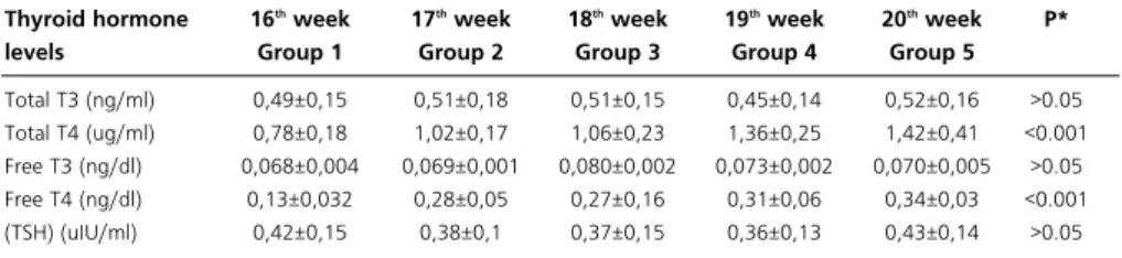
Tablo 1. Thyroid hormone values in amnion fluid, varying with gestational week.
Thyroid hormone levels Total T3 (ng/ml) Total T4 (ug/ml) Free T3 (ng/dl) Free T4 (ng/dl) (TSH) (uIU/ml) 16th week Group 1 0,49±0,15 0,78±0,18 0,068±0,004 0,13±0,032 0,42±0,15 17th week Group 2 0,51±0,18 1,02±0,17 0,069±0,001 0,28±0,05 0,38±0,1 18th week Group 3 0,51±0,15 1,06±0,23 0,080±0,002 0,27±0,16 0,37±0,15 19th week Group 4 0,45±0,14 1,36±0,25 0,073±0,002 0,31±0,06 0,36±0,13 20th week Group 5 0,52±0,16 1,42±0,41 0,070±0,005 0,34±0,03 0,43±0,14 P* >0.05 <0.001 >0.05 <0.001 >0.05
Total T3and T4free triiodothyronine (f T3), free
thyroxine (f T4) and thyroid stimulant hormone
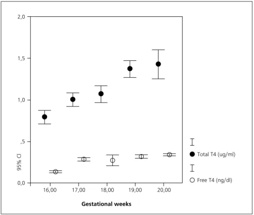
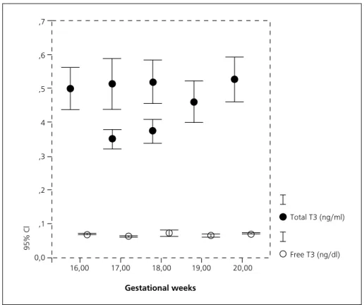
(TSH) values varying according to the gestational weeks, are shown in Table 1. Amniotic fluid levels
of total and free T4 increased progressively with
gestational age (p<0.001) (Figure 1). Although total
T3, free T3 and TSH levels did not progressively
increase with gestational age (p>0.05) (Figures 2 and 3).
Discussion
Clinical and experimental researches proved that thyroid hormones are essential for normal cerebral development in early fetal period and that has specific effects on olfactory bulbus, in sub ven-tricular zone of cerebral cortex, and in hippocam-pus.1-4
It affects thyroid hormones, mitochondrial
energy metabolism in short and long term.5 It
affects the lipid distribution of fetal thyroid hor-mones and bone differentiation in histological level.6
Rajatipi et al7observed the presence of the
thy-roid hormone receptors in fetus lung, on 13th
ges-tational week. This situation proves that thyroid hormones have a role in fetal lung development in early gestational weeks.
Fetal thyroid gland isn’t functional until 12th
week. Fetus is under the influence of maternal
thy-roid hormones within the first trimester.8Pop et al9
showed that the level of free T4in early
gestation-al period are strong indicators for motor and men-tal development of the infant after the birth.
Fetal thyroid hormones are also increased in cases of vaginal delivery, prolongation of the sec-ond gestation period, fetal umbilical fetal distress, painting amnion fluid by meconium, and forceps and vacuum usage that the fetus is exposed to the
intrauterine stress.10,11Ward et al12noted that
mater-nal cardiac diseases, preeclampsia, HIV infection, diabetes mellitus have no effect on fetal thyroid hormones.
Figure 1. Total T4 and free thyroxin levels in amnion fluid, varying with gestational
week 2,0 1,5 1,0 ,5 0,0 95% Cl 16,00 17,00 18,00 19,00 20,00 Gestational weeks Total T4 (ug/ml) Free T4 (ng/dl)
A growth in fetal serum TSH, T4 and thyroid
binding globulin levels occurs. TH, T4and T3
lev-els reach the peak level in second trimester and they tend to decrease until the next term. In last
Figure 2. Total T3 and free thyroxin levels in amnion fluid, varying with gestational week.
,7 ,6 ,5 4 ,3 ,2 ,1 0,0 95% Cl 16,00 17,00 18,00 19,00 20,00 Gestational weeks Total T3 (ng/ml) Free T3 (ng/dl)
Figure 3. Thyroid stimulant hormone TSH level in amnion fluid, varying with gestational week
16,00 17,00 18,00 19,00 20,00 Gestational weeks ,6 ,5 4 ,3 ,2 95% Cl TSH (ulU/mL)
trimester, T4and TSH levels are higher than
mater-nal levels however total T4 and T3 are lower. For
Fetal thyroid hormone metabolism, second and
third trimesters are critical transition period.13
Polk et al14
showed that total T4, and free T4
lev-els are augment along with gestational weeks and
serum T3levels are low. Klein et al15observed that
fetal serum T4, free T4and thyroid binding
globu-lin levels are in a significant increase between 26th
and 33rdgestational weeks, as from 34th gestational
week; there isn’t any change in these parameters.
Sack et al determined that amniotic fluid T4levels
are progressively elevated before 20th week,
how-ever T3levels didn’t give a progressive increase.16
In our study, total T4 levels and free thyroxin (f
T4) levels in amniotic fluid increased progressively
between 16 and 20th gestational weeks (p<0.001).
Although total T3 , free T3and TSH levels did not
increase with gestational week (p>0.05) In fetal period, normal cerebral development, fetal bone and lung differentiation and similar cases that
thy-roid hormones influence, thyroxin (T4) hormone is
effective and the need for this hormone increased
as gestational weeks go by. Although T3, free T3
and TSH levels did not progressively increase with gestational age, their presence in amnion fluid make us think that they contribute to the fetal development.
References
1. Lavado-Autric R, Auso E, Garcia-Velasco JV, Arufe Mdel C,
Escobar del Rey F, Berbel P. Early maternal hypothyroxine-mia alters histogenesis and cerebral cortex cytoarchitecture
of the progeny. J Clin Invest2003; 111: 1073-82.
2. Thorpe-Beeston JG, Nicolaides KH, McGregor AM. Fetal
thyroid function. Thyroid1992; 2: 207-17.
3. Roti E, Gnudi A, Braverman LE. The placental transport,
synthesis and metabolism of hormones and drugs which
af-fect thyroid function. Endocr Rev1983; 4: 131-49.
4. Hadj-Sahraoui N, Seugnet I, Ghorbel MT, Demeneix B.
Hypothyroidism prolongs mitotic activity in the post-natal
mouse brain. Neurosci Lett2000; 280: 79 82.
5. Wrutniak-Cabello C, Casas F, Cabello G. Thyroid hormone
action in mitochondria. J Mol Endocrinol 2001; 26: 67-77.
6. Geloso JP, Hemon P, Legrand J, Legrand C, Jost A. Some
aspects of thyroid physiology during the perinatal period.
General and Comparative Endocrinology Gen Comp Endoc-rinol1986; 10: 191-7.
7. Rajatapiti P, Kester MH, de Krijger RR, Rottier R, Visser TJ,
Tibboel D. Expression of glucocorticoid, retinoid, and thyroid hormone receptors during human lung
develop-ment. J Clin Endocrinol Metab2005; 90: 4309-4314.
8. Calvo RM, Jauniaux E, Gulbis B, Asuncion M, Gervy C,
Contempre B, Morreale de Escobar G. Fetal tissues are ex-posed to biologically relevant free thyroxine concentrations
during early phases of development. J Clin Endocrinol
Me-tab2002; 87: 1768 1777.
9. Pop VJ, Kuijpens JL, van Baar AL, Verkerk G, van Son MM,
de Vijlder JJ, Vulsma T, Wiersinga WM, Drexhage HA, Va-der HL. Low maternal free thyroxine concentrations during early pregnancy are associated with impaired psychomotor
development in infancy. Clin Endocrinol1999; 50: 149 155.
10. Fukuda S. Correlation between function of the pituitary thyroid axis and metabolism of catecholamines by the fetus
at delivery. Clin Endocrinol1987; 27: 331–338.
11. Chan LY, Leung TN, Lau TK Influences of perinatal factors
on cord blood thyroid-stimulating hormone level. Acta
Obs-tet Gynecol Scand2001; 80: 1014-8.
12. Ward LS, Kunii IS, de Barros Maciel RM. Thyroid-stimula-ting hormone levels in cord blood are not influenced by
non-thyroidal mothers’ diseases. Sao Paulo Med J 2000;
118:144-7.
13. Hume R, Simpson J, Delahunty C, van Toor H, Wu SY, Wil-liams FL, Visser TJ. Human fetal and cord serum thyroid
hormones: developmental trends and interrelationships. J
Clin Endocrinol Metab2004; 89: 4097-103.
14. Polk DH. Thyroid hormone metabolism during
develop-ment. Reprod Fertil Dev1995; 7: 469-77.
15. Klein AH, Oddie TH, Parslow M, Foley TP Jr, Fisher DA. Developmental changes in pituitary-thyroid function in the
human fetus and newborn.Early Hum Dev1982; 6: 321-30.
16. Sack J, Fisher DA, Hobel CJ, Lam R. Thyroxine in human