Electronic Thesis and Dissertation Repository
8-23-2013 12:00 AM
Alignment, Mass and Orthoses in Medial Compartment Knee
Alignment, Mass and Orthoses in Medial Compartment Knee
Osteoarthritis
Osteoarthritis
Rebecca MoyerThe University of Western Ontario
Supervisor
Dr. Trevor Birmingham
The University of Western Ontario
Graduate Program in Health and Rehabilitation Sciences
A thesis submitted in partial fulfillment of the requirements for the degree in Doctor of Philosophy
© Rebecca Moyer 2013
Follow this and additional works at: https://ir.lib.uwo.ca/etd
Part of the Orthopedics Commons
Recommended Citation Recommended Citation
Moyer, Rebecca, "Alignment, Mass and Orthoses in Medial Compartment Knee Osteoarthritis" (2013). Electronic Thesis and Dissertation Repository. 1515.
https://ir.lib.uwo.ca/etd/1515
This Dissertation/Thesis is brought to you for free and open access by Scholarship@Western. It has been accepted for inclusion in Electronic Thesis and Dissertation Repository by an authorized administrator of
Alignment, Mass and Orthoses in Medial Compartment Knee Osteoarthritis
(Thesis Format: Integrated-Article)
by
Rebecca F. Moyer
Graduate Program in Physical Therapy
A thesis submitted in partial fulfillment of the requirements for the degree of
Doctor of Philosophy
The School of Graduate and Postdoctoral Studies The University of Western Ontario
London, Ontario, Canada
ii
Abstract
iii
of considering alignment and the distribution of loads across the knee during walking when developing intervention strategies for knee OA. The present findings provide rationale for future research examining the combined use of different interventions that target biomechanics, including orthoses tailored to maximize biomechanical effects while maintaining patient comfort.
iv
Co-Authorship
v
Acknowledgements
Firstly, I would like to thank Dr. Trevor Birmingham for his continued support and guidance throughout the five years of this program. You have expanded my skills as a researcher in numerous directions and I am very appreciative for all of the past, present and future opportunities that you have made possible. Thank you. I would also like to thank my advisory committee members, Drs. Dianne Bryant, Bert Chesworth and J.R. Giffin. You have provided invaluable input to my thesis and have strengthened my abilities as a researcher. To Mr. Ian Jones, thank you for your knowledge and assistance in the lab. You have enlightened me with your data collection and analysis wisdom, which I will carry with me for the rest of my career.
I would also like to thank the Fowler Kennedy Sport Medicine Clinic for their welcoming environment, scheduling flexibility and assistance with patient recruitment. Without your support, these studies could not be possible. To my fellow graduate students in Engineering, Physical Therapy and WOBL, you have made the lab exceptionally enjoyable throughout the past five years. I am grateful for your positive attitudes and encouragement. Research wouldn’t be the same without you.
vi
Table of Contents
Abstract and Keywords ... ii
Co-Authorship ... iv
Acknowledgements ... v
List of Tables ... ix
List of Figures ... x
List of Appendices ... xii
List of Abbreviations ... xiii
1. Introduction: Background and Rationale ... 1
1.1 Knee Osteoarthritis ... 1
1.2 Obesity and Knee Osteoarthritis ... 3
1.3 Malalignment and Knee Osteoarthritis ... 3
1.4 Three-Dimensional Gait Analysis and Knee Osteoarthritis ... 6
1.5 Knee and Foot Orthoses in the Treatment of Knee Osteoarthritis ... 7
1.5.1 Valgus Knee Braces ... 8
1.5.2 Lateral Wedge Foot Orthotics ... 8
1.6 Thesis Outline ... 10
1.7 References ... 12
2. Alignment, Body Mass and Their Interaction on Dynamic Knee Joint Load in Patients with Knee Osteoarthritis ... 22
2.1 Summary ... 22
2.2 Introduction ... 23
2.3 Methods ... 25
2.3.1 Participants ... 25
2.3.2 Gait Analysis ... 25
2.3.3 Radiographic Analysis ... 27
2.3.4 Statistical Analysis ... 28
2.4 Results ... 29
2.5 Discussion ... 32
2.5.1 Study Limitations ... 35
vii
3. Systematic Review and Meta-Analysis of the Biomechanical and Clinical Effects of Valgus Knee Bracing in Patients with Medial Compartment Knee Osteoarthritis
... 40
3.1 Summary ... 40
3.2 Introduction ... 41
3.3 Materials and Methods ... 42
3.3.1 Inclusion Criteria and Exclusion Criteria ... 42
3.3.2 Search Strategy ... 42
3.3.3 Determining Inclusion ... 43
3.3.4 Methodological Quality Assessment of Included Studies ... 43
3.3.5 Outcome Measures and Data Extraction ... 44
3.3.6 Data Analysis ... 44
3.4 Results ... 45
3.4.1 Search Results ... 45
3.4.2 Characteristics of Included Studies ... 46
3.4.3 Methodological Quality Assessment of Included Studies ... 51
3.4.4 Biomechanical Effects ... 52
3.4.5 Patient-Reported Outcomes ... 58
3.4.6 Complications ... 60
3.4.7 Compliance ... 61
3.4.8 Long Term Brace Use ... 62
3.5 Discussion ... 62
3.5.1 Study Limitations ... 65
3.6 Conclusions ... 65
3.7 References ... 67
4. Combined Effects of a Valgus Knee Brace and Lateral Wedge Foot Orthotic on the External Knee Adduction Moment in Patients with Varus Gonarthrosis ... 73
4.1 Summary ... 73
4.2 Introduction ... 74
4.3 Methods ... 78
4.3.1 Participants ... 78
4.3.2 Valgus Knee Brace Fitting ... 79
4.3.3 Lateral Wedge Foot Orthotic Fitting ... 79
4.3.4 Testing Protocol ... 80
4.3.5 Gait Analysis ... 81
4.3.6 Data Analysis ... 84
4.4 Results ... 84
4.5 Discussion ... 88
4.5.1 Study Limitations ... 90
viii
4.7 References ... 92
5. Summary and General Discussion ... 97
5.1 Summary of Results ... 97
5.2 Implications ... 99
5.3 Limitations and Future Research ... 103
5.4 Recommendations ... 106
5.5 References ... 107
6. Appendices ... 109
ix
List of Tables
Table 2.1: Participants’ demographic, gait and clinical characteristics (n=487). ... 30 Table 2.2: A summary of regression models (dependent variable: peak knee adduction moment). ... 30 Table 2.3: Mean (SD) for peak knee adduction moment (Nm) for subgroups of patients based on tertiles of mechanical axis angle (MAA) and mass. Negative MAA values represent varus alignment. ... 30 Table 2.4: Regression coefficients and total explained variance in the peak adduction moments for mass tertiles. ... 31 Table 2.5: Regression coefficients and total explained variance in the peak adduction moments for mechanical axis angle (MAA) tertiles. Negative MAA values represent varus alignment.. ... 31 Table 3.1: A detailed summary of included (A) laboratory-based studies, (B)
x
List of Figures
xi
xii
List of Appendices
Appendix A: MEDLINE database search strategy ... 110
Appendix B: The Preferred Reporting Items for Systematic Reviews and Meta-Analyses (PRISMA) 2009 Checklist ... 112
Appendix C: Methodological Quality Assessment for non-randomized and randomized trials using a modified Downs and Black scale ... 115
Appendix D: Data Extraction Form ... 117
Appendix E: Copyright permission and reproduction of journal article ... 123
xiii
List of Abbreviations
AP - Anteroposterior BMI – Body mass index CI – Confidence interval CoM – Centre of mass CoP – Centre of pressure GRF – Ground reaction force
ICC – Intraclass correlation coefficient KAM – Knee adduction moment
KL Grade – Kellgren and Lawrence grade of OA severity KOOS – Knee Injury and Osteoarthritis Outcome Score MAA – Mechanical axis angle
NRS – Numeric rating scale Nm – Newton-meters OA – Osteoarthritis
RCT – Randomized controlled trial SD – Standard deviation
SMD – Standardized mean difference VAS – Visual analog scale
1. Introduction: Background and Rationale
The purpose of this chapter is to provide the background and rationale for the thesis objectives. A general description of knee osteoarthritis (OA) is presented, followed by a description of the importance of obesity and lower limb malalignment. Gait analysis and non-surgical treatments targeting those risk factors are also described. Lastly, a brief overview of thesis chapters 2-5 is provided.
1.1 Knee Osteoarthritis
Approximately 17% of people ≥ 45 years of age and 5% ≥ 26 years of age have symptomatic knee OA1. Often accompanied by other chronic disabling health conditions, OA is the most common musculoskeletal disease consuming more than 10% of Canada’s total economic burden2-5. Coinciding with growing life expectancies among an aging population and increasing incidence of obesity1, the prevalence of arthritis in society is expected to increase substantially, accompanied by a slow deterioration in physical function. Therefore, limiting OA disease progression has become an important public health strategy6. Understanding modifiable risk factors for OA and identifying intervention strategies that promote disease self-management and physical independence is paramount.
increases and physical activity decreases, abnormal joint biomechanics lead to irregular cartilage wear patterns, cartilage degradation, structural changes and bone deformations 8-11. Weight-bearing joints, such as the knee, are highly susceptible to cartilage degradation. In knee OA, the majority of the degeneration occurs in the medial compartment of the tibiofemoral joint, largely because of how the knee is loaded during walking. In healthy knees with neutral alignment, approximately 70-80% of the weight-bearing load passes through the medial compartment compared to the lateral compartment, and can increase to 100% of the load in the presence of varus malalignment and cartilage breakdown12-14.
1.2 Obesity and Knee Osteoarthritis
Based on measures of mass (kg) or BMI (kg/m2), obesity is an important modifiable risk factor for OA that has the potential for impact at the population level. Convincing evidence implicates obesity as a main precursor to the development and progression of radiographic disease16-17. In obese patients, the development of OA promotes sedentary lifestyles and immobility, which leads to further obesity and further OA progression16. This spiral of functional decline associated with obesity suggests that weight loss may protect against incident knee OA18 and increasing physical activity levels may protect against disease progression19. Additional treatment strategies that enable patients with knee OA to engage in activity to achieve these protective benefits are necessary.
Although obesity is considered to be both a systemic and biomechanical risk factor for knee OA, this thesis focuses only on its role in biomechanics. Obesity increases axial loads and can exceed the normal cyclical loads required to maintain natural cartilage function. In OA, the ability to carry increased loads associated with increased body mass can be further compromised, exacerbating knee pain and disability. Therefore, exercise and weight-loss intervention studies are imperative and have received a great deal of attention in the knee OA literature20-24.
1.3 Malalignment and Knee Osteoarthritis
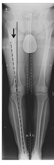
of alignment do exist, yet the MAA is considered the gold standard measure of static lower limb alignment and can be highly reliable using digital software programs25-27 (Figure 1.1). Malignment in the varus direction, also known as bow-legged, is more common in medial compartment knee OA; whereas, malalignment in the valgus direction, also known as knock-kneed, is more common in lateral compartment knee OA28-32. Although both forms of knee OA exist, medial compartment knee OA is more common due to the greater loads borne by that compartment during walking13-14.
knee joint load distribution25,34,36,40-45. In patients with varus malalignment, the distribution of load that is normally greater in the medial compartment is exaggerated further. This can lead to degradation of the medial tibiofemoral articular cartilage30,46-47, medial joint space narrowing and a further increase in varus alignment. Several authors have previously described this vicious cycle in medial compartment knee OA37,48 (Figure 1.2). A strong relationship has been consistently identified between varus malalignment and radiographic OA progression28,39. Although less consistent, recent evidence suggests that varus alignment is also associated with incident knee OA39,49-51. In addition to its independent effects, there is limited evidence to suggest that lower limb malalignment may also interact with other risk factors such as obesity35,37-38,52.
Obesity and lower limb malalignment both contribute to increased loads on the medial tibiofemoral compartment and are reported risk factors for the development and Figure 1.2: A vicious cycle of medial compartment knee osteoarthritis. Varus alignment creates aberrant loads on the medial compartment, leading to structural changes in the joint, decreased medial joint space and further increased varus alignment.
Increased medial compartment load
Articular cartilage degeneration Medial joint space
narrowing and bony deformations Increased varus
progression of knee OA. However, limited information exists on the potential interaction between alignment and body mass on medial compartment loading.
1.4 Three-Dimensional Gait Analysis in Knee Osteoarthritis
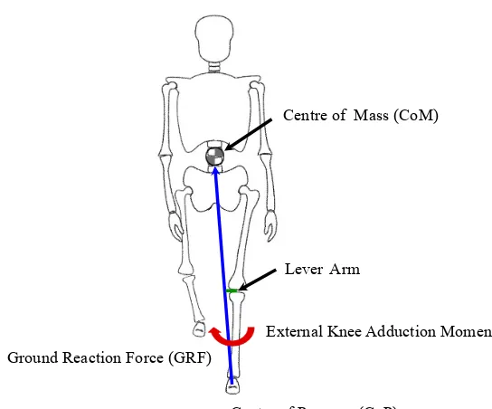
Walking is the most common activity of daily living with thousands of steps taken per day53-55. Three-dimensional gait analysis has proven to be a valuable instrument for the evaluation of biomechanical factors involved in knee OA. Knee joint kinematics and kinetics can provide particularly useful information with respect to the distribution of loads in the medial versus lateral tibiofemoral compartments. Specifically, during the stance phase of walking, the line of action of the resultant ground reaction force (GRF) is directed from the centre of pressure (CoP) under the foot and directed upwards towards the body’s centre of mass (CoM). Therefore, this GRF vector passes medial to the knee joint centre during stance, creates a lever arm in the frontal plane and an external adduction moment about the knee (Figure 1.3). In the presence of varus alignment, the frontal plane lever arm increases, the GRF shifts further away from the knee joint centre, and the external knee adduction moment increases.
1.5 Knee and Foot Orthoses in the Treatment of Knee Osteoarthritis
Clinical practice guidelines have outlined available surgical and non-surgical options for patients with symptomatic knee OA61-66. Less invasive treatment options for knee OA, with the aim to slow the rate of disease progression and improve pain and quality of life, are suggested as early treatments. Unloading the medial compartment of the knee is a common goal of conservative treatments. Through different mechanisms, valgus knee braces and lateral wedge foot orthotics both aim to decrease the external knee adduction moment. Although both knee and foot orthoses have been included in clinical practice guidelines, recommendations supporting their use are inconsistent61-66. Given the abundance of recently published literature on valgus knee bracing, a systematic review and meta-analysis is warranted.
Figure 1.3: During walking, the ground reaction force (GRF) vector originates at the foot’s centre of pressure (CoP) and passes medial to the knee towards the body’s centre of mass (CoM). This creates a lever arm in the frontal plane and an external knee adduction moment.
Centre of Pressure (CoP) Ground Reaction Force (GRF)
LeverArm
Centre of Mass (CoM)
1.5.1 Valgus Knee Braces
Varus knee braces can be used for patients with valgus alignment and lateral compartment knee OA, while valgus knee braces can be used for patients with varus alignment and medial compartment knee OA. Consistent with the greater prevalence of medial compartment OA, valgus braces are more common. These external devices are worn at the knee to provide a moment to oppose the external knee adduction moment, thereby lessening the load on the medial tibiofemoral compartment (Figure 1.4A). Off-the-shelf and custom-fit designs are available. Custom-fit braces are more expensive, but there is limited evidence to suggest that they can create greater biomechanical effects than off-the-shelf models67. Numerous published studies have evaluated various biomechanical effects of valgus braces and have reported mixed results67-92. The size of these biomechanical effects is often described as small and the carryover to clinically important benefits remains controversial67-68,70-74,76,78-80. Few clinical trials of valgus bracing have also been published and provide inconsistent conclusions67,86,78,81,93-96. While some encouraging results exist, discomfort84-85,90,97-99 and poor long-term brace use84,99-101 are also sometimes reported. Importantly, the size of biomechanical effects may be directly proportional to the angulation provided by the brace, yet greater angulations may be associated with greater discomfort71,76,90-91.
1.5.2 Lateral Wedge Foot Orthotics
decreases in the knee adduction moment and shortening of the lever arm in the frontal plane has been established102. Decreases in the external knee adduction moment have been reported76,103-110 with no diminishing effects after one month of wear108, yet the evidence remains inconclusive. Alternatively, randomized clinical trials have not supported the use of lateral wedge foot orthotics due to the lack of clinical improvements in pain and function110-112. Although greater wedge inclinations may be associated with greater reductions in the external knee adduction moment, patients have previously reported discomfort with inclinations larger than 10° 104.
Although not previously investigated, it is theoretically possible that valgus knee braces and lateral wedge foot orthotics have additive effects on decreasing the external knee adduction moment during walking. Specifically, a valgus knee brace may alter the position of the knee joint center medially, while a lateral wedge orthotic may alter the orientation of the ground reaction force laterally, when worn concurrently.
Figure 1.4: (A) Valgus knee brace and (B) full-length lateral wedge orthotic
1.6 Thesis Outline
The overall purpose of this thesis was to examine the interaction between lower limb alignment and body mass on dynamic knee joint loading, and to examine the effects of knee and foot orthoses, in patients with knee OA. The thesis consists of three studies. All studies were completed in the Wolf Orthopaedic Biomechanics Laboratory, Fowler Kennedy Sport Medicine Clinic, Western University.
Chapter 2 (Study 1): Clinical and biomechanical rationale suggest that the effect of body mass on knee joint loading may depend on lower limb alignment, although this potential interaction has not been previously described. The objective of this study was to examine the interaction and relative contributions of frontal plane alignment and body mass on measures of knee joint loading during gait. Results from this study provided further rationale for studying interventions aimed at altering malalignment, including valgus knee braces and lateral wedge foot orthotics.
Chapter 4 (Study 3): The primary objective of this proof of concept study was to test the hypothesis that a custom-fit valgus knee brace and custom-made lateral wedge foot orthotic would have greatest effects on decreasing the external knee adduction moment during gait when used concurrently. The secondary objective was to explore changes in the frontal plane ground reaction force and its lever arm.
1.7 References
1. Lawrence RC, Felson DT, Helmick CG, Arnold LM, Choi H, Deyo RA, et al. Estimates of the prevalence of arthritis and Other Rheumatic Conditions in the United States. Arthritis and Rheumatism 2008;58:26-35.
2. Badley EM. Arthritis in Canada: What do we know and what should we know? The Journal of Rheumatology. Supplement 2005;32:32-41.
3. Hawker GA, Badley EM, Croxford R, Coyte PC, Glazier RH, Guan J, et al. A population-based nested case-control study of the costs of hip and knee replacement surgery. Medical Care 2009;47:732-41.
4. Tarride JE, Haq M, O’Reilly DJ, Bowen JM, Xie F, Dolovich L, et al. The excess burden of osteoarthritis in the province of Ontario, Canada. Arthritis and Rheumatism 2012;64(4):1153-61.
5. Health Canada. Arthritis in Canada: an ongoing challenge. Ottawa: Health Canada; 2003.
6. Health Canada. Life with Arthritis in Canada. A personal and public health challenge. Ottawa: Health Canada; 2010.
7. Altman R, Asch E, Bloch D, Bole G, Borenstein D, Brandt K, et al. Development of criteria for the classification and reporting of osteoarthritis: classification of osteoarthritis of the knee. Arthritis and Rheumatism 1986;29:1039–49.
8. Boegård TL, Rudling O, Petersson IF, Jonsson K. Magnetic resonance imaging of the knee in chronic knee pain. A 2-year follow-up. Osteoarthritis and Cartilage 2001; 9(5):473-80.
9. Ding C, Cicuttini F, Scott F, Boon C, Jones G. Association of prevalent and incident knee cartilage defects with loss of tibial and patellar cartilage: a longitudinal study. Arthritis and Rheumatism 2005; 52(12):3918-27.
10. Felson DT, Gale DR, Gale ME, Niu J, Hunter DJ, Goggins J, LaValley MP. Osteophytes and progression of knee osteoarthritis. Rheumatology 2005;44:100-4.
11. Anandacoomarasamy A, Smith G, Leibman S, Caterson I, Giuffre B, Fransen M, Sambrook P, March L. Cartilage defects are associated with physical disability in obese adults. Rheumatology (Oxford) 2009; 48(10):1290-3.
13. Schipplein OD, Andriacchi TP. Interaction between active and passive knee stabilizers during level walking. Journal of Orthopaedic Research 1991;9:113-9.
14. Andriacchi TP. Dynamics of knee malalignment. The Orthopaedic Clinics of North American 1994;25:395-403.
15. Kellgren JH, Lawrence JS. Radiological assessment of osteoarthrosis. Annals of the Rheumatic Diseases 1957;16:494-502.
16. Felson DT, Anderson JJ, Naimark A, Walker AM, Meenan RF. Obesity and knee osteoarthritis. The Framingham Study. Annals of internal medicine 1988;109(1):18-24. 17. Hart DJ, Doyle DV, Spector TD. Incidence and risk factors for radiographic knee osteoarthritis in middle-aged women. Arthritis Rheum 1999;42(1):17-24.
18. Felson DT, Zhang Y, Anthony JM, Naimark A, Anderson JJ. Weight loss reduces the risk for symptomatic knee osteoarthritis in women. The Framingham Study. Annals of internal medicine 1992;116(7):535-9.
19. Sharma L, Cahue S, Song J, Hayes K, Pai YC, Dunlop D. Physical functioning over three years in knee osteoarthritis: role of psychosocial, local mechanical, and neuromuscular factors. Arthritis and Rheumatism 2003;48(12):3359-70.
20. Manninen P, Riihimaki H, Heliövaara M, Suomalainen O. Weight changes and the risk of knee osteoarthritis requiring arthroplasty. Annals of the rheumatic diseases 2004;63(11):1434-7.
21. Messier SP, Loeser RF, Mitchell MN, Valle G, Morgan TP, Rejeski WJ, et al. Exercise and weight loss in obsess older adults with knee osteoarthritis: A preliminary study. Journal of the American Geriatric Society 2000;48(9):1062-72.
22. Jadelis K, Miller ME, Ettinger WH, Messier SP. Strength, balance, and the modifying effects of obesity and knee pain: results from the Observational Arthritis Study in Seniors (OASIS). Journal of the American Geriatrics Society 2001;49(7):884-91. 23. Messier SP, Loeser RF, Miller GD, Morgan TM, Rejeski WJ, Sevick MA, et al. Exercise and dietary weight loass in overweight and obese older adults with knee osteoarthritis. Arthritis and Rheumatism 2004;50:1501-10.
24. Aaboe J, Bliddal H, Messier SP, Alkjaer T, Henriksen M. Effects of an intensive weight loss program on knee joint loading in obese adults with knee osteoarthritis. Osteoarthritis and Cartilage 2011;19(7):822-8.
26. Kraus VB, Vail TP, Worrell T, McDaniel G. A comparative assessment of alignment angle of the knee by radiographic and physical examination methods. Arthritis and Rheumatism 2005;52(6):1730-5.
27. Hinman RS, May RL, Crossley KM. Is there an alternative to the full!leg radiograph for determining knee joint alignment in osteoarthritis?. Arthritis Care and Research 2006;55(2):306-13.
28. Sharma L, Song J, Felson DT, Cahue S, Shamiyeh E, Dunlop D. The role of knee alignment in disease progression and functional decline in knee osteoarthritis. The Journal of the American Medical Association 2001;286:188-95.
29. Cerejo R, Dunlop DD, Cahue S, Channin D, Song J, Sharma L. The influence of alignment on risk of knee osteoarthritis progression according to baseline stage of disease. Arthritis and Rheumatism 2002;46(10):2632-6.
30. Eckstein F, Wirth W, Hudelmaier M, Stein V, Lengfelder V, Cahue S, et al. Patterns of femorotibial cartilage loss in knees with neutral, varus, and valgus alignment. Arthritis Care and Research 2008;59(11):1563-70.
31. Felson DT, Niu J, Yang T, Torner J, Lewis CE, Aliabadi P, et al. Physical activity, alignment and knee osteoarthritis: data from MOST and the OAI. Osteoarthritis and Cartilage 2013;21(6):789-795.
32. Andriacchi TP. Valgus alignment and lateral compartment knee osteoarthritis: a biomechanical paradox or new insight into knee osteoarthritis? Editorial. Arthritis and Rheumatism 2013;65(2):310-3.
33. Prodromos CC, Andriacchi TP, Galante JO. A relationship between gait and clinical changes following high tibial osteotomy. Journal of Bone and Joint Surgery 1985;67:1188-94.
37. Sharma L, Lou C, Cahue S, Dunlop DD. The mechanism of the effect of obesity in knee osteoarthritis: the mediating role of malalignment. Arthritis & Rheumatism 2000;43(3):568-75.
38. Felson DT, Goggins J, Niu J, Zhang Y, Hunter DJ. The effect of body weight on progression of knee osteoarthritis is dependent on alignment. Arthritis and Rheumatism 2004;50:3904-9.
39. Brouwer GM, van Tol AW, Bergink AP, Belo JN, Bernsen RM, Reijman M, et al. Association between valgus and varus alignment and the development and progression of radiographic osteoarthritis of the knee. Arthritis and Rheumatism 2007;56:1204-11. 40. Goh JC, Khoo BC. Gait analysis study on patients with varus osteoarthrosis of the knee. Clinical orthopaedics and related research 1993;294:223-31.
41. Wada M, Maezawa Y, Baba H, Shimada S, Sasaki S, Nose Y. Relationships among bone mineral densities, static alignment and dynamic load in patients with medial compartment knee osteoarthritis. Rheumatology 2001;40(5):499-505.
42. Miyazaki T, Wada M, Kawahara H, Sato M, Baba H, Shimada S. Dynamic load at baseline can predict radiographic disease progression in medial compartment knee osteoarthritis. Annals of the Rheumatic Diseases 2002;61:617-22.
43. Birmingham TB, Hunt MA, Jones IC, Jenkyn TR, Giffin JR. Test-retest reliability of the peak knee adduction moment during walking in patients with medial compartment knee osteoarthritis. Arthritis and Rheumatism 2007;57:1012-7.
44. Hunt MA, Birmingham TB, Bryant D, Jones I, Giffin JR, Jenkyn TR, et al. Lateral trunk lean explains variation in dynamic knee joint load in patients with medial compartment knee osteoarthritis. Osteoarthritis and Cartilage 2008;16:591-9.
45. Foroughi N, Smith RM, Lange AK, Baker MK, Singh MAF, Vanwanseele B. Dynamic alignment and its association with knee adduction moment in medial knee osteoarthritis. The Knee 2010; 17(3):210-6.
46. Sharma L, Eckstein F, Song J, Guermazi A, Prasad P, Kapoor D, et al. Relationship of meniscal damage, meniscal extrusion, malalignment, and joint laxity to subsequent cartilage loss in osteoarthritic knees. Arthritis and Rheumatism 2008;58(6):1716-26. 47. Moisio K, Chang A, Eckstein F, Chmiel JS, Wirth W, Almagor O, et al. Varus– valgus alignment: reduced risk of subsequent cartilage loss in the less loaded compartment. Arthritis and Rheumatism 2011;63(4):1002-9.
49. Hunter DJ, Niu J, Felson DT, Harvey WF, Gross KD, McCree P, et al. Knee alignment does not predict incident osteoarthritis: the Framingham osteoarthritis study. Arthritis and Rheumatism 2007;56(4):1212-8.
50. Sharma L, Song J, Dunlop D, Felson D, Lewis CE, Segal N, et al. Varus and valgus alignment and incident and progressive knee osteoarthritis. Annals of the Rheumatic Diseases 2010;69(11):1940-5.
51. Sharma L, Chmiel JS, Almagor O, Felson D, Guermazi A, Roemer F, et al. The role of varus and valgus alignment in the initial development of knee cartilage damage by MRI: the MOST study. Annals of the Rheumatic Diseases 2013;72(2):235-40.
52. Niu J, Zhang Q, Torner J, Nevitt M, Lewis CE, Aliabadi P, et al. Is obesity a risk factor for progressive radiographic knee osteoarthritis? Arthritis and Rheumatism 2009;61:329-35.
53. Welk GJ, Differding JA, Thompson RW, et al. The utility of the Digi-walker step counter to assess daily physical activity patterns. Medicine Science and Sports Exercise 2000;32(9):S481-S8.
54. Tudor-Locke C, Myers AM. Methodological considerations for researchers and practitioners using pedometers to measure physical (ambulatory) activity. Research Quarterly for Exercise and Sport 2001;72(1):1-12.
55. Tudor-Locke C, Bassett Jr DR. How many steps/day are enough?. Sports Medicine 2004;34(1):1-8.
56. Zhao D, Banks SA, Mitchell KH, D’Lima DD, Colwell CW Jr. Correlations between the knee adduction torque and medial contact force for a variety of gait patterns. Journal of Orthopaedic Research 2007;25:789-97.
57. Andriacchi TP, Koo S, Scanlan S. Gait mechanics influence healthy cartilage morphology and osteoarthritis of the keee. Journal of Bone and Joint Surgery (Am) 2009;91(S1):95-101.
58. Walter JP, D'Lima DD, Colwell CW, Fregly BJ. Decreased knee adduction moment does not guarantee decreased medial contact force during gait. Journal of Orthopaedic Research 2010;28(10):1348-54.
61. Jordan KM, Arden NK, Doherty M, Bannwarth B, Bijlsma JW, Dieppe P, et al. EULAR Recommendations 2003: an evidence based approach to the management of knee osteoarthritis: Report of a Task Force of the Standing Committee for International Clinical Studies Including Therapeutic Trials (ESCISIT). Annals of the Rheumatic Diseases 2003;62(12):1145-55.
62. Zhang W, Moskowitz RW, Nuki G, Abramson S, Altman RD, Arden N, et al. OARSI recommendations for the management of hip and knee osteoarthritis, part II: OARSI evidence-based expert consensus guidelines. Osteoarthritis and Cartilage 2008;16:137-162.
63. National Institute for Health and Clinical Excellence (NICE). Osteoarthritis: national clinical guideline for care and management in adults. London, Royal College of Physicians 2008.
64. Beaudreuil J, Bendaya S, Faucher M, Coudeyre E, Ribink P, Revel M, et al. Clinical practice guidelines for rest orthosis, knee sleeves and unloading knee braces in knee osteoarthritis. Joint Bone Spine 2009;76:629-36.
65. Hochberg MC, Altman RD, April KT, Benkhalti M, Guyatt G, McGowan J, et al. American College of Rheumatology 2012 recommendations for the use of nonpharmacologic and pharmacologic therapies in osteoarthritis of the hand, hip, and knee. Arthritis Care and Research (Hoboken). 2012;64(4):465-74.
66. The American Academy of Orthopaedic Surgeons. Treatment of osteoarthritis of the knee. Evidence based guideline. 2nd Ed. 2013;p.1155.
67. Draganich L, Reider B, Rimington T, Piotrowski G, Mallik K, Nasson S. The effectiveness of self-adjustable custom and off-the-shelf bracing in the treatment of varus gonarthrosis. The Journal of Bone and Joint Surgery 2006;88-A(12):2645-52.
68. Lindenfeld TN, Hewett TE, Andriacchi TP. Joint loading with valgus bracing in patients with varus gonarthrosis. Clinical Orthopaedics and Related Research. 1997;344: 290-297.
69. Hewett TE, Noyes FR, Barber-Westin SD, Heckman TP. Decrease in knee joint pain and increase in function in patients with medial compartment arthrosis: a prospective analysis of valgus bracing. Orthopedics 1998;21:131-8.
70. Self BP, Greenwald RM, Pflaster DS. A biomechanical analysis of medial unloading brace for osteoarthritis in the knee. Arthritis Care and Research. 2000;13(4):191-7.
72. Gaasbeek RDA, Groen BE, Hampsink B van Heerwaarden RJ, Duysens J. Valgus bracing in patients with medial compartment osteoarthritis of the knee: A gait analysis study of a new brace. Gait and Posture 2007;26:3-10.
73. Schmalz T, Knopf E, Drewitz H, Blumentritt S. Analysis of biomechanical effectiveness of valugs-inducing knee brace for osteoarthritis of knee. Journal of Rehabilitation Research and Development 2010;47(5):419-30.
74. Fantini Pagani CH, Bohle C, Potthast W, Bruggemann GP. Short-term effects of a dedicated knee orthosis on knee adduction moment, pain and function in patients with osteoarthritis. Archives of Physical Medicine and Rehabilitation 2010;91:1936-41.
75. Toriyama M, Deie M, Shimada N, Otani T, Shidahara H, Maejima H, et al. (2011) Effects of unloading bracing on knee and hip joints for patients with medial compartment knee osteoarthritis. Clinical Biomechanics 2011;26:497-503.
76. Fantini Pagani CH, Hinrichs M, Bruggemann GP. Kinetic and kinematic changes with the use of valgus knee brace and lateral wedge insoles in patients with medial knee osteoarthritis. Journal of Orthopaedic Research 2011;7:1125-32.
77. Esrafilian A, Karimi MT, Eshraghi A. Design and evaluation of a new type of knee orthosis to align the mediolateral angle of the knee joint with osteoarthritis. Advances in Orthopedics 2012:p.6.
78. Jones RK, Nester CJ, Richards JD, Kim WY, Johnson DS, Jari S, et al. A comparison of the biomechanical effects of valgus knee braces and lateral wedge insoles in patients with knee osteoarthritis. Gait and Posture 2013;37:368-72.
79. Moyer R, Birmingham T, Dombrowski C, Walsh R, Leitch K, Jenkyn T, et al. Combined effects of a custom-fit valgus knee brace and lateral wedge orthotic on dynamic knee joint loading in patients with varus gonarthrosis. Archives of Physical Medicine and Rehabilitation 2013;94(1):103-12.
80. Arazpour M, Bani MA, Maleki M, Ghomshe FT, Kashani RV, Hutchins SW. Comparison of the efficacy of laterally wedged insoles and bespoke unloader knee orthoses in treating medial compartment knee osteoarthritis. Prosthetics and Orthotics International 2013;37:50-57.
81. Horlick SG and Loomer RL. (1993) Valgus knee bracing for medial gonarthrosis. Clinical Journal of Sport Medicine 3: 251-5.
83. Komistek RD, Dennis DA, Northcut EJ, Wood A, Parker AW, Traina SM. An in vivo analysis of the effectiveness of the osteoarthritic knee brace during heel strike of gait. The Journal of Arthroplasty 1999;14(6):738-42.
84. Barnes CL, Cawley PW, Hederman B. Effect of CounterForce brace on symptomatic relief in a group of patients with symptomatic unicompartmental osteoarthritis: a prospective 2-year investigation American Journal of Orthopedics 2002;31:396-401. 85. Ramsey DK, Briem K, Axe MJ, Snyder-Mackler L. A mechanical theory for the effectiveness of bracing for medial compartment osteoarthritis of the knee. The Journal of Bone and Joint Surgery 2007;89:2398-2407.
86. van Raaij TM, Reijman M, Brouwer RW, Bierma-Zeinstra A, Verhaar JAN. Medial knee osteoarthritis treated by insoles or braces: A randomized trial. Clinical Orthopaedics and Related Research 2010;468:1926-32.
87. Nadaud MC, Komistek RD, Mahfouz MR, Dennis DA, Anderle MR. In vivo three-dimensional determination of the effectiveness of the osteoarthritic knee brace: a multiple brace analysis. The Journal of Bone and Joint Surgery 2005;87-A(S2):114-9.
88. Dennis DA, Komistek RD, Nadaud MC, Mahfouz M. Evaluation of off-loading braces for treatment of unicompartmental knee arthrosis. The Journal of Arthroplasty 2006;21(4):2-8
89. Anderson IA, MacDiarmid AA, Harris ML, Gillies M, Phelps R, Walsh WR. A novel method for measuring medial compartment pressures within the knee joint in-vivo. Journal of Biomechanics 2003;36:1391-5.
90. Kutzner I, Kuher S, Heinlein B, Dymke J, Bender A, Halder AM, et al. The effect of valgus braces on medial compartment load of the knee joint – in vivo load measurements in three subjects. Journal of Biomechanics 2011;44:1354-60.
91. Fantini Pagani CH, Hinrichs M, Bruggemann GP. Kinetic and kinematic changes with the use of valgus knee brace and lateral wedge insoles in patients with medial knee osteoarthritis. Journal of Orthopaedic Research 2012;30:1125-32.
92. Katsuragawa Y, Fukui N, Nakamura K. Change in bone mineral density with valgus knee bracing. International Orthopaedics (SICOT) 1999;23:164-7.
93. Kirkley A, Webster-Bogaert S, Litchfield R, Amendola A, MacDonald S, McCalden R, et al. The effect of bracing on varus gonarthrosis. The Journal of Bone and Joint Surgery 1999;81-A(4):539-47.
95. Hunter D, Gross KD, McCree P, Li L, Hirko K, Harvey WF. Realignment treatment for medial tibiofemoral osteoarthritis: randomized trial. Annals of the Rheumatic Diseases 2012;71:1658-65.
96. Richards JD, Sanchez-Ballester J, Jones RK, Darke N, Livingstone BN. A comparison of knee braces during walking for the treatment of osteoarthritis of the medial compartment of the knee. The Journal of Bone and Joint Surgery (Br) 2005;87-B(7):937-9.
97. Liu K, Lao L, Asami T, Itoh Y, Watanabe H. Clinical care of osteoarthritis knee with knee orthoses. Fukuoka Acta Med 1998;89(10):298-302.
98. Finger S and Paulos LE. Clinical and biomechanical evaluation of the unloading brace. Journal of Knee Surgery 2002;15:155-9.
99. Squyer E, Stamper DL, Hamilton DT, Sabin JA, Leopold SS. Unloader knee braces for osteoarthritis: do patients actually wear them? Clinical Orthopaedics and Related Research 2013;47:1982-91.
100. Wilson B, Rankin H, Barnes CL. Long-term results of an unloader brace in patients with unicompartmental knee osteoarthritis. Orthopaedics 2011;34:334-7.
101. Giori NJ. Load-shifting brace treatment for osteoarthritis of the knee: A minimum 2 ½-year follow-up study. Journal of Rehabilitation Research and Development 2004;41(2):187-94.
102. Hinman RS, Bowles KA, Metcalf BB, Wrigley TV, Bennell KL. Lateral wedge insoles for medial knee osteoarthritis: effects on lower limb frontal plane biomechanics. Clinical Biomechanics (Bristol, Avon) 2012;27:27-33.
103. Maly MR, Culham EG, Costigan PA. Static and dynamic biomechanics of foot orthoses in people with medial compartment knee osteoarthritis. Clinical Biomechanics 2002;17;603-10.
104. Kerrigan DC, Lelas JL, Goggins J, Merriman GJ, Kaplan RJ, Felson DT. Effectiveness of a lateral-wedge insole on knee varus torque in patients with knee osteoarthritis. Archives of Physical Medicine and Rehabilitation 2002;83:889-93.
105. Shimada S, Kobayashi S, Wada M, Uchida K, Sasaki S, Kawahara H, et al. Effects of disease severity on response to lateral wedged shoe insole for medial compartment knee osteoarthritis. Archives of Physical Medicine and Rehabilitation 2006;87:1436-41.
107. Kakihana W, Akai M, Nakazawa K, Naito K, Torii S. Inconsistent knee varus moment reduction caused by a lateral wedge in knee osteoarthritis. American Journal of Physical Medicine and Rehabilitation 2007;86:446-54.
108. Hinman RS, Bowles KA, Bennell KL. Laterally wedged insoles in knee osteoarthritis: do biomechanical effects decline after one month of wear? BMC Musculoskeletal Disorders 2009;10:146-53.
109. Abdallah A, Radwan AY. Biomechanical changes accompanying unilateral and bilateral use of laterally wedged insoles with medial arch supports in patients with medial knee osteoarthritis. Clinical Biomechanics 2011;26:783-9.
110. Bennell KL, Bowles KA, Payne C, Cicuttini F, Williamson E, Forbes A, Hanna F, Davies-Tuck M, Harris A, Hinman RS. Lateral wedge insoles for medial knee osteoarthritis: 12 month randomized controlled trial. BMJ 2011;18:342-50.
111. Pham T, Maillefert JF, Hudry C, Kieffert P, Bourgeois P, Lechevalier D, et al. Laterally elevated wedged insoles in the treatment of medial knee osteoarthritis. A two-year prospective randomized controlled study. Osteoarthritis and Cartilage 2004;12:46-55.
A version of this manuscript has been published in Osteoarthritis and Cartilage
2. Alignment, Body Mass and Their Interaction on Dynamic Knee Joint Load in Patients with Knee Osteoarthritis
2.1 Summary
2.2 Introduction
Approximately 17% of people greater than 45 years of age and 5% greater than 26 years of age have symptomatic knee osteoarthritis (OA)1. It is a leading cause of disability and increases the risk of disability due to other medical conditions substantially2,3. Knee OA that has progressed beyond the mild stage is responsible for the majority of its burden, which is extensive2,4,5. Limiting disease progression is therefore an important public health strategy, and understanding risk factors for progression is imperative.
Malalignment of the lower limb and excess body mass are both proposed risk factors for the progression of knee OA, presumably because of their contributions to increased joint loading6-11. Although greater varus alignment is consistently reported to be strongly associated with disease progression7,11,the effect of body mass is less clear and may depend on the extent of malalignment6,8,10. A plausible biomechanical hypothesis is that alignment and body mass produce interaction effects on knee joint loading. Specifically, excess body mass may modify the well-established association between alignment and load on the medial compartment of the tibiofemoral joint6,9. We are unaware of previous research that has directly tested for an interaction between alignment and body mass on knee joint load.
malalignment, but not in knees with severe malalignment. Alternatively, Niu et al.10 reported that obesity had no effect on radiographic progression in knees with varus alignment, and suggested the excess load produced by varus knee malalignment may be sufficient by itself to cause progression. Although this hypothesis is plausible and implies a greater role of malalignment than body mass on knee load, we are unaware of previous research that has evaluated the relative contributions of alignment and body mass to knee joint loading in patients with knee OA.
Several lines of evidence suggest that quantitative gait analysis provides an appropriate means to measure knee joint load during walking. In particular, the external adduction moment about the knee, calculated as the product of the frontal plane components of the ground reaction force magnitude and the lever arm, is a valid and reliable proxy for the dynamic load on the medial compartment of the tibiofemoral joint 12-15. Importantly, in addition to being affected by one’s body mass and lower limb alignment, the knee adduction moment reflects an individual’s walking characteristics and arguably represents a functional measure of dynamic knee joint loading. Gait variables most commonly reported to be associated with reduced knee adduction moments in patients with knee OA include decreased walking speed16-18, increased toe out angle14,18-20 and increased lateral trunk lean over the stance limb17. Pain and disease severity may also influence the knee adduction moment21. It is therefore important to consider these covariates when evaluating the effects of alignment and body mass on dynamic knee joint load.
to examine the interaction and relative contributions of frontal plane alignment and body mass on knee joint loading during gait. We hypothesized that while controlling for other factors suggested to alter knee joint load, there would be a statistically significant interaction between alignment and body mass on the external knee adduction moment. In the presence of significant findings we planned to describe the interaction by controlling for effect modification from two perspectives: one where the effect modifier was body mass and the other where the effect modifier was alignment.
2.3 Methods
2.3.1 Participants
We included the first 487 participants in an ongoing gait data registry for patients diagnosed with knee OA who were referred to a tertiary care center specializing in orthopaedics. The diagnosis of knee OA was based on the criteria described by Altman et al.22. Patients with rheumatoid arthritis or a concomitant neurological condition were excluded. The study was approved by the institutional research ethics board and all participants provided informed consent.
2.3.2 Gait Analysis
orientation, and positions of joint centres of rotation for the knee and ankle. These four additional markers were removed prior to gait testing. During the gait analysis, patients were instructed to walk across the laboratory at their typical walking speed while kinetic (sampled at 1200 Hz) and kinematic data (sampled at 60 Hz) were collected during the middle of several strides. Raw data were filtered using a 4th order Butterworth low pass filter with a cutoff frequency of 6Hz.
The frontal plane component of the GRF was calculated as the resultant force vector of the vertical and mediolateral components of the GRF. The frontal plane lever arm was calculated as the perpendicular distance between the frontal plane GRF and knee joint centre of rotation using custom post-processing and data reduction techniques previously described24,25. The external adduction moment about the knee was calculated using commercial software from the kinetic and kinematic data with a process called inverse dynamics (Orthotrak 6.2.4; MAC, Santa Rosa, CA). Each lower limb segment (foot, shank and thigh) was modeled as a rigid body with a local coordinate system that coincided with anatomically relevant axes. Inertial properties of each limb segment were approximated anthropometrically and the translations and rotations of each segment were reported relative to neutral positions as defined during the initial standing static trial.
midpoint of the anterior tips of the acromion processes with respect to vertical. All gait variables were calculated by averaging across five trials for each patient. We have previously reported excellent test-retest reliability of the peak knee adduction moment (ICC2,1 = 0.86)15. We have also previously reported acceptable reliability of gait speed (ICC2,1 = 0.92), toe-out angle (ICC2,1 = 0.69), and trunk lean angle (ICC2,1 = 0.91) measurements17.
2.3.3 Radiographic Analysis
2.3.4 Statistical Analysis
We used sequential (hierarchical) linear regression models to test the hypothesis that a statistical interaction exists between alignment and mass on dynamic knee joint load, while controlling for other factors suggested to affect knee loading. Specifically we created an interaction term by multiplying mechanical axis angle by mass (MAA*mass) and tested whether it contributed significantly to a model predicting peak knee adduction moment, that also included mechanical axis angle, mass and other independent variables that affect knee loading31. We tested four, hypothesis driven models. Independent variables in the first model included age, sex, height, Kellgren and Lawrence grade, pain score during walking, gait speed, toe out angle and trunk lean angle because these variables have been previously reported to affect knee adduction moments14,16-21. We then added mechanical axis angle, mass and the interaction term (MAA*mass) in three separate sequential models to determine the contribution of each of these variables. We repeated these three sequential models while reversing the order of adding mechanical axis angle and mass.
separate step to determine its contribution to the model in each tertile of alignment. The SPSS program version 18.0 (SPSS Inc., Chicago, IL) was used for all statistical analyses.
2.4 Results
Table 2.1: Participants’ demographic, gait and clinical characteristics (n=487)
Mean (SD) Min, Max
Age (years) No. of males Mass (kg) Height (m) BMI (kg/m2) Gait speed (m/s) Toe-out angle (˚) Trunk lean (˚)
Peak adduction moment (Nm) Peak adduction moment (%BWxHT)
Mechanical axis angle (˚) No. varus/valgus limbs* Pain score during walking (0-10) KL GradeŦ
No. 1/2/3/4 46 (10) 363 (74.5%) 90.6 (18.3) 1.8 (0.1) 29.5 (5.1) 1.1 (0.2) 12.1 (6.2) 3.0 (2.7) 46.1 (20.6) 3.0 (1.1) -6.5 (5.6) 437/50 3.1 (2.7) 59/148/147/133 20.0, 76.0 - 43.2, 150.7 1.5, 2.1 18.0, 49.0
0.3, 1. 8 -6.9, 32.0 -4.9, 20.3 -3.1, 127.7 -0.2, 6.4 -21.0, 22.1 - 0.0, 10.0 - * Varus is defined as < 0˚, and valgus as > 0˚.
Ŧ Higher Kellgren and Lawrence (KL) grades indicate greater disease severity
Table 2.2: A summary of regression models (dependent variable: peak knee adduction moment)
Model Adjusted R2 R
2
Change P
Trunk Lean + Toe Out + Pain + Height + Age + OA grade + Gait Speed + Gender
Trunk Lean + Toe Out + Pain + Height + Age + OA grade + Gait Speed + Gender + MAA
Trunk Lean + Toe Out + Pain + Height + Age + OA grade + Gait Speed + Gender + MAA + Mass
Trunk Lean + Toe Out + Pain + Height + Age + OA grade + Gait Speed + Gender + MAA + Mass + (MAA*Mass)
0.25 0.62 0.68 0.70 0.25 0.37 0.06 0.02 <0.001 <0.001 <0.001 <0.001
Table 2.3: Mean (SD) for peak knee adduction moment (Nm) for subgroups of patients based on tertiles of mechanical axis angle (MAA) and mass. Negative MAA values represent varus alignment.
MAA > -5º
[mean = 0º]
MAA -5º to -9º
[mean = -7º]
MAA < -9º
[mean = -12º]
Mass < 80 kg
[mean = 72kg]
26 (10) 39 (10) 50 (13)
Mass 80 to 100 kg
[mean = 89kg]
31 (15) 47 (10) 56 (15)
Mass > 100 kg
[mean = 111 kg]
31 Table 2.4: Regression coefficients and total explained variance in the peak adduction moments for mass tertiles
Peak Knee Adduction Moment Mass < 80 kg
[R2=0.66, p<0.01]
Mass 80 to 100 kg [R2=0.69, p<0.01]
Mass >100 kg [R2=0.61, p<0.01]
Variable B-coefficient P B-coefficient P B-coefficient P
Constant Age Gender Height Gait speed Trunk lean Toe-out angle MAA* OA grade Pain
-48.5 (-87.2, -9.8) 0.1 (-.01, 0. 3)
3.5 (-0.4, 7.3) 39.3 (16.3, 62.3)
4.6 (-3.7, 12.9) -0.6 (-1.2, -0.1) -0.4 (-0.6, -0.2) -1.7 (-2, -1.5) 1.1 (-0.6, 2.7) -0.2 (-0.7, 0.4)
0.014 0.081 0.07 0.001 0.272 0.02 < 0.001 < 0.001 0.206 0.583
-94.8 (-139, -50.6) 0.3 (0.1, 0.5) -9.8 (-15.4, -4.2)
69.9 (45, 94.9) 9.4 (-0.1, 18.8) -1.4 (-2, -0.7) -0.2 (-0.5, 0.1) -2.5 (-2.8, -2.2) -2.4 (-4.1, -0.6) -0.4 (-1.1, 0.3)
< 0.001 0.002 0.001 < 0.001 0.052 < 0.01 0.149 < 0.001 0.008 0.229
-109.8 (-169.2, -50.4) -0.1 (-0.4, 0.2) -1.2 (-9.1, 6.8) 80.7 (46.3, 115.1)
9.9 (-3.2, 23) -0.9 (-1.8, 0.1)
0.2 (-0.3, 0.5) -3.2 (-3.7, -2.7)
-1.9 (-4.9, 1) -0.1 (-1.1, 0.8)
< 0.001 0.499 0.776 < 0.001 0.136 0.054 0.454 < 0.001 0.196 0.772 * The mechanical axis angle (MAA) adds 32% (mass < 80kg), 54% (mass 80 to 100kg) and 44% (mass > 100kg) of explained variance when added to the models.
Table 2.5: Regression coefficients and total explained variance in the peak adduction moments for mechanical axis angle (MAA) tertiles. Negative MAA values represent varus alignment.
Peak Knee Adduction Moment MAA > -5º
[R2=0.25, p<0.01]
MAA -5º to -9º [R2=0.47, p<0.01]
MAA < -9º [R2=0.50, p<0.01]
Variable B-coefficient P B-coefficient P B-coefficient P
Constant Age Gender Height Mass* Gait speed Trunk lean Toe-out angle OA grade Pain
3.4 (-46.4, 53.2) 0.1 (-0.1, 0.3) 8.5 (2.7, 14.4) -12 (-43.1, 18.9)
0.3 (0.1, 0.4) 19.6 (4.3, 34.9)
-0.2 (-1, 0.6) -0.5 (-0.8, -0.2)
0.6 (-1.7, 3) -0.5 (-0.9, 0.8)
0.893 0.272 0.003 0.443 < 0.001 0.012 0.615 < 0.001 0.609 0.909
-82.5 (-126.5, -38.4) 0.1 (-0.1, 0.3) 0.1 (-5.6, 5.7) 56 (26.9, 85) 0.3 (0.2, 0.5) 4.5 (-4.7, 13.6)
-0.7 (-1.5, 0) -0.2 (-0.5, 0.2)
-2 (-4, 0.1) 0.1 (-0.6, 0.8)
< 0.001 0.156 0.992 < 0.001 < 0.001 0.334 0.042 0.32 0.059 0.705
-169.5 (-227.3, -111.6) 0.2 (-0.1, 0.5) -7.5 (-14.6, -0.4) 103.8 (68.1, 139.5)
0.4 (0.2, 0.5) 19.8 (6.6, 32.9)
-0.9 (-1.7, 0) -0.1 (-0.5, 0.3)
-3.1 (-6, -0.2) 0.1 (-0.9, 1)
2.5 Discussion
The present findings describe a statistical interaction between alignment and body mass on dynamic knee joint load in patients with knee OA. Specifically, the association between frontal plane alignment and medial compartment load during walking depends on mass, with a higher association observed in patients with higher mass. For example, in the tertile with highest mass, our results suggest a 3.2 Nm (approximately 6% of the mean value) increase in knee adduction moment for every 1 degree increase in varus alignment. These findings also describe the major role of alignment in loading the knee’s medial compartment during walking. In all regression analyses, mechanical axis angle contributes substantial amounts (32-54%) of explained variance in the knee adduction moment. Even in the tertile with lowest mass, results suggest a 1.7 Nm (approximately 5% of the mean value) increase in peak knee adduction moment for every 1 degree increase towards varus alignment, while controlling for other variables in the model (Table 2.4). Similarly, the means for peak knee adduction moment in the patient subgroups with the lowest mass and more varus alignment (39 Nm and 50 Nm) are greater than in the patient subgroups with the highest mass and least varus alignment (37 Nm) (Table 2.3).
Our results are consistent with the well-established major role of alignment in dynamic knee joint loading24,32. Similarly, the described major role of alignment in knee joint loading is consistent with results of a prospective study evaluating obesity as a risk factor for progression of knee OA. Niu et al.10 report no association between obesity and progression in knees with varus alignment (Relative Risk (RR) = 0.9; 95%CI = 0.7, 0.9)
alignment alone is sufficient to produce progression, and that the excess load conferred by obesity may not be necessary as an additional factor.
Our results also suggest an increase in the knee adduction moment of up to 0.4 Nm (approximately 1% of the mean value) for every 1 kg increase in mass. Although mass explained less variance than alignment, these findings should not lessen the importance of increased mass on excessive knee joint loading, or the importance of mass reduction for patients with knee OA33-36. In fact, results from our cross-sectional study are comparable with those of Messier et al.37 who in a prospective study of mass loss in older adults with knee OA suggested a 0.5 Nm reduction in knee adduction moment for every 1 kg decrease in mass. Messier et al.37 emphasize that this equates to a four-fold reduction in knee loading per step for every one pound lost, and given the thousands of steps taken per day, is clinically important37.
on knee joint load by increasing this effect at greater body mass. Conversely, the overlapping beta coefficient confidence intervals for mass in Table 2.5 show that alignment does not appear to moderate the effect of mass on knee joint load to the same extent because the effect of mass is relatively constant across increasing amounts of varus deformity. Perhaps the clinical relevance of these two perspectives about the nature of the interaction is a function of treatment objectives. For example, when evaluating the effects of interventions intended to alter alignment as the focal independent variable, it is important to control for mass because it clearly moderates the relationship between alignment and load, as shown in Table 2.4. Conversely, Table 2.5 suggests when evaluating OA treatments intended to decrease mass as the focal independent variable, it may be less critical to control for alignment because it does not appear to moderate the effect of mass on knee joint loading. This knowledge about the nature of the interaction may be clinically useful because weight reduction interventions may not necessarily occur in a setting where knee alignment measures are easily obtained.
2.5.1 Study Limitations
2.6 References
1. Lawrence RC, Felson DT, Helmick CG, Arnold LM, Choi H, Deyo RA, et al. Estimates of the prevalence of arthritis and Other Rheumatic Conditions in the United States. Arthritis and Rheumatism 2008;58:26-35.
2. Badley EM. Arthritis in Canada: What do we know and what should we know? The Journal of Rheumatology. Supplement 2005;32:32-41.
3. Kadam UT, Croft PR. Clinical comorbidity in osteoarthritis: associations with physical function in older patients in family practice. The Journal of Rheumatology 2007;34:1899-904.
4. Cooper C, Snow S, McAlindon TE, Kellingray S, Stuart B, Coggon D, et al. Risk factors for the incidence and progression of radiographic knee osteoarthritis. Arthritis and Rheumatism 2000;43:995-1000.
5. Woolf AD, Pfledger B. Burden of major musculoskeletal conditions. Bulletin of World Health Organization 2003;81:646-56.
6. Felson DT, Goggins J, Niu J, Zhang Y, Hunter DJ. The effect of body weight on progression of knee osteoarthritis is dependent on alignment. Arthritis and Rheumatism 2004;50:3904-9.
7. Sharma L, Song J, Felson DT, Cahue S, Shamiyeh E, Dunlop D. The role of knee alignment in disease progression and functional decline in knee osteoarthritis. The Journal of the American Medical Association 2001;286:188-95.
8. Reijman M, Pols HAP, Bergink AP, Hazes JMW, Belo JN, Lievense AM, et al. Body mass index associated with onset and progression of osteoarthritis of the knee but not of the hip: The Rotterdam Study. Annals of the Rheumatic Diseases 2007;66:158-62. 9. Sharma L, Lou C, Cahue S, Dunlop D. The mechanism of the effect of obesity in knee osteoarthritis: The mediating role of malalignment. Arthritis and Rheumatism 2000;43:568-75.
10. Niu J, Zhang Q, Torner J, Nevitt M, Lewis CE, Aliabadi P, et al. Is obesity a risk factor for progressive radiographic knee osteoarthritis? Arthritis and Rheumatism 2009;61:329-35.
13. Andriacchi TP, Mundermann A. The role of ambulatory mechanics in the initiation and progression of knee osteoarthritis. Current Opinions in Rheumatology 2006;18:514-8. 14. Andrews M, Noyes FR, Hewett TE, Andriacchi TP. Lower limb alignment and foot angle are related to stance phase knee adduction in normal subjects: A critical analysis of the reliability of gait analysis data. Journal of Orthopaedic Research 1996;14:289-95. 15. Birmingham TB, Hunt MA, Jones IC, Jenkyn TR, Giffin JR. Test-retest reliability of the peak knee adduction moment during walking in patients with medial compartment knee osteoarthritis. Arthritis and Rheumatism 2007;57:1012-7.
16. Mundermann A, Dyrby CO, Hurwitz DE, Sharma L, Andriacchi TP. Potential strategies to reduce medial compartment loading in patients with knee osteoarthritis of varying severity: reduce walking speed. Arthritis and Rheumatism 2004;50:1172-8.
17. Hunt MA, Birmingham TB, Bryant D, Jones I, Giffin JR, Jenkyn TR, et al. Lateral trunk lean explains variation in dynamic knee joint load in patients with medial compartment knee osteoarthritis. Osteoarthritis and Cartilage 2008;16:591-9.
18. Prodromos CC, Andriacchi TP, Galante JO. A relationship between gait and clinical changes following high tibial osteotomy. Journal of Bone and Joint Surgery 1985;67:1188-94.
19. Chang A, Hurwitz D, Dunlop D, Song J, Cahue S, Hayes K, et al. The relationship between toe-out angle during gait and progression of medial tibiofemoral osteoarthritis. Annals of Rheumatic Diseases 2007;66:1271-5.
20. Wang JW, Kuo KN, Andriacchi TP, Galante JO. The influence of walking mechanics and time on the results of proximal tibial osteotomy. Journal of Bone and Joint Surgery 1990;72:905-9.
21. Hurwitz DE, Ryals AB, Case JP, Block JA, Andriacchi TP. The knee adduction moment during gait in subjects with knee osteoarthritis is more closely correlated with static alignment then radiographic disease severity, toe out angle and pain. Journal of Orthopaedic Research 2002;20:101-7.
22. Altman R, Asch E, Bloch D, Bole G, Borenstein D, Brandt K, et al. Development of criteria for the classification and reporting of osteoarthritis: classification of osteoarthritis of the knee. Arthritis and Rheumatism 1986;29:1039–49.
23. Kadaba MP, Ramakrishnan HK, Wootten ME. Measurement of lower extremity kinematics during level walking. Journal of Orthopaedic Research 1990;8:383-92.
25. Jenkyn TR, Hunt MA, Jones IC, Giffin JR, Birmingham TB. Toe-out gait in patients with knee osteoarthritis partially transforms external knee adduction moment into flexion moment during early stance phase of gait: a tri-planar kinetic mechanism. Journal of Biomechanics 2008;41:276-83.
26. Kellgren JH, Lawrence JS. Radiological assessment of osteoarthrosis. Annals of the Rheumatic Diseases 1957;16:494-502.
27. Specogna AV, Birmingham TB, DaSilva JJ, Milner JS, Kerr J, Hunt MA, et al. Reliability of lower limb frontal plane alignment measurements using plain radiographs and digitized images. The Journal of Knee Surgery 2004;17:203-10.
28. Paley D. Principles of deformity correction. New York: Springer; 2002.
29. Hsu RW, Himeno S, Coventry MB, Chao EY. Normal axial alignment of the lower extremity and load-bearing distribution at the knee. Clinical Orthopaedic Related Research 1990;255:215-27.
30. Moreland JR, Bassett LW, Hanker GJ. Radiographic analysis of the axial alignment of the lower extremity. Journal of Bone and Joint Surgery America 1987;69:745-9.
31. Kleinbaum DG, Kupper L, Muller KE. Applied Regression Analysis and Other Multivariable Methods, 2nd Ed. Boston, PWS-Kent Pub. Co., 1988;p.718.
32. Andriacchi TP. Dynamics of knee malalignment. The Orthopaedic Clinics of North American 1994;25:395-403.
33. Christensen R, Astrup A, Bliddal H. Weight loss: the treatment of choice for knee osteoarthritis? A randomized clinical trial. Osteoarthritis and Cartilage 2005;13:20-7. 34. Huang MH, Chen CH, Chen TW, Weng MC, Wan WT, Wang YL. The effects of weight reduction on the rehabilitation of patients with knee osteoarthritis and obesity. Arthritis Care and Research 2000;13:398-405.
35. Messier SP, Loeser RF, Miller GD, Morgan TM, Rejeski WJ, Sevick MA, et al. Exercise and dietary weight loass in overweight and obese older adults with knee osteoarthritis. Arthritis and Rheumatism 2004;50:1501-10.
36. Zhang W, Moskowitz R, Nuki G, Abramson S, Altman R, Arden N, et al. OARSI recommendations for the management of hip and knee osteoarthritis, Part I: Critical appraisal of existing treatment guidelines and systematic review of current research evidence. Osteoarthritis and Cartilage 2007;15:981-1000.
38. Aiken LS, West SG. Multiple Regression: testing and interpreting interactions. Thousand Oaks, Sage Publications 1991.
39. Jaccard J and Turrisi R. Interaction Effects in Multiple Regression. 2nd Edition. Sage University Papers Series on Quantitative Applications in the Social Sciences, Thousand Oaks 2003:p.72.
3. Systematic Review and Meta-Analysis of the Biomechanical and Clinical Effects of Valgus Knee Bracing in Patients with Medial Compartment Knee Osteoarthritis
3.1 Summary
3.2 Introduction
Osteoarthritis (OA) imposes a substantial burden on individuals and society1,2. While there is no known cure, clinical practice guidelines have outlined the available treatment options for patients with symptomatic knee OA3-8. Risk factors for disease progression, patient needs and preferences should modulate which approach to consider5. Initial treatments for knee OA include both pharmacological and non-pharmacological options, while surgical therapies are available to those patients that fail to respond to non-surgical treatment4-5. Physical therapy, patient education and joint protection modalities such as valgus knee braces encourage disease self-management to minimize physical disability and improve quality of life for patients, and expose patients to less risk of side effects than pharmacological interventions9-11.
Valgus knee braces are external, removable devices aimed to redistribute knee loads about the tibio-femoral joint. While varus braces do exist and are intended to shift the load away from the lateral compartment, valgus braces are more common largely due to the greater loads borne by the medial tibio-femoral compartment during walking12. Although braces are popular4,13, their biomechanical and clinical effectiveness in the management of knee OA is still debated. Numerous studies have assessed the proposed mechanisms of bracing; however, results from these biomechanical studies vary widely 14-21. Similarly, the clinical significance of valgus bracing is unclear despite promising findings from clinical trials with respect to pain and function22-25.
Several bracing studies have been published recently and may contribute to a better understanding of these devices18-21,24-27,29-35. Additionally, improving the level of evidence informing future clinical practice guidelines for valgus knee braces might be achieved by conducting a systematic review, with meta-analyses where possible. We are unaware of any previously published meta-analyses examining the biomechanical and clinical effectiveness of valgus knee braces. Therefore, the objective of this systematic review and meta-analysis was to investigate biomechanical effects, patient-reported outcomes, complications, and compliance with valgus brace use in patients with medial knee OA.
3.3 Materials and Methods
3.3.1 Inclusion Criteria and Exclusion Criteria
Studies examining the effectiveness of valgus knee braces in patients with medial compartment knee OA published as full text, English language journal articles since 1990 were included. There were no restrictions on the development or severity of knee OA. Follow-up duration was also not restricted. Subject matter not pertaining to valgus knee bracing, as well as editorials, comments, letters, abstracts, review articles, unpublished material such as theses and dissertations, and animal or cadaveric studies were also excluded.
3.3.2 Search Strategy