Chronic Wound Care Management and Research
Dovepress
R e v i e W
open access to scientific and medical research
Open Access Full Text Article
Stem cells and chronic wound healing: state of
the art
Tripp Leavitt Michael S Hu
Clement D Marshall Leandra A Barnes Michael T Longaker H Peter Lorenz
Hagey Laboratory for Pediatric Regenerative Medicine, Division of Plastic Surgery, Department of Surgery, Stanford University School of Medicine, Stanford, CA, USA
Correspondence: H Peter Lorenz Hagey Laboratory for Pediatric Regenerative Medicine, Division of Plastic Surgery, Department of Surgery, Stanford University School of Medicine, 257 Campus Drive, MC 5148, Stanford, CA 94305, USA
Tel +1 650 725 4297 Fax +1 650 725 6605 email plorenz@stanford.edu
Abstract: Currently available treatments for chronic wounds are inadequate. A clearly effective
therapy does not exist, and treatment is often supportive. This is largely because the cellular and molecular processes underlying failure of wound repair are still poorly understood. With an increase in comorbidities, such as diabetes and vascular disease, as well as an aging population, the incidence of these intractable wounds is expected to rise. As such, chronic wounds, which are already costly, are rapidly growing as a tremendous burden to the health-care system. Stem cells have garnered much interest as a therapy for chronic wounds due to their inherent ability to differentiate into multiple lineages and promote regeneration. Herein, we discuss the types of stem cells used for chronic wound therapy, as well as the proposed means by which they do so. In particular, we highlight mesenchymal stem cells (including adipose-derived stem cells), endothelial progenitor cells, and induced pluripotent stem cells. We include the results of recent in vitro and in vivo studies in both animal models and human clinical trials. Finally, we discuss the current studies to improve stem cell therapies and the limitations of stem cell-based thera-peutics. Stem cells promise improved therapies for healing chronic wounds, but further studies that are well-designed with standardized protocols are necessary for fruition.
Keywords: stem cells, chronic wounds, cell therapy, wound healing
Introduction
Chronic wounds represent a significant burden both financially and in terms of lost quality of life, affecting both individual patients and the health-care system as a whole. In North America alone, management of complex wounds, which include chronic wounds, pressure ulcers, and nonhealing surgical wounds, carries an annual price tag of US$10 billion. Worldwide, the global wound care market is projected to surpass US$22 billion per year by 2020.1 Whereas acute wounds are expected to eventually
heal, chronic wounds are defined by a physiologically impaired healing response. Current wound management strategies remain unable to adequately treat chronic wounds, resulting in a vigorous pursuit of novel therapies, including those utilizing stem cells. The inherent regenerative capabilities of stem cells make them ideal targets for addressing the unmet clinical needs associated with chronic wounds, as well as expanding our understanding of the wound healing process. Though numerous clinical trials have measured the effects of stem cells in wound healing, most have been in the context of treating a more systemic illness such as critical limb ischemia (CLI); wound healing is instead often relegated to secondary outcome measures, making analysis of their clinical efficacy more difficult. This review will cover the clinically relevant sources of stem cells for chronic wound therapy, the means by which they modulate
Chronic Wound Care Management and Research downloaded from https://www.dovepress.com/ by 118.70.13.36 on 20-Aug-2020
For personal use only.
Number of times this article has been viewed
This article was published in the following Dove Press journal: Chronic Wound Care Management and Research
Dovepress Leavitt et al
wound healing, and the results of recent in vitro and in vivo studies conducted in animals and humans. Both published and ongoing clinical trials will be discussed, in addition to where the field is headed. While this review focuses specifi-cally on stem cells as they relate to chronic wound therapy, the reader is directed to the review by Li and Fu2 for a more
in-depth discussion of stem cell mechanisms involved in physiological wound healing.
Stem cell sources for regenerative
medicine
Stem cells are defined by their ability to self-renew and dif-ferentiate into multiple cell types. They are categorized as either multipotent or pluripotent based on the variety of cell lineages to which they may give rise. Multipotent cells are equivalent to adult stem cells, with the ability to differentiate into several different lineages. Pluripotent cells are embry-onic stem cells (ESCs), with an even greater capacity for differentiation, able to form all of the functional cell types of an organism. Various stem cell populations have been the subjects of significant research efforts. The most clini-cally relevant stem cell populations include mesenchymal stem cells (MSCs), endothelial progenitor cells (EPCs), and induced pluripotent stem cells (iPSCs).
Mesenchymal stem cells
Under suitable conditions, MSCs have the ability to differ-entiate into bone, fat, cartilage, and muscle. Their ability to adhere to polystyrene tissue culture plastic remains a crude but effective means of isolating what is now understood to be a very heterogeneous population of progenitor cells. In addi-tion to the aforemenaddi-tioned characteristics, the Internaaddi-tional Society for Cell Therapy has included cell surface expres-sion in its guidelines for defining MSCs: CD73+, CD90+,
CD105+, CD11b/14–, CD19/CD73b–, CD34–, CD45–, and
HLA-DR–.3 CD271+ has also been described as the most
specific marker for bone marrow-derived mesenchymal stem cells (BM-MSCs) and adipose-derived stem cells (ASCs).4
However, consensus has yet to be reached with regard to their antigen expression profile.5 The inconstant nature of
surface marker expression as these cells are removed from their native environment and expanded ex vivo merely wors-ens the surrounding controversy.6 Lack of standardization
can thereby impede our ability to draw comparisons across studies. Despite these shortcomings, MSCs are understood to play a significant role in coordinating the wound healing response as it advances through phases of inflammation, proliferation, and remodeling.7 In addition to their capacity
for self-renewal, MSCs have immunomodulatory effects at the local environments into which they are transplanted,8,9
supporting native cells via various paracrine mechanisms that promote cell survival, migration, and proliferation.10,11
This regenerative potential has garnered significant interest for applying MSCs in clinical interventions.
Within the larger group of cells termed MSCs, there are several described subpopulations, differentiated largely by their tissue of origin, but also in terms of sometimes-controversial differences in phenotype. MSCs reside in all mesenchymal tissues, a few of which are more practical for clinical purposes. BM-MSCs are one of the most studied populations. However, harvest of these cells necessitates painful bone marrow aspiration. This, in addition to the scarcity of BM-MSCs (1 in 10,000 mononuclear bone mar-row cells)12 has shifted favor toward the more accessible and
abundant ASCs. With regards to cell-surface expression, the International Fat Applied Technology Society together with the International Society for Cell Therapy distinguish ASCs from MSCs by their positivity for CD36 and negativity for CD106 in culture.13 ASCs are generally harvested as part
of lipoaspirate within the stromal vascular fraction (SVF), which itself has been used for regenerative wound therapy.14
The SVF contains a diverse population of cells, including endothelial, hematopoietic, and pericytic lineages, as well as MSCs. ASCs are believed to account for up to 3% of total cells within isolated SVF, which is orders of magnitude higher than the corresponding proportion of MSCs in bone marrow.15
The relative dearth of MSCs in bone marrow aspirate has also led to the utilization of bone marrow mononuclear cells (BMMNCs) in clinical studies, which demonstrate their potential for accelerating wound healing. BMMNCs are also a heterogeneous group, thought to contain EPCs and MSCs in addition to an array of growth factors and cytokines, together promoting angiogenesis.16,17 Amniotic fluid, placental
tis-sue, and umbilical cord (including Wharton’s jelly and cord blood) are further sources of MSCs. Peripheral blood is also considered by some to be a source of MSCs following mobi-lization by granulocyte-colony stimulating factor (G-CSF) injection, though these mobilized cells would generally be classified as BMMNCs or peripheral blood mononuclear cells (PBMNCs). Recently, Li et al18 described a coculture system
that routinely produces MSCs from peripheral blood without mobilization. Despite sharing the MSC moniker, populations from varying sources differ in terms of cell surface markers and differentiation efficiency, though they maintain their overall multipotent characteristics.
Chronic Wound Care Management and Research downloaded from https://www.dovepress.com/ by 118.70.13.36 on 20-Aug-2020
Dovepress Stem cells and chronic wound healing
While research has often focused on addressing the issues associated with a heterogeneous and poorly characterized cell population, the improved wound healing associated with MSC administration may in fact depend, at least in part, on multiple cell types working in concert. Rodriguez-Menocal et al19 demonstrated that whole bone marrow is more
effec-tive at stimulating angiogenesis relaeffec-tive to cultured bone marrow cells or MSCs, resulting in faster wound healing. A clinical study comparing BMMNCs to MSCs corroborated these findings in patients with chronic wounds.20 Similarly,
BMMNCs demonstrate superior osteogenic and angiogenic differentiation potential compared with isolated CD34+
cells.21 Administering heterogeneous groups of stem cells
may allow for communication between different cell types, facilitating improved tissue regeneration, as is seen with coculture of MSCs together with EPCs.22
Applied to chronic wound therapies, both MSCs23,24 and
ASCs25 have demonstrated an ability to improve wound
healing in experimental diabetic models. Findings from in vitro experiments have elucidated a number of ways that BM-MSCs promote tissue regeneration, including produc-tion of growth factors, cytokines, collagens, and matrix metalloproteinases.26,27 Their direct interactions with other
cells, such as keratinocytes, also accelerate wound healing.28
Despite their ability to differentiate into various cell types, the mechanism of action of MSCs is largely paracrine in nature.29
For example, ASC-conditioned media can stimulate the migration of vascular endothelial cells, fibroblasts, and kera-tinocytes, suggesting that the secretome can promote wound healing even in the absence of the cells themselves.30
endothelial progenitor cells
Recruited from the bone marrow, EPCs circulate in the blood expressing hematopoietic and endothelial surface markers, localizing to sites of tissue injury and ischemia.31 At the wound
bed they contribute to vasculogenesis, the process by which new vessels are formed from circulating progenitor cells. Like MSCs and ASCs, EPCs represent another heterogeneous population, with variations in classification that impede standardization of their clinical applications. The definition of EPCs overlaps with PBMNCs and BMMNCs (Figure 1), which may lead to confusion. In terms of clinical applications, EPCs are harvested from the peripheral blood (except Wettstein et al32 who harvested from bone marrow), often following
G-CSF mobilization from the bone marrow, and are commonly enriched for CD34 positivity. The angiogenic potential of these cells has made them promising targets for treating chronic wounds with underlying ischemic pathologies.
induced pluripotent stem cells
In the search for regenerative cell therapies, ESCs provide an excellent early possibility given their pluripotent nature. However, ethical concerns regarding embryological tissue usage have limited their applications. Furthermore, when used in adults, these cells would be allogeneic, potentially resulting in immune-mediated rejection. In 2007, the first human iPSCs were produced in vitro.33,34 iPSCs circumvent
the aforementioned barriers to ESC use in that they are derived from adult somatic cells, allowing for autologous tissue generation without need of posttransplant immu-nosuppressive therapy. While the first human iPSCs were derived from adult fibroblasts, more recent advances have allowed for faster and more efficient production from ASCs,35
demonstrating further potential of MSCs in regenerative medicine.
iPSCs may overcome current limitations in wound healing therapies, but their use is not without risk. A common concern associated with cell-based therapies is that in utilizing cells with a nearly unlimited ability to self-renew and regenerate, there is the potential for malignant transformation. Reverting cells from a fully differentiated to a pluripotent state requires the use of reprogramming factors. Despite demonstrating reduced tumorigenicity over previous viral integration meth-ods,36 alternative adenoviral, plasmid-based, and recombinant
protein-based strategies37,38 continue to rely on protooncogenic
reprogramming factors.39 More studies are thus needed to SVF
MSC
ASC UC-MSC
CD90+
BM -M
SC
ALDHbr BMMN
Cs
PBM NCs
EPCs
CD3 4 + G-CSF
Figure 1 Stem cell populations administered in clinical trials.
Note: Dashed lines represent more ambiguously characterized populations.
Abbreviations: ALDHbr, aldehyde dehydrogenase bright cell; ePC, endothelial
progenitor cell; ASC, adipose-derived stem cell; BMMNC, bone-marrow mononuclear cell; BM-MSC, bone-marrow mesenchymal stem cell; SvF, stromal vascular fraction; PBMNC, peripheral blood mononuclear cell; G-CSF, granulocyte-colony stimulating factor; UC-MSC, umbilical cord mesenchymal stem cell.
Chronic Wound Care Management and Research downloaded from https://www.dovepress.com/ by 118.70.13.36 on 20-Aug-2020
Dovepress Leavitt et al
establish a safety profile for iPSC interventions. As such, there are currently no clinical trials utilizing iPSCs underway in the US. However, worldwide, their first use in a clinical trial began in 2014. While this Japanese study is aimed at treating age-related macular degeneration, the findings from this study may hopefully assuage initial concerns allowing for further clinical study, including iPSC use in chronic wound therapy.
Interactions between stem cells and
chronic wounds
Chronic wound characteristics
Physiological wound healing occurs within a microenvironment conducive to tissue repair; high levels of growth factors and mitigated degradative enzymes promote the functionality of fibroblasts, keratinocytes, and vascular endothelial cells, which are key instigators of wound healing.40 Conversely, the chronic
wound bed is an environment of unabated inflammation, low mitogenic activity, excessive matrix metalloproteinases, extra-cellular matrix degradation, reduced angiogenesis, and prema-ture fibroblast senescence, resulting in an overall delayed time to healing.41–44 While chronic wounds have a variety of causes
such as pressure, diabetes, and peripheral arterial disease, the majority of chronic wounds share at least some of these patho-logical mechanisms.45 Additionally, failed reepithelialization
may perpetuate these processes.46 Chronic wounds demonstrate
a pathological level of underhealing. The use of stem cells in addressing these lesions is geared toward restoring the wound’s ability to heal, either by supplanting ineffective healing mecha-nisms or by augmenting muted physiological processes.
Inflammation
One of the proposed advantages of stem cell therapies is that their immunomodulatory effects can shift the wound equi-librium away from degradation toward tissue synthesis. In diabetic and venous ulcers for example, the cellular infiltrate and extracellular matrix demonstrate a lower CD4+ (T-helper)/
CD8+ (cytotoxic) T-cell ratio relative to acute wounds, as well
as increased B-cells and plasma cells.47 This
proinflamma-tory state can be reversed through BM-MSC suppression of T-lymphocytes48 and B-cells.49 However, the effects of chronic
wounds on stem cells may impair their ability to modulate the wound microenvironment. For example, relative to acute wound fluid, chronic wound fluid is less chemotactic to ASCs and inhibits rather than stimulates their proliferation.50
infection
The chronic inflammatory state is in part related to bacte-rial colonization. Interestingly, not only do MSCs decrease
inflammation at the wound bed, they also enhance bacterial clearance and improve survival in sepsis via secretion of the antimicrobial peptide LL-37.51,52 However, there may be
limitations to this antimicrobial effect. Long-term bacterial colonization of chronic wounds is often facilitated by poly-saccharide biofilms, which may negatively impact MSCs. Exposure to biofilm-conditioned media as well as isolated soluble biofilm factors alone are both sufficient to impair MSC migration and differentiation while promoting apop-tosis.53 These findings suggest that chronic wounds provide
a suboptimal environment for transplanted stem cells, which may thus impede the utility of stem cell therapy in chronic wounds, at least in those with ongoing bacterial colonization with biofilm forming organisms.
Hypoxia
MSCs have a high tolerance for oxidative stress in vitro, which suggests that they are ideally suited to treating isch-emic pathology, promoting tissue regeneration, and reducing reactive oxygen species burden.54 ASC survival and ensuing
tissue regeneration in nonvascularized fat grafts via adipo-genesis and angioadipo-genesis suggests this phenomenon also applies in vivo.55 Additionally, MSCs may promote wound
healing in response to hypoxia, increasing paracrine secre-tion of transforming growth factor-β1, which in turn can restore fibroblast wound healing functionality inhibited under hypoxic conditions.56 Despite tolerance, and even activation
of stem cells in hypoxic environments, byproducts of tissue hypoxia may be detrimental to their regenerative capabili-ties. As an example, elevated lactate levels, as are found in chronic wounds, are associated with gene expression in MSCs associated with inflammation and apoptosis.57
Fibroblasts
Fibroblasts are fundamental to the wound-healing cascade, but in the diabetic wound environment they display decreased proliferation and migration.47,58 However, restoration of
fibroblast function is possible via paracrine signals from MSCs, such as those elucidated by Shabbir et al.59 Exosomes
secreted by MSCs are taken up by fibroblasts from both normal and diabetic wounds leading to increased cellular migration, with a greater increase observed in the chronic wound fibroblasts.59
Cytotoxicity
Patients undergoing chemotherapy experience suboptimal wound healing. Paclitaxel was found to be more cytotoxic to ASCs compared to fibroblasts,60 suggesting patients
Chronic Wound Care Management and Research downloaded from https://www.dovepress.com/ by 118.70.13.36 on 20-Aug-2020
Dovepress Stem cells and chronic wound healing
undergoing chemotherapy may demonstrate a diminished response to stem cell-based therapies. Conversely, in an off-label trial, plerixafor was found to be better than G-CSF at mobilizing hematopoietic stem cells in diabetic patients, despite being indicated for lymphoma and multiple myeloma.61 Of note, however, is that the patients in this trial
were not undergoing chemotherapy for cancer treatment, and thus they do not represent the traditional patient sample receiving this drug. Radiation is another cancer-related treat-ment with numerous side effects, including chronic ulcer formation. Using a rat model, Huang et al62 demonstrated
that ASCs have the ability to accelerate healing of these ulcers. Impairments in wound healing vary greatly across patients undergoing cancer therapy. This variability may lead to a wide variety of wound healing responses to stem cells, necessitating further study.
Stem cells harvested from patients with
chronic disease
Further potential hurdles to autologous stem cell use in chronic wound therapy relate to the quality and quantity of cells harvested from patients with systemic disease. Systemic disease may be linked to depletion of angiogenic precursor cells both in the bone marrow and peripheral circulation.63 In
vitro experiments by Cianfarani et al64 found ASCs isolated
from diabetic mice to have lower proliferative and migration potential, muted stem cell surface marker expression, and less paracrine secretion of cytokines involved in wound healing, including vascular endothelial growth factor-A, hepatocyte growth factor, and insulin-like growth factor-1. Compared to ASCs harvested from nondiabetic mice, this also translates into a blunted improvement in diabetic wound healing.64
Human ASCs harvested from ischemic limbs of diabetic patients also display muted functionality.65 Similarly, bone
marrow cells harvested from chronic wound patients and EPCs from diabetic patients demonstrate reduced growth in culture and diminished stem cell potency relative to healthy controls.66,67 In stratifying patients receiving BMMNCs for
CLI based on clinical outcome, Altaner et al68 found that
patients who responded to therapy have higher CD44 and CD90 MSC expression and greater interleukin-4 and inter-leukin-6 secretion. However, the diabetic status of the patients in this trial did not correlate with response,68 which is
surpris-ing given recent studies showsurpris-ing diabetes impairs the ability of ASCs to promote neovascularization (via subpopulation depletion).69 Not all studies have shown that chronic systemic
disease impairs stem cell functionality. Smadja et al70 noted
that BM-MSCs harvested from CLI patients and healthy
controls demonstrate similar proangiogenic effects when transplanted into CLI-induced mice. Increasing age and BMI are risk factors for chronic disease. Therefore, it is understand-able that they also correlate negatively with stem cell yield and proliferative capacity.71–73 Overall, the effects of long-term
exposure to chronic pathophysiology on endogenous stem cell properties is likely quite variable, with corresponding differences in response to autologous cell therapy between patients. It is unfortunate that the patients most in need of stem cell-based wound therapies are also the least likely to have an optimal progenitor population to draw from.
Review of the clinical evidence
To the authors’ knowledge, no systematic review has been conducted that includes all types of stem cells used to manage chronic wounds. Several meta-analyses of stem cell therapies of CLI exist, but other forms of chronic wounds are omitted. Inconsistencies across clinical trials are often cited as making statistical comparisons difficult, if not impossible. Our own findings echo this sentiment, with variations between studies that include (but are not limited to) type of stem cell; method of cell harvest, purification, expansion, and administration; number of cells administered; graft immunophenotype; targeted pathophysiology; nebulous outcome measures (eg, complete wound closure vs improved wound healing vs time for given reduction in wound area); and follow-up time. We have identified 45 published trials measuring the effects of stem cell therapies on chronic wound healing, the major-ity of which are early Phase I trials assessing safety and efficacy, without sample randomization or placebo control. Many patients with wounds refractory to treatment over months to years may be considered to provide fairly adequate internal historical controls, though the inherent decrease in validity precludes their incorporation into meta-analysis. The difficulty of conducting randomized controlled trials has also spurred some investigators, such as Lu et al,20 to
use contralateral ischemic limbs within the same patient as an internal control. Because of the overall small number of true randomized trials, these have sometimes been included in meta-analyses. This practice is likely to introduce con-founding given the importance of paracrine-mediated MSC functionality.10 Given the incredible variability in trial designs
that include stem cell-based wound therapy, our literature search was systematic (Figure 2, Tables S1–S3), but statistical meta-analysis was not performed. The full list of relevant studies can be found in Table 1.
The type of cell used in chronic wound therapy varies widely across clinical trials, with different degrees of overlap
Chronic Wound Care Management and Research downloaded from https://www.dovepress.com/ by 118.70.13.36 on 20-Aug-2020
Dovepress Leavitt et al
between them, further complicated by often-controversial classification standards. We have attempted to categorize the cell types used in the 45 trials identified as being relevant to the use of stem cells in chronic wound therapy (Figure 1, Table 1). BMMNCs are a frequently used source of progenitor cells for clinical use comprising a heterogeneous population that includes MSCs and EPCs. Numerous study protocols, including Huang et al,74 harvest cells originally localized to
the bone marrow, but which have been mobilized by G-CSF into the peripheral blood (characterizing them as PBMNCs), circumventing the need for more invasive bone marrow aspi-ration. Within a given cell population, certain studies also include populations enriched for particular markers, such as aldehyde dehydrogenase bright cells (ALDHbr), CD90+,
or CD34+ cells.75–77 MSCs are another population frequently
used in clinical trials, sourced from various tissues includ-ing bone marrow, adipose tissue, and the umbilical cord. SVF represents an additional collection of progenitor cells, including MSCs and EPCs.78
Of the 45 selected trials, only 17 include independent placebo controls (3 additional studies relied on internal con-trols) (Table 1). To date, the JUVENTAS trial is the largest published randomized controlled trials utilizing stem cells in
wound healing. Results from this study indicate that there is no statistical significance between the placebo and treatment groups.79 The fact that this study categorized major
amputa-tions as nonhealing ulcers is likely to have skewed outcomes defined as complete wound healing relative to other studies, which traditionally exclude these patients from analysis. However, the measurements of wound area reduction, which did exclude patients who underwent amputation, also dem-onstrated a clinically insignificant advantage of BMMNC administration.
The two largest ongoing trials, NCT01245335 and NCT02099500, predict enrollments of 200 or more each, and utilize cells from bone marrow and adipose tissue, respectively. NCT01245335 is a double-blinded, random-ized, placebo-controlled trial. Though it does not specifi-cally include wound healing as an outcome, patients must have Rutherford Category 5 peripheral artery disease to qualify for enrollment, which is defined as minor tissue loss (nonhealing ulcer, focal gangrene with diffuse pedal ischemia).80 Given that the primary outcome is improvement
in Rutherford classification, this study will likely provide valid data as to the effect of bone marrow stem cells on wound healing in a much larger population than previously
NCT#s on PubMed Condition: ulcer/chronic wounds
Intervention: stem cells
112 413
16
Publications from completed trials (clinicaltrials.gov) 28
Filter duplicates
Screening and critical appralsal
498
45 Last searched August 8, 2015
Inclusion criteria:
- Condition: chronic wound - Intervention: stem cells - Outcome: wound size or healing
- Acute wounds Exclusion criteria:
Double publication of RESTORE–CLI trial population (Powell et al76,139) - Full-text not available in English - Keratinocyte or fibroblast therapy - Activation of resident stem cells (eg, with growth factor therapy) Central
PubMed
Figure 2 Literature search flowchart.
Abbreviations: NCT, National Clinical Trial; CLi, critical limb ischemia.
Chronic Wound Care Management and Research downloaded from https://www.dovepress.com/ by 118.70.13.36 on 20-Aug-2020
Dovepress Stem cells and chronic wound healing Table 1 Published studies Author Graft Cell type Ulcer type
Route of administration Sample size (Control) Controlled trial
P
,
0.05
Results
Holzinger et al
119
Autologous
APC (PBMNC)
ischemic, venous
Direct
PAD: 21; venous: 12 (30)
Y
*
Complete wound healing in 29 of 33 ulcers at 4.6 ±1.9 weeks vs 17 of 30 ulcers in control group at 8.1±
1.2 weeks (
P
,
0.01)
a.
Direct application: topical
Huang et al
74 Autologous APC (PBMNC) Diabetic/ischemic iM 18 (18) Y *
Complete wound healing in 14 of 18 limb ulcers (77.8%) vs 8.9% (7 of 18) in control (
P
=
0.016).
Ozturk et al
120 Autologous APC (PBMNC) Diabetic/ischemic iM 9 (8) Y *
Complete wound healing in 6 of 9 patients vs no change in control group (
P
,
0.01)
a.
Losordo et al
77 Autologous APC (PBMNC-CD34 +) ischemic iM 7 (7) Y
Loss to follow-up in 4 treatment group patients and 5 controls at final timepoint prevented conclusion from being drawn.
Mohammadzadeh et al
121 Autologous APC (PBMNC) ischemic iM 7 (14) Y impr ov ed w ound healing: w ound ar ea (cm
2) change
ov
er 3 months:
treatment: 14.2 ± 4.1 → 8.50 ± 6.15
vs control: 1
5. 8 ± 17.0 → 18.16 ± 23.20; P = 0. 29 0 fo r
wound areas at follow-up.
Szabo et al
122 Autologous APC (PBMNC) ischemic iM 8 (7) Y
Complete wound healing in 2 and partial healing of 4 wounds at 3 months (vs 1 healed in control group). Complete wound healing in 3 of 8 in treatment group at 2 years.
Wettstein et al
32 Autologous APC (BM-CD34 +) Pressure Direct 3 (3) (Y) W ound v olume decr ease: 48% ±
3% (vs 40%
±
6% on
the sham side) Direct application:
injection
Wang et al
96
Autologous
APC (PBMNC)
ischemic
iM (+PNS
iv
)
27/25 [±PNS] (0)
N
“*”
Ulcer area improvement greater in PBMNC
+
PNS
group vs PBMNC alone (
P
=
0.004) at 16 weeks. No
statistically significant difference in terms of number of patients with healed ulcers 8 of 27 (
+
PNS, 29.6%)
vs 4 of 25 (–PNS, 16.0%;
P
=
0.244).
Kawamoto et al
123 Autologous APC (PBMNC-CD34 +) ischemic iM unknown (0) N
Improved wound healing: ulcer size significantly smaller at week 12 (
P
=
0.001) than at baseline with
no difference between 3 dosing groups. 17 patients included in study; number with chronic wounds not included.
Kinoshita et al
124 Autologous APC (PBMNC-CD34 +) ischemic iM
TAO: 7 PAD: 3 (0)
N
Complete wound healing in 7 of 7 (TAO) and 2 of 3 (PAD) patients by week 52. Remaining PAD ulcer healed by week 208. No ulcer recurrence by week 208.
Mutirangura et al
125 Autologous APC (PBNM-CD34 +) ischemic iM 6 (0) N
Complete wound healing in 4 ulcers/amputation stump.
Tanaka et al
126 Autologous APC (PBMNC-CD34 +) Diabetic iM 5 (0) N
Complete wound closure at average of 18 weeks
(
Continued
)
Chronic Wound Care Management and Research downloaded from https://www.dovepress.com/ by 118.70.13.36 on 20-Aug-2020
Dovepress Leavitt et al
Table 1 ( Continued ) Author Graft Cell type Ulcer type
Route of administration Sample size (Control) Controlled trial
P
,
0.05
Results
Bura et al
127 Autologous ASC ischemic iM 6 (0) N
Decreased ulcer number and size.
Lee et al
128 Allogeneic ASC ischemic iM 9 (0) N
Wound healing in 6 of 9 patients (66.7%).
Marino et al
129 Autologous ASC ischemic Direct 10 (0) N
Complete wound healing in 6 of 10 cases and decrease in diameter and depth in all cases. Direct application: injection
Cervelli et al
14 Autologous e-S v F Mixed Direct 10 (10) Y *
improved wound healing: 97.9%
±
1.5%
reepithelialisation (vs 87.8%
±
4.4% in control group;
P
,
0.05).
Direct application: injection
Dash et al
130
Autologous
BM-MSC
Diabetic, ischemic (TAO)
iM
+
Direct
DFU: 3 (3); TAO: 9 (9)
Y
*
Buerger
improved wound healing: 97.9%
±
1.5%
reepithelialisation (vs 87.8%
±
4.4% in control group;
P
,
0.05).
Direct application: injection’s disease: 71% decrease in ulcer surface area (vs 23% in control group) at 12 weeks (
P
,
0.001).
Diabetic foot: 73% decrease as compared to a 30% decrease in control (
P
,
0.001).
Direct application: injection
+
topical
Gupta et al
131 Allogeneic BM-MSC ischemic iM 7 (6) Y
Complete wound healing in 6 of 7 patients in treatment group vs 6 of 6 in control.
Das et al
132 Allogeneic BM-MSC ischemic iA 4 (0) N
improved wound healing ranging from complete healing to approximately 70% reduction in the ulcer area.
Falanga et al
93 Autologous BM-MSC Diabetic, venous Direct 6 (0) N
Complete wound healing (n
=
1), 40% mean
reduction in area (n
=
4), no change (n
=
1). A strong
direct correlation was found between the number of cells applied (greater than 1x10
6 cells/cm 2 of
wound area) and the subsequent decrease in chronic wound size (
P
=
0.0058).
Direct application: fibrin polymer spray
Ravari et al
133 Autologous BM-MSC Diabetic Direct 8 (0) N
Complete wound healing in 3 out of 8 at 4 weeks (significantly reduced wound in remaining 5). Direct application: injection
+
topical
v
ojtassak et al
134
Autologous
BM-MSC
Diabetic
Direct + biograft
1 (0)
N
Decrease in wound size, increased vascularity and thickness of dermis at wound bed at 29 days. Direct application: injection
+
topical
Wang et al
135 Allogeneic UC-MSC Dermatomyositis iv 1 (0) N
improvement in chronic non-healing skin ulcers.
Li et al
136 Autologous BMMNC ischemic iM 19 (17) Y *
improved wound healing (
.
50% decrease in
wound area) in 5 of 19 at 6 months (26% vs control 0 of 17 [0%];
P
=
0.047).
Chronic Wound Care Management and Research downloaded from https://www.dovepress.com/ by 118.70.13.36 on 20-Aug-2020
Dovepress Stem cells and chronic wound healing
Walter et al
137
Autologous
BMMNC
ischemic
iA
40 (21)
Y
*
improved wound healing (ulcer area [cm
2] 3.2
±
4.7
to 1.89
±
3.5;
P
=
0.014 vs placebo, 2.92
±
3.5 to
2.89
±
4.1;
P
=
0.5) at 3 months.
Jain et al
138
Autologous
BMMNC
Diabetic
Direct
25 (23)
Y
“*”
Complete wound healing in 40% of cases (vs 29.2% in control group;
P
,
0.05 – though 3 patients in
treatment group [30% of this group’s healed cases] and 1 control patient [14.3% of healed wounds in this group] had a skin graft). improved wound healing: average wound area reduction of 17.4% at 2 weeks (vs 4.84% in control group;
P
,
0.05). Difference at 12 weeks not
statistically significant (36.4% treatment vs 27.32% control group;
P
.
0.05).
Direct application: injection
+
topical
Powell et al
76
Autologous
BMMNC-CD90
+
ischemic
iM
29 (16)
Y
“*”
Time to failure longer in treatment group vs control (
P
,
0.0001). Treatment failure events: 13
of 29 in treatment group vs 14 of 16 in control group (
P
=
0.0098).
Wound healing not considered independently.
Powell et al
139
Autologous
BMMNC-CD90
+
ischemic
iM
13 (6)
Y
Complete wound healing in 31% of patients at 12 months (vs 13% in control group;
P
.
0.05).
Barc et al
140
Autologous
BMMNC
ischemic
iM
14 (15)
Y
Complete wound healing in 5 of 14 cases (8 of 14 showed improvement vs 3 of 15 in control group; P=0.06 a).
Teraa et al
79
Autologous
BMMNC
ischemic
iA
51 (50)
Y
No treatment-related trends in terms of wound healing observed postinjection at 6 or 12 months, including between high and low dose groups. Complete wound healing in 19 out of 51 (37%) at 6 months (vs 14 out of 50 [28%]; RR
=
1.33; 95% C
i,
0.75–2.35). Ulcer area tended to decrease in both groups without a difference between the groups (difference in ulcer area at 6 months –0.1 cm
2;
95%
C
i, –6.1 to 5.9)
Franz et al
141
Autologous
BMMNC
ischemic
iA
8 (0)
N
Complete wound healing in 5 of 8 patients at 3 months.
Kirana et al
89
Autologous
BMMNC vs BMMNC- CD90
+
Diabetic/ischemic
iM &
iA
12 / 10 (0)
N
Complete wound healing in 10 of 12 (83%) BMMNC group patients vs 8 of 10 in CD90
+
enriched group; (
P
=
0.47).
No statistical difference between routes of administration.
(
Continued
)
Chronic Wound Care Management and Research downloaded from https://www.dovepress.com/ by 118.70.13.36 on 20-Aug-2020
Dovepress Leavitt et al
Table 1
(
Continued
)
Author
Graft
Cell type
Ulcer type
Route of administration Sample size (Control) Controlled trial
P
,
0.05
Results
Klepanec et al
88
Autologous
BMMNC
ischemic
iM vs
iA
iM: 13 iA: 14
N
improved wound healing in 27 of 37 cases at 6 months. No difference between routes of administration.
Matoba et al
84
Autologous
BMMNC
ischemic (include TAO)
iM
PAD 43; TAO 28 (0)
N
improved wound healing – median area (cm
2)
decreased after 6 months (
P
,
0.001):
PAD: 3.5 (range 0–77)
→
0.0 [# patients with
ulcers 43
→
17]
TAO: 1.8 (range 0–35)
→
0.0 [# patients with
ulcers 28
→
15]
Wounds remained at the smaller size at 2-year follow-up.
Napoli et al
142
Autologous
BMMNC
ischemic
iA
7 (0)
N
improved wound healing (
$
1 cm decrease of the
ulcer) in 6 of 7 patients (at 6–12 months).
Perin et al
75
Autologous
BMMNC vs ALDH
br
ischemic
iM
BMMNC: 4 ALDH
br: 2 (0)
N
No significant changes from baseline noted in ischemic ulcer grade.
Procházka et al
143
Autologous
BMMNC
ischemic
iM
42 (0)
N
Complete wound healing in 28 of 42 patients at 90 days.
Ruiz-Salmeron et al
144
Autologous
BMMNC
Diabetic/ischemic
iA
10 (0)
N
Complete wound healing in 9 of 10 patients at 12 months.
Sarasua et al
145
Autologous
BMMNC
Pressure
Direct
22 (0)
N
Complete wound healing in 19 of 22 patients (86.36%) after an average of 21 days. During a mean follow-up of 19 months, none of the resolved ulcers recurred. Direct application: topical
Takagi et al
146
Autologous
BMMNC
Systemic sclerosis
iM
11 (0)
N
Complete wound healing in 9 of 11 patients at 2-year follow-up. Skin grafting was performed over ulcers in addition to IM injection in all but one case
Dubsky et al
147
Autologous
BMMNC or PBMNC
Diabetic/ischemic
iA
31 PTA: 30 (23)
Y
*
Complete wound healing in 84% of patients (BMMNC and PBMNC groups combined) at 12 months (vs 57.7% in PTA group and 44.4% in control;
P
=
0.042).
Dubsky et al
148
Autologous
BMMNC or PBMNC
Diabetic
iM
25 (18)
Y
*
Complete wound healing in 14 of 25 (56%) combined treatment group patients at 6 months (vs 3 of 18 [16.7%] in control group;
P
=
0.0093).
Chronic Wound Care Management and Research downloaded from https://www.dovepress.com/ by 118.70.13.36 on 20-Aug-2020
Dovepress Stem cells and chronic wound healing
Lu et al
20
Autologous
BMMNC vs BM-MSC
Diabetic/ischemic
iM
BMMNC: 11 BM-MSC: 11 ("21")
(Y)
(*)
Ulcer healing rate (defined as number of patients with healed ulcers/total patients in group) higher in BMMNC group vs BM-MSC and control group: Complete wound healing in 6 of 11 BM-MSC group vs 2 of 21 in control;
P
=
0.006 at 4 weeks.
Complete wound healing in 11 of 11 BMMNC group vs 11 of 21 in control;
P
=
0.001 at 12 weeks.
Complete wound healing in 10 of 11 BM-MSC group vs 5 of 11 in BMMNC;
P
=
0.022 at 6 weeks.
Complete healing of all ulcers occurred at 8 and 12 weeks in BM-MSC and BMMNC groups, respectively.
Huang et al
74
Autologous
BMMNC vs PBMNC
ischemic
iM
BMMNC: 74 PBMNC: 76 (0)
N
No difference in ulcer healing between the different cell therapies.
Lasala et al
150
Autologous
BMMNC
+
BM-MSC
ischemic
iM
8 (0)
N
Significant improvement or complete wound healing in 6 of 8 patients.
Jimenez et al
151
Autologous
Hair follicles
Mixed
Direct
6 ("6")
(Y)
(*)
improved wound healing: 27.1% reduction of ulcer area within experimental square at 18 weeks (vs 6.5% in control square;
P
=
0.046).
Direct follicular graft transplant
Notes:
aWhen
not
stated
in
original
publication,
clinical
outcomes
were
analyzed
with
Fisher’s
exact
test
to
calculate
P
-value.
(Y),
Internal
control
used
eg,
different
area
of
wound
or
contralateral
limb;
(*),
statistically
significant
but
used
internal control; “*”, statistical significance not directly related to improved wound healing in cell therapy group vs control. Abbreviations:
Y,
yes;
N,
no;
ALDH
br, aldehyde
dehydrogenase
bright
cell;
APC,
angioge
nic
progenitor
cell;
ASC,
adipose-derive
d
stem
cell;
BMMNC,
bone-marrow
mononuclear
cell;
BM-MSC,
bone-marrow
mesenchymal
stem
cell;
DFU,
diabetic
foot
ulcer;
e-S
v
F,
enhanced
stromal
vascular
fraction;
iA,
intra-arterial;
iM,
intramuscular;
iv
, intravenous;
PAD,
peripheral
arterial
disease;
PBMNC,
peripheral
blood
mononuclear
cell;
PBNM,
peripheral
blood
non-mobilized
cells; PNS,
Panax notoginseng
saponins; PTA, percutaneous transluminal angioplasty; TAO, thromboangiitis obliterans (Buerger’s disease); UC-MSC, umbilical cord mesenchymal stem cell.
Chronic Wound Care Management and Research downloaded from https://www.dovepress.com/ by 118.70.13.36 on 20-Aug-2020
Dovepress Leavitt et al
studied. While NCT02099500 addresses the relative scarcity of published clinical trials using ASCs for chronic wound therapy and includes a large sample size, this trial lacks a placebo control arm. Overall, with regard to large studies involving placebo controls, 4 of 11 ongoing trials with an enrollment $50 do not have a placebo or sham control
arms (NCT02099500, NCT01903044, NCT02089828, and NCT01456819).
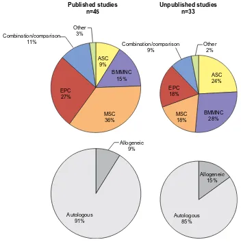
Comparisons of published studies with ongoing or unpublished studies (Table 2) demonstrate a shift in focus (Figure 3). The relative increase in studies utilizing ASCs coincides with a more recent appreciation for the higher yield
Table 2 Unpublished studies
NCT Number Recruitment Enrollment Diabetes Ischemia Venous Pressure Hypertensive Intervention
NCT01595776 Completed 8 Autologous APC/
PBMNC
NCT00919516 Completed 49 Autologous BMMNC
NCT00883870 Completed 20 Allogeneic BM-MSC
NCT00221143 Completed 15 Autologous APC
NCT01065337 Completed 30 Autologous BMMNC
NCT00616980 Completed 28 Autologous APC
NCT00523731 Completed 6 Autologous APC
NCT00392509 Completed 20 Autologous BMMNC
vs ALDHbr
NCT00468000 Completed 86 Autologous BMMNC
(CD90+ enriched)
NCT01232673 Completed 96 Autologous BMMNC
NCT00872326 Completed 20 Autologous BMMNC
NCT00371371 Completed 160 Autologous BMMNC
NCT00282646 Completed 40 Autologous BMMNC
NCT00535548 Completed 3 Autologous APC
NCT00677404 Completed 20 Autologous BMMNC
NCT00797056 Completed 32 Autologous APC
NCT01584986 Completed 22 Autologous APC
NCT01480414 Completed 20 Autologous BMMNC
NCT01408381 Completed 38 Autologous BMMNC
NCT00955669 Completed 40 Autologous BMMNC
vs BM-MSC NCT02287831 Active, not
recruiting
30 Allogeneic UC-MSC
NCT01745744 Active, not recruiting
33 Autologous ASC
NCT01049919 Active, not recruiting
152 Autologous BMMNC
NCT01472289 Active, not recruiting
15 Autologous BMMNC
NCT01245335 Active, not recruiting
210 Autologous BMMNC
NCT01751282 Active, not recruiting
66 Autologous BMMNC
in fibrin spray NCT01305863 Active, not
recruiting
60 Device: ASC coated
ePTFe vascular graft
NCT02394886 Recruiting 5 Allogeneic ASC
NCT01484574 Recruiting 126 Allogeneic BM-MSC
NCT01932021 Recruiting 10 Autologous adipose
tissue graft
NCT02474381 Recruiting 60 Autologous APC
NCT02454231 Recruiting 38 Autologous APC vs
BMMNC
NCT02099500 Recruiting 200 Autologous ASC
NCT02092870 Recruiting 25 Autologous ASC
NCT01937416 Recruiting 10 Autologous BMMNC
Chronic Wound Care Management and Research downloaded from https://www.dovepress.com/ by 118.70.13.36 on 20-Aug-2020
Dovepress Stem cells and chronic wound healing
Table 2 (Continued)
NCT Number Recruitment Enrollment Diabetes Ischemia Venous Pressure Hypertensive Intervention
NCT01572376 Recruiting 30 Autologous BMMNC
NCT01456819 Recruiting 50 Autologous BMMNC
± BM-MSC
NCT02089828 Recruiting 50 Autologous CD34+
enriched vs PBMNC
NCT02304588 Recruiting 20 Autologous MSC
NCT01833585 Recruiting 10 Autologous PBMNC
NCT02145897 Recruiting 60 Autologous SvF vs
ASC
NCT01916369 Recruiting 9 CTX DP (Human
neural stem cell product)
NCT01686139 Not yet recruiting 10 Allogeneic BM-MSC
NCT01558908 Not yet recruiting 15 Allogeneic
endometrial-MSC
NCT01353937 Not yet recruiting 27 Autologous APC
NCT02375802 Not yet recruiting 12 Autologous ASC
NCT02477540 Not yet recruiting 10 Autologous BM-MSC
NCT01903044 Not yet recruiting 60 Autologous BMMNC
Notes: Results of clinicaltrials.gov search showing ongoing trials or unpublished studies pertaining to stem cell therapies in patients with chronic wounds, verified within
the last 4 years.
Abbreviations: ALDHbr, aldehyde dehydrogenase bright cell; APC, angiogenic progenitor cell; ASC, adipose-derived stem cell; BMMNC, bone-marrow mononuclear cell;
BM-MSC, bone-marrow mesenchymal stem cell; SvF, enhanced stromal vascular fraction; PBMNC, peripheral blood mononuclear cell; UC-MSC, umbilical cord mesenchymal stem cell; ePTFE, expanded polytetrafluoroethylene.
Other 3%
Other 2% Combination/comparison
9% Combination/comparison
11%
EPC 18% EPC
27%
MSC 18% MSC
36%
BMMNC 28% BMMNC
15% ASC24%
ASC 9%
Unpublished studies n=33 Published studies
n=45
Autologous 85% Autologous
91%
Allogeneic 15% Allogeneic
9%
Figure 3 Relative proportions of stem cell population use in analyzed published and unpublished clinical studies.
Note: Chart area is proportional to sample size.
Chronic Wound Care Management and Research downloaded from https://www.dovepress.com/ by 118.70.13.36 on 20-Aug-2020
Dovepress Leavitt et al
of readily accessible stem cells existing within adipose tissue. Harvesting these cells from what is generally classified as biohazardous waste following liposuction may allow patients to forgo painful bone marrow aspiration. Additionally, BMMNCs appear to be favored in ongoing or unpublished trials, potentially due to several studies documenting the superiority of more heterogeneous cell transplants.19–21 The
number of studies and their respective sample sizes receiv-ing allogeneic cells is also greater among ongoreceiv-ing trials, reflecting an understanding that autologous stem cell potency may be blunted in patients with chronic systemic illness. Conversely, allogeneic cells have been shown to significantly enhance diabetic wound healing.81
Taken together, the published studies demonstrate that stem cell therapies can indeed lead to healing of chronic wounds resistant to traditional therapy. Meta-analyses corrob-orate this impression; most recently Liew et al82 calculated an
odds ratio of 2.90 (95% confidence interval [CI], 1.44–5.82) when comparing stem cell therapies with control treatment for complete ulcer healing. However, the 2015 meta-analysis by Liu et al83 suggests that this benefit may wane with longer
follow-up times. While there is sufficient evidence to support the belief that stem cells improve chronic wound healing in clinical trials, the limited number of placebo control groups and inconsistent means of reporting wound healing (eg, using median wound area84 rather than complete wound closure)
prevents us from establishing the true extent of this benefit. In our literature search, 9 of the 17 placebo-controlled tri-als showed a statistically significant improvement in wound healing with cell therapy. Interestingly, these studies include an array of methods to administer cells (intramuscular, intra-arterial, and direct application) and cell populations (PBMNCs, SVF, BM-MSCs, and BMMNCs). This suggests that many tissues and extraction methods offer means of reli-ably harvesting cells that can augment the healing process.
Improving stem cell yield, efficacy,
and lifespan – current and future
techniques
Unfortunately, though MSCs have demonstrated an ability to improve wound healing, their lifespans are short in vitro, reducing the efficacy of ex vivo expansion. Furthermore, they demonstrate suboptimal engraftment, survivability, and reten-tion at the wound when transplanted,85,86 with several of the
underlying mechanisms discussed previously. Many studies rely on intradermal or intramuscular injection to administer stem cells in suspension. Though technically simple, there is a relative loss of therapeutic efficacy, potentially caused by
subsequent anoikis in the absence of cell–matrix attachment or shear forces during the injection.87 When compared, stem
cells delivered intramuscularly and intra-arterially demon-strate no significant difference in terms of improved wound healing.88,89 Conversely, intravenous administration of stem
cells is uncommon in chronic wound therapy because of cell entrapment in the pulmonary vasculature (the pulmonary “first-pass” effect).90 Stem cell localization to the wound
bed is notably impaired in chronic (but not acute) wounds partly due to downregulated stromal cell-derived factor 1 (a chemokine attracting MSC to wound bed) secondary to uncontrolled inflammation.91 The resulting decrease of ASC
migration to the wound bed further elaborates on the difficul-ties of systemic stem cell therapy for chronic wound healing, necessitating consideration when developing mechanisms of administration for translational medicine. Advances in stem cell surface modification offer a potential solution to this problem by targeting cells to specific tissues.92
Poor cell engraftment and survivability are problem-atic considering the dose–effect relationship observed by Falanga et al,93 though the number of administered cells
has not universally been shown to correlate with response.77
Defining dose in heterogeneous cell populations can also be difficult, but perhaps subpopulation composition may be less relevant (given BMMNC:MSC ratios resulted in similar clinical improvements).94 Efficacy is most likely related to a
minimum required dose,93 while the lack of consensus may
be related to inconsistent methodologies (eg, wound type, cell type, harvest, expansion, and administration). Increasing stem cell potency with adjuvants such as platelet-rich plasma or Panax notoginseng saponins are possible alternatives for decreasing the minimum required dose.95,96
Means of prolonging transplanted cell lifespan are now heavily sought, as MSCs must survive to influence healing. Numerous possibilities have arisen, such as hyperoxic and pan-caspase pretreatment, which reduces MSC apoptosis in ischemic microenvironments.97 Hypoxic preconditioning has
also been shown to increase paracrine secretion by MSCs.98
Mohanty et al99 showed that small molecule-induced prion
protein upregulation also results in increased lifespan and yield of MSCs in culture, as well as improved engraftment. Gene therapy provides further opportunity, such as protein kinase G1α overexpression via adenovirus vector to promote MSC survival.100 Low-level light irradiation is another recent
tool for increasing stem cell wound healing potency.101
Endogenous wound healing pathways provide further means by which to optimize MSC survival. In the presence of proapoptotic cytokines (FasL, ubiquitous in chronic
Chronic Wound Care Management and Research downloaded from https://www.dovepress.com/ by 118.70.13.36 on 20-Aug-2020
Dovepress Stem cells and chronic wound healing
wound microenvironments), endothelial growth factor (EGF) molecules tethered to growth scaffolds demonstrated an ability to improve MSC survival via activation of the EGF-receptor.102 Surface-tethered EGF generated a superior MSC
response relative to saturating concentrations of soluble EGF,103 supporting the use of other endogenous mediators
such as the matrix protein Tenascin-C combined with biosyn-thetic scaffolds to enhance survival of transplanted MSCs.104
Such combinations may reduce the inflammatory response to the scaffolds themselves.105
Scaffolds are valuable additions to stem cell-based wound therapy as they provide an external niche for transplanted cells outside of the hostile wound environment, while still allowing them to facilitate wound healing. In an excisional wound model, Rustad et al87 showed both a faster time to
complete wound closure and a return of skin appendages in wounds treated with a biomimetic pullulan–collagen hydrogel scaffold seeded with MSCs. Moreover, this hydrogel led to longer MSC viability, increased engraftment efficiency, and enhanced angiogenesis. Clinically, Yoshikawa et al106 used a
composite graft of BM-MSCs incorporated into a collagen sponge to successfully treat decubitus ulcers refractory to artificial skin grafting. ASCs embedded in silk fibroin scaf-folds and fibrin gels have demonstrated a similar ability to accelerate wound healing in vivo.107,108 Autologous MSCs
applied with a fibrin spray system also resulted in some improvement in patients with chronic ulcers.93 Scaffolds
offer a viable means for enhancing stem cell engraftment and survivability. It is therefore likely that their incorpora-tion into cell-based therapies will increase markedly in the near future.
Clinical implementation of cell-based therapies has opened a new frontier in the development of biomedical devices aimed at optimizing current therapies.109 The
funda-mental risk of contamination associated with cell products has spurred the development of closed systems for harvesting and/or culturing cells.110 Widespread clinical use also
neces-sitates scalable technologies, such as tissue bioreactors.111
Increased automation of stem cell harvest, isolation, and expansion will allow for more standardized therapies, and subsequently more generalizable results. Novel approaches to cell population characterization such as kinome analysis may also improve clinical efficacy, or at least provide a better measure of prognosis.112
Promoting stem cell yield, survival, and efficacy at the wound bed are worthy goals. However, it is also possible that truly successful chronic wound therapy requires a deeper understanding of how the various types of stem cell therapies
can modulate systemic pathophysiology. For example, in type 2 diabetes, inhibiting the local proinflammatory phenotype at the wound bed may be insufficient to completely restore wound healing; restoration of an anti-inflammatory M1/M2 macrophage equilibrium is required to allow for physiologi-cal wound healing.113
Limitations
Some of the limitations of stem cells in wound therapies have already been discussed, such as phenotypic drift in culture, heterogeneity of cell populations, and the variable quality of cells depending on their source. Beyond barriers to therapeu-tic efficacy, there are also potential risks, as is the case with any medical intervention. The possibility of malignant trans-formation exists whenever stem cells are transplanted. While this may be a greater risk with pluripotent iPSCs, study of the more commonly used multipotent MSCs has generated less of a concern. While malignant transformation has been observed in long-term culture,114 the larger body of evidence suggests
that the risks of malignant transformation are low, especially prior to MSCs undergoing senescence.115 Follow-up to one
of the original studies thought to demonstrate spontaneous MSC transformation has since shown a small number of malignant cells to be the culprit.116 Furthermore, the
ben-eficial immunomodulatory properties of stem cells are also not without theoretical risks. MSC immunomodulation and homing to different target organs can increase risks of oppor-tunistic or disseminated infections, as well as susceptibility to malignancy.117,118 Finally, transplant of biological material
also carries risks of directly transmitting infectious agents.117
Overall, clinical trials demonstrate that stem cell applications to wound healing are safe, but physicians must continue to reevaluate the risks and benefits of their use as the results of more long-term follow-up studies are published.
Conclusion
Stem cells can provide the next step in advancing wound care, particularly for chronic wounds resistant to current therapies. Meta-analyses consistently show that stem cells provide a safe and effective means for promoting chronic wound healing. However, the statistically similar improve-ment observed in both trial arms of the large JUVENTAS study offers a solid reminder of the importance of placebo controls for measuring the true efficacy of stem cell therapy. As development of commercial devices for stem cell therapies increases, standardization of protocols will allow for greater study validity. Therapies can then be fine-tuned and catered to specific pathologies, whereas currently the different stem cell
Chronic Wound Care Management and Research downloaded from https://www.dovepress.com/ by 118.70.13.36 on 20-Aug-2020
Dovepress Leavitt et al
populations and routes of administration provide only roughly comparative results across several head-to-head studies. As we move forward, stem cells are likely to become a common tool available to clinicians for wound management, but clini-cal practice must follow evidence of safety and efficacy, so frequent reevaluation of the literature will be critical as new therapies are described.
Disclosure
The authors report no conflicts of interest in this work.
References
1. Tricco AC, Cogo E, Isaranuwatchai W, et al. A systematic review of cost-effectiveness analyses of complex wound interventions reveals optimal treatments for specific wound types. BMC Med. 2015;13:90. 2. Li H, Fu X. Mechanisms of action of mesenchymal stem cells
in cutaneous wound repair and regeneration. Cell Tissue Res. 2012;348(3):371–377.
3. Dominici M, Le Blanc K, Mueller I, et al. Minimal criteria for defining multipotent mesenchymal stromal cells. The International Society for Cellular Therapy position statement. Cytotherapy. 2006;8(4):315–317.
4. Alvarez-Viejo M, Menendez-Menendez Y, Otero-Hernandez J. CD271 as a marker to identify mesenchymal stem cells from diverse sources before culture. World J Stem Cells. 2015;7(2):470–476.
5. Ho AD, Wagner W, Franke W. Heterogeneity of mesenchymal stromal cell preparations. Cytotherapy. 2008;10(4):320–330.
6. Baer PC. Adipose-derived mesenchymal stromal/stem cells: an update on their phenotype in vivo and in vitro. World J Stem Cells. 2014;6(3):256–265.
7. Maxson S, Lopez EA, Yoo D, Danilkovitch-Miagkova A, Leroux MA. Concise review: role of mesenchymal stem cells in wound repair. Stem Cells Transl Med. 2012;1(2):142–149.
8. Gebler A, Zabel O, Seliger B. The immunomodulatory capacity of mesenchymal stem cells. Trends Mol Med. 2012;18(2):128–134. 9. Aggarwal S, Pittenger MF. Human mesenchymal stem cells modulate
allogeneic immune cell responses. Blood. 2005;105(4):1815–1822. 10. Chen L, Tredget EE, Wu PY, Wu Y. Paracrine factors of mesenchymal
stem cells recruit macrophages and endothelial lineage cells and enhance wound healing. PLoS One. 2008;3(4):e1886.
11. Matsumoto R, Omura T, Yoshiyama M, et al. Vascular endothelial growth factor-expressing mesenchymal stem cell transplantation for the treatment of acute myocardial infarction. Arterioscler Thromb Vasc Biol. 2005;25(6):1168–1173.
12. Jones EA, Kinsey SE, English A, et al. Isolation and characterization of bone marrow multipotential mesenchymal progenitor cells. Arthritis Rheum. 2002;46(12):3349–3360.
13. Bourin P, Bunnell BA, Casteilla L, et al. Stromal cells from the adipose tissue-derived stromal vascular fraction and culture expanded adipose tissue-derived stromal/stem cells: a joint statement of the International Fed-eration for Adipose Therapeutics and Science (IFATS) and the International Society for Cellular Therapy (ISCT). Cytotherapy. 2013;15(6):641–648. 14. Cervelli V, Gentile P, Angelis B, et al. Application of enhanced stromal
vascular fraction and fat grafting mixed with PRP in post-traumatic lower extremity ulcers. Stem Cell Res. 2011;6(2):103–111.
15. Fraser JK, Zhu M, Wulur I, Alfonso Z. Adipose-derived stem cells. Methods Mol Biol. 2008;449:59–67.
16. Durlik M, Olszewski WL. Biological effects of bone marrow in trans-planted limb – a review. Ann Transplant. 2004;9(4):26–31.
17. Li WW, Talcott KE, Zhai AW, Kruger EA, Li VW. The role of therapeutic angiogenesis in tissue repair and regeneration. Adv Skin Wound Care. 2005;18(9):491–500; quiz 501-492.
18. Li S, Huang KJ, Wu JC, et al. Peripheral blood-derived mesenchymal stem cells: candidate cells responsible for healing critical-sized calvarial bone defects. Stem Cells Transl Med. 2015;4(4):359–368.
19. Rodriguez-Menocal L, Shareef S, Salgado M, Shabbir A, Van Badiavas E. Role of whole bone marrow, whole bone marrow cultured cells, and mesenchymal stem cells in chronic wound healing. Stem Cell Res Ther. 2015;6:24.
20. Lu D, Chen B, Liang Z, et al. Comparison of bone marrow mesenchymal stem cells with bone marrow-derived mononuclear cells for treatment of diabetic critical limb ischemia and foot ulcer: a double-blind, random-ized, controlled trial. Diabetes Res Clin Pract. 2011;92(1):26–36. 21. Yasuhara S, Yasunaga Y, Hisatome T, et al. Efficacy of bone marrow
mononuclear cells to promote bone regeneration compared with iso-lated CD34+ cells from the same volume of aspirate. Artif Organs. 2010;34(7):594–599.
22. Li Q, Wang Z. Influence of mesenchymal stem cells with endothelial progenitor cells in co-culture on osteogenesis and angiogenesis: an in vitro study. Arch Med Res. 2013;44(7):504–513.
23. Wu Y, Chen L, Scott PG, Tredget EE. Mesenchymal stem cells enhance wound healing through differentiation and angiogenesis. Stem Cells. 2007;25(10):2648–2659.
24. Kuo YR, Wang CT, Cheng JT, Wang FS, Chiang YC, Wang CJ. Bone marrow-derived mesenchymal stem cells enhanced diabetic wound healing through recruitment of tissue regeneration in a rat model of streptozotocin-induced diabetes. Plast Reconstr Surg. 2011;128(4):872–880.
25. Kim EK, Li G, Lee TJ, Hong JP. The effect of human adipose-derived stem cells on healing of ischemic wounds in a diabetic nude mouse model. Plast Reconstr Surg. 2011;128(2):387–394.
26. Pittenger MF, Mackay AM, Beck SC, et al. Multilineage potential of adult human mesenchymal stem cells. Science. 1999;284(5411):143–147. 27. Fathke C, Wilson L, Hutter J, et al. Contribution of bone
marrow-derived cells to skin: collagen deposition and wound repair. Stem Cells. 2004;22(5):812–822.
28. Akino K, Mineda T, Akita S. Early cellular changes of human mesen-chymal stem cells and their interaction with other cells. Wound Repair Regen. 2005;13(4):434–440.
29. Rehman J, Traktuev D, Li J, et al. Secretion of angiogenic and antiapoptotic factors by human adipose stromal cells. Circulation. 2004;109(10):1292–1298.
30. Hu L, Zhao J, Liu J, Gong N, Chen L. Effects of adipose stem cell-conditioned medium on the migration of vascular endothelial cells, fibroblasts and keratinocytes. Exp Ther Med. 2013;5(3):701–706. 31. Asahara T, Murohara T, Sullivan A, et al. Isolation of putative
pro-genitor endothelial cells for angiogenesis. Science. 1997;275(5302): 964–967.
32. Wettstein R, Savic M, Pierer G, et al. Progenitor cell therapy for sacral pressure sore: a pilot study with a novel human chronic wound model. Stem Cell Res Ther. 2014;5(1):18.
33. Takahashi K, Tanabe K, Ohnuki M, et al. Induction of pluripotent stem cells from adult human fibroblasts by defined factors. Cell. 2007;131(5):861–872.
34. Yu J, Vodyanik MA, Smuga-Otto K, et al. Induced pluripo-tent stem cell lines derived from human somatic cells. Science. 2007;318(5858):1917–1920.
35. Sun N, Panetta NJ, Gupta DM, et al. Feeder-free derivation of induced pluripotent stem cells from adult human adipose stem cells. Proc Natl Acad Sci U S A. 2009;106(37):15720–15725.
36. Okita K, Ichisaka T, Yamanaka S. Generation of germline-competent induced pluripotent stem cells. Nature. 2007;448(7151):313–317. 37. Zhou H, Wu S, Joo JY, et al. Generation of induced pluripotent stem
cells using recombinant proteins. Cell Stem Cell. 2009;4(5):381–384. 38. Stadtfeld M, Nagaya M, Utikal J, Weir G, Hochedlinger K. Induced
pluripotent stem cells generated without viral integration. Science. 2008;322(5903):945–949.
39. Liu SV. iPS cells: a more critical review. Stem Cells Dev. 2008;17(3): 391–397.
Chronic Wound Care Management and Research downloaded from https://www.dovepress.com/ by 118.70.13.36 on 20-Aug-2020