Open Access
Research
Combined effects of hyperglycemic conditions and HIV-1 Nef: a
potential model for induced HIV neuropathogenesis
Edward A Acheampong
1, Cassandra Roschel
2, Muhammad Mukhtar
3,
Alagarsamy Srinivasan
4, Mohammad Rafi
5, Roger J Pomerantz
6and
Zahida Parveen*
1Address: 1The Dorrance H. Hamilton Laboratories, Division of Infectious Diseases and Environmental Medicine, PA 19107, USA, 2Bioscience Technologies - Biotechnology, Thomas Jefferson University, Philadelphia, PA 19107, USA, 3Department of Biochemistry, Pir Mehr Ali Shah Arid Agriculture University Rawalpindi, 46300 Pakistan, 4NanoBio Diagnostics, West Chester, PA 19382, USA, 5Department of Neurology, Jefferson Medical College, Thomas Jefferson University, Philadelphia, PA 19107, USA and 6Tibotec Inc. 1020 Stony Hill Road, Suite 300, Yardley, PA 19067, USA
Email: Edward A Acheampong - [email protected]; Cassandra Roschel - [email protected]; Muhammad Mukhtar - [email protected]; Alagarsamy Srinivasan - [email protected]; Mohammad Rafi - [email protected]; Roger J Pomerantz - [email protected];
Zahida Parveen* - [email protected] * Corresponding author
Abstract
Hyperglycemic conditions associated with diabetes mellitus (DM) or with the use of antiretroviral therapy may increase the risk of central nervous system (CNS) disorders in HIV-1 infected patients. In support of this hypothesis, we investigated the combined effects of hyperglycemic conditions and HIV-1 accessory protein Nef on the CNS using both in vitro and in vivo models. Astrocytes, the most abundant glial cell type required for normal synaptic transmission and other functions were selected for our in vitro study. The results show that in vitro hyperglycemic conditions enhance the expression of proinflammatory cytokines including caspase-3, complement factor 3 (C3), and the production of total nitrate and 8-iso-PGF2 α as reactive oxygen species (ROS) in human astrocytes leading to cell death in a dose-dependent manner. Delivery of purified recombinant HIV-1 Nef protein, or Nef expressed via HIV-1-based vectors in astrocytes showed similar results. The expression of Nef protein delivered via HIV-1 vectors in combination with hyperglycemia further augmented the production of ROS, C3, activation of caspase-3, modulation of filamentous protein (F-protein), depolarization of the mitochondria, and loss of astrocytes. To further verify the effects of hyperglycemia and HIV-1 Nef protein on CNS individually or in combination, in vivo studies were performed in streptozotocin (STZ) induced diabetic mice, by injecting HIV-1 Nef expressing viral particles into the sub-cortical region of the brain. Our in vivo results were similar to in vitro findings indicating an enhanced production of caspases-3, ROS (lipid oxidation and total nitrate), and C3 in the brain tissues of these animals. Interestingly, the delivery of HIV-1 Nef protein alone caused similar damage to CNS as augmented by hyperglycemia conditions. Taken together, the data suggests that HIV-1 infected individuals with hyperglycemia could potentially be at a higher risk of developing CNS related complications.
Published: 30 October 2009
Virology Journal 2009, 6:183 doi:10.1186/1743-422X-6-183
Received: 4 May 2009 Accepted: 30 October 2009
This article is available from: http://www.virologyj.com/content/6/1/183 © 2009 Acheampong et al; licensee BioMed Central Ltd.
Introduction
Antiretroviral therapy has been linked to insulin resist-ance and dyslipidemia in HIV infected individuals under treatment [1-4]. Since glucose is a major nutrient utilized by the brain[5], diabetes or HAART-associated hyperglyc-emic conditions may become a potential risk factor in the brain [6-8], and could lead to a series of devastating clini-cal conditions in the CNS of HIV-1 infected individu-als[9]. Several studies have described hyperglycemia-induced neuronal and astrocytic glial cell death leading to various neurological disorders in diabetic patients[7,10,11]. However, limited information is avail-able regarding the combined effects of hyperglycemia and HIV-1 infection on the CNS. Astrocytes play a critical role in the provision of nutrients and strength to the CNS via the foot processes protecting the blood brain barrier [12]. In this study, we selected astrocytes as target cells to eval-uate the cumulative toxic effects of hyperglycemia and HIV-1 Nef protein. Previous studies have shown that hyperglycemia increases the production of proinflamma-tory cytokines, oxidative reactive species and activation of CD4+ and CD8 T lymphocytes in the peripheral blood system [13]. Of the proteins encoded by HIV-1, Env, Vpr, Vif, Tat, and Nef are known to exhibit cytopathic effects [14-16]. Specifically, the data from previous studies sug-gest a potentially important role of Nef in cellular dys-functions and its contribution to the development of the neuropathology associated with AIDS. HIV-1 Nef expres-sion has been shown to be essential in maintaining high replication level of the virus and promoting the develop-ment of AIDS in SIV-infected monkeys[17]. Skowronski and others have shown that the expression of Nef in trans-genic mice is associated with the development of a severe AIDS like disease [18,19]. Nef and gp120 have been detected in the CSF of HIV-1 infected individuals and are known to be involved in the induction of complement factor C3 [9,20,21]. HIV-1 infection, thus affects the cellu-lar processes in the brain by activating signaling pathways and the production of cytokines [22,23]. It has been reported that extracellular release of Nef protein could exert its effects on non-infected bystander cells in brain tissues of HIV-1 infected individuals and could be detected in distant brain regions [14,17]. HIV-1 proteins also cause an increase in systemic oxidative/nitrosative stress, by enhancing the deleterious effects of secondary infections [9]. The molecular mechanism involved in HIV-1 associated neuropathogenesis is not completely understood due to the inaccessibility of the brain paren-chyma during the course of AIDS. Hence, limited infor-mation is available regarding the contributions of Nef alone and or in combination with hyperglycemic condi-tions to the pathogenesis of the CNS in the context of HIV-1 infection. The focus of this study was to evaluate the cytopathic effects of hyperglycemic conditions in the presence of HIV-1 Nef delivered either through
HIV-1-based vector systems (intracellular) or in the form of recombinant protein (extracellular) in human astrocytes (in vitro) and STZ induced diabetic mice used as an in vivo
model[24]. The delivery of Nef protein via viral injection into the STZ induced diabetic mice brain increased oxida-tive reactions as well as the production of inflammatory cytokines, complement factor C3, and depolarization of mitochondria. Induction of in vitro and in vivo hyperglyc-emia alone induced similar cytopathic effects in astrocytes and in diabetes induced mice. Further, the data involving astrocytes suggests that the presence of extracellular Nef protein further increased the risk of toxicity and cell death in a dose-dependent manner under hyperglycemic condi-tions.
Materials and methods
Cell Culture
Primary cultures of human fetal brain astrocytes and astrocytes medium were purchased from Cambrex, Inc (Walkersville, MD) and Sciencell (San Diego, CA). The cells were maintained in astrocyte media (AM) in a water-jacketed incubator at 37°C, with 5% CO2 in a humid envi-ronment. The cells were passaged at a confluence of 80-85%. The human glioblastoma/astrocytoma cell line U87-MG, and human kidney cell line 293-T were obtained from American Type Culture Collection (ATCC) and cultured in Dubelcco's Modified Eagle's Medium (DMEM) supplemented with 10% fetal bovine serum (Sigma Aldrich, St. Louis, MO), penicillin-streptomycin (100 U/ml and 100 μg/ml, respectively), and 2 mM L-glutamate (Mediatech Corp, MD).
Generation of Nef expressing viral particles andtransduction
HIV-1 Nef expressing recombinant viral particles were generated by triple transfection of plasmids using Cal-cium phosphate transfection kit (Promega Corp, Madi-son, WI) following the manufacturer's protocol. Briefly, 293 T cells were seeded in 100 mm culture plates over-night. The cells were transfected with reagents of Mamma-lian Calcium Phosphate transfection kit (from Promega) in the presence of HIV-1 based vectors DNA; pHR'CMV Nef, pHR CMV delta 8.2, and pMD.G encoding VSV.G as an envelope protein. In addition, viral particles expressing HIV-1 Nef generated from spleen necrosis virus (SNV) packaging vector pZP32, transfer vector expressing HIV-1
Nef pZP35, and envelope vector VSV.G [14,25] were used
(Perkin Elmer, Boston, MA). Based on the quantification, equal amount of viral particles were used for the experi-ments. Culture supernatants without hyperglycemia and Nef were collected from the astrocytes and used as a source for mock treatment. Astrocytes were plated at 60% confluency over night before tranduction. Primary human astrocytes or U87-MG cells were distributed into 4-well chamber slides or plates at a cell density of 1.0 × 10⁵ cells
per well and allowed to stabilize in AM media for 24 hours prior to the addition of glucose media. After the sta-bilization period, the cells in each well were washed with pre-warmed 1× PBS. To mimic the in vivo hyperglycemia, glucose stock solutions were added to glucose-free media (contained 1.0 mM sodium pyruvate, 1% strep/pen, and 5% FBS) to achieve 10 mM, 15 mM, and 20 mM glucose concentrations. Of note, 10, 15, and 20 mM glucose rep-resents the 180, 200, 350 mg glucose/dl blood in diabetic patients. The medium with 5.0 mM glucose was used as a control. The astrocytes were exposed to in vitro hyperglyc-emic conditions for 12 hours and washed with 1× PBS. The astrocytes were then transduced with viral superna-tant mixed with 8 ug/ml polybrene for astrocytic cell line(U87-MG) and 4 ug/ml for primary astrocytes for 3 hours followed by washing to remove the virus and incu-bated with complete medium. Astrocytes were harvested and supernatants were collected after 48 hours for various analyses. Non- transduced astrocytes were used as a con-trol.
In vitro effects of hyperglycemia and recombinant Nef protein on human astrocytes
Individual and cummulative effects of hyperglycemia and recombinant Nef protein on primary human fetal astro-cytes were evaluated by observing changes in the F-actin, a protein involved in mitochondrial and cellular integrity [26]. Astrocytes were seeded into 4-well chamber slides at a cell density of 1.0 × 10⁵ cells per well and exposed to
var-ious hyperglycemic conditions for 12 hours, followed by extensive washing with 1× PBS. The cells were fixed with 4% paraformaldehyde for 10 minutes and washed several times with 1× PBS to remove the fixative. The astrocytes were then stained with BODIPY phallacidin (Invitrogen Corporation, Carsbad, CA) cytoskeleton staining dye fol-lowing protocol suggested by the manufacturer, and observed under fluorescence-microscope.
The effect of recombinant Nef protein on mitochondria was studied by using Mitotracker dye (Invitrogen Corpo-ration, Carsbad, CA). The dye stains mitochondria only upon depolarization. For this, astrocytes were seeded in chamber slides in AM medium and allowed to attach for 24 hours prior to Nef protein treatments. Recombinant Nef protein generated in our laboratory [14] was added at concentrations of 1 nM, 3 nM, and 25 nM in 500 μl of glu-cose free DMEM containing 1.0 mM sodium pyruvate, 1%
strep/pen, and 5% FBS and incubated with astrocytes for 24 hours. The cells were washed with 1XPBS and stained live with 200 nM Mitotracker Red fluorochrome (Invitro-gen Corp., Carlsbald, CA), to detect the depolarization of mitochondria.
Induction of Diabetes
Mice with C57/BL6 genetic background were purchased from Jackson Laboratories to study the effects of hypergly-cemic variations either alone or in combination with intracellularly expressed HIV-1 Nef protein in vivo[27]. To rule out the effect of other accessory proteins associated with HIV-1 virus, SNV vector virus encoding Nef was also included in the study. The exact physiological concentra-tion of Nef is not clear in HIV-1 infected individuals. However extracellular recombinant Nef protein generated in our laboratory was added to astrocytes at concentra-tions mentioned in previous studies [28]. Diabetes was induced in 12 mice by a single subcutaneous injection of 40 mg/kg body weight streptozotocin (Sigma Aldrich Corp., St.Louis, MO) dissolved in freshly prepared 0.1 M citrate buffer pH 4.5. The blood glucose level was assessed by a glucometer using a drop of blood drawn at 1, 2, 4, 6, 8, 10, and 12 hours post injection. The mean elevated glu-cose level was 325 mg/dl after injection of STZ. Upon con-firmation of induction of hyperglycemia in mice, 2-ul of concentrated viral particles (1 × 10⁷) generated through
HIV-1 and SNV-based vectors systems were injected into the brain of mice via the cortex as described previously [29]. Age-matched non-diabetic and STZ treated (diabe-tes) mice injected with an equal volume of citrate buffer, served as controls. The animals were housed under path-ogenic free conditions in Thomas Jefferson Animal Facil-ity. Mice from the ages of 1-2 weeks of the same sex were used in the experiments. All procedures were conducted in accordance with federal guidelines using animal protocols approved by the Thomas Jefferson University Institutional Animal Care and Use Committee (IACUC). The mice were sacrificed eight weeks post-injection and their brains were detached, washed in cold 1× PBS and used for various analyses (25).
Western Blot Analyses
The membranes were washed in PBS containing 0.01% Tween 20 (Sigma-Aldrich, St. Louis, MO.). Non-specific proteins were blocked with PBS-based blocking buffer (Pierce Biotechnologies, Rockford, IL) and the mem-branes were probed with specific monoclonal antibodies against GFAP at a concentration of 1:1000, mouse anti-Caspase 3 antibody at a concentration of 1:1000 as pri-mary antibodies and horseradish peroxidase labeled anti-mouse immunoglobulin G (heavy plus light chains) as secondary antibodies. The protein-antibody complexes were visualized by autoradiography of the membranes after incubating with the ECL blotting detection system (Pierce Biotechnologies, Rockford, IL) and subsequently exposing them to BioMax MS (Kodak, Rochester, N.Y.) film. HIV-1 Nef protein was detected in the transduced astrocytes by treating the blots with HIV-1 Nef anti-body (NIH AIDS Repository). For in vivo analysis of the expression of Nef, viral vector expressing Nef was identi-fied from mice brain tissues using immunoprecipitation method. The Seize X Immunoprecipitation Kit (Pierce Biotechnologies, Rockford, IL) was used following the manufacturer's protocol. The purified Nef protein was then subjected to Western Blot analysis using the method described earlier.
Enzyme Linked Immunosorbent Assays (ELISAs)
The production of nitric oxide and lipid oxidation reac-tion in the form of total nitrate and 8-iso- PGF-2α, respec-tively, were measured to determine the level of reactive oxidative species induced as a result of exposure to either hyperglycemic conditions alone or in combination with HIV-1 Nef protein or Nef alone. The U87-MG cells were exposed to various concentrations of glucose (10, 15, 20, 25 mM glucose solutions) followed by transduction with HIV-1 Nef expressing viral particles. Control astrocytes were cultured in normal medium. For in vivo studies, 10-day-old mice were injected with a single dose of STZ for induction of hyperglycemia followed by delivery of HIV-1 based Nef expressing virus via injection in the brain tis-sues. Non-diabetic mice, hyperglycemic mice, or HIV-1 Nef injected mice were used as controls. To rule out the influence of other accessory proteins of HIV-1 and for exclusive effect of Nef protein, mice were also injected with virus generated by SNV vector systems [27]. The cell/ tissue lysates and supernatants from the treated cells as well as from brain tissues from the hyperglycemic and Nef treated mice were collected. Samples were analyzed for the presence of nitric oxide (NO),
8-isoprostaglandin-F2-α, or complement factor C3 using respective ELISA kits (Stressgen Biotechnologies, Victoria, BC, Canada) as well as the manufacturer's suggested protocol [30].
Results
In this study, we utilized in vitro and in vivo models to eval-uate the combined cytopathic effects of hyperglycemia
and HIV-1 proteins on the CNS to mimic the conditions in individuals with diabetes or hyperglycemia associated with the use of highly active antiretroviral therapy (HAART). For in vitro studies, U87-MG/primary astrocytes were exposed to various hyperglycemic conditions by adding the appropriate amount of glucose in medium [31] and tranduced with HIV-1 Nef expressing virus. For the in vivo studies, diabetes was induced in mice with STZ and recombinant viral particles expressing HIV-1 Nef were injected in both STZ induced diabetic and normal mice brains. The combined cytopathic effects of hyperglycemia and HIV-1 Nef protein on the CNS were determined by evaluating the expression of complement factor 3, pro-duction of oxidative species (ROS), caspase activity, changes in F-actin protein, and the depolarization of mitochondria.
Effect of Hyperglycemia and HIV-1 Nef on Complement Factor 3(C3)
Detection of Reactive Oxygen Species (ROS)
Effect of Hyperglycemia and HIV-1 Nef on Nitric Oxide Production
The ability of hyperglycemia to induce reactive oxygen species (ROS) thereby enhancing the production of nitric oxide (total nitrate) and lipid peroxidation in the form of 8-iso-prostaglandin F2 alpha (8-iso-PGF2 alpha) are well
documented, and have been previously used as biological markers to detect the oxidative stress levels [14,32,33]. In this study, we investigated the production of reactive oxy-gen species by determining the level of total nitrates pro-duced due to in vitro hyperglycemic conditions or due to the expression of HIV-1 Nef protein in astrocytes or com-Figure 1
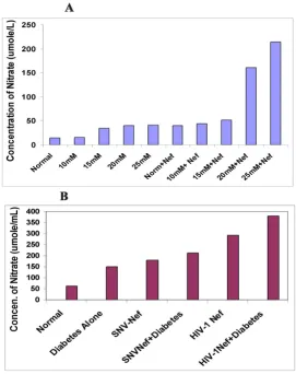
bination of both, as well as in vivo in diabetic mice. Figure 2A shows that hyperglycemia doubled the concentration of total nitrate in astrocytes upon exposure to 15, 20 and 25 mM glucose respectively, with the exception of 10 mM glucose showing similar nitrate level as observed in astro-cytes cultured under normal glycemic conditions. U87-MG astrocytes transduced with Nef expressing virus alone showed more than 2-fold increase in total nitrate. The combination of hyperglycemia with Nef expressing virus in astrocytes resulted in a dose- dependent increase in total nitrate. Astrocytes exposed to 10, 15, 20 and 25 mM glucose, and transduced with HIV-1 Nef expressing virus, increased the total nitrate from 3, 3.5, 11 and 15 fold respectively. These results suggest that hyperglycemia and Nef alone or in combination induce oxidative stress in the CNS in dose-dependent fashion.
To confirm our in vitro results in vivo, a total of 24 mice were used in the study. Diabetes was induced in 12 well-characterized C57/BL6 genetic background mice by inject-ing a sinject-ingle dose of STZ. HIV-1 Nef expressinject-ing viral parti-cles or SNV based viral partiparti-cles expressing Nef were injected into the cortex region of eight mice brains while the remaining four diabetic mice continued to grow for eight weeks. In addition, six normal mice were injected with HIV-1 Nef and SNV Nef expressing viral particles into the cortex region of the brain. Four untreated normal mice were used as controls. Eight weeks later, the mice were sac-rificed to analyze the effects of hyperglycemia alone or in combination with HIV-1 Nef protein expressed via HIV-1 based vectors on the CNS. The results illustrated in Figure 2B show similarities in the in vitro and in vivo increase in total nitrate. The hyperglycemic conditions also increased (2-fold) the total nitrate in diabetic mice brain. Delivery of HIV-1 based Nef expressing virus into the brain of the diabetic mice further enhanced the total nitrate produc-tion (more than 6-fold), in comparison to non-diabetic control mice. HIV-1 Nef expressing particles delivered into normal mice brain showed more than 4-fold increase in the production of total nitrate as compared to the nor-mal mice. Overall, our in vivo results are in agreement with those obtained through in vitro studies in astrocytes. Fur-ther, to rule out the impact of other HIV-1 accessory pro-teins, Nef expressing recombinant retroviral particles were generated using spleen necrosis virus vectors and injected into the cortex of mice brain and the results are depicted in Figure 2B. These results are close to those obtained when viral particles expressing HIV-1Nef were injected into the brain of mice. Delivery of SNV-based Nef express-ing virus alone increased more than 2-fold of total nitrate in normal mice brain, while diabetic mice showed 3-fold increase in total nitrate. These results are suggestive of an exclusive effect of Nef protein on astrocytes.
Effect of hyperglycemia and HIV-1 Nef on 8-iso-PGF2 α, production
The effect of hyperglycemia and HIV-1 Nef on lipid oxida-tion in astrocytes was determined by measuring the 8-iso-prostaglandin (8-iso-PGF2 alpha) using ELISA techniques. Astrocytes treated with various hyperglycemic conditions were either analyzed within seventy-two hours post treatment or were transduced with Nef expressing viral particles. The supernatants were collected and ana-lyzed for the production of 8-iso-PGF2-α. Similarly, dia-betes-induced mice were either left untreated or injected with HIV-1 Nef expressing virus. Eight weeks post-injec-tion, the mice were sacrificed and the brains were removed to analyze the cortex region of the brain for the production of 8-iso-PGF2 α. The results of both experi-ments are presented in Figure 3A and 3B. Fig. 3A, depicts the effect of hyperglycemia alone or in combination with HIV-1 Nef protein, in astrocytes indicating an enhanced production of 8-iso-PGF2 αin a dose-dependent manner. Hyperglycemic conditions alone increased the release of 8-iso-PGF2 α, ranging from 2 to 3-fold in astrocytes exposed to 10,15 20 and 25 mM glucose. The combina-tion of hyperglycemia and HIV-1Nef both resulted in more than 3 to 4-fold increase in lipid peroxidation reac-tion. The astrocytes treated with 25 mM glucose and trans-duced with Nef were indicating increase in cell death. Fig 3B, depicts the effect of diabetes and Nef on the produc-tion of 8-iso-PGF2 α. Our results indicate that induction of hyperglycemia in mice brain increased the production and release of 8-iso-PGF2 α, and delivery of HIV-1 based Nef expressing particles further enhanced it (2-fold further increase), as compared to the control mice. Furthermore, the use of SNV-based Nef expressing virus as a means of ruling out the possible added effects of other HIV-1 pro-teins and also to demonstrate the exclusive effect of HIV-1 Nef protein, produced similar results (Figure 3B). The expression of Nef alone in the brain of mice showed a similar increase (8-fold) in 8-iso-PGF2 αas compared to the control mice.
Effect of Hyperglycemia and HIV-1 Nef on Cytoskeleton and Mitochondria
under normal glycemia (Figure 4, panel A1). Exposure of astrocytes to various concentrations of glucose ranging from 15 mM to 20 mM enhanced the visibility of F-actin protein with significant changes in the cytoskeletal struc-ture as depicted in Figure 4 panels A2 and A3. The actin-network in astrocytes exposed to 15 mM glucose was very visible with expanded cell structure. Exposure to 20 mM glucose further enhanced the visibility with a higher
degree of disorganization of actin-network cell expansion, and increase in intracellular space indicating loss of astro-cytes (Figure 4, panel A3).
The mitochondrial depolarization was determined by MitoTracker Red fluorochrome detection method [35,36]. Figure 4 panel B2-3, depict an increase in the depolariza-tion of mitochondria in a dose-dependent manner with Figure 2
Figure 3
the addition of 3 nM or 25 nM/ml of recombinant Nef protein. The addition of 3 nM/ml Nef protein caused the depletion of astrocytes, whereas addition of 25 ng Nef completely damaged the astrocytes layer (Figure 4 panel B3) suggesting that intracellular accumulation of Nef (due to an increase in HIV-1 replication) in astrocytes could trigger apoptosis and a non-reversible damage of the mitochondria [37].
Effect of Hyperglycemia and Nef on caspases
To determine whether HIV-1 Nef and hyperglycemic con-ditions induced apoptosis, intracellular activity of caspase -3 was analyzed in primary astrocytes exposed to HIV-1 Nef particles, via Western blot and the results are depicted in Figure 5 panel A and B. The figure illustrates the impact of hyperglycemia and Nef on mice brain (in vivo) and in vitro on U87-MG astrocytes respectively. Panel A, lane 1 represents the pro-caspase 3 in normal mice brain while lane 2 represents the activated caspase-3 as a result of HIV-1 Nef expressing viral particles. Lane 3 also depicts the activation of caspase -3 by hyperglycemia. To ensure that the apoptosis observed is the exclusive effect of HIV-1 Nef protein, we subjected the brain lysates from diabetic mice injected with SNV- based Nef particles to western blot analyses and compared the results with brain lysates of mice injected with HIV-1 Nef expressing particles (Figure 5 panel A lane 4 and lane 5). These results suggest that hyperglycemia and Nef have an additive effect on caspase-3 activity, which could induce apoptosis. In panel B, the
in vitro results of hyperglycemic treated astyrocytes trans-duced with Nef exhibited dose-dependent activation of caspase-3 as depicted in Figure 5 panel B (lanes 2, 3 and 6), suggesting the apoptotic potential of hyperglycemic conditions which were dramatically augmented and syn-ergized by Nef (Figure 5 panel B lanes 2, 3 and 6). We also observed that the expression of Nef alone triggers the acti-vation of caspase -3 as illustrated in figure 5 panel B and lane 1. Similar observations were made in our in vivo stud-ies as well. The apoptotic effect of hyperglycemia and HIV-1 Nef on astrocytes and on CNS was also determined by quantifying the glial fibril acidic protein (GFAP) using GFAP specific antibody. The western blot analyses of astrocytes and mice brain exposed to hyperglycemia and/ or Nef are shown in figure 5 panels C and D respectively. These results indicate that astrocytes exposed to hypergly-cemia have reduced GFAP expression as shown in panel C lane 2 compared to normal astrocytes in Figure 5 panel C and lane 1. The results also indicate that Nef alone is capa-ble of down-modulating the expression of GFAP to a great extent in astrocytes than the hyperglycemia alone (Figure 5, panel C lane 3). Astrocytes exposed to various glucose solutions and transduced with HIV-1 Nef showed a dose-dependent decrease in GFAP protein expression (Figure 5 panel C lanes 4, 5 and 6) suggesting that hyperglycemic variations and Nef combination may synergistically and
adversely affect the expression of GFAP in astrocytes. The GFAP expression in STZ treated mice brain with and with-out Nef expression was also evaluated and the results are presented in Figure 5 panel D. These in vivo results are in agreement with our in vitro results as evident in lane 1, 2, and 3 illustrating the expression of GFAP in normal mice brain, diabetic mice and mice brain injected with Nef expressing particles (Figure 5 Panel D, lanes 4 and 5). It is evident from our results that HIV-1 Nef is more efficient in down-modulating the expression of GFAP than hyper-glycemic conditions. The data presented here also suggest that even low expression of HIV-1 Nef could affect astro-cytes by reducing the GFAP expression [38]. The expres-sion of Nef was also detected in astrocytes and in mice brain delivered via SNV based vectors, as shown in Figure 5 Panel E and F. All these results shown here are represent-ative of at least three independent experiments and repeated several times.
Discussion
The use of highly active antiretroviral therapy (HAART) has reduced the mortality and morbidity rates in HIV-1 infected individuals [39]. However, many disorders related to glucose metabolism and fat redistribution are becoming prevalent in HAART receiving patients [1-4,40,41]. Diabetes is an increasingly common disorder and causes a variety of central nervous system (CNS) com-plications including cognitive dysfunctions [6,7,10,32,42]. Glucose is one of the major nutrients uti-lized by the brain. Hyperglycemia/diabetes may allow the entry of immune cells into the CNS through impaired BBB, causing a series of devastating clinical conditions in the central nervous system (CNS) [6,8,11,32].
Figure 4
In addition to the increased production of C3, we also have identified nitric oxide (NO) as a source of cellular oxidative stress induced in both astrocytes and brain tis-sues isolated from diabetes-induced mice. Under increased hyperglycemia, we observed increased expres-sion of total nitrate in astrocytes in a dose-dependent manner compared to the control non-glucose treated astrocytes. Similarly, our in vivo diabetes-induced mice model also showed increase in nitrate as compared to that of normal mice. Furthermore, astrocytes exposed to hyperglycemic conditions particularly exposure to 20 and 25 mM glucose with HIV-1 Nef virus showed a synergistic increase in nitrate production in comparison to the con-trol astrocytes. Similar results were obtained when HIV-1 Nef expressing virus was injected into the brains of dia-betic mice and compared to non-diadia-betic mice injected with HIV-1 Nef virus (Figure 2B). Non-glucose treated astrocytes transduced with Nef virus also showed an increase in total nitrate, however, the level of production was relatively lower than that observed in astrocytes with hyperglycemia. These results are fully consistent with the results of other studies, which have shown that hypergly-cemic conditions may contribute to CNS malformation via oxidative stress[33,47].
HIV-1 proteins have been shown to be involved in exacer-bating oxidative and nitrosative stress [48-51], and our results also demonstrate that HIV-1 Nef increases oxida-tive stress both in vivo and in vitro models. Indeed, the development of HIV-1 associated dementia has been directly attributed to HIV-1-induced oxidative stress and the accompanying overproduction of several toxic factors, including prostaglandins, CD95 ligand, and free radicals [52-58].
We are reporting for the first time that in vitro hyperglyc-emia and/or HIV-1 Nef enhance the lipid oxidation by releasing 8-iso-PGF2-alpha in astrocytes in addition to increased production of total nitrate. In this study, we observed that the production and release of 8-isoPF2-alpha was increased in glucose treated astrocytes in a dose-dependent manner as depicted in Figure 3A. The expres-sion of Nef also increased more 8-isoPGF2-alpha in non-glucose treated control astrocytes. Various hyperglycemic conditions ranging from 10 to 20 mM glucose in combi-nation with Nef significantly increased the production of 8 iso-PGF2-alpha in astrocytes released into medium. The
in vivo results suggest a similar pattern, however the differ-ence in iso-PGF-2-alpha production was higher between normal and diabetic mice brain. We also found that Nef expressed through HIV-1 based vectors or by SNV vectors showed a similar increase in the production of iso-PGF-2-alpha, indicating the exclusive effect of Nef protein on generating lipid oxidation reaction in CNS cells. Taken together, the results of the present study suggest the likely
interactions between HIV-1 proteins and diabetes in inducing deleterious oxidative stress effects[6,9].
It has been reported that increased levels of ROS cause the loss of mitochondrial membrane permeability, which could induce alterations in F-actin dynamics [59]. Our results indicate that astrocytes under normal glycemic condition showed a dense cytoskeletal networking of F-actin in primary astrocytes, and variation in glycemic con-ditions caused a polarization of F-actin (figure 4A2) lead-ing to disassembly (figure 4A2 and 4A3) in a dose-dependent manner. Similarly, exposure of astrocytes to various amounts of recombinant Nef protein resulted in depolarization of mitochondria in a dose-dependent manner, suggesting that the presence of extra Nef in astro-cytes cause oxidation reaction in mitochondria, which may trigger caspase activity leading to apoptosis and cell death [34,38,60]. It has been reported that apoptotic-mediated stress-activation may occur by two distinct routes: one from the cell surface and the other from mito-chondria as observed in this study (figure 4B2 and 4B3).
We also observed an upregulation in caspase-3 activity in a dose dependent fashion (figure 5B lanes 2 and 4) in astrocytes exposed to various glucose concentrations. The activation of caspase-3 was further enhanced by the addi-tion of HIV-1 Nef [34]. The combinaaddi-tion of hyperglyc-emia and Nef further activated the caspases in astrocytes (Figure 5B lane 6) as well as in diabetic mice, suggesting that Nef independently or in combination with hypergly-cemia induces the apoptosis via caspases, which has been reported by our laboratory and other groups previ-ously[14,38,60]. Interestingly, the expression of HIV-1 Nef alone was capable of activating caspase-3 in astro-cytes. Similar observations have been reported by Lee et al
(2005) in a study, demonstrating that Nef induced cas-pase-dependent apoptosis modulate the immune responses [60].
accel-Figure 5 (see legend on next page)
Effect of hyperglycemia and HIV-1 Nef on caspases and GFAP protein
Figure 5 (see previous page)
Effect of hyperglycemia and HIV-1 Nef on caspases and GFAP protein. For in vivo studies, 10 day old and STZ induced diabetic mouse pups were injected with 1 × 10⁷ HIV-Nef
erated progression of HIV-1 associated disorders including HAD. Finally, we are of the opinion that this study may provide new insights into the overall under-standing of how hyperglycemia or diabetic conditions and HIV-1 protein Nef could interact with various cellular pathways in astrocytes.
Competing interests
The authors declare that they have no competing interests.
Authors' contributions
EAA carried out major molecular biology work including Western blots for caspases, HIV-1 Nef, GFAP (astrocytes marker), ELISAs and analysis of in vitro and in vivo data. CR generated the preliminary data for in vitro study including F-actin and mitochondrial staining in astro-cytes. MM helped coordinating the study. AS contributed in manuscript and his suggestions were crucial for the study. MR participated in in vivo part of the study. His efforts include induction of diabetes in pups, viral injec-tion in brain and the housing of mice. RP critically reviewed the study and gave his input. ZP designed and executed the study. She prepared HIV-1 and SNV based viral particles for in vitro and in vivo work, participated in viral injection in mice pups brain, supervised the entire study and drafted the manuscript. All authors have read and approved the final manuscript.
Acknowledgements
The authors would like to thank Ms. Benita Plummer, (Business Manager), Sarah Mukhtar and Mabila Manu for excellent assistance. This work was supported by grant MH074359 awarded by NIH/NIMH.
References
1. Stein JH, Klein MA, Bellehumeur JL, McBride PE, Wiebe DA, Otvos JD, Sosman JM: Use of human immunodeficiency virus-1 pro-tease inhibitors is associated with atherogenic lipoprotein changes and endothelial dysfunction. Circulation 2001,
104:257-262.
2. Kilby JM, Tabereaux PB: Severe hyperglycemia in an HIV clinic: preexisting versus drug-associated diabetes mellitus. J Acquir Immune Defic Syndr Hum Retrovirol 1998, 17:46-50.
3. Monier PL, Wilcox R: Metabolic complications associated with the use of highly active antiretroviral therapy in HIV-1-infected adults. Am J Med Sci 2004, 328:48-56.
4. Behrens G, Dejam A, Schmidt H, Balks HJ, Brabant G, Korner T, Stoll M, Schmidt RE: Impaired glucose tolerance, beta cell function and lipid metabolism in HIV patients under treatment with protease inhibitors. Aids 1999, 13:F63-70.
5. Schurr A, Rigor BM: Brain anaerobic lactate production: a sui-cide note or a survival kit? Dev Neurosci 1998, 20:348-357. 6. Mooradian AD: Pathophysiology of central nervous system
complications in diabetes mellitus. Clin Neurosci 1997,
4:322-326.
7. Kolev OI, Milanov I: Central nervous system impairment in dia-betic patients. Electromyogr Clin Neurophysiol 1999, 39:479-484. 8. Mooradian AD: Central nervous system complications of
dia-betes mellitus--a perspective from the blood-brain barrier. Brain Res Brain Res Rev 1997, 23:210-218.
9. Masliah E, Ge N, Mucke L: Pathogenesis of HIV-1 associated neurodegeneration. Crit Rev Neurobiol 1996, 10:57-67.
10. Sima AA, Kamiya H, Li ZG: Insulin, C-peptide, hyperglycemia, and central nervous system complications in diabetes. Eur J Pharmacol 2004, 490:187-197.
11. Gao Q, Gao YM: Hyperglycemic condition disturbs the prolif-eration and cell death of neural progenitors in mouse embry-onic spinal cord. Int J Dev Neurosci 2007, 25:349-357.
12. Aschner M: Immune and inflammatory responses in the CNS: modulation by astrocytes. Toxicol Lett 1998, 102-103:283-287. 13. Stentz FB, Kitabchi AE: Hyperglycemia-induced activation of
human T-lymphocytes with de novo emergence of insulin receptors and generation of reactive oxygen species. Biochem Biophys Res Commun 2005, 335:491-495.
14. Acheampong EA, Parveen Z, Muthoga LW, Kalayeh M, Mukhtar M, Pomerantz RJ: Human Immunodeficiency virus type 1 Nef potently induces apoptosis in primary human brain microv-ascular endothelial cells via the activation of caspases. J Virol
2005, 79:4257-4269.
15. Koedel U, Kohleisen B, Sporer B, Lahrtz F, Ovod V, Fontana A, Erfle V, Pfister HW: HIV type 1 Nef protein is a viral factor for leu-kocyte recruitment into the central nervous system. J Immu-nol 1999, 163:1237-1245.
16. Cheng X, Mukhtar M, Acheampong EA, Srinivasan A, Rafi M, Pomer-antz RJ, Parveen Z: HIV-1 Vpr potently induces programmed cell death in the CNS in vivo. DNA Cell Biol 2007, 26:116-131. 17. Renkema GH, Saksela K: Interactions of HIV-1 NEF with
cellu-lar signal transducing proteins. Front Biosci 2000, 5:D268-283. 18. Carl S, Greenough TC, Krumbiegel M, Greenberg M, Skowronski J,
Sullivan JL, Kirchhoff F: Modulation of different human immun-odeficiency virus type 1 Nef functions during progression to AIDS. J Virol 2001, 75:3657-3665.
19. Skowronski J, Greenberg ME, Lock M, Mariani R, Salghetti S, Swigut T, Iafrate AJ: HIV and SIV Nef modulate signal transduction and protein sorting in T cells. Cold Spring Harb Symp Quant Biol
1999, 64:453-463.
20. Bruder C, Hagleitner M, Darlington G, Mohsenipour I, Wurzner R, Hollmuller I, Stoiber H, Lass-Florl C, Dierich MP, Speth C: HIV-1 induces complement factor C3 synthesis in astrocytes and neurons by modulation of promoter activity. Mol Immunol
2004, 40:949-961.
21. Speth C, Stockl G, Mohsenipour I, Wurzner R, Stoiber H, Lass-Florl C, Dierich MP: Human immunodeficiency virus type 1 induces expression of complement factors in human astrocytes. J Virol 2001, 75:2604-2615.
22. Cota M, Kleinschmidt A, Ceccherini-Silberstein F, Aloisi F, Mengozzi M, Mantovani A, Brack-Werner R, Poli G: Upregulated expression of interleukin-8, RANTES and chemokine receptors in human astrocytic cells infected with HIV-1. J Neurovirol 2000,
6:75-83.
23. Geleziunas R, Xu W, Takeda K, Ichijo H, Greene WC: HIV-1 Nef inhibits ASK1-dependent death signalling providing a poten-tial mechanism for protecting the infected host cell. Nature
2001, 410:834-838.
24. Kim S, Shin JS, Kim HJ, Fisher RC, Lee MJ, Kim CW: Streptozo-tocin-induced diabetes can be reversed by hepatic oval cell activation through hepatic transdifferentiation and pancre-atic islet regeneration. Lab Invest 2007, 87:702-712.
25. Naldini L, Blomer U, Gallay P, Ory D, Mulligan R, Gage FH, Verma IM, Trono D: In vivo gene delivery and stable transduction of non-dividing cells by a lentiviral vector. Science 1996, 272:263-267. 26. Abd-El-Basset EM, Fedoroff S: Upregulation of F-actin and
alpha-actinin in reactive astrocytes. J Neurosci Res 1997, 49:608-616. 27. Parveen Z, Mukhtar M, Rafi M, Wenger DA, Siddiqui KM, Siler CA,
Dietzschold B, Pomerantz RJ, Schnell MJ, Dornburg R: Cell-type-specific gene delivery into neuronal cells in vitro and in vivo. Virology 2003, 314:74-83.
28. Liu X, Schrager JA, Lange GD, Marsh JW: HIV Nef-mediated cel-lular phenotypes are differentially expressed as a function of intracellular Nef concentrations. J Biol Chem 2001,
276:32763-32770.
29. Parveen Z, Krupetsky A, Engelstadter M, Cichutek K, Pomerantz RJ, Dornburg R: Spleen necrosis virus-derived C-type retroviral vectors for gene transfer to quiescent cells. Nat Biotechnol
2000, 18:623-629.
30. Acheampong E, Mukhtar M, Parveen Z, Ngoubilly N, Ahmad N, Patel C, Pomerantz RJ: Ethanol strongly potentiates apoptosis induced by HIV-1 proteins in primary human brain microv-ascular endothelial cells. Virology 2002, 304:222-234.
Publish with BioMed Central and every scientist can read your work free of charge
"BioMed Central will be the most significant development for disseminating the results of biomedical researc h in our lifetime."
Sir Paul Nurse, Cancer Research UK
Your research papers will be:
available free of charge to the entire biomedical community
peer reviewed and published immediately upon acceptance
cited in PubMed and archived on PubMed Central
yours — you keep the copyright
Submit your manuscript here:
http://www.biomedcentral.com/info/publishing_adv.asp
BioMedcentral reactive oxygen species mediate apoptosis in aortic
endothelial cells through Janus kinase 2. Vascul Pharmacol 2005,
43:320-326.
32. Aronson D: Hyperglycemia and the pathobiology of diabetic complications. Adv Cardiol 2008, 45:1-16.
33. Jacob BA, Porter KM, Elms SC, Cheng PY, Jones DP, Sutliff RL: HIV-1-induced pulmonary oxidative and nitrosative stress: exac-erbated response to endotoxin administration in HIV-1 transgenic mouse model. Am J Physiol Lung Cell Mol Physiol 2006,
291:L811-819.
34. Rasola A, Gramaglia D, Boccaccio C, Comoglio PM: Apoptosis enhancement by the HIV-1 Nef protein. J Immunol 2001,
166:81-88.
35. Stumbo AC, Cortez E, Rodrigues CA, Henriques MG, Porto LC, Bar-bosa HS, Carvalho L: Mitochondrial localization of non-histone protein HMGB1 during human endothelial cell-Toxoplasma gondii infection. Cell Biol Int 2008, 32:235-238.
36. Strasberg P, Bridge P, Merante F, Yeger H, Pereira J: Normal mito-chondrial DNA and respiratory chain activity in familial dys-autonomia fibroblasts. Biochem Mol Med 1996, 59:20-27. 37. Shi Y: A structural view of mitochondria-mediated apoptosis.
Nat Struct Biol 2001, 8:394-401.
38. Saito Y, Sharer LR, Epstein LG, Michaels J, Mintz M, Louder M, Gold-ing K, Cvetkovich TA, Blumberg BM: Overexpression of nef as a marker for restricted HIV-1 infection of astrocytes in post-mortem pediatric central nervous tissues. Neurology 1994,
44:474-481.
39. Schambelan M, Benson CA, Carr A, Currier JS, Dube MP, Gerber JG, Grinspoon SK, Grunfeld C, Kotler DP, Mulligan K, et al.: Manage-ment of metabolic complications associated with antiretro-viral therapy for HIV-1 infection: recommendations of an International AIDS Society-USA panel. J Acquir Immune Defic Syndr 2002, 31:257-275.
40. Mondal D, Pradhan L, Ali M, Agrawal KC: HAART drugs induce oxidative stress in human endothelial cells and increase endothelial recruitment of mononuclear cells: exacerbation by inflammatory cytokines and amelioration by antioxi-dants. Cardiovasc Toxicol 2004, 4:287-302.
41. Gomez-Vera J, de Alarcon A, Jimenez-Mejias ME, Acosta D, Prados D, Viciana P: Hyperglycemia associated with protease inhibitors in HIV-1-infected patients. Clin Microbiol Infect 2000, 6:391-394. 42. Marfella R, Quagliaro L, Nappo F, Ceriello A, Giugliano D: Acute
hyperglycemia induces an oxidative stress in healthy sub-jects. J Clin Invest 2001, 108:635-636.
43. Fischer M, Joos B, Hirschel B, Bleiber G, Weber R, Gunthard HF: Cel-lular viral rebound after cessation of potent antiretroviral therapy predicted by levels of multiply spliced HIV-1 RNA encoding nef. J Infect Dis 2004, 190:1979-1988.
44. Schrager JA, Marsh JW: HIV-1 Nef increases T cell activation in a stimulus-dependent manner. Proc Natl Acad Sci USA 1999,
96:8167-8172.
45. Guerri C, Renau-Piqueras J: Alcohol, astroglia, and brain devel-opment. Mol Neurobiol 1997, 15:65-81.
46. Fiala M, Rhodes RH, Shapshak P, Nagano I, Martinez-Maza O, Diagne A, Baldwin G, Graves M: Regulation of HIV-1 infection in astro-cytes: expression of Nef, TNF-alpha and IL-6 is enhanced in coculture of astrocytes with macrophages. J Neurovirol 1996,
2:158-166.
47. Hockett PK, Emery SC, Hansen L, Masliah E: Evidence of oxidative stress in the brains of fetuses with CNS anomalies and islet cell hyperplasia. Pediatr Dev Pathol 2004, 7:370-379.
48. Sacktor N, Haughey N, Cutler R, Tamara A, Turchan J, Pardo C, Var-gas D, Nath A: Novel markers of oxidative stress in actively progressive HIV dementia. J Neuroimmunol 2004, 157:176-184. 49. Turchan J, Pocernich CB, Gairola C, Chauhan A, Schifitto G,
Butter-field DA, Buch S, Narayan O, Sinai A, Geiger J, et al.: Oxidative stress in HIV demented patients and protection ex vivo with novel antioxidants. Neurology 2003, 60:307-314.
50. Otis JS, Ashikhmin YI, Brown LA, Guidot DM: Effect of HIV-1-related protein expression on cardiac and skeletal muscles from transgenic rats. AIDS Res Ther 2008, 5:8.
51. Price TO, Ercal N, Nakaoke R, Banks WA: HIV-1 viral proteins gp120 and Tat induce oxidative stress in brain endothelial cells. Brain Res 2005, 1045:57-63.
52. Brunner T, Mogil RJ, LaFace D, Yoo NJ, Mahboubi A, Echeverri F, Martin SJ, Force WR, Lynch DH, Ware CF, et al.: Cell-autonomous
Fas (CD95)/Fas-ligand interaction mediates activation-induced apoptosis in T-cell hybridomas. Nature 1995,
373:441-444.
53. Dobmeyer TS, Findhammer S, Dobmeyer JM, Klein SA, Raffel B, Hoe-lzer D, Helm EB, Kabelitz D, Rossol R: Ex vivo induction of apop-tosis in lymphocytes is mediated by oxidative stress: role for lymphocyte loss in HIV infection. Free Radic Biol Med 1997,
22:775-785.
54. Fiebich BL, Hull M, Lieb K, Gyufko K, Berger M, Bauer J: Prostaglan-din E2 induces interleukin-6 synthesis in human astrocytoma cells. J Neurochem 1997, 68:704-709.
55. Grillo CA, Piroli GG, Rosell DR, Hoskin EK, McEwen BS, Reagan LP:
Region specific increases in oxidative stress and superoxide dismutase in the hippocampus of diabetic rats subjected to stress. Neuroscience 2003, 121:133-140.
56. Montine TJ, Sidell KR, Crews BC, Markesbery WR, Marnett LJ, Rob-erts LJ, Morrow JD: Elevated CSF prostaglandin E2 levels in patients with probable AD. Neurology 1999, 53:1495-1498. 57. Bukrinsky MI, Nottet HS, Schmidtmayerova H, Dubrovsky L, Flanagan
CR, Mullins ME, Lipton SA, Gendelman HE: Regulation of nitric oxide synthase activity in human immunodeficiency virus type 1 (1)-infected monocytes: implications for HIV-associated neurological disease. J Exp Med 1995, 181:735-745. 58. Akaike T: Role of free radicals in viral pathogenesis and
muta-tion. Rev Med Virol 2001, 11:87-101.
59. Gourlay CW, Ayscough KR: A role for actin in aging and apop-tosis. Biochem Soc Trans 2005, 33:1260-1264.
60. Lee SB, Park J, Jung JU, Chung J: Nef induces apoptosis by activat-ing JNK signalactivat-ing pathway and inhibits NF-kappaB-depend-ent immune responses in Drosophila. J Cell Sci 2005,