R E S E A R C H
Open Access
Tumor characteristics and the clinical outcome of
invasive lobular carcinoma compared to
infiltrating ductal carcinoma in a
Chinese population
A-Yong Cao
1,2, Liang Huang
1,2, Jiong Wu
1,2, Jin-Song Lu
1,2, Guang-Yu Liu
1,2, Zhen-Zhou Shen
1,2,
Zhi-Ming Shao
1,2and Gen-Hong Di
1,2,3*Abstract
Background:We sought to compare the baseline demographics, standard pathologic factors and long-term clinical outcomes between ILC and infiltrating ductal carcinoma (IDC) using a large database.
Methods:Clinicopathologic features, overall survival (OS), and recurrence/metastasis-free survival (RFS) were compared between 2,202 patients with IDC and 215 patients with ILC.
Results:ILC was significantly more likely to be associated with a favorable phenotype, but the incidence of contralateral breast cancer was higher for ILC patients than for IDC patients (8.4%vs.3.9%;P=0.001). The frequencies of recurrence/metastasis (P= 0.980) and death (P= 0.064) were similar among patients with IDC and patients with ILC after adjustment for tumor size and nodal status. The median follow-up was 42.8 months. Conclusions:Chinese women with ILCs do not have better clinical outcomes than their counterparts with IDC. Management decisions should be based on individual patient and tumor biologic characteristics, and not on lobular histology.
Keywords:Invasive lobular carcinoma, Tumor characteristics, Clinical outcome
Background
Lobular carcinoma is the next most common invasive breast cancer histology after ductal carcinoma, accounting for 8% to 14% of all breast cancers [1-4]. Invasive lobular carcinomas (ILC) are believed to be more frequently mul-ticentric and bilateral and may be distinguished from infil-trating ductal carcinoma (IDC) histologically by its cell type and pattern of invasion, as well as by its immunohis-tochemical profile [5,6]. ILC is also reported to be more difficult to palpate and to visualize with mammography and has a distinctive pattern of metastatic spread [7,8]. Some epidemiologic studies have shown that for unknown
reasons, the incidence of this type of breast cancer is increasing, especially among postmenopausal women.
The morphologic features of ILC that are distinct from those of IDC include small, round cells that are bland in appearance and have scant cytoplasm [9]. Because ILC is substantially less common than IDC, knowledge about the clinical outcome of ILC has been based on analysis including relatively small numbers of cases. ILC has been reported to be associated with a poor, similar, or better prognosis than IDC [10-12]. Only limited data have been reported on the biologic features of lobular carcinomas within the context of their clinical outcome. However, some features such as age at diagnosis, tumor size, lymph node status, histological grade, and stage of disease are confirmed to be important prognostic factors for survival in IDC patients [13-15].
We therefore undertook an extensive comparison of ILC and IDC using a large database to provide a more * Correspondence:dgh_2011@yahoo.cn
1Breast Cancer Institute, Cancer Centre/Cancer Institute, Shanghai, PR, China 2
Department of Oncology, Shanghai Medical College, Institutes of Biomedical Science, Fudan University, Shanghai, PR, China
Full list of author information is available at the end of the article
complete and reliable assessment of their biologic phe-notypes and clinical behaviors. The present population-based study elucidated the prognosis of women with ILC and IDC with respect to recurrence/metastasis-free survival (RFS) and overall survival (OS) among Chinese women.
Methods
Patients and follow-up
This study enrolled 2,417 patients, who were identified histopathologically and treated at the Department of Breast Surgery at Cancer Hospital/Institute, Fudan Uni-versity (Shanghai, China) during the period from January 1, 1999 to October 1, 2010. These patients were all female and divided into to an ILC group (215 cases) and an IDC group (2,202 cases). Tumors were classified histologically as ILC or IDC only according to the criteria described by World Health Organization (WHO) classification. Each patient was free of distant metastasis at the time of the first diagnosis, and exhibited infiltra-tive carcinoma. ILC was not further subtyped in these databases, and patients with mixed ILC and IDC were excluded. Also excluded patients with special histologic types (pleomorphic, tubulo-lobular, or other variants such as mucinous, medullary, and in situ cancer), and patients with no histological confirmation of the diagno-sis, cases identified from autopsy reports only, and patients who did not undergo surgical treatment. Histo-logic grade and lymphovascular invasion were not ana-lyzed in the present study because in many cases this information was not available, and E cadhaerin test was used in part individuals of our cohort.
All patients were required to undergo a complete physical examination, bilateral mammography, chest radioscopy, ECG, ultrasonography of the breasts, axillary fossa, cervical parts, abdomen and pelvis, and routine blood and biochemical tests before surgery and accom-panying adjuvant therapy, according to the standards that were used during surgery. Some patients with early-stage breast cancer were selected for SLNB. The SLN was identified with blue dye (Methylthioninium Chloride Injection, Jiangsu Jumpcan Pharmaceutical Co., Ltd., Shanghai, China) and/or radiocolloid (99 m-Technetium sulfur colloid, CIS US Inc., Bedford, MA, USA). All of the patients at risk of relapse received adjuvant chemotherapy for four to six cycles followed by local radiotherapy (if required) and/or hormonotherapy (if required) according to the standard of therapy at the time of surgery. Follow-up data were collected annually from medical records for breast cancer recurrence, new primary cancers, and death. Personal contact with the patient including routine corres-pondence or telephone visits was used to follow the patients. The follow-up examinations were performed at
the Cancer Hospital of Fudan University every 3 months during the first 2 years, every 6 months during the next 2 years, and once a year thereafter.
Methods for biological characteristics
The immunohistochemical status of each postoperative paraffin-embedded tumor sample was defined through immunohistochemical staining, including antibodies to estrogen receptor (ER), progesterone receptor (PR), and human epidermal growth factor receptor 2 (HER2/neu). All of the primary monoclonal antibodies were purchased from Dako, Hamburg, Germany. The detailed staining procedures were performed strictly according to the man-ufacturer’s instructions. Negative controls were obtained by incubating parallel slides without primary antibodies. Sections known to be stained positively in each run served as positive controls. The percentage and intensity score of stained tumor cells (ER, PR, HER2/neu) were determined by at least two independent pathologists. The percentage was interpreted as follows: 0, no staining observed; 1, ≤25% of cells with positive staining; 2, 25% to 50%; 3, 50% to 75%; and 4, 75%. In terms of the intensity score, a score of 0 referred to a negative result, 1 to a weakly positive result, 2 to a moderately positive result and 3 to a strongly positive result. Those two scores were combined and pro-duced a final score. For all these markers except HER2/ neu staining, a score of 0 was defined as negative and 1 to 12 as positive, while strong membranous staining scores of 9 to 12 (DAKO score 3+) were defined as positive.
Statistical analysis
Table 1 Proportion of invasive lobular and infiltrating ductal histologic types according to baseline characteristics and treatment
Characteristics Invasive lobular Infiltrating ductal Pvalue
(n= 215) (n= 2,202)
n % n %
Age (years)
≤50 112 52.1 1,138 51.7 0.483
>50 103 47.9 1,064 48.3
Bilateral involvement
Yes 18 8.4 85 3.9 0.001
No 165 76.7 2,086 94.7
Unknown 32 14.9 31 1.4
Tumor size
T≤2 103 47.9 623 28.3 <0.001
2<T≤5 96 44.7 1,277 58.0
T>5 15 7.0 170 7.7
Unknown 1 0.5 132 6.0
Nodal status
0 104 48.4 1,130 51.3 <0.001
1-3 53 24.7 559 25.4
4-10 26 12.1 347 15.8
>10 29 13.5 110 5.0
Unknown 3 1.4 56 2.5
TNM
I 61 28.4 433 19.7 <0.001
II 91 42.3 1,537 69.8
III 60 27.9 215 9.8
Unknown 3 1.4 17 0.8
ER status
Negative 74 34.4 1,167 53.0 <0.001
Positive 134 62.3 1,028 46.7
Unknown 7 3.3 7 0.3
PR status
Negative 78 36.3 1,100 50.0 <0.001
Positive 130 60.5 1,053 47.8
Unknown 7 3.3 49 2.2
HR status
Negative 54 25.1 785 35.6 0.004
Positive 154 71.6 1,395 63.4
Unknown 7 3.3 22 1.0
HER2/neu status
Negative 170 79.1 1,512 68.7 0.001
Positive 31 14.4 546 24.8
Results
Patient characteristics
Most patients with ILC presented with a palpable mass (191 cases, 84.9%) or nipple discharge (4 cases, 1.9%); 13.2% (20 cases) of the patients only presented with mammographic microcalcifications. We further reviewed the manifestations on mammograms of every ILC patient presenting with a palpable mass and found that 183 cases (85.1%) were accompanied with abnormal radiological changes, including 29 cases (13.5%) with malignant micro-calcifications. When the IDC group was concerned, the main primary manifestations at first diagnosis included palpable mass (2,035 cases, 92.4%), microcalcifications detected on mammograms (39 cases, 1.8%), nipple dis-charge (72 cases, 3.3%), and breast pain, necrosis of the nipple, or other rare symptoms (56 cases, 2.5%). The same evaluations were also performed in those IDC cases presenting with a palpable mass, and a similar positive rate (92.7%, 1,886 cases) of mammographic fea-tures was finally observed.
The ILC group comprised 215 cases that were between the ages of 27 years and 85 years; the mean age was 52.5 years. The IDC group was composed of 2,202 cases aged from 23 years to 90 years; the mean age was 51.8 years. Table 1 (shown in supporting information) summarizes the clinical and biologic tumor characteristics according to histologic type. Compared with IDCs, ILCs were much more likely to be bilateral (P= 0.001), and ILCs were slightly smaller on average (47.9% smaller than 2 cm)
than IDCs (28.3% smaller than 2 cm;P= 0.000). Further-more, ILCs had a stronger association with early breast cancer (TNM = I&II) (P<0.001) but presented a relatively higher fraction of disease involving ≥4 axillary nodes (P <0.001). More revealingly, the two groups seemed not to be different in terms of the rate of lymph node involvement (48.4% of patients with negative lymph nodes among those with ILCs and 51.3% among those with IDCs, respectively).
Despite the higher rate of bilateral involvement, ILCs had more favorable biologic characteristics (Table 1). The proportion of ER-positive tumors was 62.3% for ILCs, but 46.7% for IDC (P<0.001). PR was expressed in 60.5% of ILCs and in 47.8% of IDCs (P <0.001). Regarding HR (ER or PR) status, the ILC group also had a relatively higher HR-positive rate (P= 0.004). As for HER2/neu amplification status, ILCs were much more likely to be negative (P= 0.001).
With regard to the adjuvant treatment methods, similar proportions of ILC and IDC patients received adjuvant chemotherapy. Likely due to the higher hormone receptor content, adjuvant endocrine therapy was more frequently given to patients with ILC (69.8%) than to those with IDC (43.6%,P<0.001). However, the number of patients with ILC who received adjuvant radiotherapy was slightly higher (27.9% in ILC patients vs. 18.3% in IDC patients,
P= 0.001).
The patterns of metastatic dissemination in ILCs and IDCs are shown in Table 2. Lung or pleura, bone, distant
Table 1 Proportion of invasive lobular and infiltrating ductal histologic types according to baseline characteristics
and treatment(Continued)
Surgery
Mastectomy 202 94.0 2,122 96.4 0.079
BCS 13 6.0 80 3.6
Chemotherapy
Undo 25 11.6 230 10.4 <0.001
MTX included 25 11.6 857 38.9
Anthracycline included 128 59.5 980 44.5
Taxane included 32 14.9 30 1.4
Others 4 1.9 10 0.5
Unknown 1 0.5 95 4.3
Radiation therapy
Undo 152 70.7 1,718 78.0 0.001
Do 60 27.9 402 18.3
Unknown 3 1.4 82 3.7
Hormonotherapy
Undo 61 28.4 1,216 55.2 <0.001
Do 150 69.8 961 43.6
Unknown 4 1.9 25 1.1
node, and liver involvement were frequently observed in both ILCs and IDCs. As metastasis to unusual sites such as the gastrointestinal tract and the ovaries did not appear in ILCs, the difference in the incidence of these metastatic diseases could not be determined. Informa-tion on contralateral breast tumors was also available
among the subset of 2,354 patients in whom sites of breast cancer distant from the primary site could be assessed. Contralateral breast cancers in this group were more frequent among those with ILC (9.8%) than among those with IDC (3.9%;P= 0.001).
Univariate survival analysis
The median follow-up was 42.8 months (range, 5 to 135 months), 41.2 months for the ILC groupvs. 42.9 months for the IDC group. Contact with 63 patients was lost during the follow-up period.
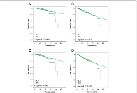
In our cohort, women diagnosed with ILC had a similar likelihood of experiencing recurrence/metastasis (P= 0.980) and death (P= 0.064) as those with IDC. Des-pite having more favorable biologic characteristics, the 5-year OS and RFS were not better for ILC (83% and 82%) than for IDC (89% and 79%, Figure 1A and B).
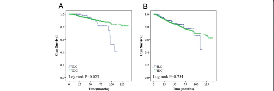
When TNM stage was included in the analysis, ILCs with higher TNM stage had a tendency to increase the risk of death compared to those among IDCs (P= 0.057 for OS and P= 0.653 for RFS) (Figure 2A and B). ILC was also one of the major predictors of worse overall survival in the ER-negative group (Figure 3A,P= 0.007),
Figure 1Overall survival (A) and recurrence/metastasis-free survival (B) according to histologic type; overall survival (C) and recurrence/metastasis-free survival (D) in higher TNM (TNMII&III) stage.*Adjusted for tumor size (≤2 vs.>2 cm) and nodal status (positive vs. negative).
Table 2 Distant sites of first recurrence/metastasis in this study
Sites ILC IDC Pvalue
(n = 183) (n = 2171)
Lungs/pleura 5 49 0.811
CNS 1 17 0.726
Ovary 0 3 NA
Gastrointestinal tract 0 1 NA
Nodes 3 53 0.478
Bone 10 107 0.892
Skin 4 33 0.566
Liver 3 37 0.798
Adrenal gland 1 1 0.170
PR-negative group (Figure 3C, P= 0.021), HR-negative
group (Figure 3E, P= 0.021), and HER2/neu-negative
group (Figure 3G,P= 0.040).
Additionally, ILC women that received chemotherapy had a higher rate of death (P= 0.023) compared to their counterparts (Figure 3A).
Multivariate survival analysis
Multivariate survival analysis was performed to determine whether ILC was an independent prognostic factor for recurrence/metastasis and death. The factors included in these analyses were histological type, age, tumor size, nodal status, TNM stage, ER status, PR status, HR status, HER2/neu status, and adjuvant treatment methods. From among these variables, the factors that independently associated with recurrence/metastasis, as well as with survival in the general population, were as follows: nodal status, TNM stage, ER status, PR status, and HR status (Table 3).
In addition, to determine whether traditional prognos-tic factors for IDC would be of value in patients with ILC, a second set of multivariate analyses of RFS and OS were performed in patients with ILC and IDC, respect-ively (Table 3). For ILC, only nodal status and TNM stage retained their independent prognostic value, while hormone receptor status remained as a prognostic factor in the IDC group, which is consistent with the findings in the general population.
Once adjustments were made based on tumor size and nodal status, histologic type did not emerge as an important prognostic factor. Thus, the lack of
prognostic significance related to ILC vs.IDC in uni-variate analyses is confirmed by the results of the multivariate analyses.
Discussion
The present retrospective study demonstrated that ILC has distinctive clinicopathologic characteristics com-pared with IDC in a Chinese population. Largely in agreement with other series, we revealed that ILC in Chinese women was more likely to be smaller in size, of a lower TNM stage, ER or PR positive, and HER2/neu negative. Despite a substantially less aggressive biologic phenotype, recurrence/metastasis and survival were very similar between ILC and IDC patients.
Our database included the cases at an interval of 11 years, and the majority of these cases were treated be-tween 1999 and 2005. The proportion of patients who had breast mastectomy was very high (>95%) in this period. Breast conservation has been increasingly used during the past 5 years in our hospital, and up to 20% of all patients underwent breast-conserving surgery (BCS) in 2011. We found in our cohort that ILCs were treated with BCS as often as IDCs, which was not consistent with other published reports [16-18]. The recommenda-tions against BCS for ILC were based on the consensus that ILC was known to more often be multifocal, multi-centric, and bilateral. The clinicians always preferred mastectomy to BCS in the treatment of ILC. However, there was recent evidence that BCS in ILC was not asso-ciated with increased local relapse rates at 5 years when compared with mastectomy [19,20]. In the current (See figure on previous page.)
Figure 2Overall survival (A) and recurrence/metastasis-free survival (B) according to ER negative; overall survival (C) and recurrence/ metastasis-free survival (D) according to PR negative; overall survival (E) and recurrence/metastasis-free survival (F) according to HR negative; overall survival (G) and recurrence/metastasis-free survival (H) in HER2/neu negative.*Adjusted for tumor size (≤2vs.>2 cm) and lymph node status (positivevs. negative).
analysis, a total of 13 ILC patients underwent BCS; we did not identify any recurrence/metastasis or death events at an average follow-up period of 39.0 months. Although in practice, the histologic type appears to play a role in the choice of surgical procedure selected, ILC can indeed be treated with BCS (and radiotherapy) when
clear margins can be achieved. In the current study, MRI test was used to evaluate every ILC patient receiving BCS, and no one had received re-excision due to a positive incised margin.
Concerning the pattern of metastatic spread, it seemed that the ILC group of Chinese women was not different
Table 3 Cox’s proportional hazards regression models for the general population, ILC and IDC groups
Variables RFS OS
RR (95% CI) Pvalue RR (95% CI) Pvalue
For general
Histological type 1.32 (0.82-2.12) 0.248 1.71 (0.91-3.21) 0.098
Age (years)≤50 1.23 (0.94-1.59) 0.131 1.43 (0.75-2.73) 0.284
Tumor size (cm)>2 1.47 (0.90-2.31) 0.124 1.71 (0.73-4.00) 0.215
Nodes positive 1.61 (1.37-1.88) <0.001 1.40 (1.08-1.81) 0.012
Higher TNM stage 1.56 (1.01-2.24) 0.016 1.78 (1.03-3.07) 0.039
ER positive 0.62 (0.43-0.91) 0.016 0.55 (0.31-1.00) 0.065
PR positive 0.59 (0.40-0.89) 0.011 0.51 (0.28-0.94) 0.031
HR positive 0.54 (0.32-0.91) 0.022 0.46 (0.21-1.02) 0.054
HER2/neu positive 1.21 (0.93-1.58) 0.157 1.24 (0.83-1.85) 0.304
Chemotherapy 0.57 (0.32-1.03) 0.060 0.57 (0.24-1.35) 0.200
Radiotherapy 0.54 (0.40-0.75) <0.001 1.21 (0.74-2.02) 0.414
Hormonotherapy 0.68 (0.51-0.90) 0.008 0.73 (0.47-1.13) 0.159
For ILC
Age (years)≤50 5.72 (0.83-39.32) 0.076 0.92 (0.81-1.04) 0.182
Tumor size (cm)>2 2.51 (0.61-10.41) 0.204 2.21 (0.97-5.01) 0.058
Nodes positive 2.87 (1.06-7.69) 0.038 5.55 (0.93-33.33) 0.060
Higher TNM stage 4.79 (2.79-11.85) 0.002 3.31 (2.15-9.45) 0.006
ER positive 1.04 (0.175-6.21) 0.964 1.98 (0.15-25.38) 0.600
PR positive 0.52 (0.10-2.58) 0.420 0.28 (0.02-3.24) 0.308
HR positive 0.66 (0.05-9.24) 0.760 0.39 (0.01-20.20) 0.643
HER2/neu positive 1.45 (0.41-5.18) 0.569 0.85 (0.21-3.42) 0.822
Chemotherapy 0.29 (0.02-4.73) 0.383 0.32 (0.12-1.05) 0.060
Radiotherapy 0.86 (0.27-2.78) 0.809 1.22 (0.26-5.72) 0.796
Hormonotherapy 1.38 (0.42-4.54) 0.595 0.90 (0.21-3.76) 0.889
For IDC
Age (years)≤50 1.21 (0.92-1.59) 0.186 1.55 (1.00-2.34) 0.049
Tumor size (cm)>2 1.39 (0.84-2.32) 0.201 2.34 (0.84-6.55) 0.105
Nodes positive 1.62 (1.37-1.91) <0.001 1.66 (1.27-2.17) <0.001
Higher TNM stage 1.52 (1.03-2.23) 0.056 1.93 (1.07-3.48) 0.029
ER positive 0.62 (0.42-0.93) 0.021 0.51 (0.27-0.97) 0.040
PR positive 0.60 (0.39-0.91) 0.016 0.47 (0.25-0.91) 0.025
HR positive 0.53 (0.31-0.92) 0.023 0.38 (0.16-0.88) 0.025
HER2/neu positive 1.19 (0.91-1.57) 0.206 1.26 (0.82-1.94) 0.292
Chemotherapy 0.60 (0.33-1.10) 0.099 0.82 (0.33-2.03) 0.662
Radiotherapy 0.51 (0.38-0.69) <0.001 0.47 (0.30-0.75) 0.002
Hormonotherapy 0.64 (0.47-0.86) 0.004 0.67 (0.42-1.08) 0.101
from their counterparts with IDC group. ILC as well as IDC were likely to affect the lungs or pleura, bone, and liver. More frequent metastasis to unusual sites such as the vulva and the gastrointestinal tract were previously reported in ILC [21,22], but we could not address this issue because of the limited cases analyzed in our data-base. We found that the incidence of contralateral breast cancer in women with ILC was nearly double that in women with IDC; this finding could make a compelling case for the use of tamoxifen to prevent contralateral breast cancer in women with lobular primaries.
Most studies have demonstrated that ILC tumors tended to be large [23-25], but 47.9% of ICLs were found not to be in excess of 2 cm compared with only 28.3% of IDCs in our data. We further revealed that a palpable mass was observed in 202 cases (93.9%), and this was the most frequent complaint, which might encourage patients to participate in early screenings and obtain a precise diagnosis when the tumor is smaller. Except for the slightly smaller size of the ILCs, the rate of lymph node involvement was comparable in each group; there-fore, many more early-stage breast cancers were pre-sented in the ILC group. To our knowledge, very few previous studies had described these findings.
The current population-based study among Chinese women also definitively validated the findings of some published studies indicating that lobular carcinomas are significantly more likely to be steroid receptor positive than are IDCs [22]. We also evaluated the well-studied growth factor receptors, HER2/neu. No more than 15% of tumors classified as ILC over-expressed HER2/neu; however, 24.8% of IDC patients displayed tumors that overexpressed this receptor. Together these findings sug-gest that ILC is biologically different from IDC, and has more favorable biologic characteristics.
The RFS and OS curves showed an early advantage for the ILC cohort, but after 4 years, an advantage emerged for the IDC cohort. After adjustment for tumor size and nodal status, there was no prognostic difference between IDC and ILC in the current analysis, which indicated that the more favorable prognostic factors of ILC did not translate into a long-term survival advantage for patients with ILC. We further clarified this important finding through a multivariate analysis in different groups. The favorable prognostic factors such as hor-mone receptor status identified in the general population and IDC group were not applicable in lobular carcin-oma. In spite of the lack of survival differences between ILC and IDC in most large datasets, the univariate ad-verse prognostic effects of ILC phenotype had appeared to be restricted to women with HR and HER2/neu-nega-tive breast cancer, the majority of whom received adju-vant chemotherapy. We therefore inferred ILC might be associated with more aggressive biologic behavior than
IDC in some subgroups and induce a lack of responsive-ness to chemotherapy treatment.
Two inherited limitations in this study should be addressed. One potential weakness was the relatively small sample of ILC, so the results in our analysis might not comprehensively account for all the distinct biology and exact prognosis of ILC. The other was that patho-logic information for lymphovascular invasion and histo-logic grade were excluded from the analysis, but these variables could have an effect on survival. Therefore, fur-ther studies with larger datasets and more complete pathologic details will be necessary to validate our findings.
Conclusions
Despite the fact that ILCs are epidemiologically and phenotypically different from IDCs, these patients do not have better clinical outcomes than do patients with IDC. At present, management decisions should be based on individual patient and tumor biologic characteristics, and not on lobular histology.
Competing interests
There is no any conflict of interest about the study.
Authors’contributions
GD carried out the study conception and design. AC was responsible for data collecting and manuscript writing. MH, ZMS and JW participated in the technique support and results analysis. GYL and JSL participated in the design of the study and performed the statistical analysis. ZZS conceived of the study, and participated in its design and coordination and helped to draft the manuscript. All authors read and approved the final manuscript.
Acknowledgments
The authors thank the patients for their willingness to cooperate with our study. This study was supported in part by grants from the National Natural Science Foundation of China (81102000), and Shanghai Science and Technology Committee (11ZR1407500).
We thank Deric Corlew who provided medical writing services on behalf of American Journal Experts (AJE).
Author details
1Breast Cancer Institute, Cancer Centre/Cancer Institute, Shanghai, PR, China. 2
Department of Oncology, Shanghai Medical College, Institutes of Biomedical Science, Fudan University, Shanghai, PR, China.3Breast Cancer Institute, Cancer Hospital/Cancer Institute; Department of Oncology, Shanghai Medical College, Fudan University, 270 Dong’an Road, Shanghai, PR 200032, China.
Received: 1 April 2012 Accepted: 29 June 2012 Published: 17 July 2012
References
1. Borst MJ, Ingold JA:Metastatic patterns of invasive lobular versus invasive ductal carcinoma of the breast.Surgery1993,114:637–641. 2. Sastre-Garau X, Jouve M, Asselain B, Vincent-Salomon A, Beuzeboc P, Dorval
T, Durand JC, Fourquet A, Pouillart P:Infiltrating lobular carcinoma of the breast. Clinicopathologic analysis of 975 cases with reference to data on conservative therapy and metastatic patterns.Cancer1996,77:113–120. 3. Arpino G, Bardou VJ, Clark GM, Elledge RM:Infiltrating lobular carcinoma of the breast: tumor characteristics and clinical outcome.Breast Cancer Res2004,6:R149–R156.
5. Cocquyt V, Van Belle S:Lobular carcinoma in situ and invasive lobular cancer of the breast.Curr Opin Obstet Gynecol2005,17:55–60. 6. Sasson AR, Fowble B, Hanlon AL, Torosian MH, Freedman G, Boraas M,
Sigurdson ER, Hoffman JP, Eisenberg BL, Patchefsky A:Lobular carcinoma in situ increases the risk of local recurrence in selected patients with stages I and II breast carcinoma treated with conservative surgery and radiation.Cancer2001,91:1862–1869.
7. Quan ML, Sclafani L, Heerdt AS, Fey JV, Morris EA, Borgen PI:Magnetic resonance imaging detects unsuspected disease in patients with invasive lobular cancer.Ann Surg Oncol2003,10:1048–1053.
8. Franceschini G, Manno A, Mulè A, Verbo A, Rizzo G, Sermoneta D, Petito L, D'alba P, Maggiore C, Terribile D, Masetti R, Coco C:Gastro-intestinal symptoms as clinical manifestation of peritoneal and retroperitoneal spread of an invasive lobular breast cancer: report of a case and review of the literature.BMC Cancer2006,6:193.
9. Yu J, Dabbs DJ, Shuai Y, Niemeier LA, Bhargava R:Classical-type invasive lobular carcinoma with HER2 overexpression: clinical, histologic, and hormone receptor characteristics.Am J Clin Pathol2011,136:88–97. 10. Fortunato L, Mascaro A, Poccia I, Andrich R, Amini M, Costarelli L, Cortese G,
Farina M, Vitelli C:Lobular breast cancer: same survival and local control compared with ductal cancer, but should both be treated the same way? Analysis of an institutional database over a 10-year period.Ann Surg Oncol2012,19:1107–1114.
11. Jayasinghe UW, Bilous AM, Boyages J:Is survival from infiltrating lobular carcinoma of the breast different from that of infiltrating ductal carcinoma?Breast J2007,13:479–485.
12. Toikkanen S, Pylkkänen L, Joensuu H:Invasive lobular carcinoma of the breast has better short- and long-term survival than invasive ductal carcinoma.Br J Cancer1997,76:1234–1240.
13. Rakha EA, Reis-Filho JS, Baehner F, Dabbs DJ, Decker T, Eusebi V, Fox SB, Ichihara S, Jacquemier J, Lakhani SR, Palacios J, Richardson AL, Schnitt SJ, Schmitt FC, Tan PH, Tse GM, Badve S, Ellis IO:Breast cancer prognostic classification in the molecular era: the role of histological grade.Breast Cancer Res2010,12:207.
14. Muñoz M, Fernández-Aceñero MJ, Martín S, Schneider J:Prognostic significance of molecular classification of breast invasive ductal carcinoma.Arch Gynecol Obstet2009,280:43–48.
15. Prat A, Parker JS, Karginova O, Fan C, Livasy C, Herschkowitz JI, He X, Perou CM:Phenotypic and molecular characterization of the claudin-low intrinsic subtype of breast cancer.Breast Cancer Res2010,12:R68. 16. Hussien M, Lioe TF, Finnegan J, Spence RA:Surgical treatment for invasive
lobular carcinoma of the breast.Breast2003,12:23–35.
17. Chung MA, Cole B, Wanebo HJ, Bland KI, Chang HR:Optimal surgical treatment of invasive lobular carcinoma of the breast.Ann Surg Oncol 1997,4:545–550.
18. Raje D, Bollard R, Wilson A:Invasive lobular cancer of the breast–is breast conservation surgery a good option?Breast J2006,12:574–575. 19. Yeatman TJ, Cantor AB, Smith TJ, Smith SK, Reintgen DS, Miller MS, Ku NN,
Baekey PA, Cox CE:Tumor biology of infiltrating lobular carcinoma. Implications for management. Ann Surg1995,222:549–559.
20. Singletary SE, Patel-Parekh L, Bland KI:Treatment trends in early-stage invasive lobular carcinoma: a report from the National Cancer Data Base. Ann Surg2005,242:281–289.
21. Papaioannou N, Zervoudis S, Grammatikakis I, Peitsidis P, Palvakis K, Youssef TF:Metastatic lobular carcinoma of the breast to the vulva: a case report and review of the literature.J Egypt Natl Canc Inst2010,22:57–60. 22. Pestalozzi BC, Zahrieh D, Mallon E, Gusterson BA, Price KN, Gelber RD,
Holmberg SB, Lindtner J, Snyder R, Thürlimann B, Murray E, Viale G, Castiglione-Gertsch M, Coates AS, Goldhirsch A:International Breast Cancer Study Group: Distinct clinical and prognostic features of infiltrating lobular carcinoma of the breast: combined results of 15 International Breast Cancer Study Group clinical trials.J Clin Oncol 2008,26:3006–3014.
23. Li CI, Uribe DJ, Daling JR:Clinical characteristics of different histologic types of breast cancer.Br J Cancer2005,93:1046–1052.
24. Adams AL, Li Y, Pfeifer JD, Hameed O:Nuclear grade and survival in invasive lobular carcinoma: a case series with long-term follow-up.Breast J2010,16:445–447.
25. Bane AL, Tjan S, Parkes RK, Andrulis I, O’Malley FP:Invasive lobular carcinoma: to grade or not to grade.Mod Pathol2005,
18:621–628.
doi:10.1186/1477-7819-10-152
Cite this article as:Caoet al.:Tumor characteristics and the clinical outcome of invasive lobular carcinoma compared to infiltrating ductal carcinoma in a Chinese population.World Journal of Surgical Oncology 201210:152.
Submit your next manuscript to BioMed Central and take full advantage of:
• Convenient online submission
• Thorough peer review
• No space constraints or color figure charges
• Immediate publication on acceptance
• Inclusion in PubMed, CAS, Scopus and Google Scholar
• Research which is freely available for redistribution