Page 1 of 11
Plasma vaspin is an effective biomarker for evaluation of future
cardiovascular events in patients with chest pain: a 5-year
retrospective observational study
Shuya Ji1#, Wenxin Kou1#, Peipei Luan1, Weixia Jian2, Jianhui Zhuang1, Xiaopeng Xu1, Yifan Zhao1,
Hailing Li1, Wenhui Peng1
1Department of Cardiology, Shanghai Tenth People’s Hospital, Tongji University School of Medicine, Shanghai 200072 China; 2Department of Endocrinology, Xinhua Hospital, Shanghai Jiaotong University School of Medicine, Shanghai 200000, China
Contributions: (I) Conception and design: H Li, W Peng; (II) Administrative support: W Peng; (III) Provision of study materials or patients: Y Zhao, X Xu; (IV) Collection and assembly of data: J Zhuang, W Jian, P Luan; (V) Data analysis and interpretation: S Ji, W Kou; (VI) Manuscript writing: All authors; (VII) Final approval of manuscript: All authors.
#These authors contributed equally to this work.
Correspondence to: Hailing Li, PhD; Wenhui Peng, PhD. Department of Cardiology, Shanghai Tenth People’s Hospital, Tongji University School of Medicine, 301 Middle Yanchang Road, Shanghai 200072, China. Email: lihailin_2004@126.com; pwenhui@tongji.edu.cn.
Background: Our previous study showed that visceral adipose tissue-derived serpin (vaspin) was an independent predictor of coronary artery disease (CAD). Further, plasma vaspin levels in patients with unstable angina pectoris were lower than those in patients with stable angina pectoris. In this study, we investigated the prognostic relevance of plasma vaspin levels in patients with CAD and non-CAD.
Methods: It was a retrospective observational study. A total of 197 patients with chest pain were enrolled, of which 88 patients with CAD and 109 patients with non-CAD were confirmed by angiography. Plasma vaspin levels and clinical parameters were measured at baseline. Incidence of major adverse cardiac event (MACE) was determined on follow-up.
Results: One hundred eighty-nine patients were successfully followed up for 5 years, of which 63 patients experienced MACEs. Patients with low vaspin levels (<0.385 ng/mL) experienced a higher incidence of MACE as compared to patients with high vaspin levels (>0.385 ng/mL) (42.55% vs. 24.21%, respectively; P=0.007). In both CAD and non-CAD groups, patients with high vaspin levels showed improvement in left ventricular ejection fraction. Kaplan Meier survival curves showed that patients with low vaspin levels had an obviously higher timing of incidence of MACE in the whole population (P=0.006) and in the non-CAD subgroup (P=0.009); however, the trend was not significant in the CAD subgroup. On multivariate analyses, plasma vaspin level was found to be an independent predictor of MACE, particularly in the non-CAD group. Conclusions: Plasma vaspin may be a useful biomarker for prediction of MACE in patients with chest pain.
Keywords: Plasma vaspin; biomarker; prognosis; major adverse cardiac events; coronary artery disease
Submitted Dec 07, 2019. Accepted for publication Feb 26, 2020. doi: 10.21037/atm.2020.03.29
View this article at: http://dx.doi.org/10.21037/atm.2020.03.29
Introduction
Adipose tissue is now regarded not only as an energy reservoir, but also as an active endocrine organ, which can secrete a variety of metabolically-active adipocytokines.
Some adipokines have even been linked with metabolic diseases; for instance, adiponectin has been shown to be a robust biomarker of insulin sensitivity (1,2).
isolated from a rat model of abdominal obesity with type 2 diabetes mellitus (T2DM) (3). Previous studies showed an association between vaspin concentration and metabolic disorders, including T2DM, cardiovascular diseases (CVDs) (4-6),polycystic ovary syndrome (7),and osteoarthritis (8). Plasma vaspin concentration was showed to correlate with body mass index (BMI), gender, and physical training. An earlier study suggested a causal relationship of plasma vaspin levels with obesity (9). Also, a recent research indicated that vaspin protected against high fat diet induced bone loss, and promoted osteogenic differentiation (10). Up-regulation of vaspin was showed to protect against insulin resistance by activating the IRS/PI3K/Akt/Glut signaling pathway and inhibiting the IκBα/NF-κB signaling pathway (11). Besides, in our previous studies, patients with low concentration of vaspin were found to be at a higher risk of coronary artery disease (CAD) and acute coronary syndrome (ACS) (12,13). Further, lower concentration of vaspin was associated with poor prognosis in patients with myocardial infarction (14).
Based on these data, it is conceivable that vaspin plays an important role in the development of atherosclerosis. In our previous animal studies, vaspin was showed to protect against atherosclerosis through inhibition of vascular smooth muscle cell proliferation and chemokines is owing to decreased production of reactive oxygen species and downregulation of NF-κB signaling pathways (15). In a study by Yuan et al., vaspin played a role of cardioprotective function by down-regulating the expression of toll-like receptor 4 and inhibiting the phosphorylation of NF-κB in myocardial ischemia reperfusion injury rats and hypoxia-re-oxygenation induced H9C2 cells (16).
These findings prompted us to further investigate the potential association between vaspin levels and prognosis of patients with CVDs. Also, although some patients with chest pain were excluded CAD by angiography, previous studies found that these patients still have a risk of major adverse cardiac events (MACEs) (17).Moreover, only a limited blood biomarker was used to predict prognosis in these patients. The present study focused on the prognostic value of plasma concentration of vaspin in patients with no angiographic evidence of CAD. Furthermore, we also sought to assess whether plasma vaspin could predict events after 5 years, as 5-year follow-up is considered as a key point for evaluation of biomarkers (18). Furthermore, patients with negative coronary angiography (CAG) may also have poor prognosis, as these patients may have other risk factors such as hypertension, and diabetes. It is also important to explore whether vaspin would be an effective biomarker of
prognostic relevance in this subpopulation.
Methods
Study population
all patients prior to their enrolment. The study protocol was approved by the Ethics Committee at the Shanghai Tenth People’s Hospital.
Coronary angiography
All patients enrolled in this study were undergone CAG, which was performed through a radial artery with standard judkins technique. Significant CAD was diagnosed as the presence of at least one of luminal diameter stenosis ≥50% including the left anterior descending artery, right coronary artery, left circumflex coronary artery and their main branches. All imaging analyses were performed by two professional interventional cardiologists.
ELISA assays, biochemical investigations and echocardiographic assessment
Approximately 5 mL whole blood samples were extracted prior to CAG. Plasma was isolated from blood samples after a 10-hour overnight fast and then stored at -80°C until further processing. Human vaspin (Adipogen, Seoul, South Korea) plasma levels were measured with commercially available ELISA kit, according to the manufacturers’ instructions. The sensitivity of the vaspin ELISA was 12 pg/mL. The intra-assay coefficient of variance (percent) of the ELISA system ranged from 1.31% to 1.74%, while the inter-assay coefficient of variance ranged from 5.9% to 8.3%. White blood cell count (WBC), high sensitive C-reactive protein (hsCRP), glycosylated haemoglobin (HbA1c), total cholesterol (TC), triglyceride (TG), high-density lipoprotein cholesterol (HDL-C), and low-high-density lipoprotein cholesterol (LDL-C) were measured by colorimetric enzymatic assay systems (Roche MODULAR P-800, Swiss Confederation).
All subjects were performed transthoracic echocardiography by an echocardiograph equipped with a broadband transducer (Vivid 7®, GE VingMed Ultrasound AS; Horten, Norway). Measurement of LVEF was obtained from the modified Simpson rule in the apical two- and four-chamber views.
Follow-up
It was a retrospective observational study. Follow-up data were obtained through the following four ways: hospital record review, telephonic contact, outpatient visits and rehospitalization. Follow-up data was collected up to December 31, 2015. The duration of follow-up was 5 years.
The primary endpoint was MACE, which included cardiac death, non-fatal myocardial infarction, revascularization with percutaneous coronary intervention or coronary artery bypass grafting, ischemic stroke, or rehospitalization for severe angina. Non-fatal myocardial infarction was defined as a rise of troponin T >0.1 ng/mL along with canonical chest pain symptoms and/or characteristic electrocardiographic changes. Deaths caused by accidents were excluded (follow-up censored at the time of death) (14,17).The median concentration of vaspin was 0.385 ng/mL. All patients were categorized into two subgroups according to the median value.
Statistical analysis
Continuous variables are expressed as mean ± standard deviation. Categorical variables are presented as frequencies and percentages. For continuous variables, normal distribution was verified with the Kolmogorov-Smirnov test. Comparisons between groups were made using independent t-test, ANOVA, the nonparametric Mann-Whitney U test or Kruskal-Wallis test as appropriate. For categorical clinical variables, Chi-square test or Fisher exact test was employed. For A P value<0.05 was considered indicative of a statistically significant between-group difference. The Kaplan-Meier method was used to analyze the timing of events during follow-up. Statistical assessment was performed with the log-rank test and P<0.05 was considered significant. Univariate and multivariate analyses were performed using a Cox proportional hazards model to determine the predictors of the primary endpoint. The explanatory variables were used in the univariate analyses. Explanatory variables that were associated with P values <0.1 on univariate analyses were included in the multivariate analysis. All analyses were performed with SPSS 20 (SPSS Inc., IL, USA).
Results
Baseline characteristics of the study population in CAD and non-CAD groups
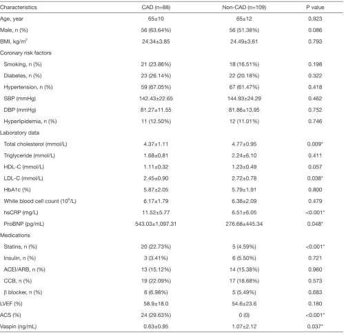
concentration of plasma vaspin in the CAD group was lower than that in the non-CAD group (0.63±0.95 vs. 1.07±2.12 ng/mL, P=0.037). No significant between-group
[image:4.595.51.551.94.579.2]differences were observed with respect to age, gender, BMI, smoking, coronary risk factors, TG, HDL-C and HbA1c levels. Serum hsCRP levels (11.52±5.77 vs. 6.51±6.05 mg/L, Table 1 Baseline characteristics of patients in CAD and non-CAD groups
Characteristics CAD (n=88) Non-CAD (n=109) P value
Age, year 65±10 65±12 0.923
Male, n (%) 56 (63.64%) 56 (51.38%) 0.086
BMI, kg/m2 24.34±3.85 24.49±3.61 0.793
Coronary risk factors
Smoking, n (%) 21 (23.86%) 18 (16.51%) 0.198
Diabetes, n (%) 23 (26.14%) 22 (20.18%) 0.322
Hypertension, n (%) 59 (67.05%) 67 (61.47%) 0.418
SBP (mmHg) 142.43±22.65 144.93±24.29 0.462
DBP (mmHg) 81.27±11.55 81.86±13.95 0.752
Hyperlipidemia, n (%) 11 (12.50%) 12 (11.01%) 0.746
Laboratory data
Total cholesterol (mmol/L) 4.37±1.11 4.77±0.95 0.009*
Triglyceride (mmol/L) 1.68±0.81 2.24±6.10 0.411
HDL-C (mmol/L) 1.11±0.32 1.23±0.49 0.057
LDL-C (mmol/L) 2.45±0.90 2.72±0.78 0.038*
HbA1c (%) 5.87±2.05 5.79±1.91 0.800
White blood cell count (109/L) 6.17±1.79 6.38±2.09 0.479
hsCRP (mg/L) 11.52±5.77 6.51±6.05 <0.001*
ProBNP (pg/mL) 543.03±1,097.31 276.68±445.34 0.048*
Medications
Statins, n (%) 20 (22.73%) 5 (4.59%) <0.001*
Insulin, n (%) 3 (3.41%) 6 (5.50%) 0.721
ACEI/ARB, n (%) 13 (15.12%) 14 (15.38%) 0.960
CCB, n (%) 19 (22.09%) 17 (18.68%) 0.573
β blocker, n (%) 6 (6.98%) 5 (5.49%) 0.683
LVEF (%) 58.9±18.0 54.6±23.6 0.180
ACS (%) 24 (29.63%) 0 (0) <0.001*
Vaspin (ng/mL) 0.63±0.95 1.07±2.12 0.037*
respectively; P<0.001) and frequency of statin usage in the CAD group were higher than that in the non-CAD group. On the contrary, serum TC and LDL-C concentrations in the non-CAD group were higher than those in the CAD group (Table 1).
Baseline characteristics of the study population disaggregated by vaspin levels
The median concentration of vaspin was 0.385 ng/mL. All patients were categorized into two subgroups according to the median value. In the CAD group, non-smokers had higher concentrations of vaspin as compared to that in smokers (34.78% vs. 11.90%, respectively; P=0.012) (Table 2). The percentage of male patients in the low-vaspin subgroup was significantly higher than that in the high-vaspin subgroup (73.91% vs. 52.38%, respectively; P=0.036). However, no significant between-group differences of cardiovascular history, such as ACS, post-PCI or coronary artery bypass graft (CABG), number of vascular lesions and degree of lesions were observed in CAD group. In the non-CAD group, higher HDL-C and insulin administration were found in high-vaspin subgroup; however, this subgroup had lower LVEF as compared to that in the low-vaspin sub-group (50.6%±27.2% vs. 59.9%±16.5%, respectively; P=0.046).
Relationship of plasma vaspin levels with 5-year MACE Eight patients were lost to 5-year follow-up. Among these, 4 patients were men and 4 patients were diagnosed with CAD. At 5-year follow-up, serum TC levels in the high-vaspin subgroup were significantly decreased in CAD group but not in the non-CAD group, as patients with CAD usually received statin therapy. Of note, high-vaspin subgroups in both the CAD and non-CAD groups showed improved cardiac LVEF (55.8%±21.7% to 64.5%±9.1% and 50.6%±27.2% to 65.1%±6.2%, respectively; P<0.05 for all). This phenomenon was not observed in low-vaspin subgroups in the two groups (Table S1).
[image:5.595.48.550.465.675.2]Although the total MACE between CAD and non-CAD groups has insignificantly difference, we found that the number of patients who experienced multiple times of MACEs in CAD group is more than non-CAD group, which could indicate that the patients with CAD has a higher risk of MACE than patients in non-CAD group. Also, we found that low- and high-vaspin subgroups of the CAD group had comparable incidence of MACE (P=0.264); however, more patients in the low-vaspin subgroup received revascularization treatment as compared to that in the high-vaspin subgroup [13 (28.26%) vs. 3 (7.89%), P=0.018]. Incidence of new-onset diseases such as diabetes or hypertension was comparable between the low- and high-vaspin subgroups in both CAD and non-CAD
Table 2 Baseline characteristics of patients in vaspin <0.385 ng/mL group and vaspin >0.385 ng/mL subgroups
Characteristics
CAD (n=88) Non-CAD (n=109)
Vaspin <0.385 (n=46)
Vaspin >0.385
(n=42) P value
Vaspin <0.385 (n=49)
Vaspin >0.385
(n=60) P value
Age, year 64±10 66±9 0.332 67±11 63±12 0.096
Male, n (%) 34 (73.91%) 22 (52.38%) 0.036* 29 (60.42%) 27 (44.26%) 0.094
BMI, kg/m2 24.36±4.09 24.31±3.58 0.955 25.12±3.37 23.88±3.75 0.088
Coronary risk factors
Smoking, n (%) 16 (34.78%) 5 (11.90%) 0.012* 7 (14.58%) 11 (18.03) 0.630
Diabetes, n (%) 11 (23.91%) 10 (26.32%) 0.800 8 (16.67%) 14 (22.95%) 0.417
Hypertension, n (%) 29 (63.04%) 30 (71.43%) 0.403 33 (68.75%) 34 (55.74%) 0.166
SBP (mmHg) 141.98±23l.86 142.93±21.53 0.845 148.15±24.84 142.35±23.73 0.219
DBP (mmHg) 83.00±13.22 79.38±9.18 0.137 81.27±15.15 82.33±13.02 0.696
Hyperlipidemia, n (%) 5 (10.87%) 6 (14.29%) 0.628 4 (8.33%) 8 (13.11%) 0.429
Table 2 (continued)
Characteristics
CAD (n=88) Non-CAD (n=109)
Vaspin <0.385 (n=46)
Vaspin >0.385
(n=42) P value
Vaspin <0.385 (n=49)
Vaspin >0.385
(n=60) P value
Laboratory data
Total cholesterol (mmol/L) 4.18±1.08 4.58±1.11 0.106 4.61±1.12 4.89±0.79 0.154
Triglyceride (mmol/L) 1.69±0.93 1.66±0.67 0.878 3.01±9.04 1.60±0.91 0.252
HDL-C (mmol/L) 1.13±0.32 1.08±0.33 0.516 1.12±0.29 1.32±0.59 0.047*
LDL-C (mmol/L) 2.35±0.82 2.57±0.99 0.281 2.67±0.91 2.75±0.66 0.643
HbA1c (%) 5.79±2.14 5.96±1.98 0.726 5.78±1.43 5.79±2.22 0.986
White blood cell count (109/L) 5.95±1.90 6.47±1.62 0.200 6.35±2.27 6.41±1.92 0.896
hsCRP (mg/L) 11.17±5.27 11.87±6.35 0.714 5.36±4.44 7.14±6.76 0.321
ProBNP (pg/mL) 461.02±952.61 631.53±1,241.69 0.740 251.21±503.95 298.09±394.12 0.640
Medications
Statins, n (%) 13 (28.26%) 6 (16.67%) 0.195 2 (4.17%) 3 (4.92%) 1.000
Insulin, n (%) 2 (4.35%) 1 (2.38%) 1.000 0 6 (9.84%) 0.033*
ACEI/ARB, n (%) 7 (15.56%) 6 (14.29%) 0.905 10 (23.81%) 4 (8.16%) 0.077
CCB, n (%) 8 (17.78%) 11 (26.83%) 0.312 10 (23.81%) 7 (14.29%) 0.245
β blocker, n (%) 4 (8.89%) 2 (4.88%) 0.760 4 (9.52%) 1 (2.04%) 0.271
Cardiovascular history
ACS (%) 13 (28.89%) 13 (28.89%) 0.870 0 0 1.000
Post-PCI or CABG (%) 21 (45.65%) 11 (28.95%) 0.117 0 0 1.000
Number of vascular lesions
1 19 (41.30%) 22 (52.38%) 0.582
2 12 (26.09%) 9 (21.43%)
≥3 15 (32.61%) 11 (26.19%)
Degree of stenosis
Lowly stenosis (50%≤ stenosis <75%)
16 (34.78%) 23 (54.76%) 0.059
Highly stenosis (stenosis ≥75%) 30 (65.22%) 19 (45.24%)
LVEF (%) 61.8±13.2 55.8±21.7 0.152 59.9±16.5 50.6±27.2 0.046*
groups. In the non-CAD group, more patients in the low-vaspin subgroup experienced MACE as compared to that in the high-vaspin subgroup [20 (41.67%), vs. 11 (19.30%); P=0.012]. Especially, in this group, the low-vaspin subgroup had a higher risk of stroke than that in the high-vaspin subgroup [7 (14.58%) vs. 1 (1.75%); P=0.036] (Table 3).
Relationship of plasma vaspin levels with prognosis
The Kaplan-Meier method was used to analyze the timing of incidence of MACE during follow-up. Kaplan Meier survival curves showed that patients in the low-vaspin group had an obviously higher timing of incidence of MACE in the whole population (log rank test 7.578, P=0.006, Figure 1). Notably, patients in the low-vaspin group had a significantly higher timing of incidence of MACE as compared to those in the high-vaspin group in the non-CAD subgroup (log rank test 6.741, P=0.009). A similar trend was observed in the CAD group, although it was not statistically significant (log rank test 1.255, P=0.263). In the
CAD group, there were insignificant difference between patients with vaspin <0.385 ng/mL and those with vaspin >0.385 ng/mL both in lowly stenosis and highly stenosis group (Figure S1).
Multivariate analysis
After inclusion of clinical characteristics in the Cox proportional hazards model, low vaspin levels (hazard ratio, HR 0.387, P=0.001) and age (HR 1.034, P=0.012) independently predicted the occurrence of MACE. Furthermore, in the non-CAD group, low vaspin levels (HR 0.260, P=0.002), specify the gender (HR 2.920, P=0.010), age (HR 1.054, P=0.005) and TG levels (HR 0.462, P=0.044) were found to be independent predictors of MACE (Table 4).
Discussion
[image:7.595.47.552.97.405.2]In this study, low plasma concentration of vaspin was Table 3 Prognostic characteristics with 5 years MACEs between low and high vaspin subgroups
Characteristics
CAD (n=84) Non-CAD (n=105)
Vaspin <0.385 (n=46)
Vaspin >0.38
(n=38) P value
Vaspin <0.385 (n=48)
Vaspin >0.38
(n=57) P value
MACE, n (%) 32 (38.10) 31 (29.52) 0.214
0 52 (61.90) 74 (70.48) 0.028*
1 16 (19.75) 21 (20.00)
2 3 (3.57) 7 (6.67)
3 9 (10.71) 3 (2.86)
4 4 (4.76) 0
New disease
Diabetes, n (%) 3 (6.52) 2 (5.26) 1.000 3 (6.67) 1 (1.75) 0.492
Hypertension, n (%) 0 0 1.000 1 (2.08) 0 0.457
Total MACE, n (%) 20 (43.48) 12 (31.58) 0.264 20 (41.67) 11 (19.30) 0.012*
Cardiac death, n (%) 2 (4.35) 3 (7.89) 0.825 2 (4.17) 1 (1.75) 0.880
Non-fatal myocardial infarction, n (%) 6 (13.04) 7 (18.42) 0.498 3 (6.25) 5 (8.77) 0.908
Revascularization, n (%) 13 (28.26) 3 (7.89) 0.018* 2 (4.17) 3 (5.26) 1.000
Stroke, n (%) 5 (10.87) 3 (7.89) 0.929 7 (14.58) 1 (1.75) 0.036*
Rehospitalisation, n (%) 13 (28.26) 10 (26.32) 0.842 11 (22.92) 9 (15.79) 0.354
All-cause death, n (%) 4 (8.70) 4 (10.53) 1.000 4 (8.33) 7 (12.28) 0.735
Figure 1 The Kaplan-Meier method was used to analyse the timing of incidence of MACEs during follow-up. (A) Patients with vaspin <0.385 ng/mL had a significantly higher timing of incidence of MACE than those with vaspin >0.385 ng/mL group (log rank test 7.578, P=0.006) in the whole population; (B) patients with low vaspin had a significantly higher timing of incidence of MACE than those with high vaspin (log rank test 6.741, P=0.009) in the non-CAD group; (C) there were insignificant between two groups (log rank test 1.255, P=0.263) in the CAD group.
found to predict the prognosis of patients after CAG. Furthermore, we found that low vaspin level could predict the prognosis in non-CAD patients. On Cox regression analysis, low vaspin level was found to be an independent risk factor for MACE.
In a study by Kameshima et al., vaspin was showed to prevent the increase in SBP, an effect that was attributed to its antioxidant and anti-inflammatory roles in smooth muscle cells of peripheral blood vessels (21). In addition, vaspin was indicated to protect vascular endothelial cells from free fatty acid induced apoptosis through up-regulation of the PI3-kinase/Akt signaling pathway (22). Further, administration of vaspin to diet-induced obese mice was showed to improve glucose tolerance, insulin sensitivity, and to alter the expression of genes implicated in the causation of insulin resistance (3,23).Also, vaspin prevented myocardial injury in rats model of diabetic cardiomyopathy by suppressing NLRP3 inflammasome activation and promoting autophagy (24).Besides, it was reported that statin therapy could increases plasma vaspin levels (25).All these studies suggest a protective effect of vaspin on vascular biology as well as that against insulin resistance. Some other reports also showed a correlation between vaspin levels and atherosclerotic lesions. An early study demonstrated that serum vaspin level was positively associated with carotid intima-media thickness (26). Further, in a study by Kameshima et al., vaspin was showed
to suppress inflammatory phenotypes by down-regulating NF-κB in human macrophages and oxidized low-density lipoprotein-induced foam cell formation (27).
Although experimental studies have indicated that vaspin is a vasculoprotective adipocytokine, its’ specific role and clinical relevance in CAD is not clear (28).In this study, presence of risk factors for CAD (such as hypertension, hyperlipidemia and diabetes) was not different between low- and high -vaspin subgroups, both in the CAD and non-CAD groups; this suggests that vaspin may be a more sensitive biomarker for atherosclerosis such as CAD. In the follow-up study, we found that patients with high vaspin levels had improved cardiac function, which suggests that patients with high vaspin levels may have a better prognosis. In addition, increased incidence of MACE may also be attributable to development of co-morbid conditions such as diabetes and hypertension. However, we did not find any significant increase in new onset diabetes or hypertension in both CAD and non-CAD groups; this implied that increase in the incidence of MACE was associated with decreased vaspin levels, and was not due to new-onset diabetes or hypertension. Gulcelik et al. found that plasma vaspin levels in diabetic patients with chronic complications (including retinopathy and nephropathy) were lower than those in their counterparts without complications (29). In our previous study, low plasma concentration of vaspin was found to be a risk factor for progression of T2DM (4). These results
1.0 0.8 0.6 0.4 0.2 0.0 1.0 0.8 0.6 0.4 0.2 0.0 1.0 0.8 0.6 0.4 0.2 0.0
No MACEs No MACEs No MACEs
0 500 1000 1500 2000 2500
vaspin <0.385 ng/mL vaspin >0.385 ng/mL vaspin <0.385 ng/mL-censored vaspin >0.385 ng/mL-censored
vaspin <0.385 ng/mL vaspin >0.385 ng/mL vaspin <0.385 ng/mL-censored vaspin >0.385 ng/mL-censored
vaspin <0.385 ng/mL vaspin >0.385 ng/mL vaspin <0.385 ng/mL-censored vaspin >0.385 ng/mL-censored
0 500 1000 1500 2000 2500 0 500 1000 1500 2000 2500 Days
All
P=0.006 P=0.009 P=0.263
Non-CAD CAD
Days Days
indicated that diabetic patients with low vaspin levels may have poorer outcomes and higher prevalence of micro- or macro-vascular complications over the longer term.
The exact reasons for the variability in plasma vaspin concentrations remain debatable. In the present study, we focus on the prognostic value of vaspin in patients with non-CAD; our findings suggest that vaspin plays an important role in the complex function of organism. In our previous study, we found decreased plasma vaspin levels in patients with CAD and those with severe ischemic symptoms (12,13). In clinical settings, some “vulnerable patients” who present with chest pain but who exhibit negative findings on CAG may not be correctly diagnosed as patients with “vulnerable plaque”. In our previous study, the average plasma vaspin concentration among patients in the non-CAD group who
experienced a MACE (0.43 ng/mL) was far lower than that in the healthy population (1.78 ng/mL) (30). Patients with negative CAG and low concentration of vaspin are at an increased risk of MACE and need more care.
[image:9.595.48.549.92.479.2]The study limitations include its retrospective nature, which may introduce an element of recall bias. Furthermore, the sample size of this study is also relatively small. Finally, this was a single-institution study and, as such, requires external validation. However, to our knowledge, the follow-up time is sufficient enough and the study confirmed the predictive value of plasma vaspin for future cardiovascular events in patients, particularly in the non-CAD group. This parameter warrants further validation as a potential selection criterion for risk factor-stratified patient management in the non-CAD population. Table 4 Univariate and multivariate analyses of predictors of MACEs in total subjects and non-CAD group
Variable Hazard ratio 95% CI P value
Model for total subjects
Univariate analysis
Vaspin 0.494 0.295–0.825 0.007
Gender 1.560 0.951–2.558 0.078
Age 1.050 1.024–1.077 <0.001
Triglyceride (mmol/L) 0.681 0.465–0997 0.048
Multivariate analysis
Vaspin (ng/mL) 0.387 0.220–0.679 0.001
Gender 1.673 0.981–2.852 0.059
Age 1.034 1.007–1.061 0.012
Triglyceride (mmol/L) 0.678 0.455–1.010 0.056
Model for non-CAD group
Univariate analysis
Vaspin 0.390 0.187–0.815 0.012
Gender 2.346 1.123–4.900 0.023
Age 1.079 1.040–1.119 <0.001
Triglyceride (mmol/L) 0.509 0.261–0.992 0.047
Multivariate analysis
Vaspin (ng/mL) 0.260 0.109–0.618 0.002
Gender 2.920 1.290–6.613 0.010
Age 1.054 1.016–1.094 0.005
Triglyceride (mmol/L) 0.462 0.218–0.980 0.044
Conclusions
Plasma vaspin may prove to be a clinically relevant biomarker for prediction of future cardiovascular events in patients, particularly in those with no evidence of CAD on coronary angiography. In this study, patients with low vaspin levels were at a higher risk of a MACE.
Acknowledgments
Funding: This study is supported by grants from the Chinese National Natural Science Foundation No. 81670746, 81700291, 81370391, 81670230 and Youth Foundation of Shanghai Pudong Gongli Hospital No. 2018YQNJJ-14.
Footnote
Conflicts of Interest: All authors have completed the ICMJE uniform disclosure form (available at http://dx.doi. org/10.21037/atm.2020.03.29). The authors have no conflicts of interest to declare.
Ethical Statement: The authors are accountable for all aspects of the work in ensuring that questions related to the accuracy or integrity of any part of the work are appropriately investigated and resolved. All procedures performed in studies involving human participants were in accordance with the ethical standards of Shanghai Tenth People’s Hospital (SHSY-IEC-4.0/17-110/01) and with the 1964 Helsinki declaration and its later amendments or comparable ethical standards. Informed consent was obtained from all individual participants included in the study.
Open Access Statement: This is an Open Access article distributed in accordance with the Creative Commons Attribution-NonCommercial-NoDerivs 4.0 International License (CC BY-NC-ND 4.0), which permits the non-commercial replication and distribution of the article with the strict proviso that no changes or edits are made and the original work is properly cited (including links to both the formal publication through the relevant DOI and the license). See: https://creativecommons.org/licenses/by-nc-nd/4.0/.
References
1. Gao H, Fall T, van Dam RM, et al. Evidence of a causal relationship between adiponectin levels and insulin sensitivity: a Mendelian randomization study. Diabetes
2013;62:1338-44.
2. Ahlstrom P, Rai E, Chakma S, et al. Adiponectin improves insulin sensitivity via activation of autophagic flux. J Mol Endocrinol 2017;59:339-50.
3. Hida K, Wada J, Eguchi J, et al. Visceral adipose tissue-derived serine protease inhibitor: a unique insulin-sensitizing adipocytokine in obesity. Proc Natl Acad Sci U S A 2005;102:10610-5.
4. Jian W, Peng W, Xiao S, et al. Role of serum vaspin in progression of type 2 diabetes: a 2-year cohort study. PLoS One 2014;9:e94763.
5. El-Lebedy DH, Ibrahim AA, Ashmawy IO. Novel adipokines vaspin and irisin as risk biomarkers for
cardiovascular diseases in type 2 diabetes mellitus. Diabetes Metab Syndr 2018;12:643-8.
6. Ostrowska Z, Ziora K, Oswiecimska J, et al. Vaspin and selected indices of bone status in girls with anorexia nervosa. Endokrynol Pol 2016;67:599-606.
7. Kohan L, Zarei A, Fallahi S, et al. Association between vaspin rs2236242 gene polymorphism and polycystic ovary syndrome risk. Gene 2014;539:209-12.
8. Bao JP, Jiang LF, Chen WP, et al. Expression of vaspin in the joint and the levels in the serum and synovial fluid of patients with osteoarthritis. Int J Clin Exp Med 2014;7:3447-53.
9. Chang HM, Lee HJ, Park HS, et al. Effects of weight reduction on serum vaspin concentrations in obese subjects: modification by insulin resistance. Obesity (Silver Spring) 2010;18:2105-10.
10. Wang H, Chen F, Li J, et al. Vaspin antagonizes high fat-induced bone loss in rats and promotes osteoblastic differentiation in primary rat osteoblasts through Smad-Runx2 signaling pathway. Nutr Metab (Lond) 2020;17:9. 11. Liu S, Duan R, Wu Y, et al. Effects of Vaspin on Insulin
Resistance in Rats and Underlying Mechanisms. Sci Rep 2018;8:13542.
12. Zhang B, Peng W, Li H, et al. Plasma vaspin
concentrations are decreased in acute coronary syndrome, but unchanged in patients without coronary lesions. Clin Biochem 2013;46:1520-5.
13. Li HL, Peng WH, Cui ST, et al. Vaspin plasma concentrations and mRNA expressions in patients with stable and unstable angina pectoris. Clin Chem Lab Med 2011;49:1547-54.
14. Zhang B, Peng W, Wang K, et al. Vaspin as a Prognostic Marker in Patients with Acute Myocardial Infarction. Heart Lung Circ 2016;25:257-64.
glucose-induced vascular smooth muscle cells proliferation and chemokinesis by inhibiting the MAPK, PI3K/Akt, and NF-kappa B signaling pathways. Atherosclerosis 2013;228:61-8.
16. Yuan L, Dai X, Fu H, et al. Vaspin protects rats against myocardial ischemia/reperfusion injury (MIRI) through the TLR4/NF-κB signaling pathway. Eur J Pharmacol 2018;835:132-9.
17. Hadamitzky M, Freissmuth B, Meyer T, et al. Prognostic value of coronary computed tomographic angiography for prediction of cardiac events in patients with suspected coronary artery disease. JACC Cardiovasc Imaging 2009;2:404-11.
18. Zhang YJ, Iqbal J, van Klaveren D, et al. Smoking is associated with adverse clinical outcomes in patients undergoing revascularization with PCI or CABG: the SYNTAX trial at 5-year follow-up. J Am Coll Cardiol 2015;65:1107-15.
19. Wolk R, Berger P, Lennon RJ, et al. Body mass index: a risk factor for unstable angina and myocardialinfarction in patients with angiographically confirmed coronaryartery disease. Circulation 2003;108:2206-11.
20. Alberti KG, Zimmet PZ. Definition, diagnosis and classification of diabetes mellitus and its complications. Part 1: diagnosis and classification of diabetes mellitus provisional report of a WHO consultation. Diabet Med 1998;15:539-53.
21. Kameshima S, Sakamoto Y, Okada M, et al. Vaspin prevents elevation of blood pressure through inhibition of peripheral vascular remodelling in spontaneously hypertensive rats. Acta Physiol (Oxf) 2016;217:120-9.
22. Jung CH, Lee WJ, Hwang JY, et al. Vaspin protects vascular endothelial cells against free fatty acid-induced apoptosis through a phosphatidylinositol 3-kinase/Akt pathway. Biochem Biophys Res Commun 2011;413:264-9. 23. Nakatsuka A, Wada J, Iseda I, et al. Vaspin is an
adipokine ameliorating ER stress in obesity as a ligand for cell-surface GRP78/MTJ-1 complex. Diabetes 2012;61:2823-32.
24. Li X, Ke X, Li Z, et al. Vaspin prevents myocardial injury in rats model of diabetic cardiomyopathy by enhancing autophagy and inhibiting inflammation. Biochem Biophys Res Commun 2019;514:1-8.
25. Al-Azzam SI, Alzoubi KH, Abeeleh JA, et al. Effect of statin therapy on vaspin levels in type 2 diabetic patients. Clin Pharmacol 2013;5:33-8.
26. Esaki E, Adachi H, Hirai Y, et al. Serum vaspin levels are positively associated with carotid atherosclerosis in a general population. Atherosclerosis 2014;233:248-52. 27. Sato K, Shirai R, Yamaguchi M, et al. Anti-Atherogenic
Effects of Vaspin on Human Aortic Smooth Muscle Cell/ Macrophage Responses and Hyperlipidemic Mouse Plaque Phenotype. Int J Mol Sci 2018;19.
28. Dimova R, Tankova T. The role of vaspin in the
development of metabolic and glucose tolerance disorders and atherosclerosis. Biomed Res Int 2015;2015:823481. 29. Gulcelik NE, Karakaya J, Gedik A, et al. Serum vaspin
levels in type 2 diabetic women in relation to microvascular complications. Eur J Endocrinol 2009;160:65-70.
30. Xu X, Wen J, Lu Y, et al. Impact of age on plasma vaspin concentration in a group of normal Chinese people. J Endocrinol Invest 2017;40:143-51.
Table S1 Variation of serum lipid level and Echocardiography during 5 years between low and high vaspin subgroups
Variable
CAD (n=84) Non-CAD (n=105)
Vaspin <0.385 (n=46) Vaspin >0.385 (n=38) Vaspin <0.385 (n=48) Vaspin >0.38 (n=57)
Baseline Follow-up P value Baseline Follow-up P value Baseline Follow-up P value Baseline Follow-up P value
Laboratory data
Total cholesterol (mmol/L) 4.18±1.08 4.00±0.97 0.396 4.64±1.13 4.04±1.08 0.035* 4.61±1.12 4.51±0.93 0.688 4.85±0.79 4.56±1.32 0.204
Triglyceride (mmol/L) 1.69±0.93 1.73±1.13 0.863 1.63±0.66 1.58±0.65 0.777 3.01±9.04 1.50±0.95 0.319 1.56±0.85 1.36±0.79 0.244
LDL-C (mmol/L) 2.35±0.83 2.17±0.79 0.309 2.63±0.99 2.32±0.85 0.199 2.67±0.91 2.53±0.80 0.469 2.72±0.66 2.74±1.06 0.911
HDL-C (mmol/L) 1.13±0.32 1.09±0.28 0.510 1.11±0.33 1.00±0.22 0.130 1.12±0.29 1.21±0.31 0.225 1.33±0.61 1.16±0.33 0.117
HbA1c (%) 5.79±2.14 6.42±1.29 0.235 5.69±1.48 6.41±1.08 0.061 5.78±1.43 6.03±0.90 0.490 5.79±2.22 7.01±1.94 0.033*
Echocardiography
LVEF (%) 61.8±13.2 62.2±11.2 0.864 55.8±21.7 64.5±9.1 0.026* 59.9±16.5 64.7±8.7 0.102 50.6±27.2 65.1±6.2 <0.001*
Internal diameter of aortic sinus (mm) 34.33±2.9934.08±3.34 0.759 33.19±3.55 32.18±8.60 0.563 33.74±3.43 33.90±2.61 0.846 32.05±3.33 34.31±5.92 0.082
Left atrial diameter (mm) 42.92±5.6841.83±6.26 0.479 42.69±5.63 42.71±7.16 0.992 42.39±6.11 42.43±5.47 0.983 40.24±5.50 42.00±6.38 0.314
Left ventricular end diastole (mm) 48.97±6.5048.70±6.38 0.850 48.50±5.55 48.26±5.75 0.866 48.00±3.70 48.10±4.94 0.918 47.03±3.48 46.11±3.90 0.294
Left ventricular end systole (mm) 33.23±7.8132.04±8.61 0.575 31.56±6.60 31.71±8.32 0.948 31.08±4.32 30.10±4.13 0.850 29.46±3.02 29.50±4.53 0.970
Interventricular septal thickness (mm) 9.44±1.23 9.58±1.14 0.637 9.91±2.32 9.65±1.22 0.670 9.58±1.08 10.05±1.24 0.136 9.22±1.13 9.94±1.81 0.084
Left ventricular posterior wall thickness (mm)
9.41±1.04 9.25±0.90 0.535 9.19±0.86 9.35±0.93 0.536 9.34±0.94 9.67±1.02 0.222 8.95±0.91 9.50±1.71 0.130
Figure S1 There were insignificant differences between patients with vaspin <0.385 ng/mL and those with vaspin >0.385 ng/mL both in lowly stenosis (P=0.159) and highly stenosis group (P=0.977).
0.8
0.6
0.4
0.2
0.0
0.8
0.6
0.4
0.2
0.0
0.8
0.6
0.4
0.2
0.0
0 500 1000 1500 2000 2500 0 500 1000 1500 2000 2500 0 500 1000 1500 2000 2500
vaspin <0.385 ng/mL vaspin >0.385 ng/mL vaspin <0.385 ng/mL-censored vaspin >0.385 ng/mL-censored
vaspin <0.385 ng/mL vaspin >0.385 ng/mL vaspin <0.385 ng/mL-censored vaspin >0.385 ng/mL-censored
vaspin <0.385 ng/mL vaspin >0.385 ng/mL vaspin <0.385 ng/mL-censored vaspin >0.385 ng/mL-censored