© 2016, IJCSMC All Rights Reserved 409 Available Online atwww.ijcsmc.com
International Journal of Computer Science and Mobile Computing
A Monthly Journal of Computer Science and Information Technology
ISSN 2320–088X
IMPACT FACTOR: 5.258
IJCSMC, Vol. 5, Issue. 7, July 2016, pg.409 – 416
Ultra Sonogram Images for Thyroid
Segmentation and Volume Estimation in
Diagnosis of Thyroid Nodules
Gouri S. Yende
1, Krushil M. Punwatkar
2¹Department Electronics & Telecommunication Engineering, B. N. College of Engineering, Pusad, Ytl. , MH, India
²Associate Professor, Department Electronics & Telecommunication Engineering, B. N. College of Engineering, Pusad, Ytl. , MH, India
1 [email protected]; 2[email protected]
Abstract— A complete solution to estimate the volume of the thyroid gland directly from ultrasound (US) images is proposed in this project work. Physicians usually diagnose the pathology of the thyroid gland by its volume. However, even if the thyroid glands are found and the shapes are hand-marked from ultrasound images, most physicians still depend on computed tomography (CT) images, which are expensive to obtain, for precise measurements of the volume of the thyroid gland. This approach relies heavily on the experience of the physicians and is very time consuming. Patients are exposed to high radiation when obtaining CT images. In contrast, Ultrasound imaging does not require ionizing radiation and is relatively inexpensive. Ultrasound imaging is thus one of the most commonly used auxiliary tools in clinical diagnosis. SVM classifier is used to classification of thyroid normal and abnormal thyroid gland. The integral region is acquired by applying a specific-region-growing method to potential points of interest. The parameters for evaluating the thyroid volume are estimated using MATLAB algorithm. The result of thyroid volume is successfully calculated in pixel unit. The volume is converted in centimetre (cm) unit. Simulation results of the thyroid show that the region segmentation can be automatically achieved and the volume of thyroid nodule can be precisely measured.
Keywords- Ultrasound imaging, Thyroid, SVM, segmentation, Local-region base.
I. INTRODUCTION
© 2016, IJCSMC All Rights Reserved 410
provide a timely approach to acquire thyroid gland image, and it is useful for dispensary in remote districts or in mobile medical services. An inherent characteristic of US imaging is the presence of multiplicative speckle noise.
A. Thyroid gland
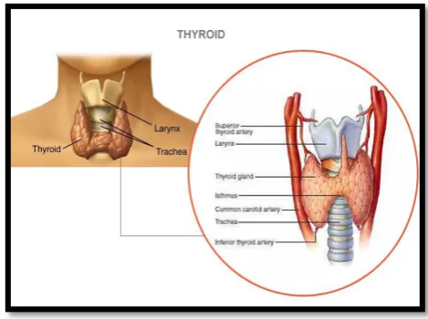
The thyroid gland is a butterfly shaped organ belonging to the endocrine system and is composed of two cone-like lobes. It controls the secretion of the thyroid hormone, which regulates the temperature of the human body, and greatly affects childhood intelligence, growth, and adult metabolism. Thyroid gland produces hormones that are helpful for the body to control metabolism. Too much or too little thyroid hormone secretion (due to a thyroid that is too large or two small, respectively) causes pathological changes and results in thyroid abnormalities. Therefore, physicians often diagnose abnormal symptoms of the thyroid gland by its volume. The thyroid gland is shown in the figure 1.
Ultrasonography is the most well accepted imaging modality for the diagnosis and follow-up of thyroid disorder. The advantages of using ultrasonic imaging include its mobility and low cost as well as the ability to measure the dimension of the gland check for the presence of masses or cysts and evaluate the structure and echogenicity of the parenchyma
.
Fig -1: Thyroid Gland
B. Application of Volume Estimation in treatment of various thyroid disorders
Volume estimation of thyroid gland plays an important role in radioiodine therapy for thyroid disorders like hypothyroidism, hyperthyroidism, etc.Medical therapy, radioiodine therapy, and surgery are the three main methods to treat patients with thyroid diseases. Radioiodine therapy is recognized as the safest, simplest, least expensive and most convenient form of treatment for adults with uncomplicated hyperthyroidism. In the treatment of thyroid diseases like hyperthyroidism and hypothyroidism with the help of radioiodine therapy needs to calculate Radioiodine dose. This radioiodine dose is calculated with the help of volume estimation of thyroid gland.
II. PROPOSED METHODOLOGY
In proposed system focuses on diagnosis of thyroid nodules based on the segmentation of thyroid region and its volume estimation. The proposed method includes image enhancement processing to remove noise, which greatly affects the segmentation results of the thyroid gland region obtained from US images. The probable thyroid gland region is located in the US image, and then, SVM classifier is used to classification of thyroid normal and abnormal thyroid gland. Region boundaries method is applied to recover an accurate shape of the thyroid gland region .The experiment results show that the proposed method can be used to segment the thyroid gland region and to estimate thyroid volume directly from US images.
III. SYSTEM IMPLEMENTATION
© 2016, IJCSMC All Rights Reserved 411
selected in database. These thyroid images are available in image gallery of Wilmington Endocrinology PA on website and some obtained from radiologist. The format of images are JPEG. The Matlab R2013 a software utilizing image processing toolbox is used for experimental work. This project working is totally dependent on this GUI as this software must be easy, simple and fast enough to operate and also the software must be compatible with all other operating system available in market. This software is specially developed for the radiologist and the doctors so the users will be from the non- technical background thus this will help them easily for detecting The software final designs is easy to understand and simple for every user, all the buttons are the pushbuttons and they call all the function using call back and also the displays are given for all the method for volume estimation of thyroid.
The diagnosis of thyroid involving thyroid region segmentation and volume estimation is carried out using image processing techniques which involves the following steps as shown in figure 2.
Fig-2 Steps of thyroid segmentation and volume estimation in US Images.
A. Gray Scale Conversion
Image formation using sensor and other image acquisition equipment denote the brightness or intensity I of the light of an image as two dimensional continuous function F(x, y) where (x, y) denotes the spatial coordinates when only the brightness of light is considered.Here the test image is color image it is converted into a grayscale image because the gray level co-occurrence matrix and wavelet decomposition matrix using a gray scale image.
1) Locating Probable Thyroid Region
Locating the probable thyroid region is the first step in detecting the thyroid abnormalities. In thyroid US images, low visual quality greatly affects the segmentation and the volume estimation results. A pre-processing step is thus required to enhance and locate the probable thyroid region.
2) Noise Reduction
Thyroid Ultrasound Image
Gray Scale Conversion and Histogram
Locating a Suspicious Thyroid Region
Feature Extraction
Classification Thyroid Images
Segmentation
© 2016, IJCSMC All Rights Reserved 412
Noise is an important factor that influences image quality. Noise reduction is necessary to do image processing in this paper the gaussian smoothing filter is used to reduce noise in an image.
B. Feature Extraction
Four discriminative textural features were then extracted from the selected ROIs
1) Histogram Feature: Histogram feature measures the texture characteristics of an M ×M block. After the pre-processing, the thyroid gland occupies most of area in the probable thyroid region. Thus, extract the intensity of the largest area.
2) Homogeneity: Measures the closeness of the distribution of elements in the GLCM to the GLCM diagonal.
Range = [0 1] Homogeneity is 1 for a diagonal GLCM.
∑ ∑ ( )
| |
Here, p (i, j) = (i, j) th entry of the co-occurrence probability matrix
3) Haar Wavelet Features: The Haar wavelet features are significant features for segmentation in US images.
4) Graycomatrix: Create gray-level co-occurrence matrix from image. Using a statistical approach such as co-occurrence matrix will help to provide valuable information about the relative position of the neighbouring pixels in an image.
C. Classification Thyroid Images
The overall literature review SVM is best classification method so, most famous classification method support vector classifiers used for this work. Support vector machine (SVM) are basically linear classifiers. The statistical features that are obtained from feature extraction are used to train the SVM. Ideally all feature samples at hand should be employed, but since most of them are redundant due to mutual correlations, an optimum number of them are selected to achieve highest accuracy. Thus the SVM is trained using these feature samples. The trained SVM is then used for testing the input images which classifies the thyroid region based on pixel classification.In this work the probable thyroid gland region is located in the US image and then, SVM is used to classification of thyroid normal and abnormal thyroid gland images and graph plot using linear kernel function.
D. Segmentation
Segmentation is a tool that used widely in many applications including image processing. One of the common applications of segmentation is in medical image analysis for clinical diagnosis that has an important role in terms of quality and quantity. Medical image segmentation methods generally have restrictions because medical images have very similar gray level and texture among the interested objects. Therefore, significant segmentation error may occur. Image segmentation is the process of partitioning an image into multiple segment or set of pixels used to locate object and boundaries. Use the proposed methodology for segmentation based algorithm is Trace region boundaries in binary image (bwboundaries) and boundary mask method that is basically to segment the local area of the images and to segment the nodule which is give the information of which type nodule exist normal and abnormal. The proper procedure of segmentation algorithm the thyroid abnormal image are segmented.
E. Volume Estimation
Since computed tomography (CT) imaging is expensive and involves hazardous radiation, US imaging is the most commonly used auxiliary tool currently utilized in clinical diagnosis. Hence, this study proposes a complete solution to estimate the volume of the thyroid gland directly from US images. Thyroid volume as measured by real-time ultrasound in cadavers was compared with direct measurements obtained by submersion. The measurements are easy to do and require no additional equipment for calculations. Volumetric analysis ofthe thyroid gland is especially necessary in assessing results of treatment and for measuring dosage in connection with radioiodine therapy.Thyroid ultrasound image of transverse view is used in this study. Therefore, the measurements only area of the thyroid region. The result of thyroid measurement is successfully calculated in pixel unit. The measurement is converted in centimetre (cm) unit.
The basic steps of the proposed methodology for volume estimation of thyroid gland are: Step 1: Display the freehand mask.
© 2016, IJCSMC All Rights Reserved 413 Step 4: BWlabel properties using finds the connected components of abinary image.
Step 5: Regionprops properties using Calculate the area in pixels, that they drew i.e. estimate the number of pixel representing the connected area.
Step6: The result of thyroid measurement is successfully calculated in pixel unit than after measurement is converted in SI unit
The thyroid area is obtained, the thyroid volume can be estimated using following formula.
Vol_cm=0.026458333*area
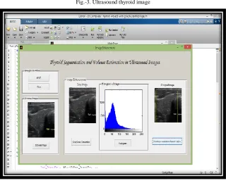
IV. SIMULATION RESULTS
Fig.-3. Ultrasound thyroid image
Fig. - 4. Detection of thyroid region
© 2016, IJCSMC All Rights Reserved 414 Fig.-5. Feature extraction and Classification of thyroid image
Fig.-6.SVM graph using linear kernel function
Fig-7. Volume estimation
© 2016, IJCSMC All Rights Reserved 415
V. CONCLUSION
In this work, an effective diagnosis of thyroid abnormalities based on image processing techniques is discussed. Segmentation
of the thyroid region and volume estimation are explained. The diagnosis is carried out by considering features such as area of
segmented region and volume. US images are a widely used tool for clinical diagnosis, although it is time consuming for
physicians to manually segment the thyroid gland region. The alternative to estimate the volume of a thyroid gland using CT
imaging is expensive and involves hazardous radiation. Thus, a convenient system for thyroid segmentation and volume
estimation in US images is of interest. The proposed method includes image enhancement processing to remove noise, which
greatly affects the segmentation results of the thyroid gland region obtained from US images. The probable thyroid gland region
is located in the US image, and then, SVM classifier is used to classification of thyroid normal and abnormal thyroid
gland.Finally, a region boundaries method is applied to recover an accurate shape of the thyroid gland region .The experiment
results show that the proposed method can be used to segment the thyroid gland region and to estimate thyroid volume directly
from US images
.
It can be help to estimate the volume of thyroid gland in radioiodine therapy.
REFERENCES
[1] Chuan-Yu Chang, Yue-Fong Lei, Chin-Hsiao Tseng, and Shyang-Rong Shih: “Thyroid Segmentation and Volume
Estimation in Ultrasound Images”, IEEE TRANSACTIONS ON BIOMEDICAL ENGINEERIN, Volume: 57, No. 6, June- 2010,
PP. 1348-1357.
[2] AarthipoornimaElangovan, Jeyaseelan.T“DESIGN OF ULTRAFAST IMAGING SYSTEM FOR THYROID NODULE DETECTION”, International Research Journal of Engineering and Technology(IRJET), Volume: 03, Issue: 02, Feb-2016, PP.
1221-1225.
[3] Chuan-Yu Chang and Yong-Cheng Hong: “A Neural Network for Thyroid Segmentation and Volume Estimation in CT Images”, IEEE COMPUTATIONAL INTELLIGENCE MAGAZINE20, October -2011, PP. 43-55.
[4] Dimitris E. Maroulis, Michalis A. Savelonas, Dimitris K. Iakovidis,Stavros A. Karkanis and Nikos Dimitropoulos: “Variable Background Active Contour Model for Computer-Aided Delineation of Nodules in Thyroid Ultrasound Images”,
IEEE TRANSACTIONS ON INFORMATION TECHNOLOGY IN BIOMEDICINE, Volume: 11, No.5, September-2007, PP.
537-543.
[5] Sheeja Agustin A, S. Suresh Babu: “Thyroid Segmentation on US Medical Images: An Overview”, IJETAE, Volume: 2,
Issue: 12, December- 2012, PP. 398-404.
[6] Nikita Singh, Alka Jindal: “A Segmentation Method and Comparison of Classification Methods for Thyroid Ultrasound Images”, International Journal of Computer Applications (0975 – 8887) Volume: 50, No.11, July 2012, PP. 43-49.
[7] D.E. Maroulis, M.A. Savelonas, S.A. Karkanis, D.K. Iakovidis, N. Dimitropoulos: “Computer-Aided Thyroid Nodule Detection in Ultrasound Images”,IEEE Symposium on Computer-Based Medical Systems (CBMS’05).
[8] DeepikaKoundal, Savita Gupta and Sukhwinder Singh: “Computer Aided Diagnosis of Thyroid Nodule: A Review”,
International Journal of Computer Science & Engineering Survey (IJCSES) Volume: 3, No.4, August 2012.
[9] NasrulHumaimiMahmood and AkmalHayatiRusli: “Segmentation and Area Measurement for Thyroid Ultrasound Image”,
© 2016, IJCSMC All Rights Reserved 416
[10] Nikita Singh, Alka Jindal: “Ultra sonogram Images for Thyroid Segmentation and Texture Classification in Diagnosis of
Malignant (Cancerous) or Benign (Non-Cancerous) Nodules”, International Journal of Engineering and Innovative Technology
(IJEIT), Volume: 1, Issue: 5, May 2012, PP. 202-206.
[11] Ambika G. Unnikrishnan, Usha V. Menon : “Thyroid disorders in india : A epidemiological perspective”,Indian Journal of
Endocrinology and Metabolism,2011, volume:15, suppliment 2, PP 78-81.
[12] Harald T Lutz, ElisabettaBuscarini: “WHO Manual of diagnostic ultrasound”, volume: 1, Second edition”, World Health