Scholarship@Western
Scholarship@Western
Electronic Thesis and Dissertation Repository
6-2-2015 12:00 AM
Investigating the role of hydrogen sulfide in the survival, growth
Investigating the role of hydrogen sulfide in the survival, growth
and angiogenic potential of clear cell renal cell carcinoma cell
and angiogenic potential of clear cell renal cell carcinoma cell
lines and xenografts
lines and xenografts
Eric Sonke
The University of Western Ontario
Supervisor Dr. Alp Sener
The University of Western Ontario
Graduate Program in Anatomy and Cell Biology
A thesis submitted in partial fulfillment of the requirements for the degree in Master of Science © Eric Sonke 2015
Follow this and additional works at: https://ir.lib.uwo.ca/etd
Part of the Biological Phenomena, Cell Phenomena, and Immunity Commons, Cancer Biology
Commons, Medical Cell Biology Commons, and the Oncology Commons
Recommended Citation Recommended Citation
Sonke, Eric, "Investigating the role of hydrogen sulfide in the survival, growth and angiogenic potential of clear cell renal cell carcinoma cell lines and xenografts" (2015). Electronic Thesis and Dissertation Repository. 2879.
https://ir.lib.uwo.ca/etd/2879
This Dissertation/Thesis is brought to you for free and open access by Scholarship@Western. It has been accepted for inclusion in Electronic Thesis and Dissertation Repository by an authorized administrator of
CARCINOMA CELL LINES AND XENOGRAFTS
(Thesis format: Monograph)
by
Eric Sonke
Graduate Program in Anatomy and Cell Biology
A thesis submitted in partial fulfillment of the requirements for the degree of
Master of Science
The School of Graduate and Postdoctoral Studies The University of Western Ontario
London, Ontario, Canada
iii
Abstract
Clear cell renal cell carcinoma (ccRCC) is characterized by Von Hippel-Lindau
(VHL)-deficiency, resulting in pseudohypoxic, angiogenic and glycolytic tumours. Hydrogen sulfide
(H2S) is an endogenously-produced gasotransmitter that accumulates under hypoxia and has
been shown to be pro-angiogenic and cytoprotective in cancer. It was hypothesized that H2S
levels are elevated in VHL-deficient ccRCC, contributing to survival, metabolism, and
angiogenesis. Using H2S-specific probes, it was found that H2S levels were higher in
VHL-deficient ccRCC cell lines compared to cells with wild-type VHL. Inhibition of H2
S-producing enzymes could reduce the proliferation, metabolism and survival of ccRCC cell
lines, as determined by live-cell imaging, XTT/ATP assay, and flow cytometry, respectively.
Using the chorioallantoic membrane angiogenesis model, it was found that systemic
inhibition of endogenous H2S production was able to decrease vascularization of
VHL-deficient ccRCC xenografts. Endogenous H2S production is an attractive new target in
ccRCC due to its involvement in multiple aspects of disease.
Keywords
iv
Co-Authorship Statement
The following persons contributed significantly towards the experimental design,
experimental preparation, data acquisition, and analysis of experimental data contained
within this thesis and recently-submitted manuscript:
Megan Verrydt: experimental design (CAM assays) and data acquisition (viability assays)
Carl O. Postenka: sample processing (CAM assays)
Siddika Pardhan: experimental preparation (CAM assays)
Chantalle J. Willie: experimental preparation (CAM assays)
Clarisse R. Mazzola: experimental design (CAM assays)
Ian Lobb: experimental design (viability assays)
Nicholas E. Power: revision
Ann F. Chambers: experimental design (CAM assays) and revision
Hon S. Leong: experimental design (CAM assays) and revision
v
Acknowledgments
In addition to the aforementioned co-authors, I would like to thank lab technicians Dr.
Manujendra Saha and Amy Mok, as well as all of the past and present students from the
Sener lab for their everyday support and expertise during my graduate and undergraduate
studies. I would also like to thank my committee members Dr. Alison Allan, Dr. Nicholas
Power and Dr. Lakshman Gunaratnam for their scientific expertise, constructive criticism
and translational approach that shaped this project. This work would also not be possible
without the aid of Xizhong Zhang, Alex Pavlovsky and Ola Ismail from the Matthew Mailing
Centre for Transplant Studies. Likewise, I would like to thank scientists and personnel in the
labs of Dr. Ann Chambers and Dr. Hon Leong at the London Regional Cancer Program who
provided me with extensive mentorship and training. I would also like to thank Dr. Anthony
Jevnikar, Dr. Michael Pluth, Dr. Lakshman Gunaratnam and Dr. John McCormick for usage
of equipment and/or provision of reagents. I would like to thank Dr. Jean-Baptiste Lattouf
and Dr. Aaron Haig who were instrumental in the processing of human samples. Of course,
without the funding provided by the department of Anatomy and Cell Biology and the
Lawson Health Research Institute this work would not be possible. Finally, I would like to
thank my supervisor Dr. Alp Sener for his un-ending optimism, encouragement, guidance
and scientific mentorship. Your passion, creativity and inquisitive nature are a daily source
vi
Table of Contents
Abstract ... iii
Co-Authorship Statement... iv
Acknowledgments... v
Table of Contents ... vi
List of Figures ... x
List of Appendices ... xii
List of Abbreviations ... xiii
1. Introduction ... 1
1.1 Renal Cell Carcinoma (RCC) ... 1
1.1.1 Clear Cell RCC and Von Hippel-Lindau ... 2
1.1.2 Past and Present (cc)RCC Therapies ... 5
1.1.3 ccRCC Metabolism – The Warburg Effect ... 9
1.2 Hydrogen Sulfide: The Third Gasotransmitter ... 14
1.2.1 H2S Production in the Kidneys: The Transsulfuration Pathway and Oxygen Sensing ... 15
1.2.2 H2S Production, Reactivity and Signalling ... 16
1.2.3 H2S: Vasodilator and Angiogenic Factor ... 18
1.2.4 H2S: Antioxidative and Cytoprotective ... 20
1.2.5 H2S: Metabolic Substrate and Regulator ... 21
1.3 H2S and Cancer ... 25
1.3.1 Exogenous H2S as a Cancer Treatment? ... 25
1.3.2 Role of Endogenous H2S Production in Cancer ... 25
1.3.3 Role of H2S in kidney cancer ... 27
vii
1.4.2 Objective I: Is Endogenous H2S Production Enhanced in VHL-deficient
ccRCC Cell Lines? ... 29
1.4.3 Objective II: Does Endogenous H2S Production Contribute to the Proliferation, Metabolism and Survival of ccRCC Cell Lines? ... 30
1.4.4 Objective III: Does Endogenous H2S Production Contribute to Neovascularization of ccRCC Xenografts? ... 31
1.4.5 Objective IV: Is Endogenous H2S Production Enhanced in human RCC tumours? ... 31
2. Materials and Methods ... 32
2.1 Cell Culture ... 32
2.2 Treatments... 32
2.3 Measurement of Endogenous H2S Production ... 33
2.3.1 Live Cell Imaging with Fluorescent Probe ... 33
2.3.2 Methylene Blue Assay ... 33
2.4 Protein Expression Analysis ... 34
2.4.1 Protein Isolation ... 34
2.4.2 SDS-PAGE and Western Blotting ... 34
2.4.3 Imaging and Analysis ... 34
2.5 Cell Proliferation Assay ... 35
2.6 Cell Viability Assay ... 35
2.7 XTT Cytotoxicity Assay ... 35
2.8 ATP Quantitation Assay ... 36
2.9 Avian Xenograft Vascularization Model ... 36
2.9.1 Lentiviral Infection ... 36
2.9.2 Avian Chorioallantoic Membrane Xenografting ... 37
2.9.3 Xenograft Treatment and Processing ... 37
viii
2.10Human Tissue Microarray Analysis ... 38
2.11Statistical Analyses ... 38
3. Results ... 40
3.1 Objective I – Is Endogenous Production of H2S Enhanced in VHL-deficient ccRCC Cell Lines? ... 40
3.1.1 Levels of H2S are increased in VHL-deficient ccRCC cell lines ... 40
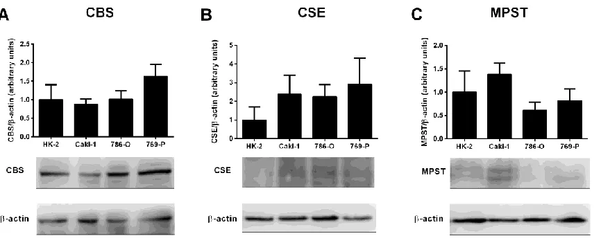
3.1.2 VHL-deficient ccRCC cell lines do not display increased expression of CBS, CSE, or MPST ... 43
3.2 Objective II – Does Endogenous H2S Production Contribute to the Proliferation, Metabolism and Survival of ccRCC Cell Lines? ... 50
3.2.1 Endogenous H2S production can be targeted, though not stimulated, in VHL-deficient ccRCC cell lines ... 50
3.2.2 Endogenous H2S production contributes to the proliferation of ccRCC cell lines ... 53
3.2.3 Endogenous H2S production contributes to the survival of ccRCC cell lines ... 56
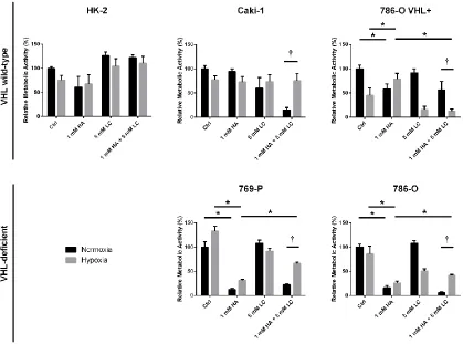
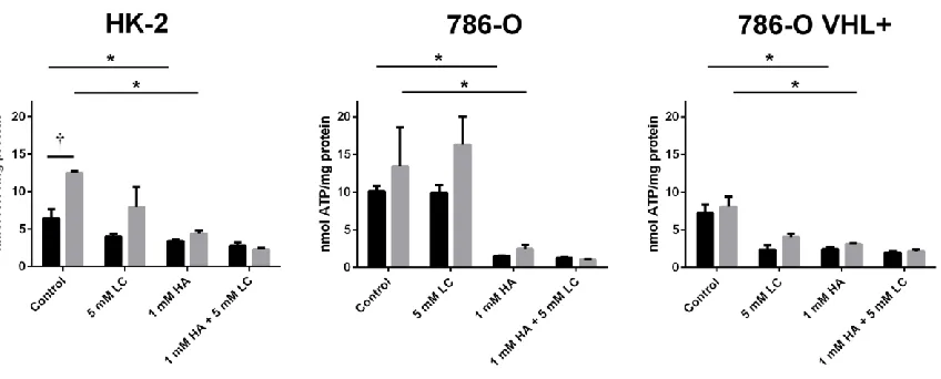
3.2.4 Endogenous H2S production contributes to the metabolism of ccRCC cell lines ... 59
3.3 Objective III – Does Endogenous H2S Production Contribute to the
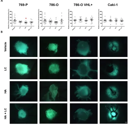
Neovascularization of ccRCC Xenografts? ... 62
3.3.1 Inhibition of endogenous H2S production restricts the neovascularization of ccRCC xenografts ... 62
3.3.2 Inhibition of endogenous H2S production restricts the growth of ccRCC xenografts ... 65
3.4 Objective IV – Is Endogenous H2S Production Enhanced in Human ccRCC Tumours? ... 67
3.4.1 Expression of CBS or CSE is not upregulated in human ccRCC tumours 67
ix
4.1 Relation to Initial Hypothesis ... 70
4.2 VHL, H2S and Oxygen Sensing ... 71
4.3 Crosstalk between HIF-1/2α and H2S ... 72
4.4 H2S in ccRCC Proliferation, Metabolism and Survival ... 74
4.5 H2S in ccRCC Angiogenesis ... 77
4.6 Translational Applications ... 78
4.7 Recommendations ... 80
5. Conclusions ... 83
References ... 84
Appendices ... 97
Curriculum Vitae ... 100
x
List of Figures
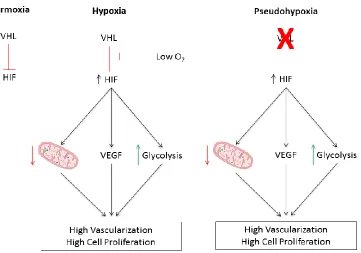
Figure 1. VHL/HIF signalling under normoxic, hypoxic, and pseudohypoxic conditions in
ccRCC ... 13
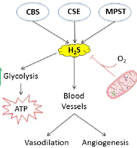
Figure 2. Cellular H2S metabolism and signalling ... 24
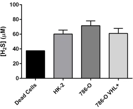
Figure 3. Baseline endogenous H2S production is greater in VHL-deficient ccRCC cell lines
than in VHL WT malignant and non-malignant renal cell lines ... 41
Figure 4. Effect of VHL knock-in on endogenous H2S production in 786-O cells, as
measured by methylene blue assay ... 42
Figure 5. Baseline normoxic expression of H2S-producing enzymes is unaltered in
VHL-deficient ccRCC cell lines when compared to malignant and non-malignant VHL WT renal
cell lines ... 44
Figure 6. Hypoxic induction of H2S-producing enzymes in malignant, and non-malignant
renal cell lines ... 46
Figure 7. Normoxic expression and hypoxic induction of H2S-producing enzymes in
wild-type 786-O cells (786-O) and VHL knock-in 786-O cells (786-O VHL+) ... 49
Figure 8. Inhibition of endogenous H2S production reduces elevated levels of H2S in
VHL-deficient ccRCC cell lines ... 52
Figure 9. Inhibition of endogenous H2S production attenuates proliferation of ccRCC cell
lines ... 55
Figure 10. Inhibition of endogenous H2S production selectively kills ccRCC cells over
non-malignant renal cells ... 58
Figure 11. Inhibition of endogenous H2S production reduces overall metabolic activity of
ccRCC cell lines ... 60
Figure 12. Inhibition of endogenous H2S production reduces ATP production in malignant
xi 2
reduces the vascularization of VHL-deficient ccRCC xenografts ... 64
Figure 14. Systemic inhibition of endogenous H2S production with hydroxylamine (HA)
restricts the growth of VHL-deficient ccRCC xenografts. ... 66
Figure 15. Diffuse and localized expression patterns of CBS and CSE in human renal
xii
List of Appendices
xiii
List of Abbreviations
786-O VHL+ 786-O Von Hippel-Lindau knock-in
Akt protein kinase B
CAM chorioallantoic membrane
CBS cystathionine β-synthase
ccRCC clear cell renal cell carcinoma
cGMP cyclic guanosine monophosphate
CO carbon monoxide
CSE cystathionine γ-lyase
EGFP extreme green fluorescent protein
EGFR epidermal growth factor receptor
eNOS endothelial nitric oxide synthase
ETC electron transport chain
GAPDH glyceraldehyde 3-phosphatase
GC guanylyl cyclase
GFR glomerular filtration rate
Glut-1 glucose transporter 1
H2S hydrogen sulfide
HA hydroxylamine
Hcy homocysteine
HIF hypoxia-inducible factor
HRE hypoxia response element
IFN-α interferon alpha
IL-2 interleukin 2
KATP ATP-dependent K+
Keap1 kelch-like ECH-associated protein-associated protein-1
LC L-cysteine
LCA lens culinaris agglutinin
LDH lactate dehydrogenase
mAb monoclonal antibody
MAX MYC-associated factor X
MMP matrix metalloproteinase
xiv
mTOR mammalian target of rapamycin
mTORC mammalian target of rapamycin complex
MXI1 MAX-interacting protein 1
NAD nicotinamide adenine dinucleotide
NADPH nicotinamide adenine dinucleotide phosphate
Nampt nicotinamide phosphoribosyltransferase
NF-κB nuclear factor kappa B
NKA Na+/K+ ATPase
NKCC Na+/K+/2Cl− co-transporter
NO nitric oxide
Nrf2 NF-E2 p45-related factor 2
OCT optimal cutting temperature
ORR objective response rate
OS overall survival
p38 mitogen-activated protein kinase p38
PAG propargyl glycine
PBS phosphate buffered saline
PD pyruvate dehydrogenase
PDE5 phosphodiesterase type 5
PDGF platelet-derived growth factor
PDK pyruvate dehydrogenase kinase
pf post-fertilization
PFS progression-free survival
PGC-1β peroxisome proliferator-activated receptor gamma coactivator 1-beta
PHD prolyl hydroxylase
PI3K phosphoinositide 3-kinase
PK pyruvate kinase
PKC protein kinase C
PKG protein kinase G
PN partial nephrectomy
PPP pentose phosphate pathway
xv
RBF renal blood flow
RCC renal cell carcinoma
RN radical nephrectomy
RNS reactive nitrogen species
ROS reactive oxygen species
RTK receptor tyrosine kinase
RTKI receptor tyrosine kinase inhibitor
SMI small molecule inhibitor
SOU sulfide oxidation unit
SQR sulfide quinone reductase
TCA tricarboxylic acid
TGF-α transforming growth factor alpha
TNF-α tumour necrosis factor alpha
VEGF vascular endothelial growth factor
VHL Von Hippel-Lindau
1. Introduction
1.1 Renal Cell Carcinoma (RCC)
As with other cancers, kidney cancer can be categorized into multiple sub-types. The
most prevalent form of kidney cancer is known as renal cell carcinoma (RCC) and
accounts for 80-90% of kidney cancer cases and 2-3% of all cancer cases (1). RCC arises
as a result of mutations to the epithelial cells of nephrons – the functional units of the
kidney. Increasing incidence rates for RCC have been reported over the last few decades,
and this has been attributed to popular usage of CT and ultrasound imaging for other
conditions (2). Some studies suggest that increasing incidence of RCC risk factors like
diabetes, hypertension and obesity may also be responsible for the increasing prevalence
of RCC, though these conditions have not been established as true etiological factors of
RCC (3-7). Despite the increase in incidental detection of RCC and timely removal of
the primary tumour, the cancer-specific mortality rate of RCC remains high at 30-40%
(2). This high mortality rate is due to the high likelihood – approximately 20-30% – that
the disease has already metastasized at the time of detection (2). Unfortunately,
metastatic RCC (mRCC) has proven to be highly resistant to both chemotherapy and
radiation therapy, and this resistance contributes to the high mortality rates observed (8).
While it is true that novel growth-targeting therapies offer improvements in overall
survival (OS) and progression-free survival (PFS), these therapies do not offer a cure for
the disease (1).
RCC can be further sub-divided into various histotypes based on tumour morphology,
genetic signature, and clinical manifestation. The area of the nephron from which the
tumour arises (proximal tubule, distal tubule, collecting duct, etc.) also plays a role in the
classification of RCCs (9). Whether sporadic or inherited, genetic mutations in RCC
often affect pathways involved in oxygen, nutrient, or energy sensing and for this reason
RCC is often described as a metabolic disease (9,10). In today’s clinical landscape of
targeted therapies, identifying key targets within these metabolic pathways is essential
(1,11). While most subtypes of RCC seem to subvert cellular metabolic pathways to their
patient, varies widely (12). Identifying pathways that are commonly disrupted in RCC
and developing therapies that target these pathways has proven to be an effective
treatment strategy in RCC in that they improved PFS when compared to chemotherapy
and radiation therapy (12).
1.1.1
Clear Cell RCC and Von Hippel-Lindau
The most common histotype of RCC is the clear cell histotype (ccRCC) and accounts for
75-80 % of all RCC cases (2). The characteristic “clear” cytoplasm of these cancer cells
is due to increased production and accumulation of deposits containing cholesterol esters,
fatty acids, and glycogen which makes histological identification relatively easy. While
the exact mechanisms that lead to accumulation of these lipid deposits are still being
investigated, we do know that a crucial step in this process is inactivation of a key tumour
suppressor – the Von Hippel-Lindau (VHL) tumour suppressor (13,14). VHL is an E3
ubiquitin ligase that is crucial in targeting certain cellular proteins for proteasomal
degradation in response to changing cellular conditions, and is inactivated in the large
majority (90%) of ccRCC tumours (1,15). When VHL is expressed in its wild-type form
(VHL WT), it is involved in regulating a number of homeostatic pathways, including the
metabolism and storage of macromolecules. Cells that lack a functional copy of VHL
(VHL-deficient) lose control of multiple metabolic pathways, leading to decreased
β-oxidation of fatty acids, increased synthesis of fatty acids and glycogen, and increased
storage of fatty acids and esterified cholesterol (13,14). The end result is a cell with
clear, lipid-filled cytoplasm.
While VHL inactivation and subsequent lipid accumulation may prove to be an
etiological link between established risk factors of ccRCC and the disease itself, VHL
deficiency plays other, more significant roles in cancer initiation and progression. The
best understood function of VHL is as a master regulator of the hypoxic response, and
key tumour suppressor for many cells in the body (16). In combination with
hypoxia-inducible factors (HIFs) and prolyl hydroxylase (PHD), VHL ultimately controls the
expression of genes that allow cells to cope with the harmful effects of hypoxia, and
restore cellular oxygenation. HIFs are heterodimeric transcription factors composed of α
hypoxia response elements (HREs). PHD is an enzyme that hydroxylates proline
residues on various proteins, but only in the presence of an essential cofactor – oxygen.
As mentioned previously, VHL is an E3 ubiquitin ligase that specifically recognizes and
binds to proteins with hydroxylated proline residues and targets them for proteasomal
degradation through ubiquitination. HIF-β subunits are not a substrate for hydroxylation
by PHD, and therefore are consistently present under normoxic and hypoxic conditions.
HIF-α subunits on the other hand are hydroxylated by PHD and targeted for degradation
by VHL under normoxic conditions to ensure that hypoxia response genes are not
expressed unnecessarily (17). However when oxygen is absent during periods of
hypoxia, PHD cannot hydroxylate HIF-α residues, allowing them to escape recognition
by VHL, form a functional heterodimer with HIF-β, translocate to the nucleus, and
initiate the hypoxic response (16).
Genes containing HREs include vascular endothelial growth factor (VEGF),
platelet-derived growth factor (PDGF), transforming growth factor alpha (TGF- α), glucose
transporter-1 (GLUT-1), matrix metalloproteinases (MMPs), carbonic anhydrase IX,
erythropoietin, cyclooxygenase-2, c-MYC, cyclin D1, and many more (18). Induction of
many of these genes through the VHL/HIF pathway is a useful means of minimizing
injury and re-establishing tissue oxygenation during periods of hypoxia that occur
following disruptions in blood supply (19). However, when these genes are aberrantly
expressed under normoxic conditions as a result of VHL inactivation, the resulting cells
are hyperproliferative, invasive, angiogenic and cell death-resistant as a result of their
perceived hypoxia, or pseudohypoxia (Figure 1) (14). It is this pseudohypoxia, as a result
of VHL inactivation, that plays the largest role in the initiation and progression of
ccRCC.
As a tumour suppressor, VHL only loses its function after both genetic copies of the gene
have been lost as a result of deletion, silencing via methylation, or some other form of
mutation (15). In other words, spontaneous ccRCC often only occurs when an already
heterozygous cell loses its functional genetic copy of VHL following a “second hit,”
resulting in complete loss of protein function. Loss of VHL function is common in many
autosomal-dominant condition known as VHL Disease (20). The majority (80%) of
patients afflicted with VHL disease inherit a mutant copy of VHL from one of their
parents, and following mutation to the second copy begin to develop cysts and tumours of
the kidney (ccRCC), pancreas, retina, and other areas of the central nervous system (16).
It was from studying this rare hereditary disease that the tumour suppressive role of VHL
in ccRCC and other cancers was elucidated (21).
Clearly VHL loss of function is an important step in neoplastic and malignant
transformation. However this, by itself, is neither necessary nor sufficient for the
formation of ccRCC tumours (22,23). Furthermore, there has been no conclusive
evidence which suggests that VHL mutation status – regardless of its overall effect on
VHL function – holds any value as a biomarker or prognostic indicator (16). It is now
generally accepted that classification of ccRCC tumours as VHL-deficient versus VHL
WT is not sufficient. VHL-deficient tumours can be further subdivided according to the
degree to which they express the two major HIF-α isoforms (HIF-1α and HIF-2α). There
are VHL-deficient tumours which aberrantly express increased levels of 1α and
HIF-2α (H1H2), and those which express increased levels of HIF-HIF-2α only (H2) (24). Several
studies have highlighted that HIF-1α and HIF-2α have very different effects on gene
transcription in ccRCC (25), and most suggest that VHL-deficient H2 tumours are more
proliferative and angiogenic than their H1H2 counterparts (26,27).
The increased proliferation of H2 ccRCC cell lines and tumours is in part due to
autocrine growth signaling that occurs through HIF-2α, TGF-α and the epidermal growth
factor receptor (EGFR) that binds TGF-α (26,28,29). Furthermore, the activity of
oncoproteins such as c-MYC and cyclin D1 are upregulated by HIF-2α (24,26), whereas
the activity of pro-apoptotic genes such as BNip3 are upregulated by HIF-1α (26).
Angiogenesis in pseudohypoxic ccRCC cells also appears to be driven by induction of
VEGF through HIF-2α, and loss of HIF-1α may actually exacerbate this process due to a
loss of feedback inhibition that is normally mediated by HIF-1α (26,27). Not only is the
H2 phenotype more aggressive than the H1H2 phenotype, but it may actually be more
common in VHL-deficient ccRCC tumours as a result of a greater likelihood of HIF-1α
are derived from H1H2 tumours and that loss of HIF-1α is a key step in ccRCC tumour
progression (9,30).
With HIF-α transcription factors identified as mediators of ccRCC progression, the
development of therapies is now focused on inhibiting the pathways that the downstream
effectors of HIF-1/2 α (VEGF, TGF-α, etc.) feed into. The development of this targeted
therapy approach has improved PFS of patients with ccRCC and represents a small
victory in cancer treatment (31). However, these therapies are not curative, and are only
effective for a matter of months before the cancer becomes resistant to them.
1.1.2
Past and Present (cc)RCC Therapies
First-line treatment of localized RCC, regardless of histotype, is usually surgery seeing as
it is the only curative therapeutic approach (32). When determining the extent of renal
mass to be excised, a number of factors must be evaluated. The staging of the tumour
according to size and presence/absence of local/distant metastases (TNM staging) is the
first to be evaluated. Patients with T1 tumours (<7 cm, limited to kidney) are often good
candidates for a partial nephrectomy (PN), a procedure in which only the tumour and
immediate surrounding area of the kidney are removed (33). Experienced centers may
even perform this PN laparoscopically instead of opting for traditional open surgery. PN
is the preferred course of treatment where the tumour is affecting a solitary functional
kidney or the patient is in overall poor condition. In these cases, sparing as much
functional tissue as possible is a priority and active surveillance may be preferred to
surgery. Removal of the entire kidney, or radical nephrectomy (RN), is recommended in
cases of locally advanced tumour growth or unfavourable tumour location. RN is often
performed laparoscopically for T2 tumours (7-10 cm, limited to kidney), however open
surgery is recommended for T3 and T4 tumours which involve local and distant
metastases (32).
There are additional, minimally invasive first-line therapies for patients with localized
disease who may not be good surgical candidates. RCC tumours have been shown to
respond to various ablation therapies such as percutaneous radiofrequency ablation,
ablation. The local progression rates for these therapies are often higher than those of
partial and radical nephrectomy, however they are useful for treatment of incidental
lesions in the elderly, multiple bilateral tumours, recurring small renal masses, and
patients at risk of losing all renal function following nephrectomy (32). In general,
treatment of localized RCC with surgery is good, but it is not perfect. The 5-year
survival rate of patients with stage I disease (T1, N0, M0) is >90%. However relapse
occurs within 5 years of surgery for patients with stage II (T2, N0, M0) or III (T3, N0,
M0; T1-3, N1, M0) disease (34). Aggravating this situation is the fact that we have yet to
identify adjuvant therapies that might manage this risk of relapse (35,36). Managing
intermediate/high-risk RCC in its early stages is just as important as targeting stage IV
mRCC, which presents its own set of challenges (36).
When possible, removal of metastases through surgery (metastectomy) can improve
prognosis if all metastatic lesions are completely removed (37). However, systemic
therapy is often required in cases where metastases are unresectable and/or burden of
disease is too great (32). Chemotherapy and radiation therapy have, unfortunately,
proven to be ineffective for treatment of mRCC, regardless of histotype. Poor response
rates – ranging from 0-14% – to a wide range of cytotoxic agents have been
well-documented and are attributed to expression of multi-drug resistance gene 1 and its
protein product P-glycoprotein (8). The molecular mechanisms conferring resistance to
radiation therapy are less well understood, however there have been links drawn to
increased expression of HIF-2α and survivin (38,39). While investigations into these
therapeutic immunities were being made, it was found that immune-based therapies were
a viable treatment option for mRCC (2).
Alongside melanoma, RCC is considered to be one of the most immunogenic of all
human malignancies. This is based on high tumoural infiltration of cytotoxic T-cells and
other immune cells that can recognize tumour-associated antigens, the occurrence of
RCC in patients receiving immunosuppression, and rare observation of spontaneous
metastatic regression (40). Of the wide array of immunotherapies investigated,
cytokine-based therapies such as high-dose interleukin-2 (IL-2) and interferon-alpha (IFN-α) have
advent of targeted therapies. Some argue that such therapies are indeed still relevant for
treatment of mRCC given their durable responses in a subset of patients, which is an area
where newer therapies fall short (41,42). However, finding predictive biomarkers that
can identify this subset of patients is a priority given the fact that associated toxicities of
cytokine therapy are generally higher grade when compared to targeted therapies (42).
First-line treatment of low-risk mRCC with IFN-α may also be just as effective as the
currently recommended first-line targeted therapy sunitinib, but at significantly lower
cost it could improve economic efficiency (41). Combination treatments consisting of
cytokine/targeted therapy are also being investigated and present the possibility of
durable responses in a larger proportion of patients (36,41,42).
From increased understanding of the molecular pathways that are active in RCC,
specifically ccRCC, have emerged therapies that hone in on these pathways. These
targeted therapies come in two forms: small molecule inhibitors (SMIs), and monoclonal
antibodies (mAbs). Since 2007, 8 targeted therapies – sorafenib, sunitinib, temsirolimus,
bevacizumab, everolimus, pazopanib, axitinib and tivozanib – have been approved for
mRCC treatment (1,11,12). Of these drugs, only bevacizumab is a mAb and it is targeted
against VEGF. The remaining 7 are SMIs that fall into two classes, receptor tyrosine
kinase (RTK) inhibitors (RTKIs) and mammalian target of rapamycin (mTOR) inhibitors.
The RTKIs that are approved and/or in clinical trials for ccRCC – sunitinib, sorafenib,
pazopanib, axitinib and tivozanib – primarily act on endothelial cell VEGF and PDGF
receptors, inhibiting signal transduction initiated by tumour cell-secreted VEGF and
PDGF (1). The mAb bevacizumab is specific for VEGF and prevents VEGF from
interacting with its receptor on endothelial cells, thus inhibiting signaling between the
tumour and blood vessels in a similar fashion (1). In this way, RTKIs and bevacizumab
target both current metastases – by preventing the initial attraction of blood vessels
(neovascularization) to secondary tumours – and future metastases – by preventing the
growth of new blood vessels towards the primary tumour (angiogenesis) that allow
tumour cells to travel throughout the body (1). While RTKIs can induce necrotic cell
death in larger tumours as a result of decreased microvascular density (MVD), RTKIs
some off-target effects of RTKIs acting on tumour cells directly have been documented.
For example, some of sorafenib’s efficacy may stem from off-target inhibition of cyclin
D1, cyclin B1 and survivin (44). Likewise, sunitinib has been observed to enhance the
anti-tumoural immune response by recruiting regulatory T-cells (45). In general though,
RTKIs and bevacizumab target mRCC by preventing vascularization of the primary
tumour and its metastases, with the intention of enhancing cell death, reducing metabolic
output, and preventing future metastases.
Unlike RTKIs, mTOR inhibitors are more direct in their treatment of ccRCC in that they
target a signaling pathway that is overactive in the cancer cells themselves. mTOR can
exist as a member of two functionally distinct complexes, mTOR complex 1 (mTORC1)
and mTOR complex 2 (mTORC2). Both mTORC1 and mTORC2 regulate cell
proliferation, cell survival, nutrient sensing, energy sensing, overall protein translation
and angiogenesis, though their effects on these processes are not synonymous (1,46).
The mTOR inhibitors currently approved for ccRCC – temsirolimus and everolimus –
both prevent the formation of mTORC1, which results in decreased translation of
1α, cyclin D1 and c-MYC (1). However as discussed previously, it is thought that
HIF-2α is responsible for most of the tumourigenic signaling in ccRCC, and its translation is
primarily mediated by mTORC2 (47). Furthermore, inhibition of mTORC1 alone has
been shown to induce a paradoxical increase in the formation of mTORC2. Therefore,
while mTORC1 inhibitors may have some effect on cell proliferation and survival, they
could actually be exacerbating the angiogenic response through increased activity of
mTORC2, increased expression of HIF-2α and subsequent secretion of VEGF (48). For
this reason, SMIs that simultaneously target mTORC1/2 as well as the associated growth
signaling kinases phosphoinositide 3-kinase (PI3K) and protein kinase B (Akt) are
currently in development, though they have yet to reach clinical trials for management of
mRCC (1,48).
While RTKIs, mAbs that target VEGF, and mTOR inhibitors have all shown to improve
PFS and objective response rates (ORR) when compared to cytokine-based therapies,
many have noted that durable, complete responses remain rare. Furthermore, while most
have only been objectively shown for some of the targeted agents, (1,11,12,31,34). The
adverse events associated with these targeted therapies, especially next generation RTKIs
like pazopanib and tivozanib, are also considered to be less severe than those associated
with cytokine-based therapies (12,42). For these reasons, RTKIs are now the current
first-line standard of care for patients with metastatic ccRCC and a favourable risk
categorization (34). However, given the relatively recent approval of these drugs for
treatment of mRCC, there is a significant lack of prognostic information to aid clinicians
in deciding which drugs are most likely to work in which patients, and the order in which
they should be administered (12). In fact, with the exception of bevacizumab + IFN-α,
there is evidence to suggest that combined therapies – especially those combining
multiple targeted therapies – are associated with serious adverse events (11).
Significant investment has been made into developing and refining RTKI and mTOR
inhibitors in order to increase their specificity, thus increasing potency and decreasing the
likelihood of adverse events (12). However, we already know that though RTKIs
developed for ccRCC are anti-angiogenic, they are not cytotoxic in and of themselves
(unless you consider their off-target effects). Likewise, though mTOR inhibitors are
cytotoxic, they are not anti-angiogenic (they may actually be pro-angiogenic). Given the
hazards associated with combined therapy, it seems that currently available treatment
options are not capable of simultaneously targeting cell survival and angiogenesis, which
may prove to be necessary in improving overall survival in a large proportion of patients
(11). In summary, while targeted therapies have shown to be effective, investigation into
new targets that play a role in both survival and angiogenesis is something that should be
considered.
1.1.3
ccRCC Metabolism – The Warburg Effect
VHL inactivation and HIF-α stabilization occur early in the development of ccRCC,
however these events alone do not lead to ccRCC. In fact 20% of VHL-knockout mice
develop renal cysts that never progress to ccRCC (23). Additional events such as
activation of oncogenic signaling pathways, inactivation of DNA repair mechanisms, and
cancerous. It is these additional events that further facilitate tumour progression, and
might be better targets for therapy (14).
The change in metabolism that occurs in ccRCC, and many other cancers, is known as
the “Warburg Effect” and involves a shift from aerobic respiration pathways to anaerobic
respiration pathways, even when oxygen is abundant (49-51). Whereas aerobic
respiration generates ATP through the tricarboxylic acid (TCA) cycle and the electron
transport chain (ETC), anaerobic respiration generates ATP through glycolysis. The
switch to aerobic glycolysis occurs in almost all RCC histotypes as a result of HIF-1α
accumulation. While HIF-1α stabilization is due to VHL inactivation in ccRCC, its
stabilization in other histotypes is due to mutation of key metabolic enzymes that
ultimately result in inhibition of PHD (10,14). Regardless of the mechanism,
accumulation of HIF-1α subsequently results in the upregulation of key enzymes
involved in glycolysis. HIF-1α stabilization ultimately uncouples glycolysis from the
TCA cycle via upregulation of pyruvate dehydrogenase kinase (PDK). PDK inactivates
pyruvate dehydrogenase (PD) through phosphorylation, thus preventing the end product
of glycolysis, pyruvate, from heading towards aerobic metabolic pathways (14,51).
Instead of heading towards the TCA cycle, pyruvate is converted into lactate via lactate
dehydrogenase (LDH) – another HIF-1α target – which allows for the regeneration of
oxidized nicotinamide adenine dinucleotide (NAD+) from its reduced form (NADH)
(14,52). This alongside upregulation of GLUT-1 – yet another HIF-1α target – ensures
that glycolysis substrates are in ample supply (14).
From the perspective of ATP production, shifting metabolites away from oxidative
phosphorylation – ATP produced by the ETC – and towards substrate-level
phosphorylation – ATP produced by glycolysis – is energetically unfavourable when
oxygen is available. This has led cancer biologists to question the selective advantage
that the Warburg Effect provides RCC and other cancers. One selective advantage that
has been suggested is the decreased production of reactive oxygen species (ROS) that
comes with decreased oxidative phosphorylation (53). In addition to uncoupling
glycolysis from the TCA cycle by upregulating PDK, HIF-1α accumulation also leads to
glycolysis – conversion of phosphoenolpyruvate into pyruvate. PK comes in two
isoforms, PKM1 and PKM2, which have variable activities. ccRCC tumours often
express the slower-acting PKM2 isoform as a result of c-MYC-mediated alternative
splicing (54). As a result of this delay at the end of glycolysis, metabolites produced
earlier in the glycolytic pathway accumulate and are available for other processes. One
such process is the pentose phosphate pathway (PPP) which produces nicotinamide
adenine dinucleotide phosphate (NADPH) – a molecule that helps cells cope with
oxidative stress through various mechanisms – and molecules like ribose 5-phosphate and
erythrose 4-phosphate – that are required for the synthesis of proteins and nucleic acids
(53). Therefore despite reducing overall ATP production, aerobic glycolysis ensures that
the enhanced growth of RCC tumours occurs in a cellular environment in which; i)
oxidative stress is regulated and ii) there are sufficient materials for production of
biomass (53).
HIF-1/2α accumulation also appears to suppress ROS production by inhibiting
mitochondrial biogenesis, mitochondrial respiration and overall O2 consumption (55). As
mentioned earlier, ccRCC tumours often express elevated levels of c-MYC. c-MYC can
heterodimerize with MYC-associated factor X (MAX) to form a transcriptional activator
of peroxisome proliferator-activated receptor gamma coactivator 1-beta (PGC-1β) – a
gene that promotes mitochondrial biogenesis (55). On the other hand, MAX can
heterodimerize with MAX-interacting protein 1 (MXI1) to form a transcriptional
repressor of PGC-1β, thus suppressing mitochondrial biogenesis. MXI1 happens to be a
target of HIF-1/2α and is overexpressed in ccRCC (56), which results in greater
formation of MXI1:MAX than c-MYC:MAX, overall repression of mitochondrial
biogenesis, and overall decreased O2 consumption (55). There is also evidence that
HIF-1α-mediated upregulation of LDH inhibits mitochondrial respiration by competing for the
same pool of NADH that is required for maintenance of the mitochondrial membrane
potential and oxidative phosphorylation (52). Not only is LDH more abundant, but its
cytoplasmic localization and faster kinetics ensure that it is able to outcompete
mitochondrial cytochrome oxidases for the NADH being produced by glycolysis (52).
The end result is lower ETC activity, lower O2 consumption, and lower overall ROS
As the energetically preferred form of cellular metabolism, oxidative phosphorylation
comes at the cost of increased ROS production. When the earth’s atmosphere changed
from a sulfide-rich environment to an oxygen-rich environment, bacterial and archaeal
species were suddenly faced with a new molecule from which they were obligated to
harness their energy. Each species either slowly acquired this new metabolic phenotype
via advantageous genetic mutations over many generations, or perished. This concept,
known as natural selection, is the driving force behind the evolution of species over time.
However natural selection can apply to cells – malignant and benign – not just species as
Darwin originally postulated (57). Cells are able to sense changes in their environment
and react in many ways, just like organisms. Some of these mechanisms have been
known for decades, some have only just come to light, and even more are still waiting to
1.2 Hydrogen Sulfide: The Third Gasotransmitter
Gasotransmitters are small, gaseous, membrane-permeable molecules that are produced
endogenously and in a dedicated fashion within mammalian cells in order to be perceived
and invoke a response (58). Two gaseous molecules – carbon monoxide (CO) and nitric
oxide (NO) – are widely accepted to fit this definition and their effects within the body
have been recognized since the 1960s and 1980s respectively (59,60). The third and most
recently discovered gasotransmitter is hydrogen sulfide (H2S), though its classification as
a bona fide gasotransmitter is still up for debate (61). While there is no doubt that H2S is
a small, membrane-permeable molecule that is produced in a dedicated fashion in nearly
all mammalian cells, there is some disagreement over whether changing levels of H2S can
be sensed, or whether H2S itself is the sensor (61,62). Regardless of how the semantics
play out, it cannot be denied that H2S production in mammalian cells plays important
roles in normal physiology and pathophysiology (58).
It has long been known that at high enough concentrations H2S is a mitochondrial poison
that inhibits Complex IV of the ETC and eliminates aerobic respiration (63). As a result,
nobody thought that H2S could possibly be playing a meaningful role in humans, despite
the fact that there was knowledge of mammalian enzymes that produce H2S (64). The
first physiological function of H2S identified in mammals was its role as a
neurotransmitter in the brain (65). Soon after, H2S was shown to participate in
synergistic smooth muscle relaxation alongside NO (66). To this day, the functions of
H2S are best described in the context of the central nervous system (67,68) and the
cardiovascular system (69-71), although additional functions have been identified in the
digestive system (72,73), musculo-skeletal system (74), endocrine system (75-77),
excretory system (78-80), respiratory system (81,82) and reproductive system (83). Not
only does endogenous H2S production play a role in the normal functioning of these
systems, but disruption of baseline H2S production has been implicated in diseases such
as hypertension, Alzheimer’s Disease, diabetes mellitus, ulcerative colitis and end-stage
renal disease (58,84,85). Furthermore, the enzymes responsible for endogenous H2S
production are expressed ubiquitously in human cells and H2S is found in the blood at
1.2.1
H
2S Production in the Kidneys: The Transsulfuration
Pathway and Oxygen Sensing
Scientists were able to measure endogenous H2S production in vivo before it was known
to be physiologically relevant. The first documented measurements of tissue-derived H2S
were from rat liver and kidney and were simply used as a way to characterize the relative
activities of the enzymes involved in the desulfhydration of L-cysteine (LC) (64). The
three enzymes that facilitate this process are cystathionine γ-lyase (CSE), cystathionine
β-synthase (CBS) and 3-mercaptopyruvate sulfurtransferase (MPST). At the time of these
observations, it was known that the liver and kidneys were the major site for the
desulfhydration of LC, although the ultimate purpose of this process was not entirely
clear (64). We now know that these enzymes are important in the kidney because in
addition to facilitating the desulfhydration of LC, they also convert the toxic metabolite
homocysteine (Hcy) into LC (86,87). Together, the conversion of Hcy into LC, and
subsequent processing of LC into sulfane sulfur (S0), sulfites (SO32-), sulfates (SO42-) and
sulfides (S2-, HS-, H2S) is known as the transsulfuration pathway.
It was previously thought that the production of H2S by CBS, CSE and MPST in the
kidney was simply a byproduct of the transsulfuration pathway, necessary only for the
purposes of chemical stoichiometry. It has recently been shown that H2S derived from
CSE and CBS is involved in regulating baseline hemodynamics and properties of renal
tubules. Notably, renal H2S concentrations were found to correlate with renal blood flow
(RBF), glomerular filtration rate (GFR) and urinary excretion of Na+ and K+ – termed
natriuresis and kaliuresis respectively (88). While increased RBF and GFR are likely due
to the well-known vasodilatory effects of H2S, increased natriuresis and kaliuresis are
attributed to the inhibitory effect that H2S has on the Na+/K+ ATPase (NKA) and the
Na+/K+/2Cl− Co-transporter (NKCC) that maintain Na+ transport across renal epithelial
cells (88). There also appears to be a relationship between baseline H2S production,
production of ROS, and regulation of systemic blood pressure through the
renin-angiotensin system (RAS) that is regulated by mesangial cells (89,90). Although CBS
and CSE are noticeably lacking from glomerular epithelial cells, H2S produced in nearby
fashion to influence global protein synthesis through AMP-activated protein kinase and
mTORC1 (91-93). Similar paracrine functioning of H2S has been implicated in the
vasodilation of proximal tubule capillaries, which also lack CBS and CSE (91).
One postulated function of H2S within the kidney is oxygen sensing (94). This recently
proposed hypothesis builds on empirical evidence that demonstrates that the relative
proportion of sulfur in its oxidized forms (SOx) and reduced forms (HxS) depends on O2
availability (81). When O2 is available under normoxia, the oxidized forms of sulfur are
favoured. Conversely, when O2 is scarce under hypoxia, the reduced forms of sulfur,
such as H2S, are favoured. This is no doubt an oversimplified model, nonetheless there is
significant evidence demonstrating that oxidation of H2S is an effective cellular
mechanism of sensing decreases in O2 in a wide variety of tissues (62,95). Beltowski
proposes that this acute mechanism may be important in regulating oxygenation to the
renal medulla – an area of the kidney that requires tight regulation of blood flow in order
to achieve the balance between hyperosmolality and O2 supply that is required for the
concentration of urine. Maintenance of this steep concentration gradient in the medulla
requires high expression of NKCC channels and comes at the cost of significant O2
consumption (60% of total renal O2 consumption) (94). To ensure that maintenance of
the gradient does not starve medullary cells of O2, it is thought that high CBS expression
in these cells results in accumulation of H2S under hypoxia, inhibition of NKCC and
NKA to reduce O2 demand, and vasodilation of descending vasa recta to increase O2
supply (93,94).
There is much that remains to be investigated regarding the role that endogenous H2S
production plays in maintaining normal renal function, however it is clear that its
presence cannot simply be described as a byproduct of transsulfuration.
1.2.2
H
2S Production, Reactivity and Signalling
Gasotransmitters are by no means traditional signalling molecules. Unlike hormones and
neurotransmitters, gasotransmitters are not suitable for long- or even short-term storage.
There are no dedicated receptors for gasotransmitters, nor is there a need for channels to
hydrophobicity. As such, once gasotransmitters are enzymatically produced, they are
immediately able to react with various functional groups in their nearby surroundings.
Given the lack of control that the cell has on these molecules following their synthesis, it
is imperative that their production is tightly regulated at the level of enzyme expression
and activity (96).
H2S is synthesized by three enzymes, two of which are localized primarily to the
cytoplasm (CBS and CSE), the other of which is localized primarily to the mitochondria
(MPST), although CBS and CSE have been observed in the nucleus and MPST in the
cytoplasm (97). CBS is primarily regulated post-translationally through various
mechanisms, although its transcription has been shown to be regulated by numerous
transcription factors, including HIF-1/2α (98,99). CBS contains a redox-sensitive heme
co-factor which can regulate enzyme activity in response to other gasotransmitters as well
as oxidative stress. While oxidative stress activates CBS, presence of NO and/or CO
inhibits CBS (100). CBS can also be allosterically activated by S-adenosylmethionine,
which is a precursor of CBS’s major substrate homocysteine (96). Regulation of MPST
is also primarily achieved through post-translational mechanisms. Various intra- and
inter-molecular disulfide bridges can form between cysteine residues under oxidative
conditions, inactivating the enzyme. The presence of certain reducing substrates may
regulate MPST activity by helping to break these inhibitory disulfide bonds under
conditions of oxidative stress (101). Conversely, little is known regarding
post-translational control of CSE activity, although its transcription may be inhibited by
n-MYC (98).
Once produced, H2S exists in an equilibrium with its anionic form (HS-) and is also
capable of forming reactive polymerized structures known as polysulfides, the effects of
which may be restricted to the cells in which they are produced (67,96). While some
believe that the effects of H2S may actually be mediated by these other reactive sulfur
species, it is generally accepted that H2S exerts its effects by modifying protein activity
post-translationally by reacting with thiol groups of cysteine residues (102). This
reaction – known as S-sulfhydration – results in the formation of a sulfhydryl group
S-sulfhydration is very similar to the reaction by which NO interacts with cysteine residues
– known as S-nitrosylation – however the biological effects of these two
gasotransmitter-mediated post-translational modifications are very different (96,103). Whereas
S-nitrosylation often results in the inhibition of target proteins, S-sulfhydration can be both
inhibitory and stimulatory. Often, a protein’s activity depends on whether key cysteine
residues have been S-nitrosylated (inactive) or S-sulfhydrated (active) (96). This can be
said for glyceraldehyde 3-phosphatase (GAPDH), a key regulator of glycolysis.
S-Sulfhydration of GAPDH at Cys150 results in a 700% increase in its glycolytic activity
whereas S-nitrosylation of this same residue inhibits the enzyme’s glycolytic function
(102). Competition between H2S, NO and ROS for cysteine residues is likely a way by
which cells buffer the effects of any one of these gaseous molecules, ensuring that a
response is triggered only when levels of one of these molecules significantly outweighs
the others (61).
In addition to altering the activity of phosphatases like GAPDH, H2S also affects the
activity of kinases, ion channels and transcription factors that are involved in regulating
cell survival, cell proliferation, membrane polarization, angiogenesis and inflammation
(104). S-sulfhydration of ATP-dependent K+ (KATP) channels is a stimulant of
vasodilation in the cardiovascular system that may be more potent than NO-mediated
vasodilation (105). H2S has also been shown to promote the anti-apoptotic signaling
cascade initiated by tumour necrosis factor alpha (TNF-α) through S-sulfhydration of the
p65 subunit of the transcription factor nuclear factor kappa B (NF-κB) (106). Finally,
H2S can activate various kinases that promote proliferation and angiogenesis – such as
Akt, protein kinase C (PKC), mitogen-activated protein kinase p38 (p38) and
extracellular signal-related protein kinase (ERK 1/2) – although S-sulfhydration of
kinases is rarely documented in the literature (104).
1.2.3
H
2S: Vasodilator and Angiogenic Factor
The synergistic effects of H2S and NO in the cardiovascular system have been known for
quite some time (66). As previously mentioned, both of these gasotransmitters are potent
vasodilators, and their effects may be mutually dependent on one another. Recent work
angiogenesis (107). Both vasodilation and angiogenesis are important physiological
responses that occur in response to disruption of normal blood supply following various
ischemic injuries. Whereas vasodilation involves increasing blood flow via relaxation of
smooth muscle cells that surround blood vessels, angiogenesis increases blood flow via
the growth and recruitment of new vascular networks, which requires the proliferation
and migration of endothelial cells. At first glance these responses appear to be quite
distinct, however closer inspection reveals that H2S and NO coordinate these responses
using the same pathway (107).
Activation of protein kinase G (PKG) is the junction at which gasotransmitter production
and the induction of angiogenesis/vasodilation meet. In order for PKG to phosphorylate
its target proteins, the co-factor cyclic guanosine monophosphate (cGMP) is required.
Production of cGMP is accomplished via guanylyl cyclase (GC), and degradation of
cGMP is accomplished via phosphodiesterase type 5 (PDE5). cGMP levels rise in the
presence of NO and H2S because NO activates GC – increasing cGMP production – and
H2S tonically inhibits PDE5 – decreasing cGMP degradation (107). Knock-out/inhibition
of either one of the major endothelial enzymes responsible for NO and H2S synthesis –
endothelial nitric oxide synthase (eNOS) and CSE respectively – results in complete loss
of the angiogenic response to VEGF and the vasodilatory response to cholinergic
stimulation (107,108). This suggests that baseline production of H2S and NO in
endothelial cells is a necessary component of these cardiovascular responses.
Focusing on the angiogenic response, several groups have shown that exogenous H2S can
induce endothelial cell proliferation, tube formation and migration via PKG-mediated
phosphorylation of ERK 1/2 and p38 (107-109). What is more interesting, is that
VEGF-mediated angiogenesis appears to require endogenous H2S production, suggesting that
VEGF exerts its angiogenic effects through H2S (108). In addition to inhibiting cGMP
degradation by PDE5, it is thought that increasing levels of H2S may increase cGMP via
Akt-mediated activation of eNOS (107). This is based on evidence that Akt is activated
in response to exogenous/endogenous H2S production, albeit the mechanism is not
currently known (107-109). The exact mechanisms that link VEGF, H2S and NO are still
1.2.4
H
2S: Antioxidative and Cytoprotective
When referring to oxidative damage, one is referring to the damage that cells incur as a
result of generating ATP through oxidative phosphorylation. As the ultimate acceptor of
electrons utilized in the ETC, O2 must be present in abundance to ensure that electrons
are quenched in pairs and not in isolation. When molecules with stable electronic
configuration happen to accept a single electron, the result is an unstable free radical that
can disrupt cellular function by inappropriately reacting with and damaging DNA,
proteins and lipids. Free radicals that are most commonly formed in cells usually contain
oxygen – referred to as ROS – or nitrogen – referred to as reactive nitrogen species
(RNS). While the presence of moderate levels of ROS/RNS is normal and even
necessary for appropriate cell signaling, excessive production of ROS/RNS has been
implicated in several diseases, many of them age-associated. While mitochondrial
dysfunction and other kinds of cell stress result in oxidative damage as a result of
increased ROS/RNS production, oxidative damage can also occur as a result of decreased
degradation of ROS/RNS by antioxidative mechanisms (110).
Several studies have shown that exogenous administration and/or endogenous production
of H2S can attenuate injury and cellular senescence caused by oxidative stress (111-115).
It was originally believed that H2S mediates these antioxidative effects by reacting
directly with ROS and RNS. However, due to the relatively low abundance of H2S
compared to other cellular reductants like glutathione, as well as the low rate constants
for H2S-mediated oxidation, it seems likely that this is not the case (111). To explain the
antioxidative properties of H2S, it has been postulated that they could simply be the result
of reversible Complex IV inhibition, decreasing metabolic rate and overall ROS
production (116). H2S has also been shown to react with electrophiles that are generated
during oxidative damage and lead cells down cell death/senescence pathways. In this
way H2S blunts the cell’s response to oxidative stress, promoting damage recovery as
opposed to cell death (117). Along the same line, H2S can abrogate apoptosis by
counteracting declines in mitochondrial membrane potential and preventing caspase
Alternatively, exogenous H2S can induce the antioxidant response through regulation of
the master transcription factor NF-E2 p45-related factor 2 (Nrf2). Under normal REDOX
conditions, Nrf2 is degraded by kelch-like ECH-associated protein-associated protein-1
(Keap1), however under oxidative stress H2S inactivates Keap1 through S-sulfhydration,
allowing Nrf2 to accumulate and induce expression of antioxidative proteins (118).
Interestingly, CBS and CSE are amongst the antioxidative proteins that are induced by
Nrf-2, creating a positive feedback loop in which H2S production plays a central role
(118). This positive feedback loop could explain how H2S can pre-condition cells to
various forms of injury that involve oxidative stress (119). Regardless of the mechanism,
H2S is clearly playing an important role in responding to oxidative stress.
1.2.5
H
2S: Metabolic Substrate and Regulator
Given its toxic reputation, it is rather hard to believe that H2S could be contributing to
mammalian cellular metabolism in any meaningful way. The existence of prokaryotes
that oxidize sulfides for energy production has been known for decades. However, these
anaerobic organisms – which usually inhabit sulfide-rich environments like the deep
ocean or volcanic springs similar to primordial earth– cannot rely on oxidative
phosphorylation for ATP production like humans and other aerobic organisms do.
However, when one considers the endosymbiotic relationship that links eukaryotic
mitochondria with ancient sulfide-oxidizing prokaryotes, perhaps it is not surprising that
eukaryotes retained a sulfide oxidation pathway (120). Conversion of H2S into persulfide
(S22-), sulfite (SO32-), thiosulfate (S2O32-) and sulfate (SO42-) occurs under aerobic
conditions and is important in ensuring that H2S does not inhibit the heme co-factors of
Complex IV of the ETC, halting oxidative phosphorylation and ATP synthesis (121). In
fact, given the high flux of sulfur into H2S we know that most of the H2S being produced
under normoxic conditions is removed via mitochondrial oxidation in order to account for
the relatively low concentration of H2S in mammalian tissues (111). This is especially
true for epithelial cells of the kidney and colon which are exposed to high levels of H2S
produced by the transsulfuration pathway and H2S-producing bacteria respectively (122).
While these physiological adaptations are impressive in their own right, what is truly
liberated by sulfide oxidation into the ETC to contribute to ATP production. This makes
H2S the first inorganic substrate for oxidative phosphorylation identified in mammalian
cells (121).
The first step of sulfide oxidation in mammalian cells is the transfer of electrons from
H2S to coenzyme quinone (Q), which is catalyzed by sulfide quinone reductase (SQR)
(122). When compared to the canonical players involved in the ETC, H2S is analogous to
NADH in that they are both the primary donors of electrons, and SQR is analogous to
Complex I in that they both feed into coenzyme Q (120). Following loss of its electrons,
H2S is processed by a sulfide-oxidizing unit (SOU) consisting of a dioxygenase and
sulfurtransferase through a mechanism that requires one molecule of O2. In this way,
oxidation of 1 mole of H2S requires 1.5 moles of O2 (1 mole for SOU and 0.5 moles for
Complex IV), which is considerably more than the 0.5 moles of O2 required for oxidation
of NADH. Therefore, oxidation of sulfide in eukaryotes is an oxygen-dependent process,
which is why H2S levels rise under hypoxia (62). The differences in stoichiometry also
highlight that oxidation of H2S is energetically less favourable than oxidation of NADH
in that it only supplies half of the electrons. Furthermore, catabolism of L-cysteine into
pyruvate would yield ten times the number of electrons available for oxidative
phosphorylation when compared to catabolism of L-cysteine into H2S. This prompts the
question “Why would eukaryotic cells saturate the activity of coenzyme Q with H2S/SQR
when electron transport through NADH/Complex I is much more efficient?” (120).
The reasons for this are three-fold, and together they emphasize the role of H2S as an
‘emergency substrate.’ First and foremost, H2S is membrane-permeable so gaining
access to the mitochondria does not require additional energy expenditure for transport
and also occurs rapidly. Second, production of H2S from L-cysteine does not require
energy investment as is the case for metabolism of carbon sources which requires initial
activation before reducing agents like NADH can be produced. Finally, the SOU has a
high affinity for H2S when present at physiological concentrations which ensures that
nearly all of it is used for production of ATP (120). The role of H2S as an emergency
metabolic substrate following various stresses has been demonstrated in smooth muscle
Tackling this issue from another angle, it has also been suggested that by competing with
electron donors from carbon-based metabolism, H2S may play a role in fine-tuning
oxygen consumption. It has been shown that moderate levels of H2S (100 nM – 10 μM)
can stimulate oxygen consumption in the presence of various carbon-based metabolic
substrates in vitro, whereas higher levels of H2S (>10 μM) overwhelm the SOU, inhibit
Complex IV and decrease oxygen consumption (125). Increasing oxygen consumption
through the SOU can be useful in reducing oxidative stress associated with oxidative
phosphorylation and may be a key link between cellular REDOX potential and cellular
metabolism. With increased aerobic respiration comes increased risk of oxidative stress,
however moderate H2S production may allow cells to achieve the same metabolic output
with less oxidative damage. Production of H2S also increases glycolysis through
1.3 H
2S and Cancer
Given its many properties, the therapeutic potential of H2S has been investigated in many
disease states, including cancer. In many of these disease states endogenous H2S
production seems to have been suppressed or lost, in which case exogenous
administration of H2S has been investigated as a treatment option. In these cases,
determining the proper dose, method of administration, and even rate of release through
the use of slow-release H2S-donour molecules are major challenges due to the small
range of concentrations at which H2S is effective before it becomes toxic (85). These
same issues have led to conflicting results on the role that H2S plays in various cancers.
1.3.1
Exogenous H
2S as a Cancer Treatment?
It has been shown that exogenous H2S treatment can restrict gastric cancer growth and
survival, although the dose used in vitro was not shown to be non-toxic to non-malignant
cells and the mechanism of action in vivo was not identified (126). Exogenous H2S
treatment has also been shown to target the growth and survival of oral cancer, breast
cancer and hepatoma cell lines over non-malignant cell lines of similar origin. However
the pro-apoptotic effects of H2S in these instances are reliant on the presence/absence of
certain proteins (127,128). If one believes that proliferating cancer cells are affected by
natural selection in the same way that all organisms are, then one would expect that
endogenous H2S production would be down-regulated if H2S is indeed anti-tumoural.
However, decreased expression of H2S-producing enzymes – and decreased endogenous
H2S production – has only been shown in aggressive prostate cancer cell lines (129). In
fact, the opposite seems to be true when evaluating endogenous H2S production in most
cancer cell lines, and inhibition of this enhanced H2S production targets cell growth,
survival, metabolic output, migration, angiogenic potential and therapy-resistance.
1.3.2
Role of Endogenous H
2S Production in Cancer
Much attention has been paid to the role that endogenous H2S production plays in the
progression of colon cancer, given that the colon is rich in bacterial H2S that is
continuously being oxidized in the mitochondria of colonocytes (122). It was first shown