COMPUTATIONAL CHALLENGES IN HIGH-RESOLUTION CRYO-ELECTRON MICROSCOPY
Thesis by Peter Anthony Leong
In Partial Fulfillment of the Requirements for the Degree of
Doctor of Philosophy
California Institute of Technology Pasadena, California
2009
© 2009 Peter Anthony Leong
Acknowledgements
I would like to thank Prof. Grant Jensen for being an excellent advisor and role model for me during my time at Caltech. He has always shown tremendous understanding, kindness, and encouragement throughout, and supported me to the fullest extent in my research, career, and personal development making this a wonderful life experience.
I am also very indebted to Drs. Bernard Heymann and Andrew Rawlinson. Bernard, who was a mentor to me when I first joined the lab, taught me much about computer hardware and software. Andy, who worked closely with me during the middle of my thesis work, helped me greatly in our discussions about the mathematics and physics related to our research work. Their mentoring has been very significant in my development as a scientist. In addition, I would also like to thank all my other lab mates, both past and present, who have been wonderful colleagues and friends. This thesis work could not have been completed without their help.
I would also like to thank my thesis and candidacy committee members Profs. Scott Fraser, Douglas Rees, Brent Fultz, Robert Phillips, and Z. Hong Zhou. Their advice and feedback about my research projects and about academics in general have been extremely helpful.
Abstract
To avoid the challenges of crystallization and the size limitations of NMR, it has long been hoped that single-particle cryo-electron microscopy (cryo-EM) would eventually yield atomically interpretable reconstructions. For the most favorable class of specimens (large icosahedral viruses), two of the key obstacles are the large computational requirements of high-resolution reconstructions and the curvature of the Ewald sphere, which leads to a breakdown of the projection theorem used by conventional 3D reconstruction programs. Here, two solutions to these obstacles are presented.
Table of Contents
Title Page (not numbered)
Copyright Page ii
Acknowledgements iv
Abstract vi
Table of Contents viii
List of Figures and Tables xii
1. Introduction 1
1.1. Structural Biology 1
1.2. Structure Determination Techniques 1
1.3. Cryo-Electron Microscopy 3
1.4. Reconstruction Theory 5
1.5. Resolution Measures 6
1.6. Resolution Limitations 9
1.7. Instrumentation Progress 9
1.8. Progress in Processing Techniques 10
1.9. Approaching Atomic Resolution by Cryo-EM 11
1.10. Icosahedral Virus Structures 11
1.11. Viruses 12
1.12. Approaching Atomic Resolution by Single Particle Analysis 14 1.13. Computational Complexity of 3D Reconstruction Algorithm 15
1.15. Distributed Computation 18
1.16. Hybrid Approach 20
1.17. Depth of Field and Ewald Sphere Curvature 20 1.18. Viruses Structures Limited by Ewald Sphere Curvature 24
1.19. References 25
1.20. Figures 32
2. Peach: A Simple Perl-Based System For Distributed
Computation And Its Application To Cryo-EM Data Processing 34
2.1. Summary 35
2.2. Introduction 36
2.3. Design 38
2.3.1. Design Philosophy 38
2.3.2. Implementation 39
2.3.3. Information Flow 39
2.3.4. The Job Server 40
2.3.5. The Job Clients 41
2.3.6. Use of Existing Capabilities 41
2.3.7. Security 41
2.3.8. Peach Administration 42
2.4. Tests and Results 43
2.4.1. Installation and Test Environments 43
2.4.2. Cryo-EM Applications 44
2.4.4. Scalability 47
2.5. Discussion 48
2.6. Acknowledgements 52
2.7. References 53
2.8. Figures 56
3. Chapter 3: Prec: An Iterative Reconstruction Method For
Correction Of The Ewald Sphere 60
3.1. Abstract 61
3.2. Introduction 62
3.3. Results 64
3.3.1. The Ewald Curvature Problem and Symbols Used 64 3.3.2. The Paraboloid Method in the Context of 3-D Reconstruction
67
3.3.3. The Prec Algorithm 69
3.3.4. Implementation of the Prec Algorithm 71
3.3.5. Tests on Simulated Images 73
3.3.6. Application to the CPV,
!
"15, and DLP reconstructions 77
3.4. Discussion 79
3.5. Acknowledgements 81
3.6. References 82
3.7. Figures 86
4. Conclusion 91
4.2. Hybrid Approach to Address Lack of Computational Power 92 4.3. Paraboloid Reconstruction Alogrithm to Address Ewald Sphere Curvature
92
4.4. References 93
A. Appendix 95
A.1. Introduction 95
A.2. Prec Refinement in Practice 95
A.3. Number of Images and Effect on Ewald Sphere 96 A.4. Comparison of Ewald Sphere Resolution Limit Predictions 97
A.5. Icosahedral Symmetry Conventions 98
A.6. List of Important Programs 100
A.7. References 101
List of Figures and Tables
Figure 1-1 Flow chart of simplified reconstruction process 32 Table 1-1 Table of biological structural features observable at different resolutions
32
Table 1-2 Table of viruses known to infect humans 33
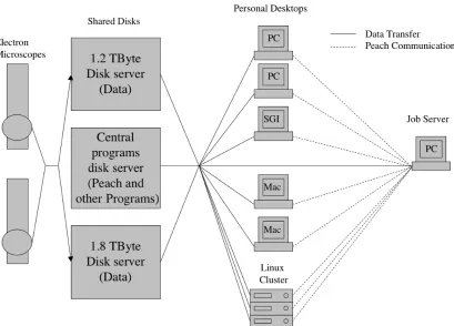
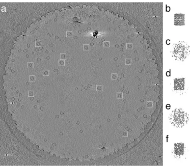
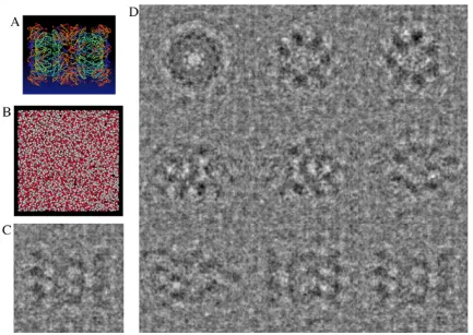
Figure 2-1 Schematic drawing of the setup and information flow in the testing of Peach 56 Figure 2-2 An example cryo-EM image processing project made feasible by Peach
57 Figure 2-3 An example image simulation project managed by Peach 58
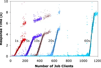
Figure 2-4 Scalability 59
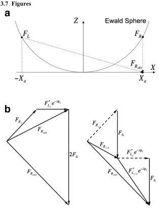
Figure 3-1 The Ewald sphere and Prec algorithm 86
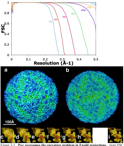
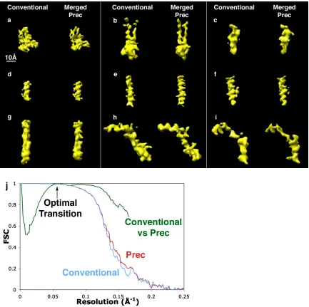
Figure 3-2 Prec overcomes the curvature problem in Ewald projections 87 Figure 3-3 Prec overcomes the curvature problem in multi-slice images and in the
presence of noise 88
Figure 3-4 Application of Prec to experimental images: 3D reconstruction of CPV 89 Figure 3-5 Reconstructions of the 754 Å diameter Reovirus from 300 kV simulated
images 90
Figure A-1 Effect of addition refinement loop 103
Figure A-2 Comparison of Ewald sphere resolution limitations 104 Figure A-3 Effect of number of images on Ewald sphere curvature resolution limit
Table A-1 Table of Euler angle conventions. 106
Table A-2 Table of orientation file formats 106
Chapter 1
Introduction
1.1 Structural Biology
Structural biology is the approach to understanding cell biology through determining the
structures of objects found in the cell. These objects range from proteins and molecular
machines to organelles. To accommodate the difference in scales of these objects, which
span from nanometers to microns, a variety of complementary imaging techniques are
used. The imaging techniques, together, determine the structures of molecular machines
and cellular structures and provide information about their quantity, distribution, and
location. Also, real-time information about processes within cells, sometimes in their
native states, can be extracted.
1.2 Structure Determination Techniques
The main techniques used in structural biology are X-ray crystallography (XRC), nuclear
magnetic resonance spectroscopy (NMR), light microscopy (LM), computational biology
and cryo-electron microscopy (Cryo-EM). These methods work together in a
complementary way to reveal information about a variety of structures in different
physical conditions.
As of June 2008, XRC has produced by far the largest number of atomic models of
proteins as compared to NMR and Cryo-EM according to the Protein Data Bank. XRC
resolution. The difficulty with this technique is that the crystallization process requires
trying numerous conditions of temperature, pH, and buffer concentrations to produce a
crystal that diffracts to sufficiently high resolution. These conditions result in structures
of the proteins in non-native states. Once such crystals can be grown, X-ray diffraction
patterns are then recorded, giving the Fourier amplitudes of the crystal. Next, the phases
need to be determined (“phase problem”) before the structures can be obtained.
NMR also produces atomic resolution structures but is limited to molecular masses of
less than 50 kDa, which includes only the smaller proteins. On rare occasions, larger
protein structures may be determined, for example, an 82-kDa enzyme in 2005
(Tugarinov, Choy et al. 2005).
LM allows for real-time imaging of live cells. Traditionally, this technique was limited
in resolution by the wavelength of light and thus could not reveal the workings of the cell
to higher resolutions. Recently, “super-resolution” techniques have been developed to
surpass the diffraction limit as described in a recent review (Hell 2007) and have reached
sub 100-nm resolutions (Juette, Gould et al. 2008; Schmidt, Wurm et al. 2008).
Computational biology techniques include comparative structure prediction, where
protein structures are predicted using known structures as a reference, and de novo
1.3 Cryo-Electron Microscopy
Cryo-EM delivers structures that span the resolution and size range between the atomic
models provided by XRC or NMR, and the imaging of entire cells by LM. Its advantages
are that samples are easily obtained, and when used in conjunction with plunge freezing
(Dubochet and Mcdowall 1981) using a Vitrobot (Iancu, Tivol et al. 2006), the proteins
or cells can be studied in their near-native state. This is achieved by first having the
sample in a buffer which is spread onto a carbon film. The film is then plunged into
liquid ethane, which cools the sample quickly enough so that the water in the sample is
frozen in vitreous form (Angell 2004). This prevents the crystallization of water, which
would damage the sample. The sample is then inserted into the microscope and imaged
with electrons, which are scattered and then focused by electron lenses to form an image
that is recorded on film or on a digital camera such as a charged-coupled device (CCD) or
CMOS detector. An advantage of cryo-EM over XRC is the recording of images instead
of just amplitudes. However, cryo-EM samples are limited to a thickness of ~ ½ micron
(Lucic, Forster et al. 2005) to prevent multiple scattering of electrons within the cell.
Also, the electron beam causes significant damage to the sample and thus the electron
dose has to be kept low in order to reduce damage. This low dose results in images with
low signal-to-noise ratios (SNRs).
There are several cryo-EM techniques available. Electron crystallography (EC) is used
when 2D crystals of proteins, which are one unit cell thick, can be formed. In such
Similarly, the imaging of helical or tubular crystals also allows for atomic structures to be
determined (Unwin 2005).
Electron cryo-tomography (ECT) is a technique which allows for the study of large
structures and even entire small cells (Henderson and Jensen 2006). ECT can image the
sample to high resolution in its native state, which is not possible with XRC, NMR, and
LM. ECT complements LM because cells can be first observed in vivo with LM and then
plunge-frozen to be imaged by ECT (Briegel, Ding et al. 2008). The ECT technique
images cells from various tilt angles along one or more tilt axes. In theory, this technique
would allow for a full reconstruction of a cell if the tilt angles ranging from -90° to +90°
could be used. In practice, a maximum tilt of about ±65° is used, resulting in an artifact
known as the “missing wedge or pyramid” (Iancu, Wright et al. 2005) in reconstructions
of the cell. This artifact arises due to a wedge or pyramid of missing information in
Fourier space. Another limitation of this technique is that the maximum dose to which
the sample can be exposed has to be shared by all images of the tilt series in order to
prevent information loss due to structural damage by the beam.
Lastly, Single particle analysis (SPA) is a technique in which many identical copies of a
specimen are imaged. The particles in solution are applied to a grid and plunge-frozen.
These grids are imaged resulting ideally in random views of these particles from all
angles, although certain types of particles have preferred orientations. The images
obtained from electron microscopes are noisy due to the low electron dose that can be
to improve the SNR and produce high-resolution reconstructions of particles through
Fourier reconstruction techniques (Crowther, Amos et al. 1970).
1.4 Reconstruction Theory
The reconstruction process can be simplified into three main stages (Figure 1-1). First,
information about the object to be reconstructed is obtained in the form of raw projection
images in various orientations, which are described by Euler angles and determined by
the common-line method (Fuller, Butcher et al. 1996) for particles of high symmetry, or
by 3D projection matching (Penczek, Grassucci et al. 1994). Secondly, corrected images
are produced by the correction of raw images, which removes artifacts that were
introduced during the imaging process due to the point spread function (PSF). This
process is called contrast transfer function (CTF) correction and is performed by taking
the 2D Fourier transform (FT) of a raw image and dividing it by the CTF, which is the FT
of the PSF, before taking the inverse FT to get a corrected image. Thirdly, a 3D
real-space reconstruction of the object is determined by a reconstruction algorithm.
where
!
p(x, y) is a projection of the object along the z-axis,
!
"(x, y,z) is the density of the
object and
!
F(X,Y,Z) is the 3D FT of the object. This derivation can be generalized for
projections in all possible directions and is called the projection theorem.
Using the property above, the 3D FT of the object can be determined by adding many
central slices with different orientations using Whittaker-Shannon interpolation
(Whittaker 1915; Shannon 1949) or by Fourier-Bessel synthesis (Klug, Crick et al. 1958).
Once the 3D FT has been sufficiently sampled, the inverse FT can be calculated to give
the reconstruction of the object.
1.5 Resolution Measures
When discussing resolution, a high resolution (or spatial frequency) corresponds to the
resolvability of features separated by small distances, while a low resolution (or spatial
frequency) corresponds to the resolvability of features separated by large distances;
Atomic resolution refers to the resolvability of the distances between atoms while
near-atomic resolution, which is slightly lower, implies that near-atomic models can be fit with the
help of additional information such as the protein sequence.
In SPA, the quality of a reconstruction is measured in terms of the resolution achieved,
which can be measured numerically or visually. Both these methods are subjective and
The most commonly used numerical resolution measure is the Fourier shell coefficient
(FSC) (Harauz and Van Heel 1986). In order to calculate the FSC, a data set consisting
of a large number of images is split randomly into two halves. Independent
reconstructions of each half of the data set are generated. The two reconstructions are
then compared by calculating the value of the FSC at each spatial frequency
!
i enumerates the set of points found at spatial frequency
!
F2i represent the values of the Fourier coefficients for each
half of the data set and
!
"1i and
!
"2i represent their phases.
A variety of factors (van Heel and Schatz 2005) can affect the value of the FSC
resolution, such as the number of additional voxels in the reconstruction which are in
excess to the object being reconstructed. Changing the size of the volume containing the
reconstruction adjusts the amount of additional voxels. Other factors that affect the
measured resolution include the types of masks and how sharp these masks are, and most
importantly the FSC threshold value, which indicates the maximum resolution of the
The resolution of a reconstruction can be determined visually if the resolution is
sufficiently high. This has recently been possible with high-resolution reconstructions of
icosahedral virus particles at ~ 4 Å resolution (Jiang, Baker et al. 2008; Yu, Jin et al.
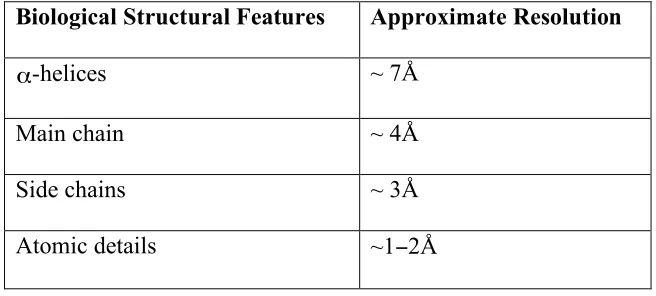
2008; Zhang, Settembre et al. 2008). Table 1-1 gives a list of biological structural
features that can be observed at various resolutions. High-resolution details can be
enhanced to a certain extent by applying an “inverse” B-factor to the reconstructions,
which adjusts the weighting of higher-resolution information by multiplication with the
following factor:
!
eB s2 (3)
where B is the B-factor and s is the spatial frequency.
However, it is important to note the FSC is not affected by the B-factor:
!
1.6 Resolution Limitations
There are two sets of resolution limitations involved in the SPA process. The first set
consists of instrumentation limitations. These include incoherent beam sources,
specimen preservation during the imaging process, and specimen charging by the electron
beam, among others. The second set of resolution limitations consist of processing
limitations, which include orientation, origin, and defocus determination and lack of
computational power. There also exists the depth of field or equivalently the Ewald
sphere curvature problem, which can be solved both computationally and instrumentally.
Further discussion of these resolution limitations can be found in cryo-EM reviews
(Baker, Olson et al. 1999; van Heel, Gowen et al. 2000).
1.7 Instrumentation Progress
Better electron sources and energy filters, more stable cooling stages, and larger, more
sensitive CCD cameras have allowed structure determination by cryo-EM to approach
near-atomic resolution by improving the recording of higher-resolution information with
fewer artifacts and increasing data throughput.
In modern electron microscopes, the electron beam source is a highly coherent field
emission electron gun (FEG). The FEG consists of a pointed field emission tip placed
near a positive electrode. This causes a strong electric field to form which allows
electrons to overcome the work function of the filament (usually tungsten) and be
emitted. FEGs are better than previous electron sources, such as the thermionic W or
because they produce better point electron sources and are colder, which reduces the
thermal energy spread, leading to more monochromatic beams, respectively. The
electron beams are focused with improved electron lenses that have lower spherical
aberrations than previously. Samples are cooled by liquid nitrogen in ECT (Iancu,
Wright et al. 2006) and by liquid helium (Fujiyoshi, Mizusaki et al. 1991) in SPA (van
Heel, Gowen et al. 2000) and EC (Hite, Raunser et al. 2007) to reduce beam damage. In
addition, energy filters are used to ensure that only elastically scattered electrons are
recorded on the CCD. Furthermore, the entire data collection process can be automated
(Potter, Chu et al. 1999).
1.8 Progress in Processing Techniques
Although the fundamentals of the reconstruction process are still the same, there now
exist several popular software packages that are used in the reconstruction of virus
particles by single particle analysis. For example, IMIRS (Liang, Ke et al. 2002) utilizes
the Fourier-Bessel synthesis method and was written for Microsoft Windows XP, while
EMAN (Ludtke, Baldwin et al. 1999), FREALIGN (Grigorieff 2007) and Bsoft
(Heymann 2001) are Cartesian-coordinate, UNIX-based packages which use a variety of
interpolations which are approximations of a full 3D Fourier interpolation (Whittaker
1915; Shannon 1949).
Fundamental improvements to the reconstruction process include CTF correction of
Improvements in computer hardware have also allowed for larger reconstructions to be
computed because of 64-bit memory addressing and faster CPU speeds.
1.9 Approaching Atomic Resolution by Cryo-EM
With these advances, near-atomic resolution of biological structures was first achieved
using EC (Henderson, Baldwin et al. 1990) and then by helical or tubular reconstructions
(Unwin 2005). Thus the next technique by Cryo-EM that will approach these high
resolutions is SPA. The alignment and orientation determination process, which is not
required for EC and helical reconstructions, is non trivial, but using particles with large
masses lessens this obstacle. In addition, high physical symmetry allows for fewer
particles to be used in the reconstruction process. Thus large icosahedral virus particles
are the best candidates for SPA to achieve atomic models.
1.10 Icosahedral Virus Structures
Virus capsids are composed of many identical copies of one or a few different capsid
proteins, and as a result, the genetic material of the virus can be smaller and the
production of a complete virus capsid quicker (Crick and Watson 1956; Caspar and Klug
1962). This use of identical proteins usually results in capsids of helical symmetry, the
best known example being the tobacco mosaic virus (Bloomer, Champness et al. 1978),
or icosahedral symmetry, for example, the herpes simplex virus (Zhou, Dougherty et al.
2000). Icosahedral symmetry is the naturally preferred structure for containing the virus
genome because it provides the largest volume using the fewest capsid units possible.
units. Furthermore, each of these asymmetric units can be composed of a number of
either identical or different subunits. The triangulation (T) number (Caspar and Klug
1962) specifies the number of subunits in each asymmetric unit.
Any image of an icosahedral virus particle can be used 60 times in the reconstruction
process because icosahedral virus particles possess 60-fold symmetry. Alternatively,
only 1/60th of the total information is required to reconstruct a virus particle. The latter
approach is more difficult to achieve in reconstruction algorithms but some progress has
been made towards it with the Fourier-Bessel reconstruction algorithm (Crowther, Amos
et al. 1970) which uses 1/10th of the information by aligning the 5-fold axis along the
z-axis and utilizing 2-fold symmetry which results in information being required only
between the azimuthal angles of 0° and 36° in a cylindrical coordinate system. Likewise,
orientation determination of icosahedral particles is also easier due to the symmetry
which allows for the use of the common-line method (Fuller, Butcher et al. 1996), which
compares intersections of the 60 central slices from each image to derive the correct
orientation.
1.11 Viruses
Virus structures are being intensively researched, as shown by a recent PubMed search
for “virus structure”, which yielded over 37,000 hits. An old review of solved
icosahedral virus structures listed over 175 reconstructions (Baker, Olson et al. 1999),
Viruses consist of genetic material enclosed in capsids, with or without envelopes. A
classification scheme was proposed (Baltimore 1971) which separated viruses into
classes depending on the type of genetic material contained within the capsids. Viruses
infect host cells either by being transported through the cellular membranes, or by
injecting their genetic material, in the form of DNA or RNA, into the cell. If viral DNA
is introduced into the cell, it is transcribed to produce RNA. The viral RNA is
subsequently translated into proteins that form the virus capsid. Despite detailed
understanding, there is still much to learn and exploit, for example, targeted viruses can
be used to cause cancer cells to kill themselves (Ito, Aoki et al. 2006).
Viruses cause a wide range of diseases, such as AIDS (human immunodeficiency virus),
cold sores (herpes virus) and even cancer (papilloma virus) (zur Hausen 2002). Greater
understanding of viruses aids us in our attempts to cure or prevent certain diseases, which
in turn would allow us to improve or save the lives of millions of people. While
reconstructions that achieve a resolution of ~ 3.5 Å allow atomic models to be fit within
the density, higher resolutions of ~ 2 Å allow predictions of the behavior and location of
the interaction surfaces of virus capsids, which in turn guide drug design in producing
drugs that target these surfaces by disrupting the original interaction surface properties,
thereby disrupting assembly of capsids.
In addition, the study of viruses as simplified cellular machines continues to improve our
horizontal gene transfer. These studies have also improved our knowledge of cell
biology.
1.12 Approach Atomic Resolution by Single Particle Analysis
3D reconstructions of virus particles from electron micrographs by Fourier synthesis
were first accomplished in 1970 (Crowther, Amos et al. 1970). Since then,
reconstruction algorithms have improved and matured, resulting in sub-nanometer
resolution in 1997 (Bottcher, Wynne et al. 1997; Conway, Cheng et al. 1997; Trus,
Roden et al. 1997).
According to Glaeser (Glaeser 1999), achieving atomic resolution, which requires the
determination of orientations from 106 images, would require an estimated 1023 floating
point operations, which would take the world’s fastest super computer with a maximum
processing power of 1.375 PFlops (June 2008, www.top500.org) over two years to
complete.. Fortunately, the 60-fold symmetry of icosahedral viruses reduces that number
by nearly two orders of magnitude.
When I first began my thesis work, several factors that limited the resolution of SPA
reconstruction had not been addressed. I attempted to address two of these challenges,
namely the lack of computing power in reconstruction algorithms and the depth of field
The resolutions of SPA reconstructions have improved significantly in the last few years
and towards the end of my thesis work in 2008, three structures reached near-atomic
resolution (Jiang, Baker et al. 2008; Yu, Jin et al. 2008; Zhang, Settembre et al. 2008).
1.13 Computational Complexity of 3D Reconstruction Algorithm
3D reconstructions are highly computationally and memory intensive. Despite the
increasing amounts of memory available, increasing speeds of processors, and the
increase in number of cores and processors per computer, the computation requirements
are still very high when trying to perform reconstructions of very large viruses to high
resolutions.
The basic reconstruction algorithm requires that the 3D FT be held in memory as samples
are applied to it, which results in a
!
O(n3) memory requirement where
!
n is the length of
one side of the transform. Due to the large memory requirements, it is necessary that the
computer performing the reconstruction possess enough RAM to meet this requirement.
Computers lacking the necessary RAM will require swapping of memory, a process that
utilizes the hard disk as additional memory. As hard disk access is several orders of
magnitude slower than RAM access, the resulting computation would not be completed
in a reasonable amount of time. The number and size of images being used in
reconstructions are very large when high-resolution reconstructions are required, due to
the smaller pixel sizes and the higher sampling of images. In practice, for a
be approximately 16, 20, and 30 GB for EMAN (Ludtke, Baldwin et al. 1999), Bsoft
(Heymann 2001), and FREALIGN (Grigorieff 2007), respectively. IMIRS (Liang, Ke et
al. 2002), which is highly optimized, would require less than 2GB. Currently, 64-bit
systems allow for access of sufficient memory for even the largest of virus particles.
Thus, memory requirements are a cost issue, which can be overcome with purchasing of
sufficient RAM.
The computation of the basic reconstruction algorithm consists of applying the value of
each pixel of the 2D FT of the images to the 3D FT making this a
!
O(m n2) computation
problem where
!
m is the number of images and
!
n is the length of one side of the 2D FT
of an image. While the problem is tractable, it does take a significant amount of time for
high-resolution structures of large virus particles, once again, due to the larger images
used in the reconstruction process. While it may seem that purchasing faster computers
can likewise solve the computation problem, it is not a good solution because CPU
speeds have already started to plateau. Fortunately, the computation problem is trivially
parallelizable for the most part and thus parallel and distributed computation are possible
solutions to solve the problem efficiently.
1.14 Parallel Computation
One approach is the parallelization of the reconstruction process, which allows for the
utilization of multiple cores or processors on a single computer or supercomputer that has
shared memory and fast access to this memory. Parallelization takes advantage of the
of increasing the speed of the processors. A program that is multi-threaded will be able
to process multiple calculations simultaneously and would take advantage of these
additional resources. This multi-threaded approach which utilizes shared memory would
require only one copy of the 3D FT to be stored in memory while allowing for the
computation time to be reduced due to the increased number of threads performing
calculations on the various processors or cores without any significant additional memory
requirements.
The most significant drawback to this approach is that when too many threads are
utilized, a bottleneck of the process occurs in the write access to the large memory
holding the 3D FT of the object. The number of memory accesses, due to sample values
being applied to the 3D FT, would be
!
O(m n2) where
!
m is the number of images used in
the reconstruction and
!
n is the length of one side of the image. These memory accesses
are essentially random in their access pattern of the memory and thus require that the
shared memory be locked before changes are made to it to prevent race conditions where
changes are inadvertently lost when multiple threads access the same memory location at
the same time. This bottleneck is encountered when the rate of samples calculated, which
scale linearly with the number of cores, exceeds the rate at which samples are applied to
the 3D FT, which is limited to the RAM access rate that is a constant. At this point,
additional cores cannot accelerate the reconstruction process any further because the
additional threads would spend increasing amounts of time waiting for access to the
Implementations of parallel optimizations to the reconstruction algorithms using
multi-threading libraries, such as the pthreads library, are described in Chapter 3.
1.15 Distributed Computation
The second available approach to reducing computation time is by distributed
computation. This means that individual processes, which are executed on multiple
computers with the necessary memory requirements, can take a subset of the data and
perform independent reconstructions that are later combined to produce the full
reconstruction. It is also possible to execute multiple processes on a single machine with
the requisite number of processors and the required multiples of RAM.
Many structural biology laboratories possess mixtures of heterogeneous workstations
purchased individually or in small sets for laboratory personnel, which constitutes a
wealth of underutilized computation capacity. This is an ideal situation for this using the
distributed approach to solve the computational problem.
This untapped resource was previously unworkable because of the effort required of
researchers to log in to multiple computers and manually distribute jobs across computers
with different operating systems. In addition, custom scripts were needed to submit jobs
one after another through the night or weekend and watch for their completion. Lastly,
computer usage had to be coordinated with laboratory colleagues so as not to impede
most workstations were still only used to a small fraction of their capacity due to the
difficulty of manually managing multiple tasks on multiple workstations.
In 2003, only a few distributed systems were available, including Open PBS from
Veridian Systems, Condor (Tannenbaum and Litzkow 1995), and BOINC, the Berkeley
Open Infrastructure for Network Computing, which mediates the SETI@home project
(Anderson, Cobb et al. 2002). These systems did not meet all our requirements for
processing jobs that had extensive read, write, and memory requirements, were
computationally intensive; had little or no fault tolerance, needed no changes to source
code, and enabled desktop harvesting.
Peach, a distributed computation system, which is described in detail in Chapter 2, was
developed in order to meet those requirements and also be simple to use and administer,
scalable, secure, robust, and as compatible as possible with the existing hardware and
software in structural biology. Essentially, Peach allows for multiple jobs to be
submitted to a heterogeneous cluster of computers and utilizes clock cycles of idle
computers. This distributed approach requires many powerful computers with sufficient
RAM when used in the reconstruction of large virus particles. Furthermore, distributed
computation is also applicable to a wide range of tasks in image processing.
The combination of the information, after the independent reconstructions are completed,
requires
!
O(log(c)n3) steps, where
!
c is the number of separate computers used in the
reconstruction and
!
of the total number of images. This combination by binary merging is significantly
quicker than in the parallel approach because results have already been accumulated by
the individual reconstructions before being combined and can be combined in parallel,
i.e., final reconstruction is left and thus require only
!
log(c) stages of combinations. If there is
availability of computers with sufficient RAM, then distributed computation is a better
solution than the parallel approach because it does not encounter the memory access
bottleneck.
1.16 Hybrid Approach
A hybrid approach, using both parallel and distributed approaches together, would be the
best solution in the reconstruction of large viruses as it utilizes computing resources
maximally by using all available cores on all available computers. This approach is
feasible with new implementations (Chapter 3) of the reconstruction algorithms in Bsoft
(Heymann 2001) and EMAN (Ludtke, Baldwin et al. 1999) that possess capabilities for
both parallel and distributed computation, and which may be used in conjunction with a
suitable distributed computation system such as Peach (Chapter 2) or by processing on
several multi-core nodes of a supercomputer.
1.17 Depth of Field and Ewald Sphere Curvature
As mentioned above, one of the resolution limitations of SPA of large virus particles is
the depth of field problem, or equivalently, the Ewald sphere curvature. The depth of
called the depth of focus, which corresponds to the distance over which the recorded
image is in focus (Fultz and Howe 2002). The depth of field can be geometrically
calculated according to the following formula:
d is the resolution, and α is the aperture angle of the lens.
For a typical transmission electron microscope, the aperture angle α is ~ 10-3 rad and the
resolution
!
d ~ 5 Å giving a depth of field
!
D of ~ 5000 Å or a ½ micron.
The geometric estimate, however, cannot be applied to high-resolution phase contrast
information because small defocus changes
!
"d, on the order of 102 Å, affect the image
intensity distribution (Reimer 1997). This effect is due to the wave aberration
!
Cs is the spherical aberration, λ is the electron wavelength, Δf is the defocus value,
The defocus gradient and Ewald sphere curvature problems were shown to be equivalent
first in 1978 (Amelinckx, Gevers et al. 1978), then in 2000 (DeRosier 2000) and again in
2004 (Wan, Chiu et al. 2004). Further elaboration about their equivalence qualitatively
and quantitatively is provided below.
Firstly to understand the situation qualitatively, consider the Ewald sphere in XRC.
Reciprocal lattice points have dimensions that are inversely proportional to the size of the
crystal. If the crystal thickness in one direction is large, then the dimension of the
reciprocal lattice point in that direction becomes small. Likewise, if we have a thin
crystal, then the dimension of the reciprocal lattice point in that direction becomes very
long and is known as a reciprocal rod or “rel-rod”. The intersections of the Ewald sphere
and reciprocal lattice points are where scattering occurs. Take the situation where
reciprocal lattice points lie along the XY plane. If the incoming beam is along the Z-axis,
then at high resolutions along the plane, there will be reciprocal lattice points which do
not intersect the Ewald sphere. If the crystal is thin, then the rel-rods stretch and intersect
the Ewald sphere. Alternatively, if instead a higher voltage is used, the Ewald sphere
flattens or has a larger radius. In this situation, the reciprocal lattice points also intersect
the Ewald sphere without needing to be rel-rods. Thus, in this situation having a crystal
thin enough will render the Ewald sphere curvature negligible. Conversely, if a crystal is
thick enough, then the Ewald sphere curvature cannot be neglected at high resolution.
The XRC example can be viewed as a simple case of what occurs in electron microscopy
amplitudes vary continuously in all directions, as opposed to discrete reciprocal lattice
points in crystallography, and scattering occurs at all points on the Ewald sphere.
Analogously, the variations in the FT are quicker with thicker EM samples and slower for
thinner EM samples in the direction of the thickness. Thus, when larger virus particles
are imaged, the effect of the Ewald sphere is significant and should not be ignored.
Alternatively, if a small virus particle is imaged, the variations of the FT are slower, so
the Ewald sphere curvature is less significant.
This can also be explained quantitatively. First let us take a point
!
broken down in real space as a set of thin slabs at different defocus values. During the
recording of the image, all the slabs contribute to
(equation 6). The difference in the defocus values,
!
"d, from the center defocus, result in
phase delays of
!
"# $d%s2 with respect to the defocus value at the center of the object. Thus contributions from the top slab of the object will have an additional phase delay of
!
"R#s2, as compared to the slab at the center.
Taking the alternative view, we assume a single defocus for the entire object. Then, the
slabs have contributed to
!
(X0,Y0,Z(X0,Y0)) without additional phase delays as all slabs
are of the same defocus. However, for each slab to have no additional phase delays, the
slabs would have to be located at the center of the object. Since the slabs are physically
phase delay according to the Fourier shift theorem, which states that
due to the curvature of the Ewald sphere. Once again, a phase delay of
!
The phase delays are identical in both cases. This indicates that considering the defocus
gradient over an object is equivalent to taking into account the Ewald sphere curvature
while assuming a single defocus value. Alternatively, if an object possesses an
insignificant defocus gradient, then curvature of the Ewald sphere can be ignored.
1.18 Virus Structures Limited by Ewald Sphere Curvature
Various studies have shown that the Ewald sphere curvature is significant for particles ~
700 Å or greater in diameter, at near-atomic resolution. In 2008, three virus structures of
this diameter were reconstructed to near-atomic resolution of ~ 4 Å (Jiang, Baker et al.
2008; Yu, Jin et al. 2008; Zhang, Settembre et al. 2008). According to Jensen and
Kornberg's envelope function (Jensen and Kornberg 2000), half of the signal in a
conventional reconstruction of such a large virus at 300 kV would be lost due to
curvature of the Ewald sphere by ~ 3.5 Å resolution. Likewise, DeRosier's formula
(DeRosier 2000) predicts that the curvature problem in this same situation would become
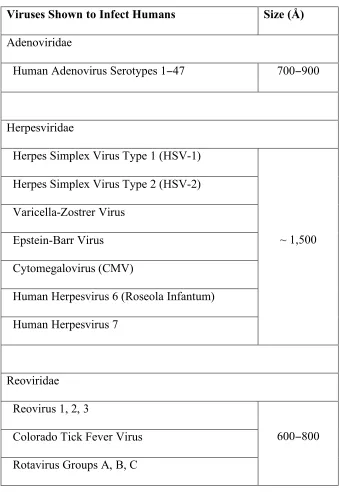
Thus, the Ewald sphere curvature will be most significant for three families of large
icosahedral viruses, namely, the adenoviridae, herpesviridae and reoviridae, as their
diameters are large enough that the curvature of the Ewald sphere will become significant
at near-atomic resolution (Table 1-2). These families are medically important as they are
responsible for a large range of diseases; for example, respiratory tract infections,
conjunctivitis, hemorrhagic cystitis, and gastroenteritis (Adenoviridae), oral and genital
herpes, chickenpox, and shingles (Herpesviridae), and human infantile gastroenteritis
(Reoviridae). For instance, the 1250 Å diameter herpes simplex virus (HSV) (Zhou,
Dougherty et al. 2000), which is currently present in over 60% of the US population, is
responsible for herpes, cowpox, cancer, and many other dangerous diseases.
To overcome the Ewald sphere curvature resolution limit, the paraboloid reconstruction
(Prec) algorithm for Cryo-EM, was developed to correct for the effects of the Ewald
sphere curvature in the context of 3D reconstructions. Details of the algorithm are
discussed in Chapter 3.
1.19 References
Amelinckx, S., R. Gevers, et al. (1978). Diffraction and imaging techniques in material
science. Amsterdam ; New York, Elsevier North-Holland.
Anderson, D. P., J. Cobb, et al. (2002). "SETI@home - An experiment in public-resource
Angell, C. A. (2004). "Amorphous water." Annual Review of Physical Chemistry 55: 559−583.
Baker, T. S., N. H. Olson, et al. (1999). "Adding the third dimension to virus life cycles:
Three-dimensional reconstruction of icosahedral viruses from cryo-electron
micrographs." Microbiology and Molecular Biology Reviews 63(4): 862−922.
Baltimore, D. (1971). "Expression of Animal Virus Genomes." Bacteriological Reviews
35(3): 235−241.
Bloomer, A. C., J. N. Champness, et al. (1978). "Protein Disk of Tobacco Mosaic-Virus
at 2.8-a Resolution Showing Interactions within and between Subunits." Nature
276(5686): 362−368.
Bottcher, B., S. A. Wynne, et al. (1997). "Determination of the fold of the core protein of
hepatitis B virus by electron cryomicroscopy." Nature 386(6620): 88−91.
Bragg, W. L. (1929). "The determination of parameters in crystal structures by means of
fourier series." Proceedings of the Royal Society of London Series A-Containing
Papers of a Mathematical and Physical Character 123(792): 537−559.
Briegel, A., H. J. Ding, et al. (2008). "Location and architecture of the Caulobacter
crescentus chemoreceptor array." Molecular Microbiology 69(1): 30−41.
Caspar, D. L. D. and A. Klug (1962). "Physical Principles in Construction of Regular
Viruses." Cold Spring Harbor Symposia on Quantitative Biology 27: 1−24.
Conway, J. F., N. Cheng, et al. (1997). "Visualization of a 4-helix bundle in the hepatitis
B virus capsid by cryo-electron microscopy." Nature 386(6620): 91−94.
Crick, F. H. C. and J. D. Watson (1956). "Structure of Small Viruses." Nature 177(4506):
Crowther, R. A., L. A. Amos, et al. (1970). "3 Dimensional Reconstructions of Spherical
Viruses by Fourier Synthesis from Electron Micrographs." Nature 226(5244): 421−425.
DeRosier, D. J. (2000). "Correction of high-resolution data for curvature of the Ewald
sphere." Ultramicroscopy 81(2): 83−98.
Dubochet, J. and A. W. Mcdowall (1981). "Vitrification of Pure Water for
Electron-Microscopy." Journal of Microscopy-Oxford 124(DEC): RP3−RP4.
Fujiyoshi, Y., T. Mizusaki, et al. (1991). "Development of a Superfluid-Helium Stage for
High-Resolution Electron-Microscopy." Ultramicroscopy 38(3−4): 241−251.
Fuller, S. D., S. J. Butcher, et al. (1996). "Three-dimensional reconstruction of
icosahedral particles - The uncommon line." Journal of Structural Biology 116(1):
48−55.
Fultz, B. and J. M. Howe (2002). Transmission electron microscopy and diffractometry
of materials. Berlin ; New York, Springer.
Glaeser, R. M. (1999). "Review: Electron crystallography: Present excitement, a nod to
the past, anticipating the future." Journal of Structural Biology 128(1): 3-14.
Grigorieff, N. (2007). "FREALIGN: High-resolution refinement of single particle
structures." Journal of Structural Biology 157(1): 117−125.
Harauz, G. and M. Van Heel (1986). "Exact Filters for General Geometry 3-Dimensional
Reconstruction." Optik 73(4): 146−156.
Henderson, G. P. and G. J. Jensen (2006). "Three-dimensional structure of Mycoplasma
pneumoniae's attachment organelle and a model for its role in gliding motility."
Molecular Microbiology 60(2): 376−385.
Henderson, R., J. M. Baldwin, et al. (1990). "Model for the Structure of
Bacteriorhodopsin Based on High-Resolution Electron Cryomicroscopy." Journal
of Molecular Biology 213(4): 899-929.
Henderson, R., J. M. Baldwin, et al. (1990). "Model for the Structure of
Bacteriorhodopsin Based on High-Resolution Electron Cryomicroscopy." Journal
of Molecular Biology 213(4): 899−929.
Heymann, J. B. (2001). "Bsoft: Image and molecular processing in electron microscopy."
Journal of Structural Biology 133(2−3): 156−169.
Hite, R. K., S. Raunser, et al. (2007). "Revival of electron crystallography." Current
Opinion in Structural Biology 17(4): 389−395.
Iancu, C. V., W. F. Tivol, et al. (2006). "Electron cryotomography sample preparation
using the Vitrobot." Nature Protocols 1(6): 2813−2819.
Iancu, C. V., E. R. Wright, et al. (2005). "A "flip-flop" rotation stage for routine dual-axis
electron cryotomography." Journal of Structural Biology 151(3): 288−297.
Iancu, C. V., E. R. Wright, et al. (2006). "A comparison of liquid nitrogen and liquid
helium as cryogens for electron cryotomography." Journal of Structural Biology
153(3): 231−240.
Ito, H., H. Aoki, et al. (2006). "Autophagic cell death of malignant glioma cells induced
by a conditionally replicating adenovirus." Journal of the National Cancer
Jensen, G. J. and R. D. Kornberg (2000). "Defocus-gradient corrected back-projection."
Ultramicroscopy 84(1−2): 57−64.
Jiang, W., M. L. Baker, et al. (2008). "Backbone structure of the infectious epsilon 15
virus capsid revealed by electron cryomicroscopy." Nature 451(7182): 1130−1134.
Juette, M. F., T. J. Gould, et al. (2008). "Three-dimensional sub-100 nm resolution
fluorescence microscopy of thick samples." Nature Methods 5(6): 527−529.
Klug, A., F. H. C. Crick, et al. (1958). "Diffraction by Helical Structures." Acta
Crystallographica 11(3): 199−213.
Liang, Y. Y., E. Y. Ke, et al. (2002). "IMIRS: a high-resolution 3D reconstruction
package integrated with a relational image database." Journal of Structural
Biology 137(3): 292−304.
Lucic, V., F. Forster, et al. (2005). "Structural studies by electron tomography: From cells
to molecules." Annual Review of Biochemistry 74: 833−865.
Ludtke, S. J., P. R. Baldwin, et al. (1999). "EMAN: Semiautomated software for
high-resolution single-particle reconstructions." Journal of Structural Biology 128(1): 82−97.
Murray, P. R., K. S. Rosenthal, et al. (2005). Medical microbiology. Philadelphia,
Elsevier Mosby.
Penczek, P. A., R. A. Grassucci, et al. (1994). "The Ribosome at Improved Resolution -
New Techniques for Merging and Orientation Refinement in 3d Cryoelectron
Potter, C. S., H. Chu, et al. (1999). "Leginon: a system for fully automated acquisition of
1000 electron micrographs a day." Ultramicroscopy 77(3−4): 153−161.
Reimer, L. (1997). Transmission electron microscopy : physics of image formation and
microanalysis. Berlin ; New York, Springer.
Schmidt, R., C. A. Wurm, et al. (2008). "Spherical nanosized focal spot unravels the
interior of cells." Nature Methods 5(6): 539−544.
Shannon, C. E. (1949). "Communication in the Presence of Noise." Proceedings of the
Institute of Radio Engineers 37(1): 10−21.
Tannenbaum, T. and M. Litzkow (1995). "The Condor Distributed-Processing System."
Dr Dobbs Journal 20(2): 40−48.
Trus, B. L., R. B. S. Roden, et al. (1997). "Novel structural features of bovine
papillomavirus capsid revealed by a three-dimensional reconstruction to 9
angstrom resolution." Nature Structural Biology 4(5): 413−420.
Tugarinov, V., W. Y. Choy, et al. (2005). "Solution NMR-derived global fold of a
monomeric 82-kDa enzyme." Proceedings of the National Academy of Sciences
of the United States of America 102(3): 622−627.
Unwin, N. (2005). "Refined structure of the nicotinic acetylcholine receptor at 4
angstrom resolution." Journal of Molecular Biology 346(4): 967−989.
Unwin, N. (2005). "Refined structure of the nicotinic acetylcholine receptor at 4
angstrom resolution." Journal of Molecular Biology 346(4): 967-989.
van Heel, M., B. Gowen, et al. (2000). "Single-particle electron cryo-microscopy:
van Heel, M. and M. Schatz (2005). "Fourier shell correlation threshold criteria." Journal
of Structural Biology 151(3): 250−262.
Wan, Y., W. Chiu, et al. (2004). "Full contrast transfer function correction in 3D
cryo-EM reconstruction". IEEE Proceedings of ICCCAS 2004 Chengdu, Sichuan,
China.
Whittaker, E. T. (1915). "On the Functions which are Represented by the Expansion of
Interpolation Theory." Proceedings of the Royal Society of Edinburgh 35: 181−194.
Yu, X. K., L. Jin, et al. (2008). "3.88 angstrom structure of cytoplasmic polyhedrosis
virus by cryo-electron microscopy." Nature 453(7193): 415−419.
Zhang, X., E. Settembre, et al. (2008). "Near-atomic resolution using electron
cryomicroscopy and single-particle reconstruction." Proceedings of the National
Academy of Sciences of the United States of America 105(6): 1867−1872.
Zhou, Z. H., M. Dougherty, et al. (2000). "Seeing the herpesvirus capsid at 8.5
angstrom." Science 288(5467): 877−880.
zur Hausen, H. (2002). "Papillomaviruses and cancer: From basic studies to clinical
1.20 Figures and tables
Figure 1-1. Flow chart of simplified reconstruction process. The reconstruction process consist of three stages: (1) Raw images from electron micrographs, (2) Corrected
images produced by CTF correction of Raw images, (3) 3D real-space reconstruction
generated by reconstruction algorithm using corrected images
Biological Structural Features Approximate Resolution
! "
α-helices ~ 7Å
Main chain ~ 4Å
Side chains ~ 3Å
Atomic details ~1−2Å
Table 1-1. Table of biological structural features observable at different resolutions.
Visual resolution of a reconstruction can be determined by the observation of various
Viruses Shown to Infect Humans Size (Å) Adenoviridae
Human Adenovirus Serotypes 1−47 700−900
Herpesviridae
Herpes Simplex Virus Type 1 (HSV-1)
Herpes Simplex Virus Type 2 (HSV-2)
Varicella-Zostrer Virus
Epstein-Barr Virus
Cytomegalovirus (CMV)
Human Herpesvirus 6 (Roseola Infantum)
Human Herpesvirus 7
~ 1,500
Reoviridae
Reovirus 1, 2, 3
Colorado Tick Fever Virus
Rotavirus Groups A, B, C
600−800
Table 1-2. Table of viruses known to infect humans. Viruses known to infect humans
(Murray, Rosenthal et al. 2005) for which the correction of the curvature of the Ewald
Chapter 2
Peach: A simple Perl-based system for distributed computation
and its application to cryo-EM data processing
Peter A. Leong1#, J. Bernard Heymann2#, and Grant J. Jensen3*
1Department of Applied Physics, California Institute of Technology, 1200 E. California
Blvd., Pasadena, California 91125
2Laboratory of Structural Biology Research, National Institute of Arthritis,
Musculoskeletal and Skin Diseases, National Institutes of Health, Bethesda, Maryland
20892
3Division of Biology, California Institute of Technology, 1200 E. California Blvd.,
Pasadena, California 91125
# These authors contributed equally
2.1 Summary
A simple distributed processing system named "Peach" was developed to meet the rising
computational demands of modern structural biology (and other) laboratories without
additional expense by using existing hardware resources more efficiently. A central
server distributes jobs to idle workstations in such a way that each computer is used
maximally, but without disturbing intermittent interactive users. As compared to other
distributed systems, Peach is simple, easy to install, easy to administer, easy to use,
scalable, and robust. While it was designed to queue and distribute large numbers of
small tasks to participating computers, it can also be used to send single jobs
automatically to the fastest currently available computer and/or survey the activity of an
entire laboratory's computers. Tests of robustness and scalability are reported, as are
three specific electron cryomicroscopy applications where Peach enabled projects that
2.2 Introduction
The availability of ever-faster computers continues to open new possibilities throughout
science and in structural biology in particular. This leads us to plan increasingly
demanding projects and gather the computational resources needed. In many structural
biology laboratories, the mixtures of heterogeneous workstations purchased individually
or in small sets for laboratory personnel in recent years constitute a wealth of
underutilized capacity. Here we report the development of a Perl-based package called
"Peach" that efficiently distributes computational tasks across such workstations without
disturbing interactive users.
The motivation for this work arose out of our own structural biological studies in electron
cryomicroscopy (cryo-EM). Modern cryo-EM has three distinct modalities: (1)
"two-dimensional crystallography", in which many crystals of a specimen only a single unit
cell thick are imaged at various tilt angles with respect to the beam; (2) "single particle
analysis," in which thousands of fields of randomly oriented particles are imaged in
projection; and (3) "tomography," in which a single, unique object is imaged iteratively
while being incrementally tilted about some axis. In each case, the resulting images are
merged to produce a three-dimensional reconstruction of the specimen, and the process
involves a large number of small, easily separable, independent calculations (for recent
reviews and some descriptions of the computational challenges in this field, see ((Walz
and Grigorieff 1998; van Heel, Gowen et al. 2000; Fernandez, Lawrence et al. 2002;
Frank 2002; Sali, Glaeser et al. 2003; Frangakis and Forster 2004; Orlova and Saibil
stating that solving the structure of a large protein complex by single particle analysis to
near-atomic resolution with current algorithms would take even a state-of-the-art teraflop
computer something like a year (Glaeser 1999). Even though we are still far away from
this resolution goal for various reasons, in a typical cryo-EM laboratory today, computer
power is already at a premium, and represents a real limitation. Researchers lose time
logging in to multiple computers, manually distributing jobs across computers with
different operating systems, generating custom scripts to submit jobs one after another
through the night or weekend, watching for their completion, and coordinating computer
usage with laboratory colleagues. Despite such efforts, most workstations are still only
used to a small fraction of their capacity due to the difficulty of manually managing
multiple tasks on multiple workstations.
To improve this situation, we searched for an inexpensive system to distribute jobs
efficiently, easily, and securely across our set of workstations. Only a few options were
available, including Open PBS from Veridian Systems and Condor (Tannenbaum and
Litzkow 1995). While we have a running version of Open PBS on our Linux cluster, it
has no "desktop harvesting", or in other words, it was not designed to take advantage of
unused time on interactive workstations, as we desired. We downloaded and installed
Condor, but disliked its complexity, as it required installation of separate executables for
each platform, a large number of different types of daemons, over twenty-five different
programs, and special submission description files. Further, source code was not
available and the documentation warned of known security issues. Another well-known
mediates the SETI@home project (Anderson, Cobb et al. 2002). BOINC was designed
for "public-resource" (as opposed to in-house, or "grid") computing, in which participants
are random individuals who donate time on their personal computers, connected to the
internet via telephone or cable modems or DSL. While wonderful for certain
applications, BOINC would not be attractive for structural biological applications
because of the large amounts of data needing to be transferred, the need for accuracy
(which in public-resource systems is achieved by redundant computing or some kind of
post-verification), and the large amounts of memory often required.
Not finding a suitable alternative, we developed Peach, a simple Perl-based distributed
computation system. The small number of scripts that constitute the system are easy to
install and require no compilation. Peach is easy to use, easy to administer, free, robust,
scalable, secure, and immediately compatible with almost any Unix operating system and
non-interactive executable. We have installed it in two laboratories, where it has
accelerated routine work and brought several structural biology projects to success that
would not otherwise have been feasible without the purchase of an expensive, dedicated
computer cluster.
2.3 Design
2.3.1 Design Philosophy From the user's point of view, the goal was to develop a
system that would accept anywhere from one to thousands of jobs and automatically
process them as fast as possible using the existing workstations in the laboratory, but
more workstations were idle, in which case a new job would be sent immediately to the
fastest one suitable, and (2) when all workstations were busy, in which case submitted
jobs would be queued and distributed later. Further, the system needed to be simple to
use and administer, scalable, secure, robust, and as compatible with the existing hardware
and software in structural biology as possible.
2.3.2 Implementation Peach was implemented following a client-server model, in
which a single job "server" daemon runs on one workstation and maintains a queue of
jobs to be done, while job "client" daemons run on all the other workstations, periodically
reporting their state and running the jobs assigned to them. Three simple "access" clients
constitute the complete user interface: (1) psubmit, which when given any executable file
with flags and options as arguments, submits that job to the system; (2) pview, which
generates reports on the status of the participating computers and submitted jobs; and (3)
pkill, which terminates and/or removes jobs. Only clients initiate communication, so new
clients can join and others terminate without disrupting the server.
2.3.3 Information Flow Work begins when a user submits a job with the psubmit
access client. psubmit writes an "execution script" on a shared disk which contains paths
to an appropriate executable for each participating operating system. Next the psubmit
client sends a message to the job server with the name of the execution script and the
identity of the user, plus optional information about preferred processors and email
addresses for reporting. The job server stores this information in memory and on disk.
status to the job server. When the job server has a job in the queue and a suitable
processor is reporting that it is idle, the server responds to the corresponding client with
the name and path of the execution script. The job client, which is owned by root, forks a
child process whose ownership is changed to the submitting user. Then the child process
runs the execution script with "niced" priority (i.e., a low priority to allow other,
interactive users better access) and writes the standard output and error files. After the
job terminates, the job server sends an e-mail message to the user if requested. If during
execution an interactive user begins to use the console, the job client immediately
suspends active Peach jobs for a configurable time period. If that period will exceed the
time the job had already been processing, the job client "releases" the job back to the job
server for reassignment elsewhere. If a process fails (returns a non-zero value to the
operating system), it is reassigned, but if it fails again, it is removed from the queue and
the owner is notified.
2.3.4 The Job Server The job server acts as a job broker, storing information about all
the submitted jobs and processors participating in the system and matching them
efficiently. The server also writes state and log files about transactions, job completions,
and job failures. In the event the server crashes, it can be easily restarted (or even
automatically restarted if desired) on any workstation, where it will read the state files
and proceed without affecting current or waiting jobs. Clients automatically find the new
IP address and port number of the server in the configuration file on the shared disk.