DECHENE, MICHELLE C. Protein Interactions: the Multiple Solvent Crystal Structures of RNAse A and Analysis of the RalA and RalBP Complex. (Under the direction of Carla Mattos).
In both structure and function, Ribonuclease A (RNAse A) and RalA are two very different
proteins. RNAse A is an extracellular digestive enzyme that catalyzes the breakdown of
3’-5’ phosphodiester linkages in single stranded RNA. RalA is a small monomeric GTPase of
the Ras family and is involved in a number of signaling pathways. While the basic fold of
RalA is similar to the rest of the Ras family, Ral proteins have a distinct effector binding
region and set of effector proteins. RalBP was the first RalA effector identified and it links
RalA to receptor mediated-endocytosis and regulation of mitosis.
RNAse A is a small kidney shaped protein with a well defined active site cleft running
between the two lobes. The active site consists of several pockets, which are responsible for
binding nucleotide bases and phosphate moieties of the RNA substrate. This enzyme is well
studied. With over 40 years of structural information available, it is an excellent model
protein for quantitatively defining the strengths of the Multiple Solvent Crystal Structures
(MSCS) Method. MSCS is an experimental method using small organic solvent molecules to
map the surface of proteins, and in addition to locating binding sites, provides information
about patterns of protein hydration and plasticity. Twenty-two solvent soaked structures
were generated revealing 16 organic solvent molecules and 12 sulfate ions clustered in the
active site, specifically in the two nucleotide-binding pockets, B1 and B2, and in the catalytic
RNAse A. Outside of the active site, many of these water molecules are involved in
stabilizing interactions, or are associated with one of the three helices of RNAse A. In the
active site, nine well-ordered water molecules, which stabilize the active site, bridge the
interaction between the ligand and the active site residues, or are displaced upon ligand
binding, were identified. These patterns of hydration are consistent with earlier analyses of
RNAse A. Finally, RMSD and the hinge angle were used as tools to quantitate the plasticity
observed at each residue and overall domain motions relative to one another, respectively. In
addition to identifying rigid residues of the active site and those exhibiting more motion, it
was found that the trends observed in the MSCS structures correlated well with those
observed in other crystal and NMR structures of RNAse A.
RalA interacts with effector proteins through its two flexible regions, termed switch I and II,
which adopt different conformations in response to the nucleotide binding state. Effector
proteins recognize RalA in the GTP-bound “on” state, and bind through these switch regions.
Where the Ras Binding Domains (RBD) of Ras effectors all adopt a similar fold and interact
with active Ras through an intermolecular β-sheet involving switch I, the recent structures of RalA-effector complex structures of RalA-Sec5 and RalA-Exo84 reveal Ral effector Ral
binding domains differ in structure and in the binding mode with RalA. In a third Ral
effector, RalBP, the Ral-binding domain is predicted to be α-helical, which is different from the β-sandwich structures of Sec5 and Exo84, suggesting the RalA-RalBP interaction presents a previously unobserved binding mode. Furthermore, structural analysis using
behavior. Significant advances have been made towards the crystallizing of the RalA-RalBP
by
Michelle C. Dechene
A dissertation submitted to the Graduate Faculty of North Carolina State University
In partial fulfillment of the Requirements for the degree of
Doctor of Philosophy
Functional Genomics
Raleigh, North Carolina
2008
APPROVED BY:
_______________________________ _______________________________
Dr. Carla Mattos Dr. Maria Celeste Sagui
Committee Chair
_______________________________ _______________________________
Michelle Dechene was raised in the suburbs of Chicago, IL. After developing an early
interest in biology, she pursued this fascination, as well as an interest in Computer Science, at
the University of Dayton, in Dayton, OH. She graduated cum laude with a B.S. in Biology
and Computer Science. Her undergraduate research project involved protein homology
modeling. Michelle then enrolled in the Ph.D. program in Functional Genomics at North
Carolina State University, in Raleigh, NC so she could pursue studies combining her two
majors. While working in the lab of Dr. Carla Mattos, she optimized the constructs and
purification of RalBP used for obtaining crystals of the RalA-RalBP complex and developed
computational tools for Multiple Solvent Crystal Structure analysis. In addition to
coursework and academic research, Michelle also gained professional experience through
two internships. She worked as a software engineering intern developing software for
in-house use at Ciphergen Biosystems and as a medicinal chemistry intern conducting research
Thank you to Dr. Carla Mattos for serving as Michelle’s advisor and providing amazing
opportunities, lab space, guidance, education, ideas, and encouragement.
The National Science Foundation Integrative Graduate Education and Research Traineeship
(NSF-IGERT) Fellowship provided key financial support during her first three years and a
later semester of graduate school. The IGERT Fellowship provided for tuition and fees, a
stipend, and supplemental funds for supplies.
Thank you to Dr. Bob Rose, Dr. Celeste Sagui, and Dr. Alex Tropsha for serving as
Michelle’s graduate advisory committee and contributing helpful suggestions and guidance.
Data were collected at the Southeast Regional Collaborative Access Team (SER-CAT) 22-ID
beamline at the Advanced Photon Source, Argonne National Laboratory. Supporting
institutions may be found at www.ser-cat.org/members.html. Use of the Advanced Photon
Source was supported by the U.S. Department of Energy, Office of Science, Office of Basic
Energy Sciences, under Contract No. W-31-109-Eng-38.
Thanks also to the past and present members of the Mattos Lab, especially, Dr. Greg
Buhrman, Dr. Senthil Kumar, Dr. Nate Nicely, Winnell Newman, and Dr. Paul Swartz. I
have learned so much from you and I appreciate your willingness to answer my questions
Thank you to Dr. Janet Thornton who welcomed me into her group at the European
Bioinformatics Institute for a month. It was this experience and the assistance and
discussions from her and Dr. Roman Laskowski, in particular, that helped start my work on
the computational analysis of the MSCS data for RNAse A.
Finally, thank you to my parents, my family, and my friends. Your support has been
List of Figures ... viii
List of Tables ... x
Chapter 1: Introduction... 1
Protein Interfaces ... 3
Protein Interaction Hot Spots... 4
The Role of Water Molecules in Protein Interactions ... 6
Protein Flexibility Associated with Protein Interactions ... 8
Intrinsically Disordered Proteins Couple Folding and Binding... 10
My Work to Study Protein Interactions ... 12
Ribonuclease A ... 13
RalA and RalBP... 14
The Multiple Solvent Crystal Structures Method ... 17
References ... 19
Chapter 2: Multiple Solvent Crystal Structures of Ribonuclease A: A Critical Assessment of the Method ... 27
Abstract... 27
Introduction... 27
Materials and Methods... 31
Crystal Growth, Cross-linking, and Solvent Soaks ... 31
Data Collection, Processing, Structure Refinement... 33
Water Renumbering... 33
Comparison of MSCS with RNAse A Structures Solved in Aqueous Solution... 36
Computational Analysis... 38
Results... 40
RNAse A MSCS Models ... 43
Analysis of Plasticity Based on Pairwise RMSD Values Between the Models... 45
Conserved Water Binding Sites ... 58
Water Molecules in the Active Site ... 68
Organic Solvent Binding Sites and Comparison with Inhibitors... 72
Solvents Bound in the B1 Subsite... 74
Solvents Bound in the P1 Subsite ... 81
Solvents Bound in the B2 Subsite... 84
Additional Active Site Solvent ... 86
Solvents in Crystal Contacts ... 86
Other Surface Solvents ... 87
Discussion... 89
Protein Plasticity ... 89
Hydration ... 91
References... 97
Chapter 3: Multiple Solvent Crystal Structures of Ribonuclease A: A Comparison of the P3221 and C2 Spacegroups ... 102
Abstract...102
Introduction ...103
Materials and Methods...106
Crystal Growth, Cross-linking, and Solvent Soaks ...106
Data Collection, Processing, Structure Refinement...107
Water Renumbering...107
Comparison of Structures and Preparation of Files ...110
Computational Analysis...111
Results...113
RNAse A Models...113
Protein Plasticity ...114
Molecular Hinge ...122
Conserved Water Binding Sites ...128
Active Site Waters ...129
Waters Outside the Active Site ...136
Water in Crystal Contacts ...138
Water Molecules Not Conserved in the P3221 MSCS Structures ...138
Organic Solvent Binding Sites...140
Solvents Bound in the Active Site ...147
Solvents Bound in Crystal Contacts ...151
Other Surface Solvents ...152
Discussion ...153
Protein Plasticity ...153
Hydration ...157
Binding of Organic Solvents...159
Conclusions...161
Acknowledgements...161
References...162
Chapter 4: The Interaction of RalA and RalBP ...165
Abstract...165
Introduction...166
Materials and Methods...171
Expression of GST-RalBP (all constructs) ...171
Batch Affinity Purification of GST-RalBP(397-518x) and Thrombin Cleavage...172
Original Purification of RalBP(397-518x)...173
Expression and Purification of RalA ...174
DNA Sequencing ...177
Mutagenesis ...178
Prediction of Naturally Disordered Regions...179
Circular Dichroism Spectroscopy...179
Engineering Truncated Forms of the Double Serine Mutant of RalBP ...181
Concentrated Thrombin Cleavage and Purification of RalBP(391-444)...182
Expression and Purification of RalA(11-178) ...184
Crystallization Trials...186
Results...187
Initial Purification of RalBP(397-518x) ...187
RalA-RalBP(397-518x) Complex Formation ...189
RalBP(397-518x) Oligomerizes Through its Two Cysteine Residues ...191
Sequencing and Double Serine Mutant...193
RalBP(397-518x) Folds Upon Binding to RalA...196
RalBP(403-499) Does Not Bind to RalA...202
Six New Truncated Constructs of RalBP Designed ...204
RalBP(397-518) Binds RalA ...206
RalBP(391-444) Binds RalA ...208
Crystal Screening...211
RalA(11-178) Binds RalBP(391-444) and Crystal Screening...213
Discussion...215
Advances Toward the Crystal Structure of the RalA-RalBP Complex ...215
RalA-Effector Interactions...219
RalA Hot Spot Identification Through Structural Analysis...220
Conformational Change at the Ral-Effector Interface ...221
Summary ...223
Acknowledgements...224
References...224
Concluding Remarks...231
Appendices...233
Appendix A: Perl Scripts ...234
Chapter 2: Multiple Solvent Crystal Structures of Ribonuclease A: A Critical
Assessment of the Method ... 27
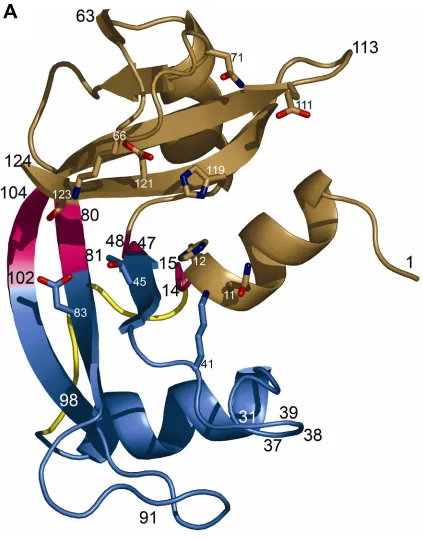
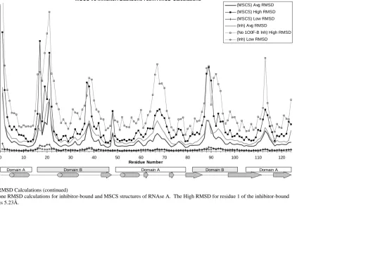
Figure 1: Structure of RNAse A ... 41-42 Figure 2: RMSD calculations: High, Low, and Average RMSD per residue.. 49-54 Figure 3: Solvation of RNAse A... 59
Figure 4: Plasticity and conserved water binding sites of RNAse A... 65
Figure 5: Conserved water molecules in the active site of RNAse A... 70
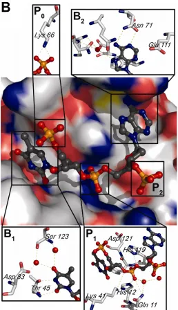
Figure 6: Organic solvent binding sites ... 73
Figure 7: Organic solvent and inhibitor binding in the active site... 80
Chapter 3: Multiple Solvent Crystal Structures of Ribonuclease A: A Comparison of the P3221 and C2 Spacegroups ... 102
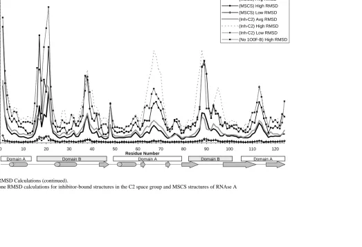
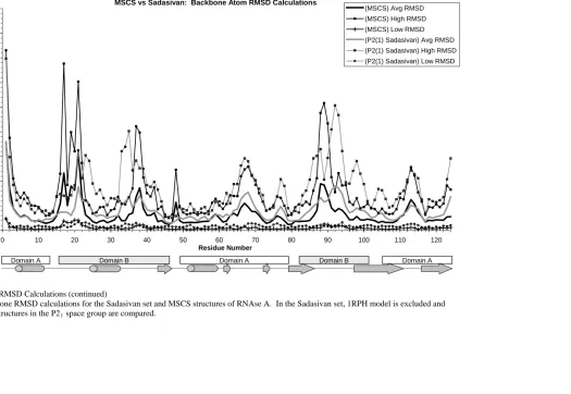
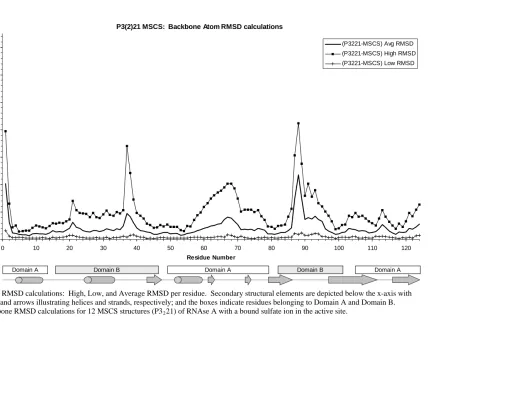
Figure 1: RMSD calculations: High, Low, and Average RMSD per residue.. 116-118 Figure 2: Hinge motions of RNAse A ... 123
Figure 3: RNAse A hinge calculations: Thr 45 N – Phe 120 N distance vs hinge Angle ... 128
Figure 4: Conserved water molecules in the active site... 133
Figure 5: Organic solvent binding sites ... 146
Figure 6: Organic solvent binding sites across all MSCS structures ... 146
Figure 7: Sulfate ion binding in the P1 pocket of the active site ... 148
Figure 8: Organic solvent and inhibitor binding in the active site... 150
Chapter 4: The interaction of RalA and RalBP ... 165
Figure 1: SDS-PAGE gel of concentrated RalBP(397-518x)... 188
Figure 2: SDS-PAGE gel showing the co-elution of RalA and RalBP(397- 518x) in size-exclusion chromatography ... 189
Figure 3: SDS-PAGE gel showing the anomalous behaviour of RalBP397- 518x) during the co-elution of RalA and RalBP(397-518x) in size-exclusion chromatography ... 190
Figure 4: SDS-PAGE gel showing RalBP(397-518x) after the second incubation with glutathione-agarose ... 191
Figure 5: Native and SDS-PAGE gels showing the oligomerization of RalBP(397-518x) ... 192
Figure 6: Translated DNA sequencing results of GST-RalBP(397-518x) ... 195
Figure 7: Translated DNA sequencing results of the GST-RalBP(397-518x) double mutant, GST-RalBP(397-518x;C411S,C451S) ... 195
Figure 8: The double serine mutant, RalBP(397-518x;C411S,C451S), binds RalA ... 197
Figure 9: PONDR predicts RalBP(397-518x) to be mostly disordered... 198 Figure 10: Circular Dichroism shows RalBP(397-518x) folds upon
Figure 12: Sequences of RalBP constructs ... 206
Figure 13: RalBP(397-518) binds RalA ... 207
Figure 14: SDS-PAGE gel of purified RalBP(391-444)... 209
Figure 15: RalBP(391-444) binds RalA ... 210
Figure 16: Select examples of High-Throughput Screening results ... 212
Figure 17: RalBP(391-444) binds RalA(11-178) ... 214
Chapter 2: Multiple Solvent Crystal Structures of Ribonuclease A: A Critical
Assessment of the Method ... 27
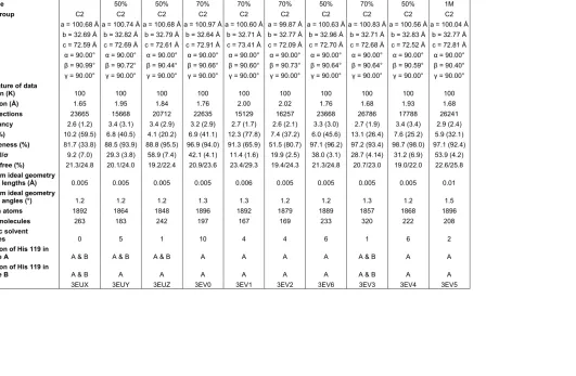
Table 1: Data Collection and Refinement Statistics for Apo-RNAse A... 34
Table 2. Percentage of residues in each domain that fall at or below the baseline of the backbone average RMSD calculations for each set of structures... 47
Table 3: Conserved Water Molecules identified by SEWS for the MSCS structures of RNAse A ... 61-62 Table 4: Water Molecules with less than 80% conservation in the MSCS Structures ... 63
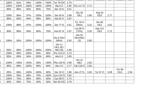
Table 5: Bound Organic Solvents and the interactions made with RNAse A .. 75-77 Chapter 3: Multiple Solvent Crystal Structures of Ribonuclease A: A Comparison of the P3221 and C2 Spacegroups ... 102
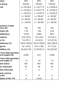
Table 1: Data Collection and Refinement Statistics for RNAse A with a Sulfate Ion in the Active Site ... 108-109 Table 2: Hinge Angle Calculations... 125-127 Table 3: Conserved Water Molecules identified by SEWS for the MSCS structures of RNAse A ... 130-132 Table 4: Water Molecules with less than 80% conservation in the MSCS structures ... 139
Table 5: Bound Organic Solvents and the interactions made with RNAse A .. 141-145 Chapter 4: The Interaction of RalA and RalBP ... 165
Table 1: Parameters used with the Jasco J-600 for Circular Dichroism ... 180
Table 2: Primers for RalBP truncation... 182
Table 3: Nomenclature of expressed and purified GST-RalBP constructs... 183
Proteins have the ability to bind with molecules in the cell, such as other proteins, nucleic
acids, polysaccharides, and additional ligands. It is through these interactions that proteins
perform many of their biological roles; malfunctions in this activity can lead to disease.
Thus, significant effort has been made to understand the fundamental features that facilitate
molecular complexes involving proteins. One approach to determining characteristics of
interaction interfaces has been done through database analysis of the protein complex
structures available in the Protein Data Bank (PDB). With approximately 44,500 crystal
structures compared to about 7,500 NMR structures, database analysis of the PDB is biased
in favor of protein interactions for which there are crystal structures. Additionally, structural
database analysis is biased towards protein interactions for which there are available
structures, which excludes most intrinsically disordered proteins.
In this dissertation, I use the X-ray crystallography based Multiple Solvent Crystals
Structures (MSCS) method to predict the binding sites and analyze the surface of
Ribonuclease A (RNAse A) in order to elucidate the interaction features between this
enzyme and its substrate. As there are 40 years of structural biology studying this protein, the
project is focused on quantitatively defining the strengths of MSCS as a tool for obtaining
binding site properties relevant to aqueous solution using organic solvents as probes. The
latter portion of this dissertation is focused on a less studied protein, RalBP, and its complex
with the GTPase RalA. I use various methods, including liquid chromatography,
RNAse A is a small extracellular enzyme that binds and cleaves RNA in its well-defined
active site cleft. While it is essentially a “lock-and-key” type enzyme, RNAse A exhibits
domain breathing motions in response to ligand binding. RalA is a GTPase belonging to the
Ras family. It is an intracellular signaling protein that interacts with effector proteins
through its flexible switch regions, which adopt different conformations based on its
nucleotide binding state. Presumably, effector molecules bind RalA when an appropriate
switch conformation is present. RalBP is multidomain protein that interacts with a number
of proteins, in addition to being an effector of RalA. Little is known about the structure of
RalBP, aside from being predicted to be highly α-helical, or how RalBP binds to RalA,
except that it is via the predicted minimum Ral-binding domain.
This treatise is composed of five chapters. Chapter 1 includes an overview of the features of
protein interactions which appear later in this dissertation. This includes hot spots, water
molecules, and protein flexibility and plasticity observed in protein interactions, and is
followed by a brief description of intrinsically disordered proteins. Additionally, RNAse A,
the interaction of RalA and RalBP, and the MSCS method are briefly introduced. Chapters 2
and 3 contain the MSCS of RNAse A. Two crystal forms were obtained for this protein and
Chapter 2 presents the results from the crystals grown with symmetry of the C2 space group
containing the apo-enzyme. These structures contain two RNAse A molecules in the
asymmetric unit. Chapter 3 compares the results from the second crystal form with
bound in the active site. Chapter 4 details the work with the complex of RalA and RalBP,
including the discovery that the Ral-binding domain of RalBP is a natively unfolded protein
in solution and folds upon binding to RalA, and the growth of an initial crystal of the protein
complex. Brief concluding remarks follow Chapter 4. The Perl scripts written for the
analysis of MSCS data of RNAse A are included in Appendix A, and references for and
descriptions of the RNAse A structures downloaded from the Protein Data Bank (PDB) are
presented in Appendix B.
Protein Interfaces
As protein interactions are critical to biological function, a substantial amount of work has
gone into studying them. A logical place to start is the protein interface where these
interactions occur, i.e., the regions of proteins designed to interact with other proteins and
substrates. An analysis of available homodimeric, heterodimeric, enzyme-inhibitor, and
antibody-protein complexes was performed to characterize the features of these interfaces
and to determine if there was a predictive element distinguishing the protein interface from
the rest of the surface (Jones and Thornton, 1996). While a number of characteristics were
examined, it was determined that there are no definitive parameters for these characteristics
and no single characteristic differentiates binding sites (Jones and Thornton, 1996; Jones and
Thornton, 1997a). Protein-protein surfaces tend to be somewhat planar and accessible, and
enzymes that bind smaller ligands tend to have a surface cleft that binds partner molecules,
the predictability of interfaces were examined, including electrostatic and shape
complementarity between interacting surfaces, the likelihood of any given residue of being
found at an interface, hydrophobicity, conformational changes upon complex formation,
solvation potential of surface patches, and the protrusion of a residue from the surface of the
protein (Jones and Thornton, 1996; Jones and Thornton, 1997a). Ensuing studies on protein
interaction interfaces have proven the complexity of protein interactions and highlighted
differences observed in different complexes.
This introduction will concentrate on the features of protein interactions that are examined in
this dissertation: protein interaction hot spots, the role of water molecules in protein
interactions, and protein flexibility associated with protein interactions. As much of what is
known about protein interfaces is derived from the available three-dimensional structures, the
topic of intrinsically disordered proteins and binding is presented following the topic of
protein flexibility.
Protein Interaction Hot Spots
Many groups have studied the interface of protein interactions and identified so-called hot
spots, but in each instance this term indicates something slightly different. In alanine
scanning experiments, a hot spot indicates a single residue that can contribute a large portion
of the binding free energy to the interface (Clackson and Wells, 1995). A later
Scott, 2003). In studies probing the surface of proteins with small organic molecules, hot
spots describe areas where the probe molecules cluster (Landon et al., 2007; Mattos et al.,
2006; Sheu et al., 2005). While the approaches and descriptions are different, the overall
idea is the same, hot spots are areas or residues on the surface of the protein, which fall in a
binding site and are important for the specificity of the interaction.
Hot spots confer specificity to the binding site. In a study examining hot spots determined by
alanine scanning, it was found that the hot spot of one protein packs against the hot spot of its
binding partner in a heterodimer interface (Bogan and Thorn, 1998). These hot spots are
enriched in tryptophan, arginine, and tyrosine residues, which are presumably preferred at
hot spots because of their ability to make multiple types of favorable interactions (e.g.
hydrogen bonding, hydrophobic, and aromatic) that can be recognized through counterpart
hot spots (Bogan and Thorn, 1998). Furthermore, it has been shown that conserved polar
residues correlate with interface hot spots (Ma et al., 2003). This is consistent with the
finding that interface residues are generally more highly conserved than other surface
residues (Yao, et al., 2003). However, the conservation of residues at interfaces compared to
the rest of the surface was determined using available structures in the PDB, and it has been
suggested that residue conservation may not be as discriminating as these studies proposed
(Caffrey et al., 2004), and, in some instances, may not be useful at all (Bradford and
numerous methods used to locate them and the functional groups that interact with these
residues. There are several computational methods, including GRID (Goodford, 1985; Wade
and Goodford, 1993), MCSS (Caflisch et al., 1993; Miranker and Karplus, 1991), and
CS-Map (Kortvelyesi et al., 2003; Silberstein et al, 2003), which use small molecular probes to
identify favorable interactions sites. MSCS (a description follows in a later section) is an
experimental crystallographic method that uses small organic solvent molecules to probe the
surface of proteins (Allen et al., 1996; Mattos et al., 2006; Ringe and Mattos, 1999). SAR by
NMR uses small organic molecules or compounds with different functional groups to bind to
subsites of the binding surface and then these molecules are optimized and linked together to
produce high affinity ligands (Hajduk et al, 1997; Shuker et al, 1996). Using small organic
compounds to probe the binding surface and then using them to build larger, more potent
molecules is the same principle behind fragment based lead discovery, which may use NMR
or crystallography (Carr et al, 2005; Hartshorn et al., 2005; Rees et al., 2004).
The Role of Water Molecules in Protein Interactions
Considering that water is the solvent of life and that proteins have evolved in this ubiquitous
solvent, it is not surprising that some water molecules have been specifically incorporated
into protein structure and interactions. Our understanding of these water molecules comes
protein or -ligand binding is entropically favorable because of the release of ordered water
into the bulk solvent. Indeed, it has been found that hot spots tend to be located at the center
of interfaces while being surrounded by “energetically unimportant” residues with the
function of protecting the hot spot residue from the bulk solvent during protein interactions
(Bogan and Thorn, 1998). Furthermore, database analysis revealed that a majority of specific
protein interaction interfaces were “dry” with a ring of water molecules around the interface
(Rodier et al., 2005). Additionally, binding sites have been found to contain certain easily
displaceable bound water molecules, which make specific interactions with the polar groups.
These interactions are reproduced by polar functional groups of the binding partner (Ringe,
1995). It is not surprising that the solvation potential of surface patches and dehydrons, or
defectively packed backbone hydrogen bonds where water exclusion would have a bond
stabilizing effect, have been used as computational predictors of protein interaction interfaces
(Jones and Thornton, 1997a; Jones and Thornton, 1997b; Fernández and Scott, 2003).
While removal of water molecules is important, it is only half of the story, and the tightly
bound ordered water molecules that stick around during complex formation have drawn
considerable interest. It has been proposed that water-mediated contacts in protein-protein
interactions are specific and add another mechanism for recognition (Papoian et al., 2003).
On average, protein-protein interfaces contain 22 water molecules with 11 water-mediated
hydrogen bonds, or about one interface water per 100 Å2 (Janin, 1999; Lo Conte et al., 1999).
in complex formation is not exclusive to protein-protein interactions. In a study analyzing
392 high resolution crystal structures of protein-ligand structures, it was found that over 85%
of these complexes have one or more bridging water molecules present at the interface (Lu et
al. 2007). Also, it is estimated that 15% of all protein-DNA hydrogen bonds are water
mediated (Luscombe et al, 2001). These water-mediated interactions are as important as
direct hydrogen bonds when it comes to stability and specificity (Janin, 1999). Water
molecules add information to the protein interface, and can be considered as part of the
binding site, particularly when they make multiple hydrogen bonds with the protein (Mattos
and Ringe, 2001; Teyra and Pisabarro, 2007). All of this illustrates that water is a key player
in protein interactions, and it is necessary to consider the role water molecules play at
interfaces.
Protein Flexibility Associated with Protein Interactions
When working with protein structures, it is easy to think of them as rigid molecules as
described by the lock-and-key representation of protein interactions. However, proteins are
flexible molecules and can adapt their structure to enhance binding to partner molecules as is
portrayed in the induced fit model (Koshland, 1960). Conformational change upon complex
formation is characteristic of protein interfaces, although there are many variations on this
theme. While some proteins may exhibit no change upon complex formation, side chain
movements, main chain segment movements, and entire domain movements have been
that enhance binding and catalysis, such as optimally orienting the substrate or squeezing out
excess water from the active site. (Hammes, 2002). In a database analysis on cases where
structures of the free and complexed forms of proteins were available, it has been found that
the size of the interaction interface is related to the conformational changes occurring upon
association. “Large” interfaces are those that bury 2000-4660 Å2 upon complex formation.
Binding at these interfaces is observed to include large conformational changes, such as
disorder-to-order transitions, large movements in loops switching to different conformations,
and changes in relative positions of domains in multidomain proteins (Lo Conte et al., 1999).
Binding at interfaces designated as “standard size” (a total buried area of 1600 (±400) Å2)
involves small conformational changes, such as shifts in surface loops, small movements of
short portions of the polypeptide chain, and adjustments in the surface side chains (Lo Conte
et al., 1999). Additionally, another database study examining side chain flexibility of
available apo- and holo- forms of protein structures revealed that only a small number of
interface side chains undergo changes, with about 85% of the cases examined showing
changes in three residues or less (Najmanovich et al., 2000).
Plasticity of interface residues contributes to specificity. While changes in conformation
seem to be important for optimizing the complex, residues that adopt a more fixed
arrangement tend to lend specificity to the interaction. This is illustrated by idea of “anchor”
and “latch” side chains in protein-protein interactions (Rajamani et al., 2004). Anchor side
structurally constrained binding groove of the other protein, an induced fit process occurs
where the flexible latch side chains adjust forming the final high affinity complex. Latch
side chains are found at the periphery of the pocket in conformations allowing for the
clamping that leads to the high affinity complex (Rajamani et al., 2004). This idea is
confirmed by molecular dynamics simulations, which found that even in the absence of a
binding partner, central interface residues tend to be less mobile than other amino acids on
the surface of the protein. Where the side chains at the periphery of the pocket demonstrate
more motion (Smith et al., 2005). A second contribution to specificity through plasticity is
through adjustments in the size of the binding site. For example, the mutants of α-Lytic
protease have a broader specificity than the wild type protein resulting from the increased
plasticity of the active site. This allows for adjustments in the size of the binding pocket,
thus accommodating both large and small substrates (Bone et al., 1989).
Intrinsically Disordered Proteins Couple Folding and Binding
Intrinsically disordered (or natively unfolded or unstructured) proteins have gained interest in
recent years. These proteins are common in eukaryotes and less so in prokaryotes, and it is
thought that possibly 30% of eukaryotic proteins are fully or partially disordered (Dyson and
Wright, 2002; Fink, 2005). The high proportion of these proteins in genomes suggests that
unstructured proteins play an important role in the cell and, in fact, there are numerous
examples with a broad range of functions, including signal transduction, cell cycle control,
Wright, 2005, Uversky, 2002a). With the growing numbers of known disordered proteins
and increasing interest in them, efforts have been made to develop methods to
computationally predict regions of disorder. IUPred predicts disordered regions by
estimating the total pair-wise inter-residue interaction energy from an amino acid sequence
(Dosztányi et al., 2005a; Dosztányi et al, 2005b). This approach is different from PONDR
VL3H and DISOPRED2, which are predictors trained on structurally determined regions of
disorder (Obradovic, 2003; Ward, 2004). These methods have had some success and an
experiment using PONDR showed that compared to eukaryotic proteins in the Swiss-Prot
database and well-ordered protein segments from the PDB, proteins involved in signaling
and those associated with cancer had much higher percentages of sequences predicted to
have long consecutive disordered regions (Iakoucheva et al., 2002).
Disordered proteins lack a stable, well-defined structure and exist in a range of states from
molten globule to random coil (Uversky, 2002b). Often, these proteins fold upon binding to
their biological targets, involving anywhere from a few residues to an entire domain (Wright
and Dyson, 1999; Dyson and Wright, 2002; Dyson and Wright, 2005). Lack of structure and
the disorder to order transition during binding of specific targets are believed to give
disordered proteins the advantages of being able to bind several different targets, and of
having precise control over the thermodynamics of the binding process (Wright and Dyson,
1999; Dyson and Wright, 2002; Uversky, 2002a, Fink, 2005). It is thought that elements of
three-dimensional structures revealed that preformed elements of structure could serve as
initial contact points, followed by folding of the unstructured protein onto the template of the
interface (Fuxreiter et al., 2004). Alternatively, a molecular dynamics study presented that
the folding of the disordered p27 protein was not caused by its own structural preferences,
but instead by the requirements to form a specific molecular interface. When bound in
complex to cyclin A, p27 adopts a β-hairpin and β-strand conformation, as opposed to its
natural folding preference for an α-helix (Verkhivker et al., 2003). This selection among
conformations adopted by a disordered protein is potentially what allows for their ability to
bind several different targets. As disordered proteins pose a major challenge for
crystallography and NMR studies, these proteins are under-represented in structural
databases, and much remains unknown about how these proteins function and interact.
My Work to Study Protein Interactions
The object of this dissertation is to study protein interactions by examining three different
proteins: Ribonuclease A, RalA, and RalBP. Ribonuclease A binds and catalyzes the
hydrolysis of RNA in a well-formed cleft, and is an example of a protein-ligand interaction.
RalA, on the other hand provides a good system to study protein-protein interactions, as it
binds to numerous effector proteins dependent upon conformational changes in its switch
regions. RalBP is an effector for RalA, and little is known about its three dimensional
Ribonuclease A (RNAse A) has been studied for almost a hundred years, beginning with the
report that the surviving active agent from boiled aqueous pig pancreatic extract could break
down yeast nucleic acid into nucleotides (Jones, 1920). The crystallization of this protein in
1939 began the modern work with RNAse A (Kunitz, 1939). As evidenced by the fact that
RNAse A can survive being boiled, RNAse A is a highly stable protein. This coupled with
its ease of collection, as it is excreted from the pancreas, purification, and crystallization,
resulted in Armour, Inc preparing over a kilogram of highly pure crystalline protein which it
then sold to the biochemical community for a small fee (Richards and Wyckoff, 1971). The
ready availability of RNAse A kicked of a flurry of research on this protein.
RNAse A is an endoribonuclease that catalyzes the breakdown of 3’5’-phosphodiester
linkages in single stranded RNA at the 3’ side of pyrimidine nucleotides. Cleavage of RNA
occurs in the two subsequent transphosphorylation and hydrolysis reactions where His12 and
His119 are involved in general acid-base catalysis. Lys41 stabilizes the
2’3’-cyclicphosphodiester intermediate. This mechanism has been well studied and has been
reviewed (Raines, 1998).
The structure of Bovine Pancreatic Ribonuclease A has been studied for over 40 years.
Fankuchen conducted the first x-ray diffraction study in 1941 (Fankuchen, 1941). This lead
to further crystallographic investigation of RNAse A, and in 1967, it was the fourth protein to
which are involved in disulfide bonds (Smyth et al., 1963). The overall shape of the protein
resembles a kidney with the active site in the cleft between the two lobes. The structure is
dominated by a β-sheet and there are three short α-helices. The two lobes have been shown
to exhibit breathing or hinge motions where the hinge closes slightly upon substrate binding
(Radha Kishan et al, 1995; Sadasivan et al, 1998; Merlino et al, 2002; Vitagliano et al, 2002;
Beach et al, 2005). The active site is divided into several subsites, used to describe substrate
binding. Phosphate moieties bind in subsites P0, P1, and P2, where P1 is the site where
cleavage occurs. Nucleotide bases bind in subsites B1 and B2, where B1 is selects for
pyrimidines (Raines, 1998). Structural studies of RNAse A have continued over the years
and there are over 100 structures of this protein deposited in the PDB, with about half
containing an inhibitor bound in the active site.
RalA and RalBP
Compared to RNAse A, RalA and RalBP are newcomers to the world of protein research.
RalA was identified a little over 20 years ago in a search of a cDNA library to find Ras
family genes (Chardin and Tavitian, 1986). Since then, work has been done to reveal the
structure of this protein and some of the ways it functions in the cell. (For a summary of
roles and binding partners of RalA, see Chapter 4). In many ways RalA is similar to Ras. It
is a small GTPase, which hydrolyzes GTP to GDP and in so doing, an accompanying
conformational change occurs in the so-called switch regions. The mechanism of the
Gly71 form hydrogen bonds with γ-phosphate oxygen atoms. With the release of the γ
-phosphate after GTP hydrolysis, the two switch regions can relax into their GDP
conformation (Vetter and Wittinghofer, 2001). It is through these switch regions that
effectors are bound, recognizing the “on” GTP-bound state.
Ras and RalA are over 50% homologous (Chardin and Tavitian, 1986), and share the same
overall structural fold (Nicely et al., 2004). RalA is 206 amino acids long and the structure is
composed of a six-stranded β-sheet, five α-helices, and ten connecting loops (Nicely et al.,
2004). Residues 40-50 of loop 2 comprise switch I, and residues 70-83 of loop 4 and part of
helix 2 comprise switch II (Nicely et al., 2004). RalA has a unique switch I sequence,
YEPTKAD, and a unique set of effectors. In addition to a unique switch I region, the
structure of RalA revealed a clustering of conserved residues on the surface of the protein,
which identifies two potential sites for protein-protein interaction. These sites are adjacent to
or modified by the switches (Nicely et al., 2004). When compared to the structures of Ras
and Rap, another Ras family member, there are structural differences in these sites,
particularly in shape and charge distribution, suggesting these sites contribute to the
specificity of binding between each of these GTPases and their respective binding partners
(Nicely et al., 2004). The first structure of RalA-effector complex was that of RalA-Sec5
(Fukai et al., 2003). Even though the structure of Sec5 is different from the standard
ubiquitin-like fold of the Ras-binding domain, it formed an intermolecular β-sheet through
The Exo84 Ral-binding domain adopts a different fold from those of Ras effectors and Sec5,
and unlike the RalA-Sec5 complex, Exo84 interacts with RalA through both switch regions
(Jin et al., 2005).
Almost 10 years after the discovery of the RalA protein, the first effector of Ral was
identified by three groups concurrently (Cantor et al., 1995; Jullien-Flores et al., 1995; Park
and Weinberg, 1995). Numerous interacting proteins and functions have been identified for
RalBP (for a summary, see Chapter 4), but there is currently no structure available for RalBP.
At 655 amino acids long, RalBP is a large multidomain protein. It was originally identified
as having a central GAP domain, a basic α-helix in the amino-terminal portion, and an acidic
α-helix and coiled-coil in the carboxy-terminal region (Cantor et al., 1995; Jullien-Flores et
al., 1995). The Ral-binding domain was predicted to overlap with the acidic α-helix and part
of the coiled coil, either from residue 391-444 (Park and Weinberg, 1995) or from residue
403-499 (Jullien-Flores et al., 1995). Since then, two ATP-binding sites have been
identified, one in the amino terminal region, and one in the carboxy terminal region (Awasthi
et al., 2001). With the structure of RalBP predicted as being highly helical, particularly in
the Ral-binding domain, the structure of the complex is likely to present a previously
The Multiple Solvent Crystal Structures (MSCS) Method uses small organic molecules to
experimentally probe the surface of a crystalline protein. This method began with the idea
that any organic inhibitor molecule can be dissected into functional groups, which can be
mimicked by small organic molecules (Ringe, 1995). To perform an MSCS experiment, a
protein crystal is crosslinked, if necessary to prevent dissolution, and transferred into a
solution of a high concentration of organic solvent. In order to allow the organic solvent
probes to compete with water, instead of other solvent molecules, only one solvent is used at
a time (Allen et al.,1996). By the end of the process, the solvent has been allowed to fill the
interstices between the protein molecules (Ringe, 1995). However, early studies revealed
that organic solvent probes are not observed covering the surface of the protein (Allen et
al.,1996). Instead, they displace water molecules in a few well-defined sites, which
correlated to the active or binding sites on the protein surface (Ringe and Mattos, 1999).
MSCS of thermolysin in increasing concentrations of isopropanol, from 2% to 100%,
revealed that the binding of organic solvents was additive. While the isopropanol molecules
were found to occupy all four of the main subsites of the active site, 75% of the subsites were
occupied only at high concentrations of solvent (English et al., 1999). These results and
those of the MSCS of thermolysin crystals soaked in acetone, acetonitrile, and phenol were
similar, with only minor changes observed in the conformation of the protein, and probe
molecules clustering in the main specificity pocket of the active site, in positions consistent
Miranker and Karplus, 1991) and GRID (Goodford, 1985; Wade and Goodford, 1993), it was
found that MSCS identified the same interactions, however, far fewer binding sites were
observed experimentally by MSCS (English et al. 2001). At that time, neither GRID nor
MCSS allowed for the flexibility of the protein surface or took into account competing water
molecules (Ringe, 1995). A more recent computational method, CS-Map (Computational
Solvent Mapping of Proteins) takes desolvation into account and when compared to GRID
and MCSS produces similar results, but with far fewer predicted hot spots (Kortvelyesi,
2003). The results of CS-Map were similar to those observed with MSCS and not only was
the consensus site of solvent molecule probes always located in the active site, but the probes
in this location formed hydrogen bonds and non bonded interactions with residues in the
same way a bound ligand would interact (Kortvelyesi et al., 2003; Silberstein et al, 2003).
Elastase was the first protein extensively mapped by MSCS (Allen et al., 1996; Mattos et al.,
2006). Similar to the results with thermolysin, the organic solvent probe molecules cluster in
the pockets of the active site, and their positions correspond with known inhibitors (Mattos et
al., 2006). In addition to probing hot spots, the elastase study revealed areas of plasticity in
the protein, particularly side chains that make subtle adjustments in response to the bound
organic solvent probes (Mattos et al, 2006). Finally, the ensemble of structures allowed for
the nearly complete first hydration shell to be visualized with over 400 unique water binding
sites. When superimposed with an inhibitor, the pattern of crystallographic water molecules
MSCS is a fast and flexible method for mapping the surface of crystalline macromolecules
generating high-resolution structures of 2Å or better (Ringe and Mattos, 1999). The strength
of this method lies in the collective analysis of the ensemble of protein structures that are
produced. In addition to providing information about organic solvent binding sites and
interactions with hot spot residues, the multiple structures provide a more complete picture of
the solvation and plasticity patterns on the surface of the protein than any single crystal
structure could. Unlike the computational methods, MSCS easily allows for the observation
of the effects of protein plasticity and competing water molecules. While this method is
powerful and generates a lot of data, it is limited to proteins with crystals that diffract to high
resolution and that can withstand the transfer into organic solvents.
References
Allen, K.N., Bellamacina, C.R., Ding, X., Jeffery, C.J., Mattos, C., Petsko, G.A., and Ringe, D. (1996). An experimental approach to mapping the binding surfaces of crystalline proteins. Journal of Physical Chemistry 100, 2605-2611.
Awasthi, S., Cheng, J.Z., Singhal, S.S., Pandya, U., Sharma, R., Singh, S.V., Zimniak, P., and Awasthi, Y.C. (2001). Functional reassembly of ATP-dependent xenobiotic transport by the N- and C-terminal domains of RLIP76 and identification of ATP binding sequences. Biochemistry 40, 4159-4168.
Beach, H., Cole, R., Gill, M.L., and Loria J.P. (2005). Conservation of μs-ms enzyme motions in the apo- and substrate-mimicked state. Journal of the American Chemical Society. 127, 9167-9176.
Bradford, J.R. and Westhead, D.R. (2003) Asymmetric mutation rates at enzyme-inhibitor interfaces: implications for the protein-protein docking problem. Protein Science 12, 2099-2103.
Caffrey, D.R., Somaroo, S., Hughes, J.D., Mintseris, J., and Huang, E.S. (2004). Are protein-protein interfaces more conserved in sequence than the rest of the protein-protein surface? Protein Science 13,190-202.
Caflisch, A., Miranker, A., and Karplus, M. (1993). Multiple Copy Simultaneous Search and construction of ligands in binding sites: Application to inhibitors of HIV-1 Aspartic Proteinase. Journal of Medicinal Chemistry 36, 2142-2167.
Carr, R.A.E., Congreve, M., Murray, C.W., Rees, D.C. (2005). Fragment-based lead discovery: leads by design. Drug Discovery Today 10, 987-992.
Cantor, S.B., Urano, T., and Feig, L.A. (1995). Identification and characterization of Ral-binding protein 1, a potential downstream target of Ral GTPases. Mol Cell Biol 15, 4578-4584.
Chardin, P., and Tavitian, A. (1986). The ral gene: a new ras related gene isolated by the use of a synthetic probe. EMBO Journal 5, 2203-2208.
Clackson, T., and Wells, J.A. (1995). A hot spot of binding energy in a hormone-receptor interface. Science 267, 383-386.
Dosztányi, Z., Csizmók, V., Tompa, P., and Simon, I. (2005a). IUPred: web server for the prediction of intrinsically unstructured regions of proteins based on estimated energy content. Bioinformatics 21, 3433-3434.
Dosztányi, Z., Csizmók, V., Tompa, P., and Simon, I. (2005b). The Pairwise Energy Content Estimated from Amino Acid Composition Discriminates between Folded and Intrinsically Unstructured Proteins. Journal of Molecular Biology 347, 827-839.
Dyson, H.J., and Wright, P.E. (2002). Coupling of folding and binding for unstructured proteins. Current Opinion in Structural Biology 12, 54-60.
isopropanol. Proteins: Structure, Function, and Genetics 37, 628-640.
English, A.C., Groom, C.R., and Hubbard, R.E. (2001). Experimental and computational mapping of the binding surface of a crystalline protein. Protein Engineering 14, 47-59.
Fankuchen, I. (1941). An x-ray and crystallographic study of ribonuclease. Journal of General Physiology 24, 315-316.
Fernández, A., and Scott, R. (2003). Dehydron: A Structurally Encoded Signal for Protein Interaction. Biophysical Journal. 85, 1914-1928.
Fink, A.L. (2005). Natively unfolded proteins. Current Opinion in Structural Biology 15, 35-41.
Fukai, S., Matern, H.T., Jagath, J.R., Scheller, R.H., and Brunger, A.T. (2003). Structural basis of the interaction between RalA and Sec5, a subunit of the sec6/8 complex. EMBO J 22, 3267-3278.
Fuxreiter, M., Simon, I., Friedrich, P., and Tompa, P. (2004). Preformed Structural Elements Feature in Partner Recognition by Intrinsically Unstructured Proteins. Journal of Molecular Biology. 338, 1015-1026.
Goodford, P.J. (1985). A computational procedure for determining energetically favorable binding sites on biologically important macromolecules. Journal of Medicinal Chemistry 28, 849-857.
Hajduk, P.J., Sheppard, G., Nettesheim, D.G., Olejniczak, E.T., Shuker, S.B., Meadows, R.P., Steinman, D.H., Carrera, G.M., Jr., Marcotte, P.A., Severin, J., Walter, K., Smith, H., Gubbins, E., Simmer, R., Holzman, T.F., Morgan, D.W., Davidsen, S.K., Summers, J.B., and Fesik, S.W. (1997). Discovery of potent nonpeptide inhibitors of stromelysin using SAR by NMR. Journal of the American Chemical Society 119, 5818-5827.
Hammes, G.G. (2002). Multiple conformational changes in enzyme catalysis. Biochemistry 41, 8221-8228.
Biology 323, 573-584.
Janin, J. (1999). Wet and dry interfaces: the role of solvent in protein and protein-DNA recognition. Structure 7, R277-279.
Jin, R., Junutula, J.R., Matern, H.T., Ervin, K.E., Scheller, R.H., and Brunger, A.T. (2005). Exo84 and Sec5 are competitive regulatory Sec6/8 effectors to the RalA GTPase. EMBO Journal 24, 2064-2074.
Jones, S., and Thornton, J.M. (1996). Principles of protein-protein interactions. Proceedings of the National Academy of Sciences USA 93, 13-20.
Jones, S., and Thornton, J.M. (1997a). Analysis of protein-protein interaction sites using surface patches. Journal of Molecular Biology 272, 121-132.
Jones, S., and Thornton, J.M. (1997b). Prediction of protein-protein interaction sites using patch analysis. Journal of Molecular Biology 272, 133-143.
Jones, W. (1920). The Action of Boiled Pancreas Extract on Yeast Nucleic Acid. American Journal of Physiology 52, 203-207.
Jullien-Flores, V., Dorseuil, O., Romero, F., Letourneur, F., Saragosti, S., Berger, R., Tavitian, A., Gacon, G., and Camonis, J.H. (1995). Bridging Ral GTPase to Rho pathways. RLIP76, a Ral effector with CDC42/Rac GTPase-activating protein activity. J Biol Chem 270, 22473-22477.
Karplus, P.A, and Faerman, C. (1994). Ordered water in macromolecular structure. Current Opinion in Structural Biology 4, 770-776.
Kartha, G., Bello, J., and Harker, D. (1967). Tertiary structure of ribonuclease. Nature 213, 862-865.
Kortvelyesi, T., Dennis, S., Silberstein, M., Brown, L., 3rd, and Vajda, S. (2003). Algorithms for Computational Solvent Mapping of Proteins. Proteins: Structure, Function, and Genetics 51, 340-351.
Koshland, D.E. Jr. (1960). The active site and enzyme action. Advances in enzymology and related subjects of biochemistry 22, 45-97.
Hot Spots within Druggable Binding Regions by Computational Solvent Mapping of Proteins. Journal of Medicinal Chemistry 50, 1231-1240.
Lo Conte, L., Chothia, C., Janin, J. (1999). The atomic structure of protein-protein recognition sites. Journal of Molecular Biology 285, 2177-2198.
Lu, Y., Wang, R., Yang, C.-Y., and Wang, S. (2007). Analysis of Ligand-Bound Water Molecules in High-Resolution Crystal Structures of Protein-Ligand Complexes. Journal of Chemical Information and Modeling 47, 668-675.
Luscombe, N.M., Laskowski, R.A., and Thornton, J.M. (2001). Amino acid-base
interactions: a three-dimensional analysis of protein-DNA interactions at an atomic level. Nucleic Acids Research 29, 2860-2874.
Ma, B., Elkayam, T., Wolfson, H., and Nussinov, R. (2003). Protein-protein interactions: structurally conserved residues distinguish between binding sites and exposed protein surfaces. Proceedings of the National Academy of Sciences USA 100,5772-5777.
Mattos, C., and Ringe, D. (2001). International Tables for Crystallography, edited by M.G. Rossmann and E. Arnold pp. 623-647. Dordrecht: Kluwer Academic Publishers.
Mattos, C., Bellamacina, C.R., Peisach, E., Pereira, A., Vitkup, D., Petsko, G.A., and Ringe, D. (2006). Multiple Solvent Crystal Structures: Probing Binding Sites, Plasticity and Hydration. Journal of Molecular Biology 357, 1471-1482.
Merlino, A., Vitagliano, L., Ceruso, M.A., Di Nola, A., and Mazzarella L. (2002). Global and local motions in ribonuclease A: A molecular dynamics study. Biopolymers 65, 274-283.
Miranker, A., and Karplus, M. (1991). Functionality maps of binding sites: A Multiple Copy Simultaneous Search Method. Proteins: Structure, Function, and Genetics 11, 29-34.
Najmanovich, R., Kuttner, J., Sobolev, V., and Edelman, M. (2000). Side-chain flexibility in proteins upon ligand binding. Proteins: Structure, Function, and Genetics 39, 261-268.
Nicely, N.I., Kosak, J., de Serrano, V., and Mattos, C. (2004). Crystal Structures of Ral-GppNHp and Ral-GDP Reveal Two Binding Sites that Are Also Present in Ras and Rap. Structure 12, 2025-2036.
in Protein-Protein Recognition Landscapes. Journal of the American Chemical Society 125, 9170-9178.
Park, S.H., and Weinberg, R.A. (1995). A putative effector of Ral has homology to Rho/Rac GTPase activating proteins. Oncogene 11, 2349-2355.
Powers, R.A., and Shoichet, B.K. (2002). Structure-based approach for binding site identification on AmpC beta-lactamase. Journal of Medicinal Chemistry 45, 3222-3234.
Radha Kishan, K.V., Chandra, N.R., Sudarsanakumar, C., Suguna, K., and Vijayan, M. (1995). Water-dependent domain motion and flexibility in ribonuclease A and the invariant features in its hydration shell. An x-ray study of two low-humidity crystal forms of the enzyme. Acta Crystallographica Section D. 51, 703-710.
Rajamani, D., Thiel, S., Vajda, S., and Camacho C.J. (2004). Anchor residues in protein-protein interactions. Proceedings of the National Academy of Sciences 101, 11287-11292.
Raines, R.T. (1998). Ribonuclease A. Chemical Reviews 98, 1045-1065.
Rees, D.C., Congreve, M., Murray, C.W., and Carr, R. (2004). Fragment-based lead discovery. Nature Reviews Drug Discovery 3, 660-672.
Richards, F.M., and Wyckoff, H.W. (1971). The Enzymes, edited by P. Boyer pp. 647-806. New York: Academic Press.
Ringe, D. (1995). What makes a binding site a binding site? Current Opinion in Structural Biology 5, 825-829.
Ringe, D., and Mattos, C. (1999). Analysis of the binding surfaces of proteins. Medicinal Research Reviews 19, 321-331.
Rodier, F., Bahadur, R.P., Chakrabarti, P., and Janin, J. (2005). Hydration of protein-protein interfaces. Proteins 60, 36-45.
Sadasivan, C., Nagendra, H.G., and Vijayan, M. (1998). Plasticity, Hydration and
Accessibility in Ribonuclease A. The Structure of a New Crystal Form and its Low-Humidity Variant. Acta Crystallographica D54, 1343-1352.
affinity ligands for proteins: SAR by NMR. Science 274, 1531-1534.
Silberstein, M., Dennis, S., Brown, L., 3rd, Kortvelyesi, T., Clodfelter, K., and Vajda, S. (2003). Identification of Substrate Binding Sites in Enzymes by Computational Solvent Mapping. Journal of Molecular Biology 332, 1095-1113.
Smith, G.R., Sternberg, M.J.E., and Bates, P.A. (2005). The Relationship between the Flexibility of Proteins and their Conformational States on Forming Protein-Protein
Complexes with an Application to Protein-Protein Docking. Journal of Molecular Biology 347, 1077-1101.
Smyth, D.G., Stein, W.H., and Moore, S. (1963). The sequence of amino acid residues in bovine pancreatic ribonuclease: revisions and confirmations. Journal of Biological Chemistry 238, 227-234.
Teyra, J. and Pisabarro, M.T. (2007). Characterization of Interfacial Solvent in Protein Complexes and Contribution of Wet Spots to the Interface Description. Proteins: Structure, Function, and Bioinformatics 67, 1087-1095.
Uversky, V.N. (2002a). What does it mean to be natively unfolded? European Journal of Biochemistry 269, 2-12.
Uversky, V.N. (2002b). Natively unfolded proteins: A point where biology waits for physics. Protein Science 11,739-756.
Verkhivker, G.M., Bouzida, D., Gehlhaar, D.K., Rejto, P.A., Freer, S.T., and Rose, P.W. (2003). Stimulating disorder-order transitions in molecular recognition of unstructured proteins: Where folding meets binding. Proceedings of the National Academy of Sciences 100, 5148-5153.
Vetter, I.R., and Wittinghofer, A. (2001). The guanine nucleotide-binding switch in three dimensions. Science 294, 1299-1304.
Vitagliano, L., Merlino, A., Zagari, A., and Mazzarella, L. (2002). Reversible substrated-induced domain motions in Ribonuclease A. Proteins: Structure, Function, and Genetics. 46, 97-104
functional analysis of native disorder in proteins from the three kingdoms of life. Journal of Molecular Biology 337, 635–645.
Wright, P.E., and Dyson, H.J. (1999). Intrinsically unstructured proteins: re-assessing the protein structure-function paradigm. Journal of Molecular Biology 293, 321-331.
Wyckoff, H.W., Hardman, K.D., Allewell, N.M., Inagami, T., Johnson, L.N., and Richards, F.M. (1967). The structure of ribonuclease-s at 3.5 Å resolution. Journal of Biological Chemistry 242, 3984-3988.
Yao, H., Kristensen, D.M., Mihalek, I., Sowa, M.E., Shaw, C., Kimmel, M., Kavraki, L., and Lichtarge, O. (2003). An accurate, sensitive, and scalable method to identify functional sites in protein structures. Journal of Molecular Biology 326, 255-261.
CHAPTER 2: Multiple Solvent Crystal Structures of Ribonuclease A: A Critical Assessment of the Method
Abstract
The Multiple Solvent Crystal Structures (MSCS) uses organic solvents to map the surfaces of
proteins. In addition to identifying binding sites, this method allows for a more thorough
examination of protein plasticity and hydration than could be achieved by a single crystal
structure. The crystal structures of Bovine Pancreatic Ribonuclease A (RNAse A) soaked in a
variety of solvents were solved (50% dioxane, 50% dimethylformamide, 70%
dimethylsulfoxide, 70% 1,6-hexanediol, 70% 2-propanol, 50% R,S,R-bisfuran alcohol, 70%
t-butanol, 50% trifluoroethanol, or 1.0 M trimethylamine-N-oxide). As RNAse A is a
well-studied protein, it serves as an excellent model for the development of quantitative tools for
MSCS data analysis. The MSCS structures of RNAse A reveal patterns of plasticity and
conserved water binding sites consistent with crystal and NMR structures available in the
Protein Data Bank (PDB). Additionally, comparison of the MSCS structures with
inhibitor-bound structures of RNAse A from the PDB reveals that the organic solvent molecules inhibitor-bound
in the active site pick out key interactions made by bound inhibitor molecules and highlight
ligand binding hot spots in the active site of RNAse A.
Introduction
The Multiple Solvent Crystal Structures (MSCS) method was first published in 1996 as an
experimental method for locating and characterizing protein binding sites for potential use in
al., 1994) and Elastase (Allen et al., 1996) in neat acetonitrile had unexpectedly revealed that
the protein was far from solvated by its organic solvent host, but rather retained a substantial
first hydration shell with only a few molecules of the solvent bound primarily in the active
site. This observation led to the idea that by soaking a protein in several distinct organic
solvents and superimposing the resulting structures, the bound organic solvent molecules
would reveal the shape and chemical complementarity in the active site that could then be
used to develop functional groups in a larger ligand. The idea of linking fragments to produce
more potent ligands has its origins in earlier pioneering work (Jencks, 1981) and was at the
time also being explored by NMR, not with organic solvents, but with small solutes in
aqueous solution (Shuker et al., 1996). The approach of screening large numbers of small
solute molecules has contributed with some degree of success to pharmaceutical research,
combining both X-ray crystallography and NMR spectroscopy in fragment-based lead
discovery and drug design (Rees et al., 2004).
The MSCS method is currently being developed as a powerful tool to experimentally locate
and determine fundamental properties of protein binding sites (Mattos, 2002; Mattos et al.,
2006). It has also played a role in the development of computational solvent mapping
(CS-Map), an in silico counterpart to MSCS used to locate hot spots for protein-ligand
interactions (Dennis et al., 2002; Sheu et al., 2005a; Silberstein et al., 2003; Silberstein et al.,
2006). Results from other computational methods, such as Multiple Copy Simultaneous
Search (MCSS) that aim to place chemical functional groups in protein active sites, have
Karplus, 1991; Stultz and Karplus, 2000). Thus, MSCS has played an important role in the
synergy that exists between experimental and computational strategies that aim to predict and
understand protein-binding sites.
MSCS has been shown to correctly locate the known substrate binding sites for both Elastase
(Ringe and Mattos, 1999) and Thermolysin (English et al., 1999; English et al., 2001).
Locating binding sites in proteins for which structures exist resulting from the various
ongoing genomics projects has become one of the great challenges facing this new era of
structural biology. This problem has preoccupied many structure prediction and protein
bioinformatics groups, resulting in a variety of computational methods for binding site
prediction and characterization, with various degrees of success (Glaser et al., 2006; Jones
and Thornton, 2004; Laurie and Jackson, 2006; Sheu et al., 2005b). MSCS is unique
however, not only because it is an experimental method, but also because in addition to
locating binding sites with the organic solvent probes it provides a picture of hydration and
plasticity on the entire surface of the protein (Mattos et al., 2006; Mattos and Ringe, 2001).
To date, however, the patterns of clustering of organic solvent molecules, plasticity and
hydration have been analyzed only qualitatively by MSCS. In the present article we use the
very well studied protein Bovine Pancreatic Ribonuclease A (RNAse A) in a detailed MSCS
analysis that can be compared with information accumulated over 40 years of structural
biology research on this protein in order to establish more quantitatively the extent to which
physical and chemical properties of protein surfaces are captured by MSCS. What aspects of
MSCS reflect those obtained from the over 100 RNAse A structures in the Protein Data Bank
(PDB) that have been solved in aqueous solution from various crystalline environments and
bound to countless inhibitors? Even more interesting, do the plasticity trends reflect the
qualitative features obtained by solution NMR? Are water molecules conserved in the MSCS
set the same as those observed to be conserved in the large number of structures solved in
aqueous solution? In short, the goal of this study is to provide an assessment of how well the
MSCS set reflects the properties of RNAse A in aqueous solution, delineating the strengths
and limitations inherent in the method. This is an important step in defining how the MSCS
method can be used to understand the details of surfaces in general and binding sites in
particular.
In addition to providing an excellent model for our current work on MSCS, RNAse A has
received significant attention as a model for members of the Ribonuclease family to which it
belongs, many of which have been shown to possess potent physiological activities
(D'Alessio, 1993). Examples of therapeutically important RNAse A homologues include
angiogenin, eosinophil-derived neurotoxin, and eosinophil cationic protein (Leonidas et al.,
2003; Leonidas et al., 2006; Leonidas et al., 1999). These studies add to a wealth of
structural information available in the PDB of RNAse A bound to inhibitors.
RNAse A catalyzes the breakdown of 3’5’-phosphodiester linkages in single stranded RNA
at the 3’ side of pyrimidine nucleotides. It uses an acid-base mechanism in which His 12 and
RNAse A provides an excellent summary for decades of work on this enzyme (Raines,
1998). The plasticity and hydration features obtained by MSCS are compared in the present
work with those published in a set of eight structures which encompasses two different
crystal forms (Sadasivan et al., 1998a), and a partially overlapping set of nine structures
representing four crystal forms (Zegers et al., 1994). None of the crystal forms included in
these two crystallographic studies of RNAse A include the form which we used for the
MSCS work (Vitagliano et al., 2000). In addition the plasticity captured by MSCS is
compared to that observed in the NMR structure consisting of 32 models deposited in the
PDB (Santoro et al., 1993). For the assessment of how well the organic solvent probes reveal
functional group binding sites for RNAse A we use 58 inhibitor-bound structures taken from
the PDB. The published RNAse A structures in aqueous solution provide a diverse and
robust set for assessment and validation of the MSCS results in terms of plasticity, hydration
and identification of hot spots on protein surfaces.
Materials and Methods
Crystal Growth, Cross-linking, and Solvent Soaks
A procedure similar to that described previously (Vitagliano et al., 2000) was used to remove
the sulfate from the RNAse A active site. Dialysis of the protein into buffers of increasing pH
results in deprotonation of the two catalytic His residues, releasing the negatively charged
sulfate molecule. Bovine Pancreatic Ribonuclease A (type XII A) was purchased from Sigma
and dissolved in de-ionized water for a final concentration of 30 mg/mL. Protein was then
repeated with a 0.15M Tris-HCl buffer at pH 8.2, and subsequently with a 0.15 M Tris-HCl
buffer at pH 9.0, each for 24 hours. RNAse A was then transferred to fresh buffer at pH 9.0
and the pH was adjusted to 5.5 over 8 hours by adding small aliquots of 1 M HCl. In the
final step, RNAse A protein was dialyzed over night into a 10mM sodium citrate buffer pH
5.0 and concentrated to 12 mg/mL. As in previous reports (Leonidas et al., 1997; Vitagliano
et al., 2000), RNAse A crystals were grown using hanging drop vapor diffusion at 18 °C with
10 μL drops containing half protein solution and half reservoir solution over a 500 μL
reservoir. The reservoir solution contained 20 mM sodium citrate buffer at pH 5.0 and
PEG4000 levels of 30% w/v, 32% w/v, or 35% w/v. All crystallization drops resulted in
high diffraction quality crystals after 5 months.
RNAse A crystals were manually transferred with a cryo-loop to a 10 μL drop of 0.08%
glutaraldehyde (8.26 % w/v in distilled water, pH 4.25, Electron Microscopy Sciences) in
stabilization buffer (0.15M HEPES, pH 7.5, 32% w/v PEG 4000) over a 300 μL reservoir
(the same glutaraldehyde-stabilization buffer as the drop) and the cross-linking reaction was
allowed to proceed at room temperature for 30 minutes. Cross-linked crystals were then
transferred with a cryo-loop to new drops containing stabilization buffer and an organic
solvent and allowed to soak for 1 to 2 hours at room temperature. Soaked crystals were then
collected, cryo-protected by dunking in stabilization buffer containing 20% glycerol, and