ABSTRACT
RHO, HELEN YOUNG. Regulation of HNF-1α by DCoH and Dyrk 1B: Equilibrium Unfolding Studies of DCoH and Characterization of an Active Dyrk 1B Construct. (Under the direction of Robert Bennett Rose).
HNF-1α is a transcription factor which plays very important roles in the
regulation of insulin synthesis and secretion as well as cellular metabolism. Mutations in this protein result in a familial form of diabetes. Regulation of HNF-1α is mediated through protein-protein interactions and post-translational modifications. Our interest in HNF-1α prompted the study of two of its regulators, dimerization cofactor of HNF-1α (DCoH) and the dual specificity tyrosine regulated kinase 1B (Dyrk 1B). DCoH enhances the transcriptional activity of HNF-1α through protein-protein interactions. Phosphorylation of HNF-1α by Dyrk 1B also enhanced HNF-1α activity.
Regulation of HNF-1α by DCoH provides a unique example of a molecular switch in which the same interface is used to generate two distinct complexes. DCoH is a bifunctional protein whose effects are spatially partitioned in vivo. DCoH interaction with HNF-1α enhances HNF-1α’s transcriptional activity in the nucleus. In the cytoplasm, DCoH functions as a dehydratase which aids in the regeneration of
propensities to interact with HNF-1α. DCoH1 has to co-fold with HNF-1α in order to interact while DCoH2 will interact with HNF-1α upon mixing. We characterized the interface through x-ray crystallography and chemical denaturation. We demonstrate that the DCoH1 homotetramer unfolding is very slow, indicating it is not thermodynamically stable, as previously thought. Rather, it is kinetically trapped. In contrast, DCoH2 unfolding is reversible. In order to study this difference, we introduced a point mutation to the DCoH1 dimer-dimer interface to resemble DCoH2. The DCoH1 mutant unfolded reversibly. The structure revealed an ordered water at the interface as in the DCoH2 structure which enhances the rate of unfolding. Unfolding analysis resembled a two-state model with an indication of a dimer intermediate. This implies that formation of the DCoH1/HNF-1α complex must occur upon folding in the cytoplasm.
occurred in the C-terminal region. The more stable active kinase construct was
Regulation of HNF-1α by DCoH and Dyrk 1B: Equilibrium Unfolding Studies of DCoH and Characterization of an Active Dyrk 1B Construct
by
Helen Young Rho
A dissertation submitted to the Graduate Faculty of North Carolina State University
In partial fulfillment of the Requirements for the Degree of
Doctor of Philosophy
Biochemistry
Raleigh, North Carolina 2010
APPROVED BY:
______________________________ ______________________________
Dr. Robert Rose Dr. Carla Mattos
Chair of Advisory Committee
______________________________ ______________________________
ii
DEDICATION
iii BIOGRAPHY
Helen grew up in the suburbs of New York. Her passion for research unknowingly began in junior high school when she was selected to participate in a summer research program. Her research evolved into a project studying multiple
sclerosis at Maimonedes Medical Center. This student assistantship spanned throughout high school and college. Helen graduated from Columbia University with a Bachelor of Science degree in Biomedical Engineering: Cellular and Tissue Engineering track. She gained additional research experience working at Duke University Medical Center studying rheumatoid arthritis. After taking a biochemistry course, Helen realized that biochemistry was the field that she wanted to pursue her graduate studies in. Under the direction of Dr. Robert Rose, she studied proteins involved in diabetes and cancer. Helen looks forward to contributing to medical research using the knowledge and skills
iv
ACKNOWLEDGEMENTS
I would like to take this opportunity to thank several people without whose support I would not have been able to reach this point in my life. I would like to thank Dr. Robert Rose, my mentor, for the opportunity to work in his lab. You have been a wonderful advisor providing guidance and learning opportunities throughout the years. I enjoyed our stimulating (and often long) scientific discussions which helped fuel my passion for medicine and research. Your subtle “nudges” helped to shape me into the scientist I am today.
I would also like to thank my committee members, Dr. Carla Mattos, Dr. William Miller, and Dr. Jun Ninomiya-Tsuji for serving as my advisory committee. Your
guidance and helpful research advice throughout the years were priceless. I feel very fortunate in that all four of my committee members were not just outstanding scientists but were also wonderful role models as people. Your compassion and guidance through graduate student life and life in general has been invaluable.
My family has always given me their unconditional love, support, and
v
loving, warm, and wonderful people for parents. Mom and Dad, I am so fortunate that you both have always impressed upon me the importance of family, strong moral values, equality among people, education in all forms, hard work, and the pursuit of happiness. For these lessons, I am eternally grateful. Insta-Party: Jean, Richard, and Joan, every minute together has been and still is fun. I admire each of your individual strengths and unique qualities that make each of you so special. I hope we continue to grow together. I’m glad Mom and Dad decided to have four of us.
I was first introduced to biochemistry by Dr. Arrel Toews, one of the best teachers I have ever had. Without Dr. Toews and his biochemistry course, I would never have discovered this passion of mine I am grateful for your friendship, unconditional support, and bright outlook on life. Dr. Misch and Dr. Tu helped me pursue graduate studies in Biochemistry, and I am grateful for your support. LiLi, I was very fortunate that you took me under your wing while we worked together; you taught me so much about research and life.
To all my friends, lab mates (past and present), the undergrads who breathed new life into the lab, and the entire Biochemistry Department: thank you for always being there to share in the excitement. Your kindness, friendship, and hugs made the good times better and softened life’s blows. C, our weekly calls really helped me through the last stretch of graduate school. Who knew that M could be learned that way? JJEGMS when will we have another go at endless popcorn, WoT with the T-ist mask and
vi
soccer, basketball, or life, your honest, gentle encouragement has always helped me push my limits farther to succeed even after high school. Alan, your inspiring words rang out clearly across the track at that meet so many years ago. They helped me achieve victory then, and they still reverberate in me after all these years.
vii
TABLE OF CONTENTS
LIST OF TABLES ... xiii
LIST OF FIGURES ... xiv
CHAPTER 1: Literature Review ... 1
Brief Introduction to Diabetes ... 3
Transcriptional Regulation of the Insulin Gene ... 7
Significance of HNF-1α in Diabetes ... 10
Role of HNF-1α in Tumor Progression ... 14
Transcriptional Coactivators ... 15
Regulation of HNF-1α by DCoH ... 16
DCoH Function ... 17
DCoH Structure ... 20
DCoH interacts with Other Factors ... 22
DCoH Homologues ... 23
Regulation of HNF-1α by Dyrk 1B ... 24
Phosphorylation in Beta Cells ... 24
Kinases ... 25
Kinase Function ... 26
viii
HNF-1α Phosphorylation by Dyrk 1B ... 27
Kinase Structure ... 33
Dyrk 1B ... 35
Identification and Cloning of Dyrk 1B ... 35
Dyrk 1B Domains ... 35
Dyrk Family of Protein Kinases ... 37
Dyrk 1B Substrates and Consensus Sequence ... 40
Dyrk 1B Function in Cell Cycle Regulation ... 41
Dyrk 1B in Differentiation ... 47
Role of Dyrk 1B in Regulation of Apoptosis ... 49
Regulation of Dyrk 1B ... 49
Goals ... 53
REFERENCES ... 55
CHAPTER 2: Kinetic Stability Determines the Interaction Dynamics of the Bifunctional Protein DCoH1, the Dimerization Cofactor of the Transcription Factor Hepatocyte Nuclear Factor-1α ... 82
ABBREVIATIONS ... 83
ABSTRACT ... 84
ix
EXPERIMENTAL PROCEDURES ... 90
DCoH Purification ... 90
Affinity Chromatography Assay ... 91
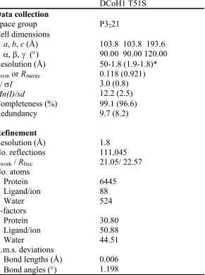
DCoH1 T51S Structure Determination ... 92
Unfolding Studies ... 93
Modeling ... 95
RESULTS ... 97
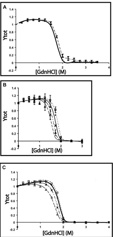
DCoH Unfolding Monitored by Intrinsic Tryptophan Fluorescence ... 97
DCoH2 Unfolding is Reversible ... 97
DCoH1 Unfolding Exhibits Hysteresis ... 106
The DCoH1 Thr 51 Ser Point Mutant Unfolds Reversibly ... 109
DCoH1 T51S Binds to HNF-1α in vitro ... 114
DISCUSSION ... 118
Comparison of DCoH2 and DCoH1 T51S Equilibrium Unfolding... 119
DCoH1 is Kinetically Stable ... 123
Implications for DCoH Regulation of HNF-1 ... 126
Conclusion ... 128
ACKNOWLEDGMENTS ... 131
REFERENCES ... 132
x
Supplemental Materials ... 139
CHAPTER 3: Characterization of an Active Dyrk 1B Construct ... 147
ABSTRACT ... 148
INTRODUCTION ... 149
MATERIALS AND METHODS ... 152
Comparison/Prediction Models Using ERK2 ... 152
Engineering of Active Dyrk 1B Kinase Domain Constructs TDKD1 and TDKD3 for Expression in E.coli ... 152
Expression and Expression Testing of Dyrk 1B Constructs in E.coli ... 155
Protein Expression and Purification ... 156
Expression and Purification of HNF-1α and DCoH-HNF-1α Fusion Protein Substrates ... 157
Kinase Assay ... 158
Mass Spectrometry ... 159
Crystallization ... 161
RESULTS ... 162
Initial Activity Test of TDKD1Using HA-Tak1/Tab1 Kinase Controls ... 163
Mass Spectrometry of TDKD1 ... 167
xi
Co-expression and Purification of TDKD3 ... 171
Activity Test of TDKD3 Reveals Enhanced Phosphorylation of HNF-1α in the Presence of DCoH1 ... 178
Mass Spectrometry of TDKD3 ... 182
Crystallization of DKD3: ... 190
DISCUSSION ... 192
Advances Toward the Purification and Characterization of Dyrk 1B Kinase ... 192
Future Directions ... 192
Regulation of HNF-1a... 193
Substrate Specificity ... 194
Regulation by Upstream Kinases ... 196
Directions for Improving Upon the Crystallization Conditions of Dyrk 1B ... 199
Afterword ... 199
ACKNOWLEDGEMENTS ... 202
REFERENCES ... 203
APPENDIX ... 208
Crystallization Trials: High-Throughput and In-House Crystallization Screening 208 Engineering of Other Dyrk 1B Constructs for Expression in E. Coli ... 216
Engineering of Dyrk 1B Constructs for Expression in Insect Cells ... 218
Expression and Expression Testing of Dyrk 1B Constructs in E.coli ... 219
xii
Purification of Dyrk 1B Constructs ... 220
Western Blotting ... 221
Expression and Purification of 20 Dyrk 1B Constructs from E. coli and Insect Cells ... 222
xiii
LIST OF TABLES
CHAPTER 1: Literature Review ... 1
Table 1. Comparison of the 2 Classes of Diabetes. ... 5
Table 2. Comparison of Type 2 Diabetes and MODY ... 9
CHAPTER 2: Kinetic Stability Determines the Interaction Dynamics of the Bifunctional Protein DCoH1, the Dimerization Cofactor of the Transcription Factor Hepatocyte Nuclear Factor-1α ... 82
Table 1: Equations for the Equilibrium Unfolding of DCoH ... 102
Table 2: Optimized Parameters Obtained from Global Fitting of DCoH2 and DCoH1T51S Fluorescence Data to a Two-State Tetramer Unfolding Model ... 105
Table 3: Buried Surface Area versus m-Values. ... 105
Table 4: Data Collection and Refinement Statistics ... 112
xiv
LIST OF FIGURES
CHAPTER 1: Literature Review ... 1
Figure 1. Model of a pancreatic beta cell and the proteins implicated in MODY ... 8
Figure 2. Schematic representation of HNF-1α. ... 13
Figure 3. DCoH is a metabolic enzyme. ... 19
Figure 4. DCoH1 homotetramer vs DCoH1 in a complex with HNF-1α. ... 21
Figure 5. Dyrk 1B enhances HNF-1α’s activity in a dose dependent manner ... 29
Figure 6. Ribbon diagram representation of a dimer of HNF-1α DNA binding domains bound to DNA. ... 30
Figure 7. Dyrk 1B interactions and effects. ... 31
Figure 8. Schematic representation of Dyrk 1B domains. ... 36
Figure 9. Phylogenetic representation of the Dyrk family kinase domains. ... 39
Figure 10. Sequences for Dyrk 1B substrates. ... 42
xv
CHAPTER 2: Kinetic Stability Determines the Interaction Dynamics of the
Bifunctional Protein DCoH1, the Dimerization Cofactor of the Transcription Factor
Hepatocyte Nuclear Factor-1α ... 82
Figure 1. Ribbon diagram representation of the tetrameric DCoH2 showing the
positions of the tryptophans. ... 98
Figure 2. Relative fluorescence emission spectra of native and denatured DCoH2 and DCoH1 at 2.1 μM protein. ... 99
Figure 3. Equilibrium unfolding of DCoH2 in GdnHCl as measured by intrinsic Trp fluorescence. ... 103
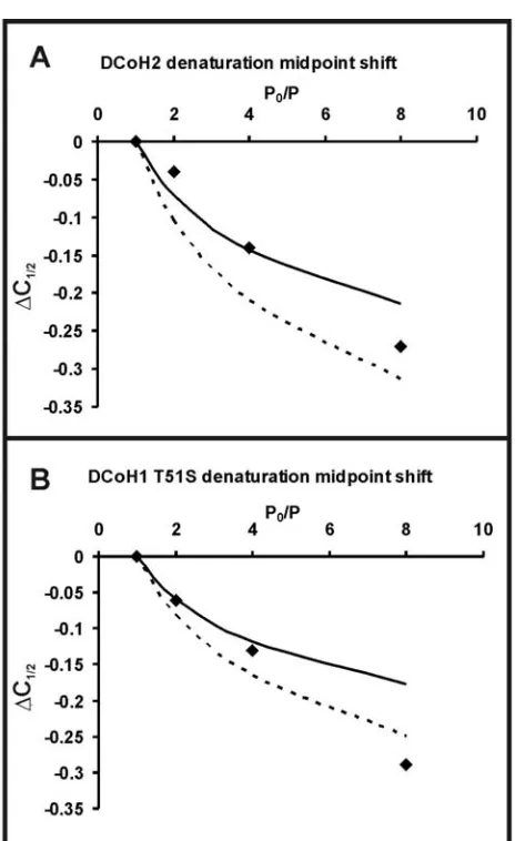
Figure 4. Change in the folding midpoint Δ(C1/2) as a function of the fractional change of protein concentration (Po/P) for DCoH2 and DCoH1 T51S. ... 107
Figure 5. Unfolding of wild type DCoH1 is not reversible. ... 111
Figure 6. An ordered water molecule binds at the tetramer interface of DCoH1 T51S. ... 113
Figure 7. Equilibrium unfolding of DCoH1 T51S in GdnHCl as measured by intrinsic Trp fluorescence. ... 116
Figure 8. The Thr 51 Ser mutation in the DCoH1 homotetramer interacts with HNF-1α in vitro. ... 117
xvi
Figure 10. The tetramer interface of DCoH1 is not tightly packed. ... 125
Figure 11. Diagram representing the free energy of unfolding of DCoH1 T51S and DCoH2 homotetramers. ... 127
Figure 12. Simulated data fit to a 3-state model. ... 145
Figure 13. Simulated data fit to a 2-state model. ... 146
CHAPTER 3: Characterization of an Active Dyrk 1B Construct ... 147
Figure 1. Ribbon diagram of ERK 2 and sequence alignment of ERK2 and Dyrk 1B. ... 164
Figure 2. Schematic representation of the Dyrk 1B constructs, TDKD1 and TDKD3. ... 166
Figure 3. Phosphorylation of MBP and DCoH/HNF-1α 1-280 by TDKD1. ... 168
Figure 4. PEST analysis of Dyrk 1B revealed high potential PEST sequences. ... 172
Figure 5. Purification of TDKD3 by nickel affinity chromatography. ... 174
Figure 6. Purified TDKD3 was proteolyzed by TEVmut produced in house. ... 177
Figure 7. TDKD3 phosphorylates MBP and DCoH/HNF-1α 1-280. ... 179
Figure 8. DCoH enhances phosphorylation of HNF-1α by TDKD3 ... 180
Figure 9. TDKD3 phosphorylates DYRKtide but not KEMPtide. ... 183
xvii
Figure 11. Example of Dyrk 1B kinase crystals obtained from crystallization trials set up in house. ... 191
Figure 12. Examples of high-throughput screening results from HWICHSTB. ... 209
Figure 13. A pH versus PEG optimization of a Hauptman-Woodward screen condition ... 213
Figure 14. Example of Dyrk 1B kinase crystals obtained from crystallization trials set up in house. ... 215
Figure 15. Schematic representation of Dyrk 1B constructs. ... 223
1
2
Diabetes is a major health problem. According to the American Diabetes Association, 7.8% of the American population (~23.6 million adults and children) have diabetes. Sadly, almost 25% of them are not even aware that they have this disease. While patients have a great deal of control over the progression of the disease and a wide variety of successful treatments are available, diabetes can still lead to major health problems and contributes to the high cost of health care. Diabetes has both genetic and environmental determinants. In general, the genetic susceptibility to diabetes is not well understood. While my other research projects also included E47 and Siah2, in this dissertation, I discuss
transcriptional regulation in molecular diabetes specifically focusing on protein-protein interactions of an important transcription factor, HNF-1α, and transcriptional regulation by a bifunctional coactivator, DCoH, and a kinase, Dyrk 1B.
DCoH is a bifunctional protein utilizing the same interface for two distinct protein-protein interactions. X-ray crystallography and unfolding studies aided in characterizing the DCoH interface and the regulation of HNF-1α. A stable construct of another bifunctional protein, Dyrk 1B survival kinase, was engineered and successfully crystallized.
3
coactivator function by measuring the stability of the DCoH homotetramer. In Chapter 3, the design, cloning, expression, purification, and crystallization of a stable and active Dyrk 1B construct are described.
Brief Introduction to Diabetes
The pancreas is both an endocrine and exocrine organ. The parenchymal tissue is composed of the endocrine islets of Langerhans, which produce hormones, and the exocrine acinar cells, which secrete digestive enzymes into the gut through ducts. The islets of
Langerhans contain insulin producing beta cells which are scattered throughout the pancreas. The other four cell types found in the pancreatic islets include alpha cells which produce glucagon, delta cells which produce somatostatin, pancreatic polypeptide cells which produce pancreatic polypeptide, and the epsilon cells which produce ghrelin (Bonner-Weir 1991; Wierup, Svensson et al. 2002; Heller, Jenny et al. 2005). Though the islets only account for 1%-2% of the pancreas, their effects are far reaching in regulating glucose metabolism.
4
or ≥ 200 mg/dL 2 hours post glucose challenge (Harris, Donahue et al. 2003; American Diabetes Association 2005). Reduced insulin action sustains elevated glucose levels, leading to blindness, kidney dysfunction, vascular damage, and early mortality. Current treatments include insulin injections for Type I and Type II as well as oral hypoglycemic agents to help improve insulin sensitivity in Type II diabetics.
Type II diabetes is the most common form of diabetes. Ninety to ninety five percent of diagnosed diabetics are Type II. The age of onset is generally greater than 35 years old and obesity is typically present. Patients have a strong genetic predisposition to developing Type II diabetes (Champe and Harvey 1994). T2D can be classified further based on the role of genetic factors into polygenic or monogenic forms (McCarthy 2002; McCarthy and
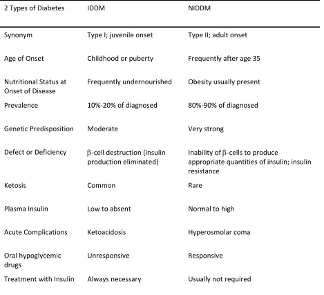
5 Table 1. Comparison of the 2 Classes of Diabetes. Adapted from (Champe and Harvey 1994)
2 Types of Diabetes IDDM NIDDM
Synonym Type I; juvenile onset Type II; adult onset
Age of Onset Childhood or puberty Frequently after age 35
Nutritional Status at
Onset of Disease
Frequently undernourished Obesity usually present
Prevalence 10%‐20% of diagnosed 80%‐90% of diagnosed
Genetic Predisposition Moderate Very strong
Defect or Deficiency β‐cell destruction (insulin
production eliminated)
Inability of β‐cells to produce
appropriate quantities of insulin; insulin
resistance
Ketosis Common Rare
Plasma Insulin Low to absent Normal to high
Acute Complications Ketoacidosis Hyperosmolar coma
Oral hypoglycemic
drugs
Unresponsive Responsive
6
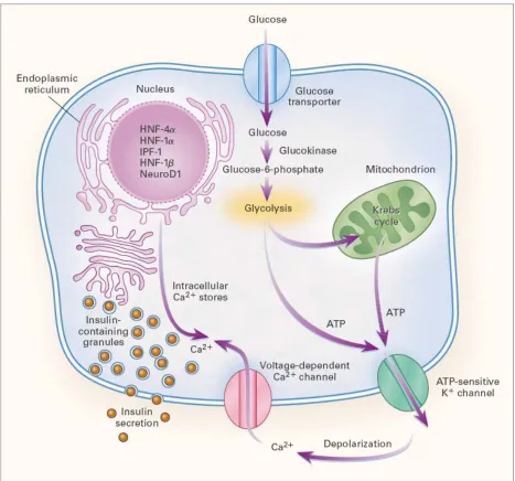
Insulin secretion in response to high blood glucose is a complex process. Glucose enters the beta cell via the GLUT2 transporter in the beta cell (Figure 1) (Fajans, Bell et al. 2001). Glucokinase phosphorylates the sugar to form glucose-6-phosphate which can enter the Krebs cycle or glycolysis. The ATP generated inhibits ATP sensitive potassium ( K+ ) channels leading to depolarization of the membrane and opening of the voltage-gated calcium ( Ca2+ ) channels. An influx of calcium along with mobilization of intracellular calcium permits the insulin-filled secretory granules to fuse with the plasma membrane and release the contents into circulation. The MODY transcription factors regulate insulin gene transcription, enzymes associated with glucose transport and metabolism, and other genes necessary for normal beta cell function.
7
dose to stimulate insulin secretion in beta cells thereby improving glycemic control (Pearson, Liddell et al. 2000).
Transcriptional Regulation of the Insulin Gene
Many efforts have been made to study insulin gene expression in order to learn how to control endogenous insulin levels to control diabetes or to understand how insulin
8
9 Table 2. Comparison of Type 2 Diabetes and MODY (Hattersley 1998).
Type 2 Diabetes MODY
Age of onset Middle/old age Childhood/adolescence/young
adult
Disease-related mortality High Variable
Availability of multiple affected 3 generation families
Very rare Frequent
Role of environment Considerable Frequently obese
Minimal Rarely Obese Pathophysiology Beta-cell dysfunction and
insulin resistance
Beta cell dysfunction
Inheritance Polygenic
10
The 5 MODY transcription factors function within a regulatory network to regulate beta cell function and maintain glucose levels (Shih and Stoffel 2001). HNF-1α is important for facilitating cross-talk between transcription factors within the liver and pancreas as well as between both organs (Kulkarni and Kahn 2004). HNF-1α is involved in insulin
transcription and secretion in pancreatic beta cells. Three HNFs (HNF-1α, HNF-4α, and HNF-6) function cooperatively in the liver and control the expression of many essential genes. Insulin binds to receptors on the liver, thus affecting transcriptional regulation by HNF-1α. The liver is also the primary site of gluconeogenesis. Gluconeogenesis is initiated when low blood glucose levels trigger glucagon release from pancreatic alpha cells. Down regulation of other beta cell transcription factors in HNF-1α knockout mice suggested that HNF-1α was part of a transcription factors network responsible for maintaining beta cell function (Boj, Parrizas et al. 2001; Servitja and Ferrer 2004). HNF-1α can interact with another transcription factor, including HNF-1β, and is part of a feedback loop with HNF-4α (Boj, Parrizas et al. 2001; Ferrer 2002), mutations in which result in another MODY.
Significance of HNF-1α in Diabetes
11
kidney, and pancreatic islet cells (Blumenfeld, Maury et al. 1991; Mendel and Crabtree 1991; Emens, Landers et al. 1992; Tronche and Yaniv 1992; Pontoglio, Barra et al. 1996; Malecki 2005). It also binds to the promoters of developmentally regulated liver specific genes including albumin and α1 antitrypsin (Courtois, Morgan et al. 1987; Lichtsteiner and
Schibler 1989; Odom, Zizlsperger et al. 2004). In pancreatic beta cell, it is expressed late in development and mediates transcription of essential genes such as insulin (Okita, Yang et al. 1999), the GLUT2 transporter (Ban, Yamada et al. 2002), and mitochondrial genes required for insulin secretion (Wollheim 2000; Wobser, Dussmann et al. 2002). Additionally, it was found to recruit coactivators involved in chromatin remodeling to beta cell genes (Parrizas, Maestro et al. 2001; Soutoglou, Viollet et al. 2001).
HNF-1α houses 4 domains (Tronche and Yaniv 1992). At the amino-terminus (amino acids 1-32) is the dimerization domain (Figure 2). HNF-1α dimerization is essential to DNA binding (Nicosia, Monaci et al. 1990). This region is followed by a linker region and then a DNA binding domain (amino acids 100-278) with POU-like and homeodomain-like motifs. The carboxy terminal houses the transactivation domain (amino acids 281-631).
MODY3 is caused by HNF-1α haploinsufficiency (Rose, Bayle et al. 2000; Thomas, Badenberg et al. 2002). This dose dependence in pancreatic beta cells may be spatially regulated as not all organs are affected by the same mutations. Mutations in HNF-1α are the most common cause of MODY but can also cause Type II diabetes as with the I27L
12
beta cell dysfunction (Chiu, Chuang et al. 2000). There are over 200 mutations found to occur within the hnf-1α gene and the promoter region (Ellard and Colclough 2006).
HNF-1α knockout mice are viable, but they exhibit reduced insulin levels in pancreatic beta cells. This suggested that HNF-1α was involved not only with insulin secretion, as was previously known, but also insulin synthesis (Lee, Sauer et al. 1998; Pontoglio, Sreenan et al. 1998). The knockout mice exhibited failure to thrive and died during or just after the weaning period after progressive wasting with pronounced
hepatomegaly (Pontoglio, Barra et al. 1996). MODY3 results in compromised carbohydrate metabolism, e.g. mitochondrial defects and down regulation of GLUT2 expression (Wang, Maechler et al. 1998; Wang, Antinozzi et al. 2000; Shih, Screenan et al. 2001), and
progressive beta cell deficiency. Beta cell deficiency may be caused by diminished beta cell proliferation and increased apoptosis (Shih, Screenan et al. 2001; Wobser, Dussmann et al. 2002).
HNF-1α was also studied to gain better understanding of differentiation. Transcription factors are important during development because they regulate gene
expression in specific tissues and cells. Homeodomain proteins produce an array of effects in the cell, all while sharing surprisingly similar DNA-binding domains (Hansen and
Crabtree 1993). What distinguishes HNF-1α from other homeodomain containing proteins is the dimerization domain at the amino terminus (Nicosia, Monaci et al. 1990) and the
13 Figure 2. Schematic representation of HNF-1α.
14 Role of HNF-1α in Tumor Progression
Misregulation not only causes diabetes but tumor progression as well. These are not MODY3 mutations, so they do not occur via the same misregulation. Biallelic somatic alterations of HNF-1α in hepatocellular adenomas and carcinomas suggested that HNF-1α may function as a tumor suppressor (Bluteau, Jeannot et al. 2002). One study found that nearly 25% of the colorectal tumors tested exhibited HNF-1α mutations (Laurent-Puig, Plomteux et al. 2003). All alterations were located in exon 4 of the poly C tract and 14 of the 15 consisted of a cytosine deletion. The resulting HNF-1α only had a dimerization domain and DNA binding domain. The most common HNF-1α mutation resulting in MODY is 872insC, overexpression of which leads to the production of a dominant negative protein (Yamagata, Oda et al. 1996; Vaxillaire, Pueyo et al. 1999). Because the 872delC mutation in colorectal cancer was monoallelic and there was no frame shift RNA observed as there was for the hepatocellular adenomas, the 872delC mutation was also thought to be dominant negative (Laurent-Puig, Plomteux et al. 2003).
15
1989; Cartwright, Meisler et al. 1990; Talamonti, Roh et al. 1993; Bonham, Ritchie et al. 2000). HNF-1α mutations result in colorectal cancers with microsatellite instability (Laurent-Puig, Plomteux et al. 2003).
Transcriptional Coactivators
One theory proposes that accessory molecules, such as a coactivator like DCoH, can lend specificity to transcription factors (Hansen and Crabtree 1993). Transcriptional co-activators are defined as proteins which can enhance transcriptional activity without binding to DNA (Spiegelman and Heinrich 2004). One of the coactivators that will be discussed in this dissertation is Dimerization Cofactor of HNF-1α (DCoH), which stimulates HNF-1α directed transcription in vivo (Mendel, Khavari et al. 1991). Within the family of
coactivators, proteins can be further subdivided. Primary coactivators are those which can bind directly to transcription factors and also have transcriptionally related enzymatic function. Proteins which bind to transcription factors but recruit other proteins with transcriptionally related enzymatic functions are known as secondary coactivators. These definitions taken together with the fact that DCoH does not have a transcriptionally related enzymatic function but has a saddle shaped groove thought to bind some yet unknown transcriptionally related proteins (Johnen and Kaufman 1997) classifies DCoH as a
16
DCoH is a bifunctional protein. It was originally identified as a contaminant in a rat PAH preparation where it was found to stimulate BH4 dependent phenylalanine
hydroxylation (Kaufman 1970). The enzymatic function was further characterized (Huang, Max et al. 1973; Citron, Davis et al. 1992; Hauer, Rebrin et al. 1993). It is also known as pterin-4a-carbinolamine dehydratase (PCD). The same protein was discovered to co-purify with HNF-1α and to regulate HNF-1α activity (Mendel, Khavari et al. 1991).
Regulation of HNF-1α by DCoH
17
dimerization of HNF-1α, thereby producing non-functional dimers. It is possible that DCoH could also bind to and stabilize these dimers, contributing to MODY3. Regulation of HNF-1α is considered further in the discussion of functional switches and plasticity.
DCoH Function
DCoH is unique in that it is a bifunctional protein. These functions correlate directly with its oligomeric states. In the nucleus, DCoH dimers interact with HNF-1α dimers to enhance HNF-1α directed transcription (Eskinazi, Thony et al. 1999). In the cytoplasm, DCoH homotetramers function as the metabolic enzyme PCD (Figure 3). The PCD activity is involved in regeneration of the biopterin cofactor utilized by NO synthase and aromatic amino acid hydroxylases (Citron, Davis et al. 1992). There is no known relation between these functions. The DCoH- HNF-1 complex exhibits 2:2 stoichiometry; the 2 dimers cannot interact with simple mixing in vitro (Rose, Pullen et al. 2004). Rather, the proteins must be co-folded (Cronk 1996).
The same interface of the DCoH dimer is utilized in the interaction with another DCoH dimer or with HNF-1α. Mutational analysis at the interface or active site revealed that these regions are distinct (Johnen and Kaufman 1997; Sourdive, Transy et al. 1997). The relationship between the 2 physiological functions of DCoH is still not fully
18
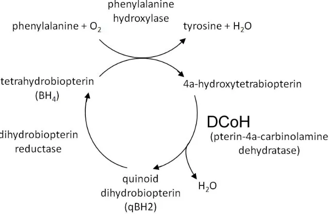
dehydratase within the regeneration of tetrahydrobiopterin, but also as a transcriptional activator of the human PAH gene (Pontoglio, Barra et al. 1996; Lei and Kaufman 1998). In fact, complete inactivation of HNF-1α leads to phenylketonuria (Pontoglio, Barra et al. 1996). Bacterial PAH function was rescued by mammalian DCoH suggesting an
evolutionarily conserved regulatory function for the protein (Endrizzi, Cronk et al. 1995). In the common bacterium Pseudomonas aeruginosa, DCoH (PhhB) and phenylalanine
hydroxylase (PhhA) are situated on the same operon (Song, Xia et al. 1999). DCoH may function through HNF-1α to regulate the expression of a partner in the recycling of aromatic amino acids. Mutations in DCoH lead to hyperphenylalanemia, a skin depigmentation
19 Figure 3. DCoH is a metabolic enzyme.
20 DCoH Structure
The structure of DCoH is often referred to as being saddle shaped, with 1 tetramer housing 2 saddles (Endrizzi, Cronk et al. 1995) (Figure 4A). Each DCoH1 monomer consists of 3 helixes and 4 antiparallel beta strands. The sequence of these secondary structural units is α1-β1-β2-α2-β3-β4-α3. The structure, beginning with helix 1, is composed of residues 10-23. Residues 15-50 form a beta hairpin and strands 1 and 2 (half of the 4 stranded antiparallel beta sheet). Four beta strands from 2 monomers contribute to form a continuous 8 stranded beta sheet. Helix 2 (residues 43-60) is the most important portion of the
Figure 4. DCoH1 homotetramer vs DCoH1 in a complex with HNF-1α.
The ribbon diagrams of the (A) DCoH1 homotetramer and (B) DCoH-HNF-1α complexes are shown above. The figures were generated in PyMol (Endrizzi, Cronk et al. 1995; Rose, Bayle et al. 2000).
22 DCoH interacts with Other Factors
The structure suggests that DCoH may function as a secondary coactivator to bridge HNF-1α to general transcriptional machinery or other coactivators. In both the
homotetrameric structure as well as in the complex with HNFp1, the saddle remains open, i.e. the open surface is accessible to other factors while it is complexed to HNF-1α. The beta strands of each dimer form a continuous 8 stranded beta sheet reminiscent of a molecular saddle as in TATA box binding protein (TBP) (Endrizzi, Cronk et al. 1995; Kim and Burley 1995). However, the TBP saddle is composed of 10 beta strands while the DCoH saddle is made of 8 (Nikolov, Hu et al. 1992; Kim and Burley 1995). Another difference is that the TBP saddle is primarily hydrophobic (Kim, Geiger et al. 1993) and accommodates the minor groove of the DNA (Kim and Burley 1994) while the DCoH saddle exhibits a central
hydrophobic region with polar residues and a perimeter of acidic and basic residues. Currently, with the exception of HNF-1α and β, no other binding partners are known.
In addition to structural comparisons and analyses, there are several studies indicating that DCoH interacts with other factors. DCoH2, a DCoH paralog, interacts with Dyrk 1B which can also phosphorylate HNF-1α (Lim, Jin et al. 2002). A yeast 2 hybrid screen using DCoH as bait with a Xenopus library identified Seven in absentia homologue 2 (Siah-2), an E3 ubiquitin ligase, as prey (Bogdan, Senkel et al. 2001). Our lab could not confirm the interaction with Siah2 in vitro (not shown).
23
lacking in the egg (Pogge v. Strandmann and Ryffel 1995). XDCoH was also found in the epithelium surrounding the embryonic eye where HNF-1α is not expressed. In all cells, XDCoH was found to be predominantly nuclear, indicating that DCoH plays a role in transcriptional regulation in the eye. Because DCoH lacks a nuclear localization signal, it demands a means of transport into the nucleus without the aid of HNF-1α implicating other proteins for this role. Other members of the homeodomain family have been suggested.
DCoH Homologues
DCoH2 (also known as DCoHm) is a homologue of DCoH enriched in muscle tissue. It was first discovered as a prey to the Dyrk 1B bait used in a yeast 2 hybrid screen of a human skeletal muscle library (Lim, Jin et al. 2002). Human DCoH1 and DCoH2 share approximately 60% amino acid identity. DCoH1 and DCoH2 can both form a complex with HNF-1α, but they differ in their availability for HNF-1α binding (Rose, Pullen et al. 2004). Only the DCoH dimers, not tetramers, may associate with HNF-1α, as the same interface is used. In vitro studies have previously shown that the DCoH2 homotetramer is less stable than the DCoH1 homotetramer (Rose, Pullen et al. 2004). This allows for DCoH2 to
dissociate into dimers to interact with HNF-1α in solution. DCoH1 requires co-folding with HNF-1α to form the DCoH1/ HNF-1α complex in vitro. The relative instability of DCoH2 homotetramers may account for the low levels of cytoplasmic DCoH2 activity and
24
(Bayle, Randazzo et al. 2002). The observed glucose intolerance was likely tempered by the compensatory actions of DCoH2. Even though both DCoH1 and DCoH2 have the same enzymatic and transcriptional activities, it was thought that the decreased stability of the DCoH2 homotetramer relative to DCoH1 might provide a mechanism for controlling the activity in the cell (Rose, Bayle et al. 2000). We measured the stability of the DCoH homotetramers in order to understand the difference in ability of DCoH1 and DCoH2 to interact with HNF-1α (Chapter 2).
Regulation of HNF-1α by Dyrk 1B
Phosphorylation in Beta Cells
25
(Khoo, Griffen et al. 2003; Al-Quobaili and Montenarh 2008). These phosphorylations enhance DNA binding and transactivation ability (Petersen, Peshavaria et al. 1998). Upon glucose stimulation, phosphorylation of PDX-1 by the SAPK/p38 pathway was also found to promote nuclear localization of PDX-1 (Macfarlane, McKinnon et al. 1999). ERK2 can also phosphorylate NeuroD1 to modulate its transactivation capacity, heterodimerization with E47, and DNA binding. Phosphorylation of E47 by ERK2 also regulates dimerization with NeuroD1 and DNA binding (Khoo, Griffen et al. 2003).
HNF-1α was also found to be phosphorylated (Lim, Jin et al. 2002). It is post-translationally modified in normal rats (liver and kidneys) as well as in diabetic mice (Barrera-Hernandez, Wanke et al. 1996; Barrera-Hernandez, Wanke et al. 1996). However, the physiological significance of HNF-1α phosphorylation in beta cells is not clear and is the subject of my research. Other HNF family members are also phosphorylated. HNF-4 and HNF-6 are phosphorylated by protein kinase A, PKA, which modulates their transcriptional activity (Viollet, Kahn et al. 1997; Streeper, Hornbuckle et al. 2001).
Kinases
Kinases are important in eukaryotic signaling in growth and development. Improper regulation of kinases can result in cell transformation and cancer. For example, the first oncogene discovered, v-Src, encodes a misregulated tyrosine kinase (Levinson, Oppermann et al. 1978).
26
the basis of their target acceptor amino acid (Hunter 1991). The protein kinase family includes serine/threonine, tyrosine, and histidine kinases (Hunter 1991; Huse and Kuriyan 2002).
Kinase Function
Protein kinases are often referred to as “molecular switches” which can adopt at least 2 disparate conformations, “ON” which is maximally active, and “OFF” which is inactive or minimally active. Phosphorylation of kinases in their activation loops induces
conformational changes that activates the kinase (Huse and Kuriyan 2002). Protein kinases catalyze the transfer of the γ-phosphate of ATP to the hydroxyl of serine, threonine, or tyrosine. Because kinases catalyze the same reaction, it is not surprising that some kinases adopt similar conformations in the active state (Huse and Kuriyan 2002).
Effect of HNF-1α Phosphorylation
27
transfection experiments (Figure 5). These co-transfections showed a synergistic increase in HNF-1α activity. However, Dyrk 1B is able to activate HNF-1α more effectively.
In Caco-2 cells (a colon carcinoma cell line), it was shown that HNF-1α was phosphorylated. However, inhibition of phosphatases 1/2 (known to regulate glucose and glycogen metabolism) increased protein turnover and reduced DNA-binding as measured by western blotting and EMSA, respectively (Carriere, Lacasa et al. 2001). These are partially attributed to decreased protein half-life and mRNA content which were measured by pulse-chase and RT-PCR. The reduced mRNA content was directly correlated with a decrease in DNA-binding by HNF-4, a known HNF-1α regulator. It is possible that phosphorylation can signal ubiquitination. Alternatively, these results suggest that the phosphatase is acting indirectly by affecting HNF-1α’s regulator, HNF-4. These contrasting results indicate that the role of phosphorylation on HNF-1α regulation is a complex process in which certain kinases, such as Dyrk 1B and MKK3, play activating roles while other phosphorylation events, mediated by other kinases, are deactivating. The kinases responsible for these inhibitory post-translational modifications remains to be identified.
HNF-1α Phosphorylation by Dyrk 1B
28
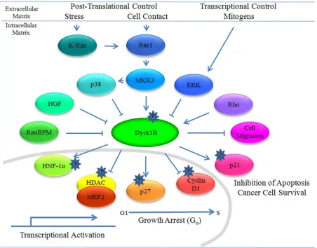
Frantz et al. 2002). This phosphorylation site is also within its cAMP-response element-binding domain which binds CBP. Phosphorylation of HNF-1α occurs when the MKK3 pathway is activated. MKK3 activation is prompted by inflammatory cytokines (e.g. tumor necrosis factor, TNF) and environmental stresses (e.g. osmotic stress, UV radiation) which then activate the MKK3 pathway and subsequently Dyrk 1B (Lim, Jin et al. 2002) (Figure 7).
A Ser247Ala mutation still resulted in phosphorylation of HNF-1α indicating at least one other Dyrk 1B phosphorylation site (Lim, Jin et al. 2002). The Ser247Ala mutant was not tested for transcriptional activation, so the activity of the transfection assays is the sum of all phosphorylations. These other phosphorylation sites are not known.
HNF-1α may be upregulated in response to stress due to its critical physiologic role. HNF-1α binds to more than 222 genes in hepatocytes and 106 genes in the pancreas (Odom, Zizlsperger et al. 2004). These genes included those required for hepatic function
29
Figure 5. Dyrk 1B enhances HNF-1α’s activity in a dose dependent manner
30
Figure 6. Ribbon diagram representation of a dimer of HNF-1α DNA binding domains bound to DNA.
31 Figure 7. Dyrk 1B interactions and effects.
33 Kinase Structure
Structural organization is similar among serine/ threonine and tyrosine kinases. Kinases are bilobate structures. The N-terminal lobe is smaller and often called the N-lobe. It is composed of β sheets and a conserved α helix, called helix αC. The C-terminal lobe, or C-lobe, is larger and consists primarily of helices.
There are 4 prominent features in kinases: the activation loop/ lip, the αC helix, the catalytic loop, and the P-loop (Chapter 3, Figure 1). Two conserved regulatory domains within the kinase are the activation loop/lip and the αC helix (Huse and Kuriyan 2002). In order to be “ON”, the kinase requires phosphorylation of its activation loop residues which then extends the loop into an open conformation to readily bind a substrate. The
phosphorylation motif (including the number of phosphorylation sites) within the activation loop varies between kinase classes. For example, it is Thr-X-Tyr in MAPK and Tyr-X-Tyr in Dyrk.
The αC helix is the only conserved helix in all kinases. The activation loop and the αC helix are structurally coupled. Activating changes in one induces similar changes in the
other. For example, phosphorylation of the activation loop can induce the helix to take on an active conformation and vice versa (Jeffrey, Russo et al. 1995; Russo, Jeffrey et al. 1996; Yamaguchi and Hendrickson 1996). The αC helix is an important regulator of kinase
34
pair with a Lys (Lys72 in PKA) which interacts with the α and β phosphates of ATP. The helix is also adjacent to the activation loop.
The catalytic loop is a highly conserved loop structure situated at the base of the active site. Several catalytic residues (Asps, Asn) are conserved among all known kinases. In PKA, these residues interact with the hydroxyl group of the substrate and guide this interaction through hydrogen bonds for proper positioning of other catalytic residues. Located at the base of the activation loop is the conserved Asp-Phe-Gly motif which is part of the conserved canonical kinase domain. The Asp-Phe-Gly motif is responsible for binding of divalent cations involved in recognizing nucleotides as well as being structurally coupled to activation loop phosphorylation. The conformation of the Asp-Phe-Gly motif is directly linked with that of the αC helix. The interaction between the αC helix, the above mentioned Lys (for ion pairing), and the Asp-Phe-Gly motif links the helical conformation with
nucleotide binding.
35 Dyrk 1B
Identification and Cloning of Dyrk 1B
Dyrk 1B/ Mirk was identified by 2 different groups. Dyrk 1B was originally
identified via nested PCR cloning from murine 3T3-L1 cells by a group interested in further characterizing Dyrk related kinases (Becker, Weber et al. 1998). Mirk was identified separately and cloned from human SW480 colorectal adenocarcinoma cDNA (Lee, Deng et al. 2000). It is identical to Dyrk 1B (Leder, Weber et al. 1999) which it will henceforth be referred to as. Dyrk 1B is the human homolog of a drosophila gene known as minibrain (Mnb) (Tejedor, Zhu et al. 1995; Kentrup, Becker et al. 1996; Song, Sternberg et al. 1996). Mnb is involved in post-embryonic neural development. Mutations in Mnb result in CNS development disorders (Tejedor, Zhu et al. 1995).
Dyrk 1B Domains
36
Figure 8. Schematic representation of Dyrk 1B domains.
37 Dyrk Family of Protein Kinases
Dyrk 1B is a member of the Dyrk/ Minibrain family of dual specificity tyrosine-regulated, arginine-directed kinases (Kentrup, Becker et al. 1996; Becker, Weber et al. 1998; Himpel, Tegge et al. 2000). This family of serine/threonine kinases is known as dual
specificity, or dual function, kinases due to their ability to phosphorylate their substrates on Ser/Thr while autophosphorylating tyrosine residues in their own activation sequence (Tejedor, Zhu et al. 1995; Kentrup, Becker et al. 1996; Song, Sternberg et al. 1996; Becker, Weber et al. 1998).
The Dyrk kinase family is part of the larger cyclin-dependent kinase (CDK), mitogen-activated protein kinase (MAPK), glycogen synthase kinase (GSK), and CDK2-like kinase (CLK), or CMGC, family of protein kinases (Manning, Whyte et al. 2002). The CMGC family is modulated through a key tyrosine phosphorylation in addition to serine/threonine phosphorylations. CDKs and MAPKs are well known proline-directed kinases involved in cell cycle regulation and signal transduction. GSK is involved in a number of processes including apoptosis, cell structure, growth, and motility (Jope and Johnson 2004). CLK kinases may be involved in RNA splicing and signal transduction during differentiation (Myers, Murphy et al. 1994; Duncan, Stojdl et al. 1997; Muraki, Ohkawara et al. 2004). Dyrks are involved in differentiation, cell survival, and cell proliferation (Garrett, Menold et al. 1991; Tejedor, Zhu et al. 1995; Yang, Ahn et al. 2001; Ewton, Lee et al. 2003).
38
of the lower eukaryotic branch are found in Schizosaccharomyces pombe, Pom1p,
Saccharomyces cerevisiae, Yak1p, and Dictyostelium discoideum, YakA (Garrett, Menold et al. 1991; Bahler and Pringle 1998; Souza, Lu et al. 1998). The animal specific Dyrks include mammalian Dyrk 1, Drosophila melanogaster, minibrain (mnb), and Caenorhabditis elegans mbk1. Phylogenetically, mammalian Dyrks, Dyrks 1-4, are further divided into 3
subfamilies as seen in Figure 9 (Becker, Weber et al. 1998; Yoshida 2008).
The amino acid sequence of the conserved kinase domain of Dyrk 1B is 56%
identical in the human forms of Dyrk 1, Dyrk 2, and Dyrk 3. Outside of the catalytic domain, the Dyrk family members do not share sequence similarity. They also differ in their substrate specificity, subcellular localization, and tissue distribution. The Dyrk family members can be more specifically classified according to subcellular localization. Both Dyrk 1A and Dyrk 1B are pancellular (i.e. reside throughout the cell) and share 85% sequence identity in the catalytic domain (Kim, Choi et al. 1998; Leder, Weber et al. 1999; Mercer and Friedman 2006). Dyrks 2-4 are cytoplasmic (Becker, Weber et al. 1998).
39
40
or HIPK2 are viable, but the HIPK1/2 double knock out is embryonic lethal indicating functional redundancy (Isono, Nemoto et al. 2006). This functional redundancy indicates importance to the organism through evolution. Dyrk 1A and Dyrk 1B have been studied in different model systems (neurogenesis and myogenesis, respectively), so the proteins must be tested in similar systems. However, it is unlikely that Dyrk 1A and Dyrk 1B are completely functionally redundant. Dyrk 1B knockout mice survived to 18 days post-conception (typical gestation period ranges from 18-22 days) (Leder, Czajkowska et al. 2003). Dyrk 1A
knockout mice exhibit a delay in embryonic growth and mid-gestation mortality (Mouse Genome Informatics). As of yet, there are no known Dyrk 1A/1B double knockouts, but the mortality of both knockouts (i.e. lack of rescue) seems to indicate individual importance and not point to redundancy.
Dyrk 1B Substrates and Consensus Sequence
41
directed and with the exception of p27kip1, Arg can be positioned at the P+/-3 position. Ser247 of HNF-1α is phosphorylated by Dyrk 1B. No other consensus Dyrk 1B (or Dyrk 1A) phosphorylation sites exist. HNF-1α phosphorylation persisted after alanine mutation of Ser247 indicating that a non-consensus phosphorylation site exists.
Dyrk 1B Function in Cell Cycle Regulation
Dyrk 1B is a versatile protein kinase involved in cell cycle regulation, survival, and differentiation (Lee, Deng et al. 2000; Lim, Jin et al. 2002; Lim, Zou et al. 2002). Dyrk 1B is highly expressed in tumor cells and normal tissue such as heart, brain, testes, and skeletal muscle. In skeletal muscle tissue, it is involved in muscle cell differentiation (Guimera, Casas et al. 1999; Leder, Weber et al. 1999; Lee, Deng et al. 2000). Low expression levels in a variety of tissues indicates a general role for Dyrk 1B in cell physiology or growth (Deng, Mercer et al. 2004). How Dyrk 1B toggles between these disparate roles in proliferation and differentiation is not clear in the published literature. This prompted investigation of Dyrk 1B’s role in cell cycle regulation (Deng, Mercer et al. 2004).
42
Substrate
Cyclin D1 T P T* D V R
HNF-1α R G V S* P S
HDAC5 R S S S* P L L R R
p21 Cip1 Y H S* K R R
p27 Kip1 S* P S L E R
Consensus
Sequence R X X S* P S*/T* X X R
Figure 10. Sequences for Dyrk 1B substrates.
The known Dyrk 1B substrates are summarized alphabetically along with their
phosphorylation sequences aligned. The consensus sequence is shaded and also shown at the bottom. The asterisk (*) indicates phosphorylation. Dyrk 1B is arginine directed; the arginine can be located 3 residues away from the phosphorylated residue in either direction (P+/-3). Proline is usually present next to the phosphorylated residue, though it is not
43
such as colon, prostate, ovarian, and nonsmall cell lung carcinomas, rhabdomyosarcoma, and pancreatic ductal adenocarcinoma (Mercer and Friedman 2006). Comparison of the cloned cDNA sequence to the genomic sequence indicates that Dyrk 1B regulation is not due to a mutation of Dyrk 1B but rather is due to an increase in transcription rate (Lee, Deng et al. 2000).
Dyrk 1B can mediate cell survival in vitro (Lee, Deng et al. 2000). Colon carcinomas stably overexpressing Dyrk 1B were able to proliferate under serum-free conditions or prior to autocrine growth factor induction early in the development of colon cancer whereas control transfectants and kinase-dead mutant transfectants were not (Lee, Deng et al. 2000). Dyrk 1B may enable cells to survive a brief period of tumor prevascularization when
carcinomas no longer have access to nutrients, especially solid tumors. Mutations in the YQY activation domain or the ATP binding domain inhibit its kinase activity rendering colon carcinoma cell incapable of survival in serum free media (Lee, Deng et al. 2000). This suggests that kinase activity is necessary for colon cancer survival.
44
Cell cycle regulation by Dyrk 1B occurs through interaction with several cell cycle regulators including cyclin D1, p21Cip1, and p27Kip1(Ewton, Lee et al. 2003; Zou, Ewton et al. 2004). Phosphorylation can induce ubiquitination and turnover of cyclins and cyclin dependent kinase inhibitors during the cell cycle. D cyclins are induced by mitogens and are expressed at high levels in G1. D cyclins aid with nuclear import of CDK4 and complex with CDK4/6 to phosphorylate Rb protein, which releases factors for transitioning from G1 to S of the cell cycle (Sherr 1993; Diehl and Sherr 1997). Phosphorylation of cyclin D is required for DNA synthesis during S phase (Guo, Yang et al. 2005). Dyrk 1B levels and activity are highest in Go in myoblasts. In skeletal muscle which has been serum deprived, Dyrk 1B arrests myoblasts in Go by phosphorylating cyclin D1 and p27kip1 (Deng, Ewton et al. 2003; Deng, Mercer et al. 2004; Zou, Ewton et al. 2004).
Cyclin-dependent kinase inhibitors (CKI), like p27Kip1 and p21Cip1, regulate cell cycling. High levels of the Cip/Kip family members halt cell cycling at Go/G1 by
complexing with G1 cyclin/CDK complexes, such as CDK2/ cyclin E, until elevated levels of mitogens induce G cyclins and subsequent cell cycling (proliferation) (Polyak, Lee et al. 1994; Toyoshima and Hunter 1994). In colon carcinomas, Dyrk 1B increased the turnover of cyclin D1 and p21Cip1 (Ewton, Lee et al. 2003).
45
for increased stabilization (Deng, Mercer et al. 2004; Besson, Gurian-West et al. 2006). This phosphorylation by Dyrk 1B is a requirement for the anti-apoptotic and cell cycle arrest protein, Bcl2 and BclXL, mediated Go quiescence in NIH3T3 and murine embryonic fibroblasts (Janumyan, Cui et al. 2008).
46
Figure 11. Dyrk 1B associates with DCoHm (DCoH2) and HNF-1α separately and in a complex.
47 Dyrk 1B in Differentiation
Dyrk 1B plays a pivotal role in regulating the transition between cell cycling
(proliferation) and differentiation. Dyrk 1B promotes cell survival during the initial phases of differentiation (Mercer, Ewton et al. 2005).
Dyrk 1B does not only maintain untransformed cells in Go arrest (Deng, Mercer et al. 2004; Zou, Ewton et al. 2004). This Go checkpoint kinase mediates differentiation, for example, in myoblasts during myogenesis (Deng, Ewton et al. 2003). Dyrk 1B
phosphorylates cyclin D1 at T288 which mediates ubiquitination and degradation of cyclin D1 (Zou, Ewton et al. 2004; Takahashi-Yanaga, Mori et al. 2006). Dyrk 1B can
phosphorylate cyclin D1 and D3 regardless of whether or not it is overexpressed (Friedman 2007). The significance of this phosphorylation is that if cyclin D1 and D3 are not degraded, they are active, so they can activate CDKs. Then p130/Rb2 becomes highly phosphorylated and can no longer sequester E2F4, a transcription factor which maintains quiescence
(Friedman 2007).
Dyrk 1B mediates differentiation by activating certain transcription factors. Previously, it was mentioned that Dyrk 1B is a transcriptional co-activator for HNF-1α. Dyrk 1B also functions indirectly as a transcriptional coactivator for the bHLH transcription factor MEF2, which is involved in myoblast differentiation (Deng, Ewton et al. 2005). MEF2 is inhibited by class II histone deacetylases (HDACs). Phosphorylation of HDACs by Dyrk 1B in the NLS region prevents nuclear localization which then permits MEF2 to
48
Supporting a role of Dyrk 1B in differentiation is the involvement of another Dyrk family member, Dyrk 1A, in neuronal differentiation (Yang, Ahn et al. 2001). Dyrk 1A phosphorylates CREB in vivo activating CRE-mediated transcription during neuronal differentiation in hippocampal progenitor cells.
Interestingly, both Dyrk 1A and Dyrk 1B are transcriptional coactivators of FKHR (FOXO1-a dependent glucose-6-phosphatase) independent of their kinase activity (von Groote-Bidlingmaier, Schmoll et al. 2003). Additionally, homeodomain-interacting protein kinases, HIPK1-3, and androgen receptor interacting protein kinase, ANPK, are Dyrk-related kinases that are also known to play a role in transcriptional regulation (Kim, Choi et al. 1998; Moilanen, Karvonen et al. 1998). HIPK1-3 and ANPK enable the transcriptional activity of specific homeoproteins involved in embryogenic development and neurogenesis and
androgen receptor activation which in turn mediates biological actions of male sex steroids, respectively (Quigley, De Bellis et al. 1995; Harvey 1996).
49 Role of Dyrk 1B in Regulation of Apoptosis
Dyrk 1B is also involved in the regulation of apoptosis. Dyrk 1B phosphorylation of p21Cip1 on Ser153 of the NLS region prevents translocation to the nucleus (Mercer, Ewton et al. 2005). Cytoplasmic localization not only prevents p21 from mediating cell cycle arrest, but also inhibits pro-apoptotic signaling via sequestration of procaspase 3 and apoptosis signaling kinase 1 (ASK1) (Asada, Yamada et al. 1999; Zhou, Liao et al. 2001; Deng, Ewton et al. 2006). Phosphomimetic p21S153D is also very effective at blocking caspase 3
activation in the cytoplasm (Mercer, Ewton et al. 2005).
Regulation of Dyrk 1B
50
Dyrk 1B interacts with a number of MAPKs. MAPKs transduce signal from growth factors, cytokines, hormones, and environmental stresses and are activated via
phosphorylation (Canagarajah, Khokhlatchev et al. 1997). MAPK phosphorylation is carried out by an upstream dual specificity protein kinase, MAPK kinase kinase or MEK, in
mammalian cells (Nakielny, Cohen et al. 1992).
51
MKK3 is also an activator of p38 and was originally thought to activate p38 which might then activate Dyrk 1B. However, p38 was unusual in that it sequestered Dyrk 1B independent of kinase function and inhibited HNF-1α activation by Dyrk 1B (Lim, Zou et al. 2002). It was therefore suggested that MKK3 may activate Dyrk 1B in response to certain stresses and that Dyrk 1B competes with p38 for activation by MKK3 (Lim, Zou et al. 2002).
Dyrk 1B levels are reduced by activated ERKs in vivo due to phosphorylation at a C-terminal serine and possibly another residue within the PEST region (Marian, Winawer et al. 1989; Lee, Deng et al. 2000). Dyrk 1B’s ability to enable proliferation under serum-free conditions or prior to autocrine growth factor induction early in the development of colon cancer suggested that elevated Dyrk 1B levels in certain colon carcinomas mediate growth and proliferation under conditions in which the ERK 1 /2 class of MAPK levels are low (Lee, Deng et al. 2000). This may enable cells to survive a brief period of tumor
prevascularization when carcinomas no longer have access to nutrients, as occurs in solid tumors.
52
53 Goals
The focus of our lab has been HNF-1α and elucidating its regulation. This required understanding regulation of the regulators. In the DCoH chapter (Chapter 2), we will show that the stability difference between 2 DCoH homologues helps to mediate interaction with HNF-1α.
Our goals for the Dyrk 1B project include structural and functional aspects (Chapter 3), particularly substrate specificity and requirement for autophosphorylation. In order to study the protein, we needed a functionally stable kinase construct to work with. Much effort went into identifying this. The structure of Dyrk 1B remains unknown but may be similar to the structure of Dyrk 1A which was recently solved.
The major question we wanted to address was how phosphorylation of HNF-1α regulated its activity. Phosphorylation on different sites of the same protein can have
54
55 REFERENCES
Adayev, T., M.-C. Chen-Hwang, et al. (2007). "Dual-Specificity Tyrosine Phosphorylation-Regulated Kinase 1A Does Not Require Tyrosine Phosphorylation for Activity in Vitro." Biochemistry 46(25): 7614-7624.
Adler, C., S. Ghisla, et al. (1992). "7-substituted pterins in humans with suspected pterin-4a-carbinolamine dehydratase deficiency. Mechanism of formation via non-enzymatic transformation from 6-substituted pterins." Eur J Biochem 208(1): 139-44.
Al-Quobaili, F. and M. Montenarh (2008). "Pancreatic duodenal homeobox factor-1 and diabetes mellitus type 2 (review)." Int J Mol Med 21(4): 399-404.
American Diabetes Association (2005). "Diagnosis and classification of diabetes mellitus." Diabetes Care 28 Suppl 1: S37-42.
Artner, I. and R. Stein (2008). Transcriptional Regulation of Insulin Gene Expression. Pancreatic Beta Cell in Health and Disease. S. Seino and G. I. Bell. Tokyo, London, Springer: 474.
Asada, M., T. Yamada, et al. (1999). "Apoptosis inhibitory activity of cytoplasmic p21(Cip1/WAF1) in monocytic differentiation." Embo J 18(5): 1223-34.
Bahler, J. and P. Nurse (2001). "Fission yeast Pom1p kinase activity is cell cycle regulated and essential for cellular symmetry during growth and division." Embo J 20(5): 1064-73.
Bahler, J. and J. R. Pringle (1998). "Pom1p, a fission yeast protein kinase that provides positional information for both polarized growth and cytokinesis." Genes Dev 12(9): 1356-70.
56
Ban, N., Y. Yamada, et al. (2002). "Hepatocyte nuclear factor-1alpha recruits the
transcriptional co-activator p300 on the GLUT2 gene promoter." Diabetes 51(5): 1409-18.
Barrera-Hernandez, G., I. E. Wanke, et al. (1996). "Effects of diabetes mellitus on hepatocyte nuclear factor 1 decrease albumin gene transcription." J Biol Chem 271(17): 9969-75.
Barrera-Hernandez, G., I. E. Wanke, et al. (1996). "Phlorizin or vanadate treatment reverses impaired expression of albumin and hepatocyte nuclear factor 1 in diabetic rats." Diabetes 45(9): 1217-22.
Bayle, J. H., F. Randazzo, et al. (2002). "Hyperphenylalaninemia and Impaired Glucose Tolerance in Mice Lacking the Bifunctional DCoH Gene." J. Biol. Chem. 277(32): 28884-28891.
Becker, W. and H. G. Joost (1999). "Structural and functional characteristics of Dyrk, a novel subfamily of protein kinases with dual specificity." Prog Nucleic Acid Res Mol Biol 62: 1-17.
Becker, W., Y. Weber, et al. (1998). "Sequence Characteristics, Subcellular Localization, and Substrate Specificity of DYRK-related Kinases, a Novel Family of Dual Specificity Protein Kinases." J. Biol. Chem. 273(40): 25893-25902.
Beckett, D. (2004). "Functional switches in transcription regulation; molecular mimicry and plasticity in protein-protein interactions." Biochemistry 43(25): 7983-91.
Benes, C., M. P. Roisin, et al. (1998). "Rapid activation and nuclear translocation of mitogen-activated protein kinases in response to physiological concentration of glucose in the MIN6 pancreatic beta cell line." J Biol Chem 273(25): 15507-13.
Besson, A., M. Gurian-West, et al. (2006). "A pathway in quiescent cells that controls
57
Blumenfeld, M., M. Maury, et al. (1991). "Hepatic nuclear factor 1 (HNF1) shows a wider distribution than products of its known target genes in developing mouse."
Development 113(2): 589-99.
Bluteau, O., E. Jeannot, et al. (2002). "Bi-allelic inactivation of TCF1 in hepatic adenomas." Nat Genet 32(2): 312-5.
Bogdan, S., S. Senkel, et al. (2001). "Misexpression of Xsiah-2 induces a small eye phenotype in Xenopus." Mech Dev 103(1-2): 61-9.
Boj, S. F., M. Parrizas, et al. (2001). "A transcription factor regulatory circuit in differentiated pancreatic cells." Proc Natl Acad Sci U S A 98(25): 14481-6.
Bonham, K., S. A. Ritchie, et al. (2000). "An alternative, human SRC promoter and its regulation by hepatic nuclear factor-1alpha." J Biol Chem 275(48): 37604-11.
Bonner-Weir, S. (1991). Anatomy of the Islet of Langerhans. The Endocrine Pancreas. S. E. New York, Raven: 15-27.
Bossemeyer, D., R. A. Engh, et al. (1993). "Phosphotransferase and substrate binding mechanism of the cAMP-dependent protein kinase catalytic subunit from porcine heart as deduced from the 2.0 A structure of the complex with Mn2+ adenylyl imidodiphosphate and inhibitor peptide PKI(5-24)." Embo J 12(3): 849-59.
Brunger, A. T., P. D. Adams, et al. (1998). "Crystallography & NMR system: A new
software suite for macromolecular structure determination." Acta Crystallogr D Biol Crystallogr 54(Pt 5): 905-21.
Cabane, C., W. Englaro, et al. (2003). "Regulation of C2C12 myogenic terminal
differentiation by MKK3/p38alpha pathway." Am J Physiol Cell Physiol 284(3): C658-66.
58
Carriere, V., M. Lacasa, et al. (2001). "Activity of hepatocyte nuclear factor 1alpha and hepatocyte nuclear factor 1beta isoforms is differently affected by the inhibition of protein phosphatases 1/2A." Biochem J 354(Pt 2): 301-8.
Cartwright, C. A., M. P. Kamps, et al. (1989). "pp60c-src activation in human colon carcinoma." J Clin Invest 83(6): 2025-33.
Cartwright, C. A., A. I. Meisler, et al. (1990). "Activation of the pp60c-src protein kinase is an early event in colonic carcinogenesis." Proc Natl Acad Sci U S A 87(2): 558-62.
Catanzano, F., G. Graziano, et al. (1998). "Guanidine-induced denaturation of
beta-glycosidase from Sulfolobus solfataricus expressed in Escherichia coli." Biochemistry 37(41): 14484-90.
Champe, P. C. and R. A. Harvey (1994). Biochemistry. Philadelphia, PA, Lippincott WIlliams &Wilkins.
Chan, H. M. and N. B. La Thangue (2001). "p300/CBP proteins: HATs for transcriptional bridges and scaffolds." J Cell Sci 114(Pt 13): 2363-73.
Chi, Y. I., J. D. Frantz, et al. (2002). "Diabetes mutations delineate an atypical POU domain in HNF-1alpha." Mol Cell 10(5): 1129-37.
Chiu, K. C., L.-M. Chuang, et al. (2000). "The I27L Amino Acid Polymorphism of Hepatic Nuclear Factor-1{alpha} Is Associated with Insulin Resistance." J Clin Endocrinol Metab 85(6): 2178-2183.
Chouard, T., M. Blumenfeld, et al. (1990). "A distal dimerization domain is essential for DNA-binding by the atypical HNF1 homeodomain." Nucleic Acids Res 18(19): 5853-63.
59
Citron, B., M. Davis, et al. (1992). "Identity of 4a-Carbinolamine Dehydratase, a Component of the Phenylalanine Hydroxylation System, and DCoH, a Transregulator of
Homeodomain Proteins." Proceedings of the National Academy of Sciences 89(24): 11891-11894.
Citron, B. A., S. Kaufman, et al. (1993). "Mutation in the 4a-carbinolamine dehydratase gene leads to mild hyperphenylalaninemia with defective cofactor metabolism." Am J Hum Genet 53(3): 768-74.
Cobb, M. H. and E. J. Goldsmith (1995). "How MAP Kinases Are Regulated." J. Biol. Chem. 270(25): 14843-14846.
Cokol, M., R. Nair, et al. (2000). "Finding nuclear localization signals." EMBO Rep 1(5): 411-5.
Collaborative Computation Project, N. (1994). "The CCP4 suite: programs for protein crystallography." Acta Cryst. D50: 760-763.
Courtois, G., J. G. Morgan, et al. (1987). "Interaction of a liver-specific nuclear factor with the fibrinogen and alpha 1-antitrypsin promoters." Science 238(4827): 688-92.
Cronk, J. (1996). Structural studies of DCoH, a bifunctional enzyme and protein-binding transcriptional coactivator. Molecular and Cell Biology. Berkeley, University of California, Berkeley.
Cronk, J., J. Endrizzi, et al. (1996). "High-resolution structures of the bifunctional enzyme and transcriptional coactivator DCoH and its complex with a product analogue." Protein Sci 5(10): 1963-1972.
Crowe, D. T. and M. J. Tsai (1989). "Mutagenesis of the rat insulin II 5'-flanking region defines sequences important for expression in HIT cells." Mol Cell Biol 9(4): 1784-9.
Curtius, H. C., C. Adler, et al. (1990). "7-Substituted pterins: formation during phenylalanine hydroxylation in the absence of dehydratase." Biochem Biophys Res Commun