Development of a Cationic Mucic
Acid Polymer-Based Nanoparticle
siRNA Delivery System
Thesis by
Dorothy Weichi Pan
In Partial Fulfillment of the Requirements for the degree of Doctor of Philosophy
CALIFORNIA INSTITUTE OF TECHNOLOGY Pasadena, California
2016
2016 Dorothy Weichi Pan ORCID: 0000-0003-4066-7750
ACKNOWLEDGEMENTS
There are numerous people to whom I am grateful who have made my thesis research possible and enriched my experience at Caltech.
First of all, I would like to thank my advisor, Professor Mark E. Davis, for his mentorship during my PhD. He makes himself available to his students, especially when they have puzzling data and questions. For an MD/PhD student aspiring to bridge the space between the laboratory and the clinic, his wealth of experience in translational medicine, business, and entrepreneurship after having brought two therapeutics to clinical trials was a valuable resource. I’d also like to thank Professor Jim Heath, the chair of my committee, for his leadership with Caltech’s NanoSystems Biology Cancer Center, as well as my other committee members Professor Judy Campbell and Professor Dave Tirrell for their insights into proposing and planning projects.
computers operating instruments in our lab which crashed during my time performing experiments. The staff at the animal facility was also critical to performing mouse experiments, especially vet tech Gwen Williams for her thorough knowledge of procedures and intuition for mouse behavior, and veterinarian Dr. Karen Lencioni for her advice on caring for mice with tumors. Alyssa Maskell, Lorena Sandoval, and John Papsys provided animal husbandry for which I am appreciative.
I would also like to thank the members of my lab for being wonderful colleagues. Han Han, Leonard Medrano, Yashodan Bhawe, and especially Devin Wiley and Jonathan Zuckerman, provided a lot of mentorship when I was getting my project started. Ben Boal worked in the chemistry hood across from me and was a source of advice in synthetic procedures. My MD/PhD colleague Andrew Clark, as well as Emily Wyatt and Dana Levine, are always willing to discuss ideas and experiments. I had the privilege of mentoring undergraduate Kristin Anderson for two summers, as well as Jan Winkler from ETH and Merle Bischoff from Aachen for their master’s theses, and I gained a lot of insight by helping them propose and guide them through their projects.
Outside the lab, I was very involved in many activities at Caltech. The Catalina Community Associates and the Resident Associates have been an amazing team to work with in building and fostering a residential community. I’d like to thank Felicia Hunt and Larissa Charnsangavej for their support of residential life, and my RA and CCA colleagues Beau Pritchett, Swarnima Manohar, Corey Reeves, Christine Morrison, Emily Wyatt, Daniel Brooks, Camille McAvoy, and many other CCA’s for their contributions, teamwork, and friendship. The Caltech Y is also a place that fosters a sense of adventure and service for the Caltech community. As a member of the outdoors committee, I’d like to thank the staff at the Y, Athena Castro, Portia Harris, Liz Jackman, and especially Greg Fletcher, for their support of all our student-led trips. Becky Schwantes, Jeremy Sandler, Casey Handmer, Howard Hui, William Frankland, Andrew Robbins, Joan Ballester, and Zoltan Tuza have been wonderful friends with whom I have led countless hikes and trips so that less experienced students could safely participate in adventures. Isaac Fees, Rebecca Rojansky, and I also led a Y hike where Rebecca (also an MD/PhD) and I had to deal with some altitude and medical issues that put our clinical reasoning to use in the wilderness!
conversations about music as well as careers – interviews, postdocs, and jobs – as they are all a step ahead of me in life after being a student. Concertmaster Sean Symon kept me company in another sound-proof practice room late at night in the music house. Zachary Erickson pestered me about the concerto competition until I finally decided to learn the Nielsen Flute Concerto and even got to play it with the orchestra. My flute teacher, Gary Woodward, is perhaps the most patient coach ever with an infinite number of ways to think about music and technical tricks up his sleeve. Hye-Sung Choe was a wonderful coach for my first chamber group at Caltech with Colin McKinney, Michael Zhang, and Jennifer Zhu playing Saint-Saens. Michael Kreiner has been a lot of fun to work with as bassoonist and coach for multiple of my chamber groups, including with Kelly Kim, Charles Cao, Jamie Rankin, and Aidan Chatwin-Davies. Aidan Chatwin-Davies was first a musical colleague, then a friend who also enjoyed hikes, but our relationship has become something more, and I appreciate him for supporting my hectic lifestyle.
ABSTRACT
Cancer chemotherapy has advanced from highly toxic drugs to more targeted treatments in the last 70 years. Chapter 1 opens with an introduction to targeted therapy for cancer. The benefits of using a nanoparticle to deliver therapeutics are discussed. We move on to siRNA in particular, and why it would be advantageous as a therapy. Specific to siRNA delivery are some challenges, such as nuclease degradation, quick clearance from circulation, needing to enter cells, and getting to the cytosol. We propose the development of a nanoparticle delivery system to tackle these challenges so that siRNA can be effective.
Chapter 2 of this thesis discusses the synthesis and analysis of a cationic mucic acid polymer (cMAP) which condenses siRNA to form a nanoparticle. Various methods to add polyethylene glycol (PEG) for stabilizing the nanoparticle in physiologic solutions, including using a boronic acid binding to diols on mucic acid, forming a copolymer of cMAP with PEG, and creating a triblock with mPEG on both ends of cMAP. The goal of these various pegylation strategies was to increase the circulation time of the siRNA nanoparticle in the bloodstream to allow more of the nanoparticle to reach tumor tissue by the enhanced permeation and retention effect. We found that the triblock mPEG-cMAP-PEGm polymer condensed siRNA to form very stable 30-40 nm particles that circulated for the longest time – almost 10% of the formulation remained in the bloodstream of mice 1 h after intravenous injection.
conjugates, so we wanted to know whether the ADCC effect is preserved when the antibody is bound to a nanoparticle, which is a much larger and complex entity. We utilized antibodies against epidermal growth factor receptor with similar binding and pharmacokinetics, cetuximab and panitumumab, which differ in that cetuximab is an IgG1 and panitumumab is an IgG2 (which does not cause ADCC). Although a natural killer cell culture model showed that gold nanoparticles with a full antibody targeting agent can elicit target cell lysis, we found that this effect was not preserved in vivo. Whether this is due to the antibody not being accessible to immune cells or whether the natural killer cells are inactivated in a tumor xenograft remains unknown. It is possible that using a full antibody still has value if there are immune functions which are altered in a complex in vivo environment that are intact in an in vitro system, so the value of using a full antibody as a targeting agent versus using an antibody fragment or a protein such as transferrin is still open to further exploration.
nanoparticle formulations were able to knock down the targeted mRNA in vitro. Mixed effects suggesting function were seen in vivo.
PUBLISHED CONTENT AND CONTRIBUTIONS
Pan, D.W. and Davis, M.E. Cationic mucic acid polymer-based siRNA delivery systems. Bioconjugate Chem. 2015, 26, 1791-1803. doi : 10.1021/acs.bioconjchem.5b00324
D.W.P participated in the conception of the project, performed synthesis and experiments, prepared the data, and participated in the writing of the manuscript.
Ahmed, M., Pan, D.W., and Davis, M.E. Lack of in vivo antibody dependent cellular cytotoxicity with antibody containing gold nanoparticles. Bioconjugate Chem. 2015, 26, 812-816. doi : 10.1021/acs.bioconjchem.5b00139
TABLE OF CONTENTS
Acknowledgements ... iii
Abstract ... vii
Published Content and Contributions ... x
Table of Contents ... xi
List of Figures ... xii
List of Schemes ... xviii
List of Structures ... xix
List of Tables ... xx
Chapter I: Introduction ... 1
Targeted Therapeutics for Cancer ... 1
Nanoparticle Therapies ... 4
siRNA Delivery: Advantages and Challenges... 9
Thesis Organization ... 12
References ... 13
Chapter II: Cationic Mucic Acid Polymer-Based siRNA Delivery Systems ... 18
Abstract ... 18
Introduction ... 19
Results and Discussion ... 22
Experimental Procedures ... 40
Acknowledgements ... 51
Supporting Information ... 51
References ... 131
Chapter III: Lack of In Vivo Antibody Dependent Cellular Cytotoxicity ... 134
Abstract ... 134
Introduction ... 135
Results and Discussion ... 136
Supporting Information ... 145
Acknowledgements ... 160
References ... 160
Chapter IV: Achieving Cell Internalization, Endosomal Escape, and Efficacy with the cMAP siRNA Nanoparticle System ... 162
Abstract ... 162
Introduction ... 162
Results and Discussion ... 163
Experimental ... 186
Supporting Information ... 202
References ... 236
LIST OF FIGURES
Number Page
Figure I.1: Cancer incidence and death by site ... 1
Figure I.2: Trastuzumab conjugated to a DM (T-DM1) is an example of an antibody drug conjugate that is FDA approved ... 3
Figure I.3: Timeline of nanoparticle development ... 5
Figure I.4: Adverse effects of camptothecin-containing drugs and CRLX101, a nanoparticle which contains camptothecin ... 8
Figure I.5: The enhanced permeation and retention (EPR) effect of nanoparticles accumulating in tumor tissues due to leaky blood vessels and poor lymphatic drainage ... 8
Figure I.6: siRNA mechanism of action ... 10
Figure I.7: siRNA nanoparticle endocytosis into an endosome necessitates an endosomal escape mechanism for siRNA to reach the cytosol where it can bind to RISC and perform its functions ... 10
Figure II.1. End groups of cMAP ... 24
Figure II.2. 1H NMR (600 MHz) of cMAP showing resonances from the methoxy group and methylene groups adjacent to the end group functionalities ... 25
Figure II.3. Percentage of siRNA encapsulated by cMAP, cMAP-PEG5k copolymer, and mPEG5k-cMAP-PEG5km triblock polymer using the RiboGreen assay ... 28
Figure II.4. CryoTEM images of NP formulations: cMAP + 5-nPBA-PEG5km (A), cMAP-PEG5k copolymer (B), cMAP-PEG5k copolymer + 5-nPBA-PEG5km (C), mPEG5k-cMAP-PEG5km (D), and mPEG5k-cMAP-PEG5km + 5-nPBA-PEG5km (E) ... 33
Figure II.5. PK of formulated siRNA NPs compared to siRNA alone ... 36
Figure II.S1: 1H NMR Spectrum of mucic acid dimethyl ester ... 52
Figure II.S2: 1H NMR Spectrum of N-boc protected mucic acid ethylenediamine ... 53
Figure II.S3: ESI of N-boc protected mucic acid ethylenediamine ... 54
Figure II.S4: 1H NMR Spectrum of Mucic Acid Ethylenediamine in D2O ... 56
Figure II.S5: 1H NMR Spectrum of Mucic Acid Ethylenediamine in DMSO ... 57
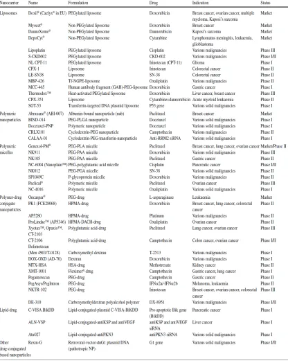
Figure II.S6: 13C NMR Spectrum of Mucic Acid Ethylenediamine in DMSO ... 58
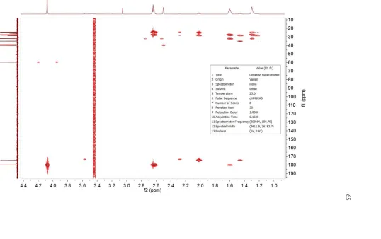
Figure II.S8: 1H NMR Spectrum of Dimethyl Suberimidate ... 63
Figure II.S9: 13C NMR Spectrum of Dimethyl Suberimidate ... 64
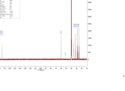
Figure II.S10: 1H-13C HMBC Spectrum of Dimethyl Suberimidate ... 65
Figure II.S11: 1H NMR Spectrum of Dimethyl Suberimidate hydrolyzed to dimethyl ester ... 66
Figure II.S12: 13C NMR Spectrum of Dimethyl Suberimidate hydrolyzed to dimethyl ester ... 67
Figure II.S13: ESI of Dimethyl Suberimidate ... 68
Figure II.S14: 1H NMR Spectrum of cMAP ... 70
Figure II.S15: 13C NMR Spectrum of cMAP ... 72
Figure II.S16: 1H-13C HSQC Spectrum of cMAP ... 74
Figure II.S17: 1H-1H COSY Spectrum of cMAP ... 76
Figure II.S18: 1H-13C HMBC Spectrum of cMAP ... 78
Figure II.S19: 1H stacked DOSY Spectrum of cMAP ... 80
Figure II.S20: 1H transformed DOSY Spectrum of cMAP ... 81
Figure II.S21: 1H NMR of cMAP-PEG5k copolymer ... 83
Figure II.S22: 1H-13C HSQC spectrum of cMAP-PEG5k copolymer ... 84
Figure II.S23: 1H DOSY transformed spectrum of cMAP-PEG5k copolymer ... 85
Figure II.S24: 1H NMR of cMAP-PEG3.4k copolymer ... 86
Figure II.S25: 13C NMR of cMAP-PEG3.4k copolymer ... 87
Figure II.S26: 1H-13C HSQC spectrum of cMAP-PEG3.4k copolymer ... 88
Figure II.S27: 1H DOSY transformed spectrum of cMAP-PEG3.4k copolymer ... 89
Figure II.S28: 1H NMR of mPEG5k-cMAP-PEG5km ... 91
Figure II.S29: 1H-13C HSQC of mPEG5k-cMAP-PEG5km ... 92
Figure II.S30: 1H DOSY transform of mPEG5k-cMAP-PEG5km ... 93
Figure II.S31: 1H NMR of mPEG2k-cMAP-PEG2km ... 94
Figure II.S32: 1H-13C HSQC of mPEG2k-cMAP-PEG2km ... 95
Figure II.S33: 1H DOSY transform of mPEG2k-cMAP-PEG2km ... 96
Figure II.S34: 1H NMR of 5-nPBA-PEGm ... 97
Figure II.S35: 1H DOSY of 5-nPBA-PEGm ... 98
Figure II.S36: 1H DOSY transform of 5-nPBA-PEGm ... 99
Figure II.S37: 11B NMR of 5-nPBA-PEGm ... 100
Figure II.S38: MALDI mass spectrum of 5-nPBA-PEGm ... 101
Figure II.S39. cMAP gel retardation assay ... 103
Figure II.S41. cMAP-PEG5k copolymer gel retardation assay ... 104
Figure II.S42. cMAP-PEG copolymer RiboGreen assay ... 105
Figure II.S43. mPEG-cMAP-PEGm triblock RiboGreen assay ... 105
Figure II.S44. Without PEG, the cMAP-siRNA NP is unstable once in 1X PBS, but is stable for 2 days when 5-nPBA-PEGm is used to stabilize the NP ... 106
Figure II.S45. Without added 5-nPBA-PEGm, the cMAP-PEG3.4k-cMAP siRNA NP formulated at a 1+/- charge ratio aggregates once in 1X PBS, but is stable when at least one 5-nPBA-PEGm per two diol groups (0.5 PEG) on cMAP is added to the formulation ... 107
Figure II.S46. Without added 5-nPBA-PEGm, the cMAP-PEG3.4k-cMAP siRNA NP formulated at a 3+/- charge ratio aggregates once in 1X PBS, but is stable when at least one 5-nPBA-PEGm per two diol groups (0.5 PEG) on cMAP is added to the formulation ... 108
Figure II.S47. Without added 5-nPBA-PEGm, the cMAP-PEG5k-cMAP siRNA NP formulated at a 1+/- charge ratio aggregates once in 1X PBS, but is stable when at least one 5-nPBA-PEGm per two diol groups (0.5 PEG) on cMAP is added to the formulation ... 108
Figure II.S48: Lognormal size distribution by DLS for the cMAP + 5-nPBA-PEGm NP ... 109
Figure II.S49: Lognormal size distribution by DLS for the cMAP-PEG copolymer NP ... 110
Figure II.S50: Lognormal size distribution by DLS for the cMAP-PEG copolymer + 5-nPBA-PEGm NP ... 111
Figure II.S51: Lognormal size distribution by DLS for the mPEG-cMAP-PEGm NP ... 112
Figure II.S52: Lognormal size distribution by DLS for the mPEG-cMAP-PEGm + 5-nPBA-PEGm NP ... 113
Figure II.S53: Size distribution by CryoTEM for the cMAP + 5-nPBA-PEGm NP ... 114
Figure II.S54: Additional CryoTEM images for the cMAP + 5-nPBA-PEGm NP ... 115
Figure II.S55: Size distribution by CryoTEM for the cMAP-PEG copolymer NP ... 116
Figure II.S56: Additional CryoTEM images for the cMAP-PEG copolymer NP ... 117
Figure II.S57: Size distribution by CryoTEM for the cMAP-PEG copolymer + 5-nPBA-PEGm NP ... 119
Figure II.S58: Additional CryoTEM images for the cMAP-PEG copolymer + 5-nPBA-PEGm NP ... 120
Figure II.S59: Size distribution by CryoTEM for the mPEG-cMAP-PEGm NP ... 121
Figure II.S60: Additional CryoTEM images for the mPEG-cMAP-PEGm NP ... 121
Figure II.S61: Size distribution by CryoTEM for mPEG-cMAP-PEGm + 5-nPBA-PEGm NP ... 123
Figure II.S63: The circulation time of the mPEG-cMAP-PEGm siRNA NP is similar
in Balb/c and nude mice. n=3 mice ... 126 Figure II.S64: Gel showing mPEG-cMAP-PEGm NPs with and without
5-nPBA-PEGm in mouse serum ... 127 Figure II.S65: Gel showing that the mPEG-cMAP-PEGm NP is intact in mouse serum ... 128 Figure II.S66: Gel in Figure II.S65, detected by Cy3 fluorophore ... 129 Figure II.S67: Gel showing that the mPEG-cMAP-PEGm NP is intact in mouse serum,
including in serum after 20 minutes of circulation after injection ... 130 Figure III.1: Effects of antibody treatment on H1975 xenograft tumors in nude mice ... 140 Figure III.2: H1975 xenograft tumor growth in mice treated with antibody containing AuNPs ... 141 Figure III.S1: Cell viabilities of H1975 cells 72 hours post-incubation with antibodies (top) and antibody-functionalized AuNPs (bottom), as determined by MTS assay... 151 Figure III.S2: ADCC in the BT474M1 cell line occurs with trastuzumab and
trastuzumab-PEG but not trastuzumab-Fab or rituximab (top); and with trastuzumab AuNPs
but not rituximab- or mPEG-AuNPs (bottom) as determined by the LDH assay ... 152 Figure III.S3: Silver enhancement of gold nanoparticles in frozen tissue sections of
tumor, liver, spleen, and kidney ... 153 Figure III.S4: CD11b staining of NK cells in tumor tissues... 157 Figure III.S5: CD45 staining of frozen tumor sections ... 158 Figure IV.1: mRNA knockdown of RRM2 in Neuro-2A cells using cMAP and
cMAP-His siRNA nanoparticles without PEG stabilization ... 172 Figure IV.2: EGFR mRNA knockdown with NPs formulated with TriB-His
containing siEGFR and targeted with 2 mol% of cetuximab targeting agents ... 177 Figure IV.3: RRM2 mRNA knockdown with NPs formulated with TriB-His
containing siRRM2 and targeted with 4 mol% of transferrin targeting agents ... 177 Figure IV.4: Detection of siEGFR as delivered by the cMAP + 5nPBA-PEGm
formulation with 0.13 mol% CTX-PEG-5nPBA targeting agent in H1975 xenografts ... 180 Figure IV.5: H1975 tumor xenograft growth in nude mice treated with siEGFR as delivered by the cMAP + 5nPBA-PEGm formulation with 0.13 mol% CTX-PEG-5nPBA targeting
Figure IV.7: M249shBRAF xenograft tumor growth in nude mice using TriB-His NP’s
containing siBRAF with 0.25 mol% Tf-PEG5k-(5nPBA)2 targeting agent ... 185
Figure IV.S1: 1H NMR of (5nPBA)2-Lysine-OMe ... 203
Figure IV.S2: 11B NMR of (5nPBA)2-Lysine-OMe ... 204
Figure IV.S3: 1H NMR of (5nPBA)2-Lysine ... 205
Figure IV.S4: 11B NMR of (5nPBA)2-Lysine ... 206
Figure IV.S5: 1H NMR of 5nPBA-PEG5k-COOH ... 207
Figure IV.S6: 1H NMR of 5nPBA-PEG5k-COOH with enlarged downfield region ... 208
Figure IV.S7: MALDI of 5nPBA-PEG5k-COOH ... 209
Figure IV.S8: 1H NMR of 5nPBA-PEG10k-COOH ... 210
Figure IV.S9: MALDI of 5nPBA-PEG10k-COOH ... 211
Figure IV.S10: 1H NMR of (5nPBA)2-PEG5k-COOH ... 212
Figure IV.S11: MALDI of (5nPBA)2-PEG5k-COOH ... 213
Figure IV.S12: 1H NMR of (5nPBA)2-PEG10k-COOH ... 214
Figure IV.S13: MALDI of (5nPBA)2-PEG10k-COOH ... 215
Figure IV.S14: MALDI of crude 5nPBA-PEG5k-Tf ... 216
Figure IV.S15: MALDI of crude 5nPBA-PEG5k-CTX ... 217
Figure IV.S16: Purification of crude 5nPBA-PEG5k-CTX by HPLC ... 218
Figure IV.S17: MALDI of purified 5nPBA-PEG5k-CTX, 158 kD ... 219
Figure IV.S18: MALDI of purified PEGylated transferrin ... 220
Figure IV.S19: 1H NMR of cMAP-PEG5k-COOH ... 221
Figure IV.S20: MALDI of cMAP-PEG5k-Tf ... 222
Figure IV.S21: 1H NMR of His-cMAP-His ... 223
Figure IV.S22: 1H NMR of mPEG-His-cMAP-His-PEGm (TriB-His) ... 224
Figure IV.S23: Titration curve of mPEG-His-cMAP-His-PEGm (TriB-His) ... 225
Figure IV.S24: Validation of the siEGFR sequence with Lipofectamine RNAiMAX transfection reagent ... 225
Figure IV.S25: Validation of the siBRAF sequence with Lipofectamine RNAiMAX transfection reagent ... 226
Figure IV.S26: Displacement binding of CTX-AF488 by cetuximab and cetuximab-PEGs to EGFR on H1975 cells ... 227
Figure IV.S28: Validation of qRT-PCR assay probe for the siEGFR sequence by
transfecting cells with siEGFR using Lipofectamine RNAiMAX transfection reagent ... 228 Figure IV.S29: Individual mice for the 0.13 mol% cetuximab-targeted
cMAP + 5-nPBA-PEGm NP formulation containing siEGFR group ... 228 Figure IV.S30: Individual mice for the cetuximab alone group ... 229 Figure IV.S31: MALDI of panitumumab-PEG5k-5nPBA, 153 kD ... 230 Figure IV.S32: Mean sizes of M249shBRAF tumor xenografts in nude mice (n=6)
comparing standard diet to doxycycline-containing diet ... 231 Figure IV.S33: Individual nude mice bearing M249shBRAF tumor xenografts (n = 5)
injected with saline control ... 232 Figure IV.S34: Individual nude mice bearing M249shBRAF tumor xenografts (n = 5) injected with 0.25 mol% Tf-PEG5k-(5nPBA)2 targeted TriB-His NP containing siBRAF at 5 mg/kg per dose.. 233 Figure IV.S35: Mean sizes of nude mice (n = 5) bearing M249shBRAF tumor comparing
the 0.25 mol% Tf-PEG5k-(5nPBA)2 targeted TriB-His NP containing siBRAF at 5 mg/kg
per dose group with that of saline control showing no statistical significance ... 234 Figure IV.S36: Mean sizes of nude mice (n = 2) bearing M249shBRAF tumor comparing
the 0.5 mol% Tf-PEG5k-adamante targeted CDP NP containing siBRAF at 5 mg/kg per
LIST OF SCHEMES
Number Page
Scheme II.1. Synthesis of cationic Mucic Acid Polymer (cMAP) ... 23
Scheme II.2: Synthesis of cMAP-PEG copolymer ... 25
Scheme II.3: Synthesis of mPEG-cMAP-PEGm triblock polymer ... 26
Scheme II.4. Synthesis of 5-nitrophenylboronic acid-PEGm ... 29
Scheme II.5. pH dependence of 5-nitrophenylboronic acid-PEGm... 29
Scheme II.6. Diagram showing the various NPs with siRNA that were formed: cMAP (I, not stable and not injected), cMAP + 5-nPBA-PEGm (A), cMAP-PEG copolymer (B), cMAP-PEG copolymer + 5-nPBA-PEGm (C), mPEG-cMAP-PEGm triblock (D), and mPEG-cMAP-PEGm triblock + 5-nPBA-PEGm (E) ... 30
Scheme III.1: Assembly of antibody containing gold nanoparticles ... 138
Scheme IV.1: pH dependence of 5-nitrophenyl boronic acid ... 165
Scheme IV.2: Synthesis of 5-nitrophenylboronic acid-PEG5k-COOH ... 165
Scheme IV.3: Conjugation of transferrin to 5-nitrophenylboronic acid-PEG-COOH ... 166
Scheme IV.4: Conjugation of cetuximab to 5-nitrophenylboronic acid-PEG-COOH ... 166
Scheme IV.5: Synthetic scheme for di(5-nitrophenylboronic acid)-PEG-COOH ... 167
Scheme IV.6: Directly conjugating a protein to cMAP via a PEG linker ... 170
LIST OF STRUCTURES
Number Page
Structure II.S1: Mucic Acid Ethylenediamine ... 55
Structure II.S2: Dimethyl Suberimidate ... 61
Structure II.S3: Dimethyl Suberimidate hydrolyzed to the dimethyl ester ... 62
Structure II.S4: Dimethyl Suberimidate with one side hydrolyzed to the carboxylate... 62
LIST OF TABLES
Number Page
Table I.1: Nanoparticle delivery systems for cancer therapy on the market
and in clinical development ... 7
Table I.2: Nanoparticles containing siRNA or other nucleic acids for cancer treatment in clinical trials ... 11
Table I.3: siRNA for non-cancer therapy applications that have reached the clinic ... 12
Table II.1. Molecular weights of cMAP-based polymers ... 27
Table II.2. Nanoparticle composition for NPs formulated at a charge ratio of 3+/- ... 32
Table II.3. Size and surface charge of formulated NPs ... 32
Table II.S1: 1H and 13C NMR peak assignments for Mucic Acid Ethylenediamine ... 59
Table II.S2: 1H NMR peak assignments for Dimethyl Suberimidate with varying degrees of hydrolysis ... 61
Table II.S3: 1H NMR peak assignments for cMAP ... 71
Table II.S4: 13C NMR peak assignments for cMAP ... 73
Table II.S5: 1H-13C HSQC NMR peak assignments for cMAP ... 75
Table II.S6: 1H-1H COSY NMR peak assignments for cMAP ... 77
Table II.S7: 1H-13C HMBC NMR peak assignments for cMAP ... 79
Table II.S8: Ratios of amine: methoxy: carboxylate end groups in 8 batches of cMAP by comparing NMR integrations ... 82
Table II.S9: Retained mass on each MWCO filter after fractionating crude cMAP-PEG3.4k copolymer ... 90
Table II.S10: Retained mass on each MWCO filter after fractionating crude cMAP-PEG5k copolymer ... 90
Table II.S11: Gel Permeation Chromatography Analysis of cMAP Batches ... 102
Table II.S12: GPC Analysis of cMAP copolymers and triblocks (other than 5 kD PEG reported in main article text)... 102
Table III.1: Properties of antibody containing AuNPs ... 139 Table III.S1: Quantification of antibodies on AuNPs surface using two different methods ... 150 Table IV.1: Binding constant of 5nPBA-PEG-COOH and (5nPBA)2-PEG-COOH to
Alizarin Red S and to cMAP ... 167 Table IV.2: Hydrodynamic diameter and surface charge of targeted nanoparticles formed
using the triblock polymer with transferrin as the targeting agent ... 169 Table IV.3: Absolute molecular weight of histidine-containing cMAP polymers as
determined by multi-angle light scattering ... 171 Table IV.4: NP size and zeta potential formulated with TriB-His without and
with transferrin targeting agent added to the formulation ... 173 Table IV.5: Hydrodynamic diameter and zeta potential of nanoparticle formulation
(building up from the cMAP polymer alone) of the CTX-targeted cMAP-siEGFR NPs
C h a p t e r 1
Introduction
Targeted therapeutics for cancer
Cancer is currently the second leading cause of death in the United States.1 With 8 million cancer-related deaths in 2012, malignancies are also one of the major causes of death and disability worldwide.2 The most common types of cancer diagnosed as well as
leading to death are those arising from sites in the lung, breast, prostate, and colon, as shown in Figure I.1.1 In addition to the human burden caused by cancer, the financial costs are considerable. In 2009, an estimated $286 billion in medical costs and in lost
productivity was caused by new cancer diagnoses, with 94% of that amount being incurred in developed countries such as the United States.3 This price tag may be due to the use of
newer therapies for cancer, which are more specific against the cancer and enable the patient to have a better quality of life that allows them to carry on most of their usual activities.
Although many toxic chemotherapeutics such as methotrexate and cisplatin are still part of many chemotherapy regimens, cancer treatment has advanced in the last 70 years from highly toxic drugs with many adverse effects to more specific therapies that cause fewer side effects. Some of the first toxic compounds used against leukemia were derivatives of nitrogen mustard and anti-folates.4,5 Many of these chemotherapeutics used for cancer treatment target cell replication. Because cancer cells generally proliferate more rapidly than other cell types in the body, these types of chemotherapeutics have a larger impact on the malignant cells. However, because some cells in the body, such as blood cells, epithelial cells lining the gastrointestinal (GI) tract and skin or hair, need to be replenished fairly frequently, there can be severe adverse effects resulting from chemotherapeutics, such as anemia, nausea, vomiting, diarrhea, sores, and hair loss.
The first targeted therapy developed was Gleevec, or imatinib, which inhibits the tyrosine kinase domain fused by bcr-abl resulting from the Philadelphia chromosome 9;22 translocation.6 Since then, numerous targeted therapies have been developed, many of which target tyrosine kinases – tyrosine kinase inhibitors (TKIs). For example, erlotinib and gefitinib are inhibitors of the intracellular tyrosine kinase domain in the epidermal growth factor receptor (EGFR) often overexpressed or mutated in non-small cell lung cancer (NSCLC), colorectal cancer, and head and neck cancers.7-10
antibody which is directed against human epidermal growth factor receptor 2 (Her2).13 Her2 is often overexpressed in up to 30% of breast cancers and some types of gastric cancer.14,15 In all cases, eventually the malignant cells mutate and become resistant to the targeted therapy.12,16-18 Therefore, newer targeted therapies are developed in an effort to overcome therapy resistance, and many of these targeted therapies are used in combination with other targeted therapies or chemotherapy, such as cetuximab with irinotecan,
fluorouracil, and leucovorin (FOLFIRI) in metastatic colorectal cancer.9,19-21
The idea to combine a targeted antibody therapeutic with a chemotherapeutic as a single drug resulted in antibody-drug conjugates (ADCs). ADCs link a cytotoxic drug to an antibody to better target the chemotherapeutic agent to the appropriate cells. They were developed to enhance the potency of the antibody after resistance to trastuzumab
developed, as well as create a sort of combination therapy with multiple modes of action in a single formulation.22 Trastuzumab conjugated to the cytotoxic microtubule disruptor emtansine (DM1) shown in Figure I.2, Kadcyla (T-DM1) is an example of an FDA approved ADC, with many more in development at Genentech and other pharmaceutical companies.23-24 Although T-DM1 initially is effective against transtuzumab-resistant
cancers, resistance to the ADC eventually develops and combinations with other therapeutics are used in an attempt to rescue its function.22-27
Perhaps it is cancer’s ability to outsmart each new therapy and the myriad side effects that these therapies cause that cancer has been dubbed “the emperor of all maladies.”28 TKIs, antibodies, and ADCs have been shown to be effective and less toxic than the cytotoxic chemotherapeutic agents, but some adverse effects remain. For example, TKIs and antibodies against EGFR can cause skin rashes and GI toxicity because EGFR is highly expressed in epithelial tissues. ADCs such as T-DM1 is also able to cause many of the side effects associated with cytotoxic chemotherapy, though it is obviously less toxic than emtansine alone, which is not tolerated by patients at all.22 In an effort to develop a therapeutic that is more potent against malignant cells but less toxic to the patient, nanoparticles with more design complexity and flexibility show promise to enable multiple therapeutic modes of action with high drug-loading capacity while having minimal penetration to normal tissue.
Nanoparticle Therapies
The earliest nanoparticle therapeutics sought to reduce the toxicity of cytotoxic chemotherapeutics by encapsulating the drug in a liposome (doxorubicin, Doxil) or coating it with albumin (paclitaxel, Abraxane).29,30 Although the development of nanoparticles
Figure I.3: Timeline of nanoparticle development. Doxil, Abraxane, and Genexol-PM (in Korea) are approved for cancer therapy. CRLX101, CALAA-01, and BIND-014 are in clinical trials for cancer therapy. Adapted from Kamaly, et al. Chem. Soc. Rev. 2012, 41, 2971-3010.
Despite the few nanoparticles that have been FDA approved, nanoparticles show efficacy and substantially decrease the toxicity of the chemotherapeutic agents that they have been formulated to deliver. For example, CRLX101, a cyclodextrin-PEG polymer containing camptothecin that forms a 30 nm diameter nanoparticle, has been shown to have few adverse effects compared to other camptothecin-containing drugs not in nanoparticle form (shown in Figure I.4). Adverse effects are seen in less than 40% of patients, with very few of the serious grade 3-4 adverse effects seen. It has been shown to preferentially localize in tumor tissue in gastric cancer without being present in adjacent normal gastric tissue.33
contributes to the accumulation of nanoparticles.34 Passive targeting of nanoparticles by the EPR effect contributes to the enhanced safety profile of nanoparticle therapeutics, and enables the use of drug combinations with the nanoparticle-encapsulated drug that would otherwise be too toxic for the patient.
Figure I.4: Adverse effects of camptothecin-containing drugs and CRLX101, a nanoparticle which contains camptothecin. From Cerulean Pharma Inc., United States Securities and Exchange Commission Annual Report
(http://www.sec.gov/Archives/edgar/data/1401914/000156459016014403/ceru-10k_20151231.htm)
siRNA Delivery: Advantages and Challenges
Nanoparticle-based delivery of macromolecular agents as a therapeutic commenced with the discovery of RNA interference (RNAi) by Fire and Mello in 1998.35 Small interfering RNA (siRNA) is a short double stranded RNA ca. 21 base pairs. It is able to very specifically cleave the messenger RNA (mRNA) complementary to it using the RNAi induced silencing complex (RISC) in the cytosol to silence an overexpressed or mutated gene, as shown in Figure I.6. siRNA sequences are now being developed with very high potency, so that the concentration required for silencing 50% of the mRNA is in the single digit picomolar scale. Because siRNA is catalytic, once it has reached the cell cytosol it will remain for a prolonged period of time subject to cell division rate.36 RNAi has the promise of being able to silence any gene, including for intracellular proteins, such as KRAS, which have been considered “undruggable.”37 Once a nanoparticle siRNA delivery system is developed, it is able to deliver any siRNA sequence, including multiple siRNA sequences in the same particle for combined silencing of multiple genes fueling the cancer cell’s growth. Silencing of multiple genes using multiple siRNA sequences in the same delivery system has been shown both in preclinical studies and clinical trials.38,39
glomerular basement membrane in the kidney.42-46 Furthermore, the siRNA needs to be internalized into cells, where it is endocytosed into an endosome. Then the siRNA needs escape from the endosome and reach the cytosol to bind to RISC and perform its functions, as shown in Figure I.7.
siRNA
mRNA
cleaved mRNA RISC siRNA
Extracellular Space
Cytosol
Figure I.6: siRNA mechanism of action
Cytosol
Extracellular Space
H+ H+
H+
Endocytosis
Endosomal Escape Mechanism
Despite the challenges that siRNA delivery faces, a number of nanoparticles containing siRNA for cancer therapy (Table I.2), as well as several other applications (Table I.3), have reached the clinic and shown to be effective, though there has yet one to be approved to date.47-51 Furthermore, these siRNA-containing nanoparticles show fairly good safety profile.41,48,52
Table I.3: siRNA for non-cancer therapy applications that have reached the clinic. From Draz, et al. Thernostics 2014, 4, 872-892.
Thesis Organization
This thesis presents a nanoparticle siRNA delivery system based upon a cationic mucic acid polymer that attempts to overcome the challenges facing siRNA therapy for cancer treatment. These challenges, as mentioned earlier, include protecting the siRNA with the delivery system, increasing the siRNA nanoparticle circulation time, getting siRNA into cells, and allowing the siRNA to escape from the endosome to the cytosol for function.
allow nanoparticle internalization: some antibodies have multiple functions which include an immune-mediated antibody-dependent cellular cytotoxicity (ADCC) effect, and we wanted to see whether this function is preserved when the antibody is attached to a nanoparticle to evaluate the value of using an antibody to target the nanoparticle with an added ADCC effect over using a protein or peptide without these immune effects to target the nanoparticle. Chapter 4 discusses achieving internalization of the cationic mucic acid polymer-based nanoparticle and endosomal escape. Finally, chapter 5 concludes with an outlook on the future of nanoparticle therapeutics and targeted cancer treatment.
References
1. Stricker, T.P.; Kumar, V. Neoplasia. In Robbins Basic Pathology, 8th Edition; Kumar, V.;
Abbas, A.K., Fausto, N., Mitchell, R., Eds.; Elsevier Health Sciences: Philadelphia, 2007; pp 173-224.
2. Forman, D.; Ferlay, J. The global and regional burden of cancer. In World Cancer Report 2014; Stewart, B.W., Wild, C.P., Eds.; International Agency for Research on Cancer: Lyon,
2014; pp 15-76.
3. Sutcliffe, S.B. National cancer control plans. In World Cancer Report 2014; Stewart, B.W.,
Wild, C.P., Eds.; International Agency for Research on Cancer: Lyon, 2014; pp 529-536. 4. Fenn, J.E.; Udelsman, R. First use of intravenous chemotherapy cancer treatment: rectifying
the record. J. Am. Coll. Surg. 2011, 212, 413-417.
5. Spain, P.D.; Kadan-Lottick, N. Observations of unprecedented remissions following novel treatment for acute leukemia in children in 1948. J. R. Soc. Med. 2012, 105, 177-181.
6. Stegmeier, F.; Warmuth, M.; Sellers, W.R.; Dorsch, M. Targeted cancer therapies in the twenty-first century: lessons from imatinib. Clinical Pharmacology and Therapeutics 2010, 87, 543-552.
7. Wu, K.; House, L.; Liu, W.; Cho, W.C.S. Personalized targeted therapy for lung cancer. Int. J. Mol. Sci. 2012, 13, 11471-11496.
non-small cell lung cancer cell lines correlates with gene copy number and EGFR mutations but not EGFR protein levels. Clin. Cancer Res. 2006, 12, 7117-7125.
9. Cutsem, E.V.; Kohne, C.-H.; Hitre, E., et al. Cetuximab and chemotherapy as initial treatment for metastatic colorectral cancer. N Engl J Med 2009, 360, 1408-1417.
10. Sok, J.C.; Coppelli, F.M.; Thomas, S.M., et al. Mutant epidermal growth factor receptor (EGFRvIII) contributes to head and neck cancer growth and resistance to EGFR targeting.
Clin Cancer Res 2006, 12, 5064-5073.
11. Patel, D.; Lahiji, A.; Patel, S.; Franklin, M.; Jimenez, X.; Hicklin, D.J.; Kang, X. Monoclonal antibody cetuximab binds to and down-regulates constitutively activated epidermal growth factor receptor vIII on the cell surface. Anticancer Research, 2007, 27, 3355-3366.
12. Diaz, L.A.; Williams, R.T.; Wu, J.; Kinde, I.; etc al. The molecular evolution of acquired resistance to targeted EGFR blockade in colorectal cancers. Nature 2012, 486, 537-540.
13. Hudis, C.A. Trastuzumab – Mechanism of action and use in clinical practice. N Engl J Med 2007, 357, 39-51.
14. Boku, N. HER2-positive gastric cancer. Gastric Cancer 2014, 17, 1-12.
15. Ruschoff, J.; Hanna, W.; Bilous, M.; et al. HER2 testing in gastric cancer: a practical approach. Modern Pathology 2013, 25, 637-650.
16. Brand, T.M.; Iida, M.; Wheeler, D.L. Molecular mechanisms of resistance to the EGFR monoclonal antibody cetuximab. Cancer Biology & Therapy 2011, 11, 777-792.
17. Kim, S.M.; Kim, J.S.; Kim, J.-H.; Yun, C.-O.; Kim, E.M.; Kim, H.K.; Solca, F.; Choi, S.-Y.; Cho, B.C. Acquired resistance to cetuximab is mediated by increased PTEN instability and leads cross-resistance to gefitinib in HCC827 NSCLC cells.
18. Bardelli, A.; Janne, P.A. The road to resistance: EGFR mutation and cetuximab. Nature Medicine 2012, 18, 199-200.
19. Cross, D.A.E.; Ashton, S.E.; Ghiorghiu, S. AZD9291, an irreversible EGFR TKI, overcomes T790M-mediated resistance to EGFR inhibitors in lung cancer. Cancer Discov. 2014, 4,
1046-1061.
20. Gibbons, D.L.; Byers, L.A. A HER 1-2 punch: dual EGFR targeting deals resistance a deadly blow. Cancer Discov. 2014, 4, 991-994.
22. Peddi, P.F.; Hurvitz, S.A. Trastuzumab emtansine: the first targeted chemotherapy for treatment of breast cancer. Future Oncol. 2013, 9, 319-326.
23. LoRusso, P.M.; Weiss, D.; Guardino, E. Trastuzumab Emtansine: A unique antibody-drug conjugate in development for human epidermal growth factor receptor 2-positive cancer.
Clin. Cancer Res. 2011, 17, 6437-6447.
24. Barok, M.; Tanner, M.; Koninki, K.; Isola, J. Trastuzumab-DM1 causes tumour growth inhibition by mitotic catastrophe in trastuzumab resistant breast cancer cells in vivo. Breast Cancer Research 2011, 13, R46.
25. Junttila, T.T.; Li, G.; Parsons, K.; Phillips, G.L.; Sliwkowski, M.X. Trastuzumab-DM1 (T-DM1) retains all the mechanisms of trastuzumab and efficiently inhibits growth of lapatinib insensitive breast cancer. Breast Cancer Res. Treat. 2011, 128, 347-356.
26. Gwin, W.R.; Spector, N.L. Pertuzumab protects the Achilles’ heel of trastuzumab-emtansine.
Clin. Cancer Res. 2014, 20, 278-280.
27. Phillips, G.D.L.; Fields, C.T.; Li, G.; et al. Dual targeting of HER2-positive cancer with trastuzumab emtansine and pertuzumab: critical role for neuregulin in antitumor response to combination therapy. Clin. Cancer Res. 2014, 20, 456-468.
28. Mukherjee, S. The emperor of all maladies: a biography of cancer. Scribner: New York,
2010, p xiv.
29. Kamaly, N.; Xiao, Z.; Valencia, P.M.; Radovic-Moreno, A.F.; Farokhzad, O.C. Targeted polymeric therapeutic nanoparticles: design, development and clinical translation. Chem. Soc. Rev. 2012, 41, 2971-3010.
30. Egusquiaguirre, S.P.; Igartua, M.; Hernandez, R.M.; Pedraz, J.L. Nanoparticle delivery systems for cancer therapy: advances in clinical and preclinical research. Clin. Transl. Oncol. 2012, 14, 83-93.
31. Wang, A.Z.; Langer, R.; Farokhzad, O.C. Nanoparticle delivery of cancer drugs. Annu. Rev. Med. 2012, 63, 185-198.
32. Cheng, Z.; Al Zaki, A.; Hui, J.Z.; Muzykantov, V.R.; Tsourkas, A. Multifunctional
nanoparticles: Cost versus benefit of adding targeting and imaging capabilities. Science 2012, 338, 903-910.
34. Peer, D.; Karp, J.M.; Hong, S.; Farokhzad, O.C.; Margalit, R.; Langer, R. Nanocarriers as an emerging platform for cancer therapy. Nature Nanotechnology 2007, 2, 751-760.
35. Fire, A.; Xu, S.; Montgomery, M.K.; Kostas, S.A.; Driver, S.E.; Mello, C.C. Potent and specific genetic interference by double-stranded RNA in Caenorhabditis elegans. Nature 1998, 19, 806-811.
36. Bartlett, D.W.; Davis, M.E. Insights into the kinetics of siRNA-mediated gene silencing from live-cell and live-animal bioluminescent imaging. Nucleic Acids Research 2006, 34, 322-333.
37. Wu, S. Y., Lopez-Berestein, G., Calin, G. A., and Sood, A. K. RNAi Therapies: Drugging the Undruggable. Sci. Transl. Med.2014,6, 240ps7.
38. Tabernero, J.; Shapiro, G.I.; LoRusso, P.M; et al. First-in-man trial of an RNA interference therapeutic targeting VEGF and KSP in cancer patients with liver involvement. Cancer Discovery 2013, 3, 406-417.
39. Yuan, T.L.; Fellmann, C.; Lee, C.-S.; et al. Development of siRNA payloads to target KRAS-mutant cancer. Cancer Discovery 2014, 4, 1182-1197.
40. Ballarin-Gonzalez, B.; Howard, K.A. Polycation-based nanoparticle delivery of RNAi therapeutics: adverse effects and solutions. Advanced Drug Delivery Reviews 2012, 64,
1717-1729.
41. Zuckerman, J. E., Gritli, I., Tolcher, A., Heidel, J. D., Lim, D., Morgan, R., Chmielowski, B., Ribas, A., Davis, M. E., and Yen, Y. Correlating animal and human phase Ia/Ib clinical data with CALAA-01, a targeted, polymer-based nanoparticle containing siRNA. Proc. Natl. Acad. Sci. U. S. A.2014,111, 11449−11454.
42. Zuckerman, J. E., Choi, C. H. J., Han, H., and Davis, M. E. Polycation-siRNA nanoparticles can disassemble at the kidney glomerular basement membrane. Proc. Natl. Acad. Sci. U. S. A. 2012, 109, 3137−3142.
43. Naeye, B., Deschout, H., Caveliers, V., Descamps, B., Braeckmans, K., Vanhove, C., Demeester, J., Lahoutte, T., De Smedt, S. C., and Raemdonck, K. In vivo disassembly of IV
administered siRNA matrix nanoparticles at the renal filtration barrier. Biomaterials2013, 34, 2350−2358.
44. Christie, R. J., Matsumoto, Y., Miyata, K., Nomoto, T., Fukushima, S., Osada, K., Halnaut, J., Pittella, F., Kim, H. J., Nishiyama, N., et al. Targeted polymeric micelles for siRNA treatment of experimental cancer by intravenous injection. ACS Nano2012, 6, 5174−5189.
endosome escape, stability, blood circulation time, and bioactivity in vivo. ACS Nano2013, 7,
8870−8880.
46. Barrett, S. E., Burke, R. S., Abrams, M. T., Bason, C., Busuek, M., Carlini, E., Carr, B. A., Crocker, L. S., Fan, H., Garbaccio, R. M., et al. Development of a liver-targeted siRNA delivery platform with a broad therapeutic window utilizing biodegradable polypeptide-based polymer conjugates. J. Controlled Release2014, 183, 124−137.
47. Davis, M. E., Zuckerman, J. E., Choi, C. H. J., Seligson, D., Tolcher, A., Alabi, C. A., Yen, Y., Heidel, J. D., and Ribas, A. Evidence of RNAi in humans from systemically administered siRNA via targeted nanoparticles. Nature2010, 464, 1067−1070.
48. Zuckerman, J.E.; Davis, M.E. Clinical experiences with systemically administered siRNA-based therapeutics in cancer. Nature Reviews Drug Discovery 2015, 14, 843-856.
49. Draz, M.S.; Fang, B.A.; Zhang, P.; et al. Nanoparticle-mediated systemic delivery of siRNA for treatment of cancers and viral infections. Theranostics 2014, 4, 872-892.
50. Bouchie, A. Companies in footrace to deliver RNAi. Nature Biotechnology 2012, 12,
1154-1157.
51. Young, S.W.S.; Stenzel, M.; Yang, J.-L. Nanoparticle-siRNA: A potential cancer therapy?
Critical Reviews in Oncology/Hematology 2016, 98, 159-169.
C h a p t e r 2
Cationic mucic acid polymer-based siRNA delivery systems
This chapter has been published as:
Pan, D.W. and Davis, M.E. Cationic mucic acid polymer-based siRNA delivery systems. Bioconjugate Chem. 2015, 26, 1791-1803. doi : 10.1021/acs.bioconjchem.5b00324
Abstract
Nanoparticle (NP) delivery systems for small interfering RNA (siRNA) that have good systemic circulation and high nucleic acid content are highly desired for translation into clinical use.Here, a family of cationic mucic acid-containing polymers is synthesized and shown to assemble with siRNA to form NPs. A cationic mucic acid polymer (cMAP) containing alternating mucic acid and charged monomers is synthesized. When combined
with siRNA, cMAP forms NPs that require steric stabilization by polyethylene glycol
(PEG) that is attached to the NP surface via a 5-nitrophenylboronic acid linkage
(5-nitrophenylboronic acid-PEGm (5-nPBA-PEGm)) to diols on mucic acid in the cMAP in
order to inhibit aggregation in biological fluids. As an alternative, the cMAP is covalently
conjugated with PEG via two methods. First, a copolymer is prepared with alternating
cMAP-PEG units that can form loops of the PEG on the surface of the formulated
siRNA-containing NPs. Second, an mPEG-cMAP-PEGm triblock polymer is synthesized that
could lead to a PEG brush configuration on the surface of the formulated siRNA-containing
NPs. The copolymer and triblock polymer are able to form stable siRNA-containing NPs
without and with the addition of nPBA-PEGm. Five formulations: (i) the cMAP with
5-nPBA-PEGm, (ii) cMAP-PEG copolymer both (a) with and (b) without 5-5-nPBA-PEGm,
5-nPBA-PEGm, are used to produce NPs in the 30-40 nm size range, and their circulation times
evaluated in mice using tail vein injections. The mPEG-cMAP-PEGm triblock polymer
provides the siRNA-containing NP with the longest circulation time (5-10% of the
formulation remains in circulation at 60 min post-dosing), even when a portion of the
excess cationic components used in the formulation are filtered away prior to injection. A
NP formulation using the mPEG-cMAP-PEGm triblock polymer that is free of excess
components could contain as much as ca. 30wt% siRNA.
INTRODUCTION
Therapeutics that use RNA interference (RNAi) as their mechanism of action have great promise for the treatment of human disease. For example, siRNA has attractive features for use as a therapeutic, including: (i) the ability to target essentially any gene (thus, all targets are in principle druggable), (ii) potent, single-digit, picomolar IC50’s
(concentration required for 50% inhibition) for mRNA inhibition in well-designed siRNAs, (iii) chemical modifications and sequence designs that can minimize off-target effects and immune stimulation without compromising potency and target specificity, and (iv) a catalytic RNAi mechanism of action, resulting in extended siRNA inhibition of mRNA target expression. Although a major obstacle to the translation of siRNA into an effective and efficient therapeutic is the delivery of the nucleic acid to the target, siRNA-based experimental therapeutics have reached the clinic.1
clinic for the treatment of cancer.3-5 This targeted NP contains a cyclodextrin-based polycation (CDP) that assembles with siRNA via electrostatic interactions between positive charges on the polymer and negative charges on the siRNA backbone. CALAA-01 was able to deliver siRNA to solid tumors in patients and release functional siRNA that inhibited the target using an RNAi mechanism (the first example in a human).4,5 While CALAA-01 revealed several positive attributes, one of its shortcomings is that it has a very limited circulation time. The fast clearance of CALAA-01 that is observed in animals (mice, rats, dogs, and non-human primates) is also observed in humans.5 We have investigated the origin of this short circulation time, and have shown that CALAA-01 disassembles at the glomerular basement membrane (GBM) in the kidney.6 We speculated that this clearance mechanism may affect any NP formulation that is primarily assembled through electrostatic interactions between cationic delivery components and anionic nucleic acids.6 Other siRNA delivery systems that use either cationic polymers or lipids have shown similar short circulation times and renal clearance.7-10
A number of the current polymeric and liposomal systems used to deliver siRNA in vivo contain excess cationic components in their formulations (positive to negative charge
ratios are commonly greater than 1), in addition to a large amount of material, e.g., polyethylene glycol (PEG), used to sterically stabilize the formed NPs. Excess cationic components can have unwanted side effects in vivo, causing adverse reactions such as platelet aggregation, complement activation, and inflammatory reactions.9,11-14
RESULTS AND DISCUSSIONS
cMAP Synthesis, NMR Characterization, and End-Group Determination. A cationic mucic acid polymer (cMAP) was synthesized by using the series of reactions schematically illustrated in Scheme II.1. The mucic acid and the intermediate reaction products leading to the preparation of mucic acid ethylenediamine were fully characterized (Supporting Information: Figures II.S1-II.S7, Structure II.S1, and Table II.S1). The condensation reaction between mucic acid ethylenediamine and DMS yielded the cMAP material. Because DMS can hydrolyze at conditions like those used for the polymerization, we investigated the reaction pathway for this reaction and the products formed (Supporting Information: Table II.S2, Structures II.S2-II.S4, Figures II.S8-II.S13). This information assisted in the characterization of the cMAP product.
Scheme II.1. Synthesis of cationic Mucic Acid Polymer (cMAP).
The cMAP end groups include methoxy of a methoxy ester, amine, and small amounts of carboxylic acid (Figure II.1). 1H NMR analysis of cMAP shows the presence of a characteristic sharp methoxy peak at 3.55 ppm (Figure II.2), and this assignment is supported by 1H-13C HSQC NMR measurements (Supporting Information: Figure II.S16). The methoxy group originates from the loss of ammonia from the imidate group of the DMS through hydrolysis (Supporting Information: Table II.S2, Structures II.S2-II.S4, Figures II.S8-II.S13), and had been previously reported.20 The methylene group adjacent to the methoxy can be observed as a triplet at 2.25 ppm in the 1H NMR spectrum
cannot be directly observed with 1H NMR. However, analysis of the NMR spectrum of the monomer (Supporting Information: Figure II.S4-5) and the HMBC NMR spectrum of cMAP (Supporting Information: Figure II.S18) enabled assignment of the triplet at 2.85 ppm to be from a methylene group adjacent to the amine functional group. Additionally, a TNBSA assay for primary amines was positive, thus confirming that the cMAP has a terminal primary amine as an end group. Lastly, there was a small amount of carboxylic acid as an end group that arises from complete hydrolysis of the methyl ester or as an impurity in the starting DMS. The methylene group adjacent to the carboxylic acid is observed as a small triplet at 2.00 ppm in the 1H NMR spectrum (Figure II.2). The ratios of these end groups in a batch of cMAP can be determined by comparing the integrations of the triplets at 2.85 (amine), 2.25 (methoxy), and 2.00 (carboxylate) ppm, and are shown for 8 batches in Supplementary Information, Table II.S8. The average values for the % amine, % methoxy, and % carboxylate are 49%, 42%, and 9%, respectively.
Figure II.1. End groups of cMAP. Polymers can have one amine and one methoxy (top), both amine (middle), or both methoxy (bottom) end groups. A small amount of carboxylic acid is also observed and would be generated from hydrolysis of one end of DMS.
H3CO
O
NH2 H
Figure II.2. 1H NMR (600 MHz) of cMAP showing resonances from the methoxy group and methylene groups adjacent to the end group functionalities.
cMAP-PEG Copolymer and mPEG-cMAP-PEGm Triblock. cMAP was reacted with activated carboxylic acid end groups on PEG, such as succinimidyl propionic acid ester (SPA) or succinimidyl valeric acid ester (SVA). cMAP reacted with di-SPA-PEG or mPEG-SVA generated copolymers or triblock polymers, respectively, with PEG lengths of 2, 3.4, or 5 kD (NMR data from these polymers are provided in the Supporting Information: Figures II.S21-II.S27).
Scheme II.2: Synthesis of cMAP-PEG copolymer.
100 kD; size distributions reported in Supporting Information: Table II.S9-10 are from the polymer yields obtained by fractionating the crude polymer through sequentially smaller molecular weight cutoff centrifugal spin filters). Because a polymer with such a large molecular weight could pose substantial toxicity in vivo, in an effort to synthesize a well-defined polymer with a reasonable length, the cMAP-PEG-cMAP triblock polymer species was isolated from the copolymer using this fractionation method. Other triblock polymers of this repeat structure of a cationic polymer flanking a PEG or PLA polymer has been explored previously for gene and iron oxide-carbon nanotube delivery.21-23
Reacting cMAP with mPEG-SVA limited the structure of the resulting product to the mPEG-cMAP-PEGm triblock polymer (Scheme II.3) (NMR characterizations provided in Supporting Information: Figures II.S28-II.S33). Some cMAP-PEGm diblock polymer was also present and separated from the desired triblock by fractionation.
Scheme II.3: Synthesis of mPEG-cMAP-PEGm triblock polymer.
Molecular Weights of Polymers by GPC. Gel permeation chromatography was used to characterize the molecular weight of cMAP. Though the elution time of the polymer can be correlated to its size, with new cationic polymers there are no ideal size standards for calibration. Therefore, we determined the absolute molecular weight of the polymers using a multi-angle light scattering detector. The advantage of this method is its dependence only on the polymer’s scattering ability and its concentration; it does not require a standard for comparison. The differential refractive index with respect to concentration, dn/dc, of cMAP
O O O
O n N O O H N N H O O OH OH OH OH N H H N NH2
NH2 H N N H O O OH OH OH OH N H H N n O O O n O O n O H N N H O O OH OH OH OH +H 3N H N NH2
NH2 H
was determined (Table II.1) and used to measure molecular weight. The average molecular weight of 9 batches of cMAP was around 6 kD with a polydispersity index (PDI) of less than 1.1 (Table II.1). The results from the individual batches can be found in Supporting Information: Table II.S11. Using a similar method, the 5k cMAP-PEG copolymer had a larger size distribution with a PDI of 1.4, and an Mw of 42 kD and Mn of 29 kD (Table II.1). The 5k mPEG-cMAP-PEGm triblock was about 21 kD with a PDI of less than 1.1 (Table II.1). Additionally, results for the 3.4 kD PEG cMAP-PEG copolymer and the 2 kD PEG mPEG-cMAP-PEGm triblock, as well as the cMAP-PEG-cMAP triblocks derived from fractionating the cMAP-PEG copolymer are all reported in Supporting Information: Table II.S12.
Polymer dn/dc (mL/g) Mn (kD) Mw (kD) PDI
(Mw/Mn) cMAP (9 batches ± std. error) 0.1806 ±
0.0002
6.30 ± 0.40
6.76 ± 0.40 1.08 ± 0.01 cMAP-PEG5k copolymer (2
batches ± std. error)
0.1660 ±
0.0003 28.72 ± 4.55 41.49 ± 14.65 1.40 ± 0.29 mPEG5k-cMAP-PEG5km (3 batches ± std. error) 0.1420 ± 0.0004 20.98 ± 0.67 21.95 ± 0.67
1.05 ± 0.02
Table II.1. Molecular weights of cMAP-based polymers.
PEG lengths in Supporting Information: Figures II.S42-II.S43. The results of the RiboGreen assay are perhaps more sensitive, but comparable to those from a gel retardation assay (shown for cMAP and cMAP-PEG copolymer in Supporting Information: Figures II.S39-II.S41).
Figure II.3. Percentage of siRNA encapsulated by cMAP, cMAP-PEG5k copolymer, and mPEG5k-cMAP-PEG5km triblock polymer using the RiboGreen assay.
Nanoparticle Formulations and Properties.
Formulations. 5-nitrophenyl boronic acid-PEGm (5-nPBA-PEGm), synthesized as shown
in Scheme II.4, contains a boronic acid group that allows one end of this 5kD PEG to bind
to vicinal diol groups on mucic acid15,16 in cMAP at a pH above 6.8 to provide steric
stabilization of the siRNA-containing NPs, as illustrated in Scheme II.5. The various NP
formulations using cMAP, cMAP-PEG copolymer, and mPEG-cMAP-PEGm triblock
polymer with or without extra 5-nPBA-PEGm are shown in Scheme II.6. A NP prepared
while stable in water, is unstable in PBS (one 5-nPBA-PEGm per diol added to the
formulation, Supporting Information: Figure II.S44).
Scheme II.4. Synthesis of 5-nitrophenylboronic acid-PEGm.
In contrast to cMAP alone, cMAP-PEG copolymer and mPEG-cMAP-PEGm
triblock polymer are able to form stable particles without additional 5-nPBA-PEGm.
However, the pure cMAP-PEG-cMAP triblock polymer isolated from the cMAP-PEG
copolymer was not able to form stable siRNA-containing NPs without added
5-nPBA-PEGm, perhaps because it does not contain enough PEG to fully shield and sterically
stabilize the NP (Supporting Information: Table II.S13 and Figures II.S45-II.S47).
Although the cMAP-PEG copolymer and mPEG-cMAP-PEGm triblock polymer
form stable NPs in PBS, formulations with additional 5-nPBA-PEGm were also prepared
to test whether the extra PEG offered greater steric stability to the NPs when tested in vivo.
The amount of PEG bound to the NPs is approximately 20% (Table II.2). The polymeric
components of the NP were mixed together with an equal volume of siRNA to form NPs at
concentrations of 0.8 – 1 mg siRNA/mL. Furthermore, the cMAP-PEG copolymer and
mPEG-cMAP-PEGm triblock polymers were able to formulate stable NPs directly in PBS,
eliminating the need to first formulate stable particles in a low salt buffer followed by
Formulation % 5-nPBA-PEGm bound to NP
% cationic polymer bound to NP
cMAP N/A 33.0 ± 0.2
cMAP + 5-nPBA-PEG5km 34.2 ± 8.7 N/A
cMAP-PEG5k copolymer N/A 46.1 ± 1.3
cMAP-PEG5k copolymer + 5-nPBA-PEG5km
21.5 ± 1.7 N/A
mPEG5k-cMAP-PEG5km N/A 34.4 ± 0.8
mPEG5k-cMAP-PEG5km + 5-nPBA-PEG5km
18.4 ± 4.5 N/A
Table II.2. Nanoparticle composition for NPs formulated at a charge ratio of 3+/-. Mean +/- S.E.M. of 3 runs (for PEG) or 2 runs (for polymer).
Formulation Hydrodynamic Diameter by DLS (nm) Diameter by CryoTEM (nm) Zeta potential (mV) in 10 mM
phosphate buffer, pH 7.4
Zeta potential (mV) in 1 mM KCl, pH 5.5
cMAP
+ 5-nPBA-PEG5km
40.9 ± 8.9 29.3 ± 12.8 -3.14 ± 0.56 0.76 ± 0.37 cMAP-PEG5k
copolymer
25.1 ± 5.6 27.0 ± 7.9 0.69 ± 0.71 1.77 ± 0.76 cMAP-PEG5k
copolymer
+ 5-nPBA-PEG5km
38.1 ± 15.3 34.4 ± 19.7 -2.25 ± 0.64 0.70 ± 0.74
mPEG5k-cMAP-PEG5km
36.8 ± 20.2 33.6 ± 16.7 0.42 ± 0.73 0.40 ± 0.64
mPEG5k-cMAP-PEG5km
+ 5-nPBA-PEG5km
29.8 ± 9.2 27.8 ± 12.9 -0.36 ± 0.64 1.44 ± 0.83
Table II.3. Size and surface charge of formulated NPs.
Nanoparticle Size. The sizes of the formulated NPs were characterized by dynamic light
scattering (DLS) and cryo-transmission electron microscopy (CryoTEM). The diameters of
these NPs are all ca. 30-40 nm as determined by both DLS and CryoTEM (Table II.3). The
NPs have a spherical morphology (CryoTEM imaging, shown in Figure II.4). Additional
images and the distributions of sizes by both DLS and CryoTEM are reported in
Nanoparticle Zeta potential. The zeta potential of the NPs (a measure of the NP surface
charge), was measured in two solutions of different pH: 10 mM phosphate buffered at pH
7.4, when 5-nPBA-PEGm would be bound to the vicinal diols on cMAP; and 1 mM KCl at
pH 5.5, when 5-nPBA-PEGm would dissociate from the diols of the mucic acid.
cMAP-siRNA NPs with 5-nPBA-PEGm have a slightly negative zeta potential at -3 mV in pH 7.4
phosphate buffer when 5-nPBA-PEGm is present on the NP. However, when these NPs are
placed in 1 mM KCl at pH 5.5, the zeta potential is about +1 mV. These results are
consistent with the boronic acid binding to diols on the mucic acid to form a tetrahedral
boronate complex at pH 7.4 that shields the positive charge on cMAP, and with the boronic
acid dissociating from the NP at acidic pH 5.5. Similar effects are observed with the
cMAP-PEG copolymer and mPEG-cMAP-PEGm triblock polymer with and without
5-nPBA-PEGm (Table II.3).
Nanoparticle Stoichiometry. The amount of cMAP and copolymers bound to the NPs is shown in Table II.2. For all three polymers (cMAP, cMAP-PEG copolymer, and mPEG-cMAP-PEGm triblock polymer), approximately 33% of the total polymer used for formulation is bound for an effective NP charge ratio of 1+/-. The amount of 5-nPBA-PEGm present on the NP formulations containing excess PEG for stabilization is also shown in Table II.2. The amount of 5-nPBA-PEGm bound to the cMAP + 5-nPBA-PEGm NP is about 34%, or one PEG per diol (Table II.2). About 20% of the PEG was found to be bound to the NP for the cMAP-PEG copolymer and the mPEG-cMAP-PEGm triblock polymer NP formulations. Considering the excess cationic polymer present when the particles were formulated at a 3 +/- charge ratio and because the effective NP charge ratio is 1+/-, this means that a little less than 1 PEG per diol is present on the NP. Virtually all of the siRNA is encapsulated in the NPs, as was shown above in the data on siRNA encapsulation (Figure II.3).
In vivo Pharmacokinetic Studies in Mice.
Stable formulations of NPs were tested in vivo via tail vein injection into Balb/c mice. At the doses injected, no toxicities (no visible signs of distress and no deaths) were observed from any formulation. The pharmacokinetics (PKs) of the various NPs were measured, and the results are illustrated in Figure II.5.
A NP composed of the cMAP polymer and siRNA mixed at a 3+/- charge ratio and stabilized with 5-nPBA-PEGm was tested, as this NP formulation is analogous to the CDP formulation that was used for clinical studies (CALAA-01).3-5 The cMAP-based NP has
AD-PEG can detach from the NP during circulation to cause the NP to lose stability. Han24 and Eriksen25 have synthesized AD2-PEG, and have shown that this compound has greater
Figure II.5. PK of formulated siRNA NPs compared to siRNA alone. (A) Comparison of siRNA alone with CALAA-01, the CDP system with AD2-PEG for stabilization, and
cMAP + 5-nPBA-PEGm, the latter of which shows greater stability than CDP with AD2
NPs formed using the cMAP-PEG copolymer can be stably formulated with siRNA in PBS at a 3+/- charge ratio into a NP without the use of 5-nPBA-PEGm (vide supra). The PEG in the cMAP-PEG copolymer is thought to form PEG loops (hypothesized by Zhong, et al.21 with a PEI-PEG-PEI polymer condensing pDNA) to shield the NP core. Additional 5-nPBA-PEGm can be used for further stabilization of the NPs. The zeta potential switching from negative at pH 7.4 to positive at pH 5.5, in addition to the 20% PEG bound to the particles by measuring the amount of excess PEG filtered away, show that the 5-nPBA-PEGm is able to interact with the cMAP-PEG copolymer in the NP formulations. The NPs formulated with cMAP-PEG copolymer did not provide for longer circulation times over the cMAP:5-nPBA-PEGm-based NPs whether or not 5-nPBA-PEGm is added (Figure II.5B).
excess triblock polymer from the 3+/- NP formulation by spin filtering the formulation with a 30 kD MWCO membrane did not result in a decrease in circulation time (Figure II.5C). Using this method of purification, it is difficult to remove all of the excess polymer. These results suggest that the polymer that is not contained within the NP does not alter the PK.
Of the polymer variations investigated here, the mPEG-cMAP-PEGm NP provides for the longest circulation time and the circulation time does not increase with additional bound 5-nPBA-PEGm. Although the 5-nPBA-PEGm interaction with the diols on cMAP are likely stronger than the interactions between adamantane and CDP, there is still likely to be some amount of PEG shedding from the NP. On the other hand, the triblock polymer, with 2 PEGs per cMAP unit, may be able to achieve a PEG density on the NP surface required for a good brush layer. However, the amount of PEG covalently linked on this triblock polymer is less than that on the cMAP + 5-nPBA-PEGm NP. The PK data from these systems suggest that the PEG shedding during circulation still occurs, but is less than what happens with the CDP-adamantane system. Previously, Han and Davis showed with an antibody targeting agent bound to a mucic acid polymer based NP containing a small molecule drug via the same 5-nPBA-PEG linkage, that some of the targeting agent stays on the NP in circulation as the presence of the targeting agent on the NP led to greater sequestration of the NPs in the spleen (function of the humanized antibody) and altered the distribution of the NPs in the tumor.16 Thus, some of the 5-nPBA-PEGm must be staying on the NPs during the circulation.
Typhoon imager. Results from these experiments are presented in the Supporting Information: Figures II.S64-II.S67, and they show that the siRNA and fluorescently tagged siRNA remain in intact NPs while circulating in vivo.
Summary
EXPERIMENTAL PROCEDURES
General.
Mucic acid and oxalyl chloride were purchased from Sigma-Aldrich, N-boc-ethylenediamine from AK Scientific, dimethyl suberimidate from Thermo Fisher Scientific or Sigma-Aldrich, and 3-carboxyl-5-nitrophenyl boronic acid from Alfa-Aesar. Polyethylene glycol reagents were purchased from either Jenkem Technology USA or Laysan Bio, Inc.
Nuclear magnetic resonance (NMR) spectra were acquired on Varian 300 MHz, 500 MHz, or 600 MHz instruments at 25 degrees Celsius, without spinning, at 500 or 600 MHz. For most 1H proton spectra, a delay time of 1-1.5 s was used; for quantitative integration of the polymer, a 25 s delay was used. 13C carbon spectra were acquired at 500 MHz with default settings. 1H-13C heteronuclear single quantum coherence (HSQC), 1H-1H correlation spectroscopy (COSY), and 1H-13C heteronuclear multiple-bond correlation spectroscopy (HMBC) spectra using default VNMRJ3.0 HSQCAD, COSY, and HMBC settings were acquired. Additionally, diffusion ordered spectroscopy (DOSY) spectra using the bipolar pulse pair stimulated echo with convection compensation (Dbppste_cc) method in VNMRJ3.0 with diffusion gradient length of 4.0 ms and diffusion delay of 100.0 ms was acquired for synthesized polymers. Acquisition parameters are listed on each spectrum in Supporting Information.
Synthesis of the Mucic Acid Containing Polymers.
1. Synthesis of cationic Mucic Acid Polymer (cMAP) (Scheme II.1). Methanol (360 mL) was added to mucic acid (15 g, 71 mmol, 1 equiv) in a 500 mL round bottom flask containing a stir bar. Concentrated sulf