Cellular Mechanotransduction in Bone
Guillaume T. Charras
A thesis submitted for the degree of
Doetor o f Philosophy
in the
University o f London
University College London
The Bone and Mineral Centre, Department of Medicine, UCL
ProQuest Number: 10014861
All rights reserved
INFORMATION TO ALL USERS
The quality of this reproduction is dependent upon the quality of the copy submitted. In the unlikely event that the author did not send a complete manuscript and there are missing pages, these will be noted. Also, if material had to be removed,
a note will indicate the deletion.
uest.
ProQuest 10014861
Published by ProQuest LLC(2016). Copyright of the Dissertation is held by the Author. All rights reserved.
This work is protected against unauthorized copying under Title 17, United States Code. Microform Edition © ProQuest LLC.
ProQuest LLC
789 East Eisenhower Parkway P.O. Box 1346
Abstract
Bone is a structure finely tuned to its mechanical environment. Strain detection in the
skeleton is thought to be effected by bone lining cells and osteocytes embedded within the
bone matrix. However, because bone is a composite material with multiple enclosed
cavities and a complex architecture, the strain to which cells are subjected to in vivo and to
which they react remains unknown.
This thesis examined the sensitivity o f cells o f the osteoblastic lineage to mechanical strain
using three different methods. First, Atomic Force Microscopy (AFM) was used to
mechanically stimulate the cells and measure their elasticity, whilst changes in intracellular
calcium concentrations were monitored with a confocal microscope. Post-hoc, the strain
applied was calculated. The proportion o f cells reacting by increasing their intracellular
calcium concentrations followed a dose response curve with 50% o f the cells reacting for
2.5% strain. Second, patch-clamp electrophysiology and simultaneous video-microscopy
were combined to provide an experimental curve relating the length o f the membrane
extension into the micropipette to the aspiration pressure and the opening o f
mechanosensitive channels in response to pressure. A finite element simulation o f the
membrane aspiration into the micropipette revealed that strains superior to 20% were
needed to open mechano-sensitive channels. Third, digital strain estimation was applied to
Green Fluorescent Protein-actin or tubulin transfected melanoma cells to provide an
estimate o f the strain magnitude needed to elicit a rise in intracellular calcium
concentration in response to micropipette poking. Intercellular communication was also
examined.
Many studies in vitro have studied the mechanisms o f cellular adaptation to mechanical
strains; however, the applied strains remain unknown and results fi*om different techniques
cannot be compared. By combining experimental determination o f cell profiles and
elasticities by AFM with finite element modelling, we calculated the cellular strains
Declaration
The studies presented in this thesis were performed by the author whilst a member o f the
Bone and Mineral Centre, Department o f Medicine, University College London, London,
UK.
Except where acknowledgement is made, this work is my own and has not been submitted
To my mother Caroline for pushing me to do Biology.
To my father Alec for pushing me to do Engineering.
“Oh! la science! On a tout repris. Pour le corps et po u r l'âme, - le viatique, - on a la médecine et la
philosophie, - les remèdes de bonnes fem m es et les chansons populaires arrangés. E t les divertissements des
princes et les je u x qu'ils interdisaient! Géographie, cosmographie, mécanique, chimie!...
La science, la nouvelle noblesse! Le progrès. Le monde marche! Pourquoi ne tournerait-il pas?
C'est la vision des nombres. Nous allons à l'Esprit. C'est très-certain, c'est oracle, ce que j e dis. Je comprends, et ne sachant m'expliquer sans paroles païennes, j e voudrais me taire.
Le sang païen revient! L'Esprit est proche, pourquoi Christ ne m'aide-t-il pas, en donnant à mon âme
noblesse et liberté. Hélas! l'Évangile a passé! l'Évangile! L'Évangile.
J'attends Dieu avec gourmandise. Je suis de race inférieure de toute éternité.
M e voici sur la plage armoricaine. Que les villes s'allument dans le soir. Ma journée est faite; j e quitte
l'Europe. L'air marin brûlera mes poumons; les climats perdus me tanneront. Nager, broyer l'herbe, chasser,
fu m e r surtout; boire des liqueurs fortes comme du métal bouillant, - comme faisaient ces chers ancêtres
autour des feux.
Je reviendrai, avec des membres de fer, la peau sombre, l'oeil furieux: sur mon masque, on me jugera d'une
race forte. J'aurai de l'or: j e serai o is if et brutal. Les fem m es soignent ces féroces infirmes retour des pays chauds. Je serai mêlé aux affaires politiques. Sauvé.
Maintenant, j e suis maudit, j'a i horreur de la patrie. Le meilleur, c'est un som m eil bien ivre, sur la grève. ”
Acknowledgements
First and foremost, I would like to thank my supervisor Prof. Mike Horton for accepting
me in the lab based on a few emails and a phone interview, and above all for giving me the
freedom and encouragement without which this thesis would never have been possible.
I would like to thank Johnson and Johnson for their financial support through a ‘Focused
Giving’ award to Prof. Mike Horton.
I would especially like to thank all o f the members o f the Bone and Mineral centre for help
and assistance over the past three years. In particular, I would like to thank Dr. Petri
Lehenkari for his help and company. Without him, I doubt that I would ever have managed
to learn how to operate the AFM so quickly (perkele!). A special thank you also to Dr.
Steve Nesbitt without whose sarcastic comments I might not have managed to maintain a
GSOH over the three years... Not to mention my friends and week-end work buddies April
Delaurier and Cecilia Prêle with whom I shared many a happy fish and chips and trip to the
“Indian Y” (and the pub).
I would like to thank the Bogue foundation who enabled me to leam patch-clamp
electrophysiology during my stay in the University o f Western Ontario, London, Canada.
My sincere thanks to Drs Beatrice Williams, Jeff Dixon and Steve Sims for helping me out
during my work there. A warm thank you to the members o f the Sims and Dixon labs for
their welcome and especially to Mary Pilkington, Tom Karkanis and Maneesh Deshpande
for the nights out on town, the midnight poutines, and for convincing me to run 1 Ok for
charity at -12°C on a Sunday m orning...
I would like to thank Thierry Charras and Jean-Paul Magnaud from the Commissariat à
l ’Energie Atomique, Saclay, France for their help with the finite element modelling and the
computational fluid dynamics and for explaining the subtleties and intricacies o f object-
oriented programming languages.
I would like to thank Dr. Christoph Ballestrem for giving us the GFP-transfected
melanoma cells used in Chapter 5 o f this study.
A big “mere/” to Florent Cayré and David Lecarpentier for advice concerning
In addition, I would like to thank my friends in London: Cyrille Alhéritière, Nina Joyce,
Malika Riche, Mylène Nouai, Astrid Hobertz, Steve Allen, the Alhéritière cousins
Eléonore and Laurence, as well as Mike and Teresa W heeler for the nights out exploring
what London has to offer.
Many thanks also to my friends outside o f London: Karel Minnaar, Stéphane Boccara,
Benoit Joly, Florent Cayré, Virginie Godet, Idriss Nouar, Jérôme Basdevant, Fabrice
Nourisson, Fabrice Dourlens, Christophe and Juliette Fiessinger, Harold Chicot, Christian
Pospiech and my brother Kevin whose visits provided a fair share o f epic week-ends out
and about.
A special thank you to the members from the UCL fencing club for providing much
needed exercise during bouts and in particular Joss and Martial for extra-fencing activities.
Last but not least, 1 would like to thank Paula Eyzaguirre for her constant support and
encouragement during these three years. Vaya con suerte, guapa frailecilla. Nunca te
Table of Contents
Abstract...2
Acknowlegements... 5
Table of Contents... 7
List of T ables... 14
List of Figures...15
List of Publications...232
CHAPTER 1 - INTRODUCTION... 18
1.1 Ov e r v ie w... 18
1.2 Bo n e Ce l l s...19
1.2.1 Osteoblasts and Bone-lining cells...19
1.2.2 Osteocytes...19
1.2.3 Osteoclasts...20
1.3 Bo n e Mic r o s t r u c t u r e... 20
1.4 Bo n e Ad a p t a t i o n... 22
1.4.1 Whole Bone and Tissue Level Adaptation...22
1.4.2 Bone Remodelling Cycle...25
1.5 De t e c t io nisa Cell-d r iv e n Pr o c e s s... 26
1.6 Wh a t St im u l u s Do Bo n e Cel ls Se n s e? ...27
1.7 St r a in Se n s in gb y Ce l l s...30
1.8 Ce l l Me c h a n ic sa n dt h e Cy t o s k e l e t o n... 32
1.9 In t r a c e l l u l a r Ca l c iu m Co n c e n t r a t io n a n dits Mo d u l a t io n... 34
1.10 In t e r c e l l u l a r Co m m u n ic a t io n...35
1.11 St r a in in g Ex p e r im e n t so n Bo n e Ce l l s ... 35
1.11.1 In Vivo Whole Bone Studies...36
1.11.2 In Vitro Organ C ultures...36
1.11.3 In Vitro Cell Culture E xperim ents...37
1.11.3.1 Substrate Stretch...37
1.11.3.2 Fluid Shear... 37
1.11.3.3 Hydrostatic Compression...38
1.11.3.4 Gene Expression Following Mechanical Stimulation in V itro...38
1.11.4 Micromanipulation Techniques...39
1.12 At o m ic Fo r c e Mic r o sc o p yof Bio l o g ic a l Ma t e r i a l s...40
1.12.1 Principles o f A F M ...40
1.12.2 Applications o f A F M ...42
1.12.2.1 Innocuity to Living C ells...42
1.12.2.2 Imaging...42
1.12.2.4 Biophysical Applications...45
1.12.2.5 AFM as a Micro-manipulator... 45
1.13 Finite El e m e n t Mo d e l l in gin Bo n e Me c h a n ic s... 46
1.13.1 Finite Elem ent M odellin g...46
1.13.2 A pplication o f Finite Elem ent M odellin g to Bone B io lo g y...47
1.13.3 A pplications o f C om putational F luid D ynam ics to B io lo g y...47
1.13.4 Finite E lem ent M odellin g o f C e lls...48
1.13.5 Com putational F luid D ynam ics o f C e lls...48
1.14 Su m m a r y... 48
1.15 Aimofth e Th e s is...50
1.16 Th esis Ou t l i n e...50
CHAPTER 2 - GENERAL METHODS...52
2.1 Bu f f e r s... 52
2 .2 Cel l Cu l t u r e... 52
2.2.1 O ste o b la sts...52
2.2.2 B 16 M ouse M elanom a C ells...53
2.3 Tr y p s in is a t io no f Ad h e r e n t Ce l l s... 55
2 .4 Fre ez in ga n d St o r a g eof Ce l l s... 55
2.5 Th a w in g OF Ce l l s... 55
2 .6 Cel l Fi x a t i o n... 55
2.6.1 P araform aldehyde F ix a tio n...55
2.6.2 F orm aldehyde F ix a tio n...56
2 .7 Im m u n o f l u o r e s c e n c e An a l y s i s... 56
2 .7.1 C ell P erm eabilisation by Triton X - 1 0 0...56
2.7.2 F luorescent Imm unostaining - G eneral p ro c ed u re...56
2.7.3 Fluorescent Imm unostaining - M ultiple stainin g...56
2.7.4 A n tib o d ies...57
2.7.5 Confocal M icro sco p y...57
2.8 Al k a l in e Ph o s p h a t a s e Ex p r e s s i o n... 57
2.9 At o m ic Fo r c e Mic r o s c o p y... 58
2.9.1 E xperim ental S etu p...58
2 .9.2 Integrating A F M with Confocal M icroscopy an d/or F ram e G rabbin g...58
2.9.3 C a lib ra tio n...59
2.9.4 Glass B ea d Cantilever P reparation...59
2.9.5 Cantilever Washing...62
2 .9 .6 Application o f a Known F o r c e...62
2 .9 .7 Evaluation o f the Cellular M a teria l P ro p e rtie s...62
2 .1 0 Liv e Cel l Co n f o c a li m a g in g...67
2.11 Me c h a n ic a l St im u l a t io n... 67
2 .1 1 .1 M icropipette P o k in g...68
2.11.2 A F M Stim ulation...68
9
2 .1 3 I n h i b i t o r s A N D M o d u l a t o r s ...69
CHAPTER 3 - ATOMIC FORCE MICROSCOPY CAN BE USED TO MECHANICALLY STIMULATE OSTEOBLASTS AND EVALUATE CELLULAR STRAIN DISTRIBUTIONS...71
3.1 In t r o d u c t io n...71
3.2 Me t h o d s... 72
3.2.1 Cell C ulture...72
3.2.2 Intracellular Calcium M easurem ents...73
3.2.3 Atom ic Force M icroscopy...73
3.2.4 Experimental procedure...73
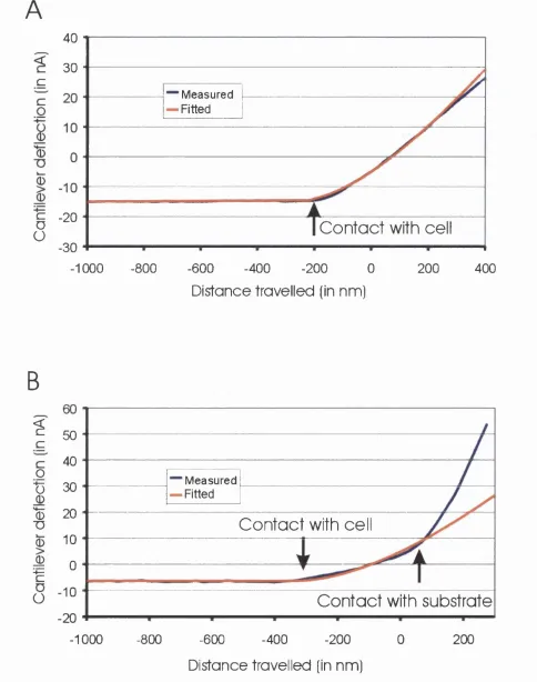
3.2.5 Measurement o f Material Properties...73
3.2.6 Determining the Surface S trains...73
3.2.7 Experimental Validation...74
3.2.8 Error A n a lysis...74
3.2.9 F E M M odelling...76
3.2.10 Statistics...76
3.3 Re s u l t s...76
3.3.1 Cellular R eactions...76
3.3.2 Surface S tra in s...78
3.3.3 Experimental validation and Error A n a lysis...80
3.3.4 Strain D istributions...80
3.3.5 Influence o f the Poisson ratio and the cell thickness on the strain distribution...82
3.4 Dis c u s s io n...82
CHAPTER 4 - SINGLE CELL MECHANOTRANSDUCTION AND ITS MODULATION ANALYSED BY ATOMIC FORCE MICROSCOPE INDENTATION...86
4.1 In t r o d u c t io n...86
4 .2 Ma t e r ia l sa n d Me t h o d s...87
4.2.1 Cell Culture a nd Histological Staining...87
4.2.2 Im m unostaining and Confocal M icroscopy...87
4.2.3 Functional Gap Junctional Communication A ssa y...88
4.2.4 Intracellular Calcium M easurem ents...88
4.2.5 Atomic Force M icroscopy...88
4.2.6 Experimental procedure...88
4.2.7 Spontaneous calcium w aves...88
4.2.8 M easurement o f Material Properties...88
4.2.9 Determining Cellular Strains...88
4.2.10 Inhibitor Studies...89
4.2.10.1 Calcium entry pathway... 89
4.2.10.2 Cytoskeletal modulation o f mechanical signal transduction... 89
4.2.11 M orphological effects o f cytoskeletal disruption...90
4.2.12 Data Analysis and Statistics...90
10
4.3.1 P henotype...91
4.3.2 Spontaneous Calcium Reactions...91
4.3.3 M echanically stim ulated calcium reactions...91
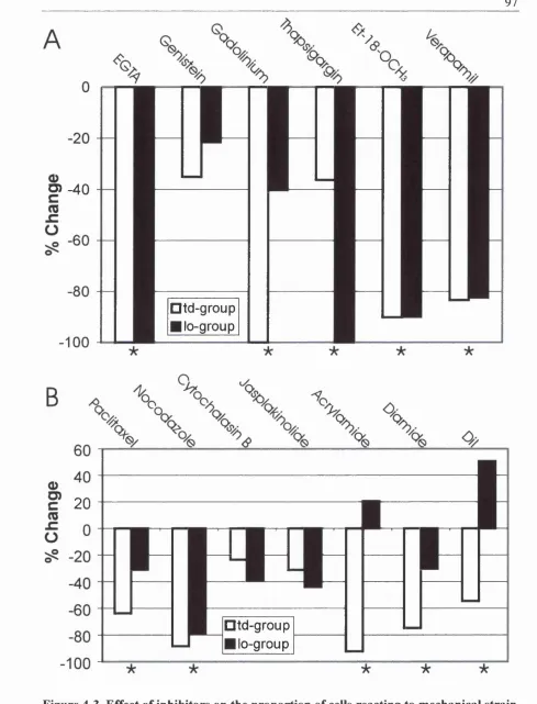
4.3.4 Propagation o f Signal and Presence o f Functional Gap-Junctions and Gap-Junctional Proteins94 4.3.5 Pathway inhibitors (Figure 4-3A )...94
4.3.6 Cytoskeletal disrupting drugs (Figure 4 -3 B )...96
4.3.7 Confocal microscopy o f the cytoskeleton after disruption...96
4 .4 Dis c u s s io n... 99
CHAPTER 5 - DIRECT ESTIMATION OF INTRACELLULAR STRAINS USING DIGITAL STRAIN ESTIMATION AND GFP TRANSFECTED CELLS... 106
5.1 In t r o d u c t io n...106
5 .2 Me t h o d s... 109
5.2.1 Cell culture...109
5.2.2 Confocal Imaging a nd intracellular calcium im aging...109
5.2.3 M echanical Stim ulation...109
5.2.4 D igital Strain Estim ation...110
5.2.5 Translation Suppression...110
5.2.6 Local Displacement Calculation...110
5.2.7 Subpixel Displacements and Sm oothing...I l l 5.2.8 D elaunay Triangulation...113
5.2.9 Strain Calculation...113
5.2.10 Validation...113
5.2.11 Analysis o f Intracellular Calcium Experim ents...113
5.2.12 Assessment o f Potential Cellular D am age...114
5.2.13 Strain Fields D ue to M icropipette Indentation in a Homogenous Continuum...114
5.2.14 Data Analysis and Statistics...114
5.3 Re s u l t s...114
5.3.1 Validation o f the method...114
5.3.2 Strain Distributions in Cells, Comparison to the Continuum M o d e l...122
5.3.3 Nuclear Behaviour in Response to M echanical Stimulation...122
5.3.4 Intracellular Strains and Calcium Concentrations...122
5.3.5 M echanically-induced Tubulin Depolymerisation andA ctin Contraction...128
5 .4 Dis c u s s io n...128
CHAPTER 6 - ESTIMATING THE SENSITIVITY OF MECHANOSENSITIVE ION CHANNELS TO MECHANICAL STRAIN... 133
6.1 In t r o d u c t io n...133
6.2 Me t h o d s... 135
6.2.1 Cell C ulture...135
6.2.2 Patch Clamp R ecordings...135
6.2.3 M echanical Stimulus and Pressure M easurem ent...136
__________________________________________________ n
6.2.5 Bleb H eight M easurem ent...137
6.2.6 Electrophysiology Data A nalysis...137
6.2.7 Finite Elem ent M odelling...138
6.2.8 Curve F ittin g...140
6.2.9 A F M Indentation and Analysis...141
6.2.9.1 Experimental procedure... 141
6.2.9.2 Importance o f Intracellular Sodium Metabolism in the Generation o f Intracellular Calcium Concentration Increases... 142
6.3 Re s u l t s...142
6.3.1 Electrophysiology...142
6.3.2 Video M icroscopy...145
6.3.3 Finite Element M odelling...145
6.3.3.1 Strain Distributions...145
6.3.3.2 Influence o f Mechanical Parameters on Linear Elastic M odels... 146
6.3.3.3 Cellular Strain Needed to Open Mechanosensitive Channels... 149
6.3.3.4 Variation o f Cellular Elasticity with Applied Strain...149
6.3.4 A F M D ata...151
6.4 Dis c u s s io n...153
Ap p e n d ix I: Th e o r e t ic a l Mo d e l... 159
CHAPTER 7 - SODIUM WAVES PRECEDE CALCIUM WAVES IN CELL SIGNALLING... 162
7.1 In t r o d u c t io n...162
7.2 Me t h o d s... 164
7.2.1 Cell Culture...164
7.2.2 Experimental P rotocol...164
7.2.2.1 Incubation and im aging... 164
7.2.2.2 Pipette Poking...166
7.2.2.3 Assessment o f Cellular Integrity... 166
7.2.3 Data A n a lysis...166
7.2.3.1 Whole Cell Time Course and Image Generation... 166
7.2.3.2 Line-Scan Time Course and Image Generation... 167
7.2.3.3 Curve A nalysis... 167
7.2.3.4 Controls for Timing Experiments... 168
7.2.4 Inhibitor Studies...168
7.2.5 Statistics...169
7.3 Re s u l t s...170
7.3.1 Response to M echanical Stim ulation...170
7.3.1.1 Stimulated c e lls ... 170
7.3.1.2 Non-stimulated c e ll s ...170
7.3.2 Inhibitor Studies...173
7.3.2.1 Effect o f Inhibitors on Increases in Sodium ...173
7.3.2.2 Effect o f Inhibitors on Increases in Calcium Concentration...173
7.3.2.3 Effect o f Inhibitors on Signal Propagation...177
12
7.3.3 Cell Integrity after M echanical Stimulation...180
7.4 Dis c u s s io n... 180
7.4.1 Cellular Integrity after M echanical Stimulation...181
7.4.2 Propagation o f Sodium and Calcium Waves...182
7.4.3 Ionic Increases in the Stimulated C ells...183
7.4.4 Signal P ropagation...185
7.4.5 Adjacent C ells...185
7.4.6 Timings o f the ionic increases...186
7.4.7 Conclusion...187
CHAPTER 8 - DETERMINATION OF CELLULAR STRAINS BY COMBINED ATOMIC FORCE MICROSCOPY AND FINITE ELEMENT MODELLING... 188
8.1 In t r o d u c t io n...188
8.2 Me t h o d s... 190
8.2.1 Experimental d ata...190
8.2.1.1 Cell Culture...190
8.2.1.2 Immuno-staining and Confocal Microscopy...190
8.2.1.3 Atomic Force M icroscopy... 190
8.2.1.4 Material Property Measurement... 191
8.2.2 Numerical M odelling fo r Whole Cell M echanical M odels...191
8.2.2.1 Generation o f Whole Cell M odels... 191
5.2.2.2 Physical M odel... 192
8.2.2.3 Boundary conditions...192
8.2.2.4 Generation o f Osteocyte M odels... 193
8.2.3 Numerical M odelling fo r F luid Shear Simulations...193
8.2.3.1 Generation o f the M odels... 194
8.2.3.2 Computational Fluid Dynamics: Physical Model and Boundary Conditions...194
8.2.3.3 Finite Element Modelling: Physical Model and Boundary Conditions... 195
8.2.3.4 Variation o f the Physical Parameters...195
8.2.4 Numerical M odelling fo r Micromanipulation M o d e ls...195
8.2.4.1 Magnetic Microbead Pulling...196
8.2.4.2 Microbead T w isting...197
8.2.4.3 Micropipette Poking...198
8.2.5 Sensitivity o f M echano-Detection M echanism s...198
8.2.6 Adaptation to M echanical Strain...199
8.2.7 Cellular Strain Resulting fro m Stray Fluid Flow...199
8.2.8 Statistics and Curve-Fitting...199
8.3 Re s u l t s...199
8.3.1 Experimentally M easured M aterial Properties o f Osteoblasts...199
8.3.2 Strain Distributions and M agnitudes...201
8.3.2.1 Whole Cell M od els... 201
8.3.2.2 Micromanipulation M odels... 206
8.3.3 Effect o f M aterial Property Changes...210
_____________________________________________________________________________________ n
8.3.3.2 Micromanipulation M odels...210
8.3.4 Effect o f Poisson Ratio Changes...210
8.3.4.1 Whole Cell M od els... 210
8.3.4.2 Micromanipulation M odels... 210
8.3.5 Effect o f the Direction o f Application o f Stim ulus...211
8.3.6 Effect o f Fluid Flow Param eters...211
8.3.7 Osteocytes...211
8.3.8 Cellular Strain Elicited by Stray Fluid F lo w...212
8.4 Dis c u s s io n...215
8.4.1 Cellular Strain Detection M echanisms...215
8.4.2 Importance o f the Cellular Poisson R atio...217
8.4.3 Cellular Adaptation to M echanical Perturbation...218
8.4.4 Strain Detection in O steocytes...219
8.4.5 Limitations o f the Integrated M easurement and M odelling Process...220
8.4.6 Conclusion...221
CHAPTER 9 - GENERAL CONCLUSIONS... 222
9.1 Su m m a r y...222
9.1.1 M echanical Stimulation o f Osteoblastic Cells by A F M and Cellular Strain D istributions...222
9.1.2 Sensitivity o f Osteoblastic Cells to M echanical Strain and Its M odulation...222
9.1.3 Experimental Determination o f Cellular Strain Distributions and Sensitivity to M echanical Strain ...223
9.1.4 Sensitivity o f Stretch-Activated Channels to M embrane Strain in O steoblasts...223
9.1.5 Intracellular and Intercellular Sodium and Calcium W aves...223
9.1.6 Estimation o f the Cellular Strains Elicited by whole Cell a nd Micromanipulation Techniques..224
9.2 M e c h a n o t r a n s d u c t i o n b yB o n e C e l l s ... 2 2 4 9.3 Cy t o m e c h a n ic s... 229
_____________________________________________________________________________________ u
List of Tables
Table 1-1. Gene expression in in vitro cell cultures in response to mechanical stimulation.
...38
Table 2-1. List o f antibodies...57
Table 2-II. List o f Inhibitors or Modulators... 69
Table 4-1. Effect o f cytoskeletal disruption on cell height and cell volume... 99
Table 5-1. Cellular Strain resulting from thermal fluctuations and mechanical stimulation. ...123
Table 8-1. Strain magnitudes resulting from whole cell mechanical stim ulation...201
Table 8-II. Strain magnitudes resulting from micromanipulation...206
Table 8-III. Strain magnitudes in an osteocyte embedded in the bone matrix... 211
Table 8-1V. Additional cellular strains induced by stray fluid flow in experimental substrate stretch systems... 212
15
List of Figures
Figure 1-1. Bone Structure...21
Figure 1-2. Bone adaptation to mechanical strain...24
Figure 1-3. Bone remodelling cycle...28
Figure 1-4. Bone modelling...29
Figure 1-5. Cellular Mechanosensory Pathways, Crystal Structure o f a Stretch-Activated Ion Channel, and Tensegrity Architecture...31
Figure 1-6. Images o f bone cells using atomic force, confocal, and scanning electron microscopies...41
Figure 1-7. Principles o f atomic force microscopy...43
Figure 1-8. Applications o f finite element modelling to biology...49
Figure 2-1. Microscopy images o f osteoblastic cells and B16 F I melanoma cells...54
Figure 2-2. AFM-inverted microscope interface...60
Figure 2-3. Modification o f the A F M tip, optical light path, and mechanical stimulation with a micropipette...61
Figure 2-4. Schematic representation o f the indentation o f an infinite half-plane by a spherical or a conical indentor...63
Figure 2-5. F or ce-deflection curves on a thick and a thin cellular material...65
Figure 3-1. Osteoblasts can be mechanically stimulated by A F M micro-indentation and react by increasing their intracellular calcium levels... 77
Figure 3-2. Strain in the membrane as a function o f distance from indentation centre...78
Figure 3-3. Indentation by a spherical indentor o f a live osteoblast loaded with Calcein-...79
Figure 3-4. Strain distributions elicited by A F M indentation...81
Figure 3-5. Effect o f the Poisson ratio and cell thickness on the mechanical strains elicited by A F M micro-indentation...83
Figure 4-1. Osteoblasts respond to a mechanical stimulus by a rise in intracellular calcium concentration...93
Figure 4-2. Mechanically-induced intracellular calcium transients can be transmitted to neighbouring cells. Osteoblasts possess gap-junctional protein connexin-43 and functional gap-junctions...95
^
Figure 4-4. Effect o f cytoskeletal treatments on cytoskeletal organisation and cellular
profile...98
Figure 4-5. Calcium entry pathways fo r contact and stress relaxation reactions.
M echanical model o f the cell....103
Figure 5-1. Flowchart o f the operations effected during digital strain estimation...112
Figure 5-2. Validation o f the use o f digital strain estimation in GFP-actin transfected cells.
...y y j
Figure 5-3. Strain distribution elicited by A F M microindentation in a GFP-tubulin
transfected cell...117
Figure 5-4. The nucleus moves as a rigid body within the cytoskeleton in GFP-actin and
GFP-tubulin cells, and is minimally deformed....119
Figure 5-5. Deformations in a homogenous continuum due to indentation by a spherical
indentor predicted by finite-elem ent modelling....121
Figure 5-6. Intracellular strain distribution measured in GFP-actin transfected cells
during mechanical stimulation giving rise to an increase in intracellular calcium...124
Figure 5-7. Intracellular strain distribution measured in GFP-actin transfected cells
during mechanical stimulation giving rise to injury and an increase in intracellular
calcium...125
Figure 5-8. Intracellular strain distribution measured in GFP-tubulin transfected cells
during mechanical stimulation giving rise to an increase in intracellular calcium...126
Figure 5-9. Mechanical stimulation can elicit catastrophic depolymerisation o f the tubulin
network and retraction o f the actin cytoskeleton...127
Figure 6-1. Pressure application through micropipette aspiration elicits opening o f
mechanosensitive channels in prim ary osteoblasts...144
Figure 6-2. Video-microscopy o f membrane aspiration into the micropipette...147
Figure 6-3. Results o f the fin ite element simulations fo r a linear elastic material...148
Figure 6-4. Results o f the fin ite element simulations fo r the models incorporating p re
strain and linear stiffening...150
Figure 6-5. Experimental data fro m Atomic Force Microscopy micro-indentation...152
Figure 6-6. Proposed model fo r the modulation o f cell sensitivity to mechanical strain
through stretch-activated channels...155
Figure Al-1. Diagram o f the aspiration o f a bleb o f membrane into a micropipette...161
_____________________________________________________________________________________ n
Figure 7-2. Effect o f inhibitors o f the sodium and calcium pathways and gap-junctional
communication on the increases in intracellular sodium concentrations in the stimulated
and adjacent cells...175
Figure 7-3. Effect o f inhibitors o f the sodium and calcium pathways and gap-junctional communication on the increases in intracellular calcium concentrations in the stimulated and adjacent cells...176
Figure 7-4. Effect o f inhibitors o f the sodium and calcium pathways and gap-junctional communication on the propagation and time difference (tca-tNo) between increases in intracellular calcium and sodium...178
Figure 7-5. Examination o f potential cellular injury during micropipette p oking....179
Figure 7-6. Proposed model o f the intracellular sodium and calcium signalling pathways in stimulated and adjacent cells...184
Figure 8-1. Characterisation and prim ary data acquisition from osteoblasts...200
Figure 8-2. The effect o f substrate stretch...203
Figure 8-3. The effect o f hydrostatic pressure...204
Figure 8-4. The effect o f fluid shear...205
Figure 8-5. The effect o f microbead pulling...207
Figure 8-6. The effect o f microbead twisting...208
1 ^
Chapter 1 - Introduction
1.1 Overview
W ith the lengthening o f life expectancy in most countries and the shift towards a more
sedentary life style, bone fracture is a prevalent risk among an increasing elderly
population. Understanding the cellular pathways involved in bone adaptation to its
mechanical usage is o f great interest for clinical applications. Indeed, bones are load-
bearing structures that adapt to their mechanical usage in a cell-driven process that results
in either the formation o f additional bone or the resorption o f excess bone. Hence, the
study o f the cellular pathways involved in the detection o f mechanical strain is o f great
importance and its understanding may lead to the discovery o f new therapies to counter the
effects o f diseases such as osteoporosis. Each year, in the United States alone, osteoporosis
is the cause o f an estimated 260 000 hip fractures in patients o f over 65 years o f age (as
cited in Keyak et al., 1998). Due to complications o f the fractures, a significant number o f
the patients die and the lifestyle o f the majority is significantly impaired. Recently, much
hope has been placed in bisphosphonate-based treatments. These drugs, which inhibit the
resorptive phase o f bone remodelling with great efficacy, slow the loss o f bone mass.
However, they interrupt the remodelling cycle and prevent the formation o f new bone -ef-
bone. Hence, the quality o f existing bone slowly degrades and micro-cracks accumulate. In
contrast to drugs such as the bisphosphonates, compounds stimulating bone formation
would both stop bone loss and maintain bone quality. For example, this could be achieved
by enhancing the sensitivity o f bone cells to mechanical strain leading to bone formation
even in response to low exercise regimen. However, before such compounds can be
discovered, new means o f quantifying cell sensitivity to mechanical strain have to be
devised.
This introduction will first give an overview o f bone biology and microstructure, then
discuss bone adaptation to mechanical strain and the cellular means o f detecting
mechanical strain. Second, it will detail applications o f Atomic Force Microscopy (AFM)
to biology. Third, the principles and biological applications o f Finite Element Modelling
(FEM) and Computational Fluid Dynamics (CFD) will be discussed. Finally, an outline o f
______________________________________________________________________________________19^
1.2 Bone Cells
There are three main types o f cells in bone: osteoblasts, osteoclasts and osteocytes. All
three are involved in the maintenance and synthesis o f the bone tissue throughout life.
They are engaged in two major processes: bone modelling whereby bone is formed or
adapted to a new mechanical environment (formation > resorption) and bone remodelling
whereby bone maintenance is performed (formation = resorption). Both processes occur as
a result o f a synchronised action between these three types o f cells (Marks and Hermey,
1996).
1.2.1 Osteoblasts and Bone-lining cells
Osteoblasts (Figs 1-6A, 1-6B and 1-6C) are mono-nucleated cuboidal cells derived from
multi-potential stem cells that can also differentiate into chondrocytes, adipocytes or
myoblasts (Alberts et al., 1994; Grigoriadis et al., 1988; Owen and Friedenstein, 1988).
They are responsible for the synthesis o f the organic components o f the bone matrix in the
form o f unmineralised osteoid (Junqueira et al., 1989). The osteoid is composed o f type I
collagen, proteoglycans and glycoproteins. Osteoblasts initiate mineralisation via alkaline
phosphatase and the osteoid becomes mineralised with hydroxyapatite crystals
(Caio(P0 4)6(OH)2). As bone matrix deposition proceeds, some osteoblasts become trapped in the newly deposited matrix and differentiate into osteocytes. Quiescent surface
osteoblasts turn into bone lining cells, which can be reactivated upon mechanical
stimulation to become active osteoblasts (Chow et al., 1998a). Bone-lining cells display a
flattened morphology, cover all o f the bone surfaces and communicate with the osteocytes
via linked cellular extensions (Miller and Jee, 1989; Palumbo et al., 1990; Yellowley et al.,
2000).
1.2.2 Osteocytes
Osteocytes, while possessing most o f the characteristics o f osteoblasts, have lost the ability
to synthesise osteoid. They reside in a cavity, called a lacuna, surrounded by a
proteoglycan-rich fluid space (Fig 1-1). Osteocytes develop filopodial processes, which
serve to link them to other osteocytes and to bone lining cells on the bone surface through
gap junctions (Kusuzaki et al., 2000; Marotti et al., 1995; Palumbo et al., 1990; Sissons et
al., 1990; Yellowley et al., 2000). These fllopodial contacts provide a mechanism whereby
the osteocytes receive nutrients and metabolites from blood vessels (Cowin et al., 1991).
The cavities in which these fllopodial processes reside are known as canaliculi. Osteocytes
^
1.2.3 Osteoclasts
Osteoclasts are large multi-nucleated cells derived from the fusion o f blood-derived
mononuclear cells (Fig 1-6C) (Junqueira et al., 1989). Their formation is controlled by the
osteoblasts through the balance in synthesis o f the membrane protein receptor activator o f
nuclear factor kB ligand (RANKL), which binds to RANK receptors on forming and
mature osteoclasts, and the soluble decoy-receptor osteoprotegerin (OPG), which competes
with RANK for RANKL binding sites (Teitelbaum, 2000). As bone resorption starts,
osteoclasts form a ring-shaped F-actin rich zone (the ‘clear zone’) which contributes to the
creation o f a well-isolated microenvironment between the cell and the bone matrix (the
seal is impermeable to molecules above 40 000 Da; Stenbeck and Horton, 2000). This
microenvironment is acidified to a pH o f approximately 4.5 through the action o f
osteoclast H^-ATPases (Sundquist et al., 1990), which degrades the mineral component o f
the bone matrix. The organic matrix o f bone is degraded using acid cathepsins (especially
cathepsin K), neutral collagenases and other proteolytic enzymes secreted by osteoclasts
(Vaananen and Harkonen, 1996). Degraded bone matrix transcytoses through the
hasolateral cell body prior to exocytose from a specialised secretory domain located at the
apical cap (Nesbitt and Horton, 1997; Salo et al., 1997). Osteoclasts have also been
proposed to degrade osteocytes present within the tissue being resorbed (Elmardi et al.,
1990).
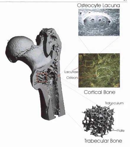
1.3 Bone Microstructure
Osteoblasts secrete two types o f bone: woven or immature bone and lamellar bone. Woven
hone is a temporary structure, which is formed through the random deposition o f collagen
fibres by osteoblasts. It possesses a lower mineral content than lamellar bone and has a
higher proportion o f osteocytes (Junqueira et al., 1989). W oven bone can be synthesised in
any connective tissue and as a consequence, it appears during the formation and the repair
o f bony tissues. In contrast, lamellar bone can only be deposited appositionally onto
previously existing bone and is a permanent tissue. It is deposited in layers (called
lamellae) o f 3 to 7 pm in thickness that are either parallel to each other (as in trabecular
bone) or concentrically organised (as in osteonal systems. Fig 1-1). Successive lamellae
coalesce via a cementing substance, known as the cement line, that consists o f mineralised
matrix with few collagen fibres (Junqueira et al., 1989). The mechanical function o f the
21
Osteocyte Lacuna
L acunae
O steon
Cortical Bone
Trabeculum
Plate
Trabecular Bone
Figure 1-1. Bone Structure.
Micro-computed tomography o f the head o f a split femur. Adult long bone is composed o f two different types o f bony tissue with specific micro-architectures. Trabecular bone (a micro-CT o f which is shown here) is found at the epiphyses o f bones. It is composed o f a complex assembly o f struts (or trabecula) and plates. Mechanically, it serves to redirect the forces applied at the joints along the long axis o f the bone. The internal spaces within the trabecular bone are filled with haemopoietic bone marrow. The external portion o f bone is composed o f a dense layer o f cortical or compact bone that is designed to work in compression. Cortical bone (a transversal cut o f which is shown here) is composed o f cylindrical units called osteons. Osteons are composed o f concentrically deposited layers o f lamellar bone separated by cement lines and centred on a blood vessel called the Haversian canal. During the synthesis o f bone, some osteoblasts get trapped within the matrix and become osteocytes. The cavity in which they reside is called an osteocyte lacuna (black areas in the region boxed in yellow).
^
Lamellar bone is found in two structures throughout the body: trabecular bone and cortical
or compact bone (Fig 1-1). Trabecular bone is found in the extremity o f long bones. It can
be pictured as an assembly o f interconnected beams and plates (Figs 1-1, 1-2B and 1-2C).
The beams are referred to as trabeculae and they are formed o f parallel lamellae o f cortical
bone. These lamellae house a number o f osteocytes. As the osteocyte nutrients are supplied
merely through diffusion, the trabeculae are limited in size to a maximum diameter o f 200
to 300 pm.
Cortical bone is composed o f a parallel assembly o f units called osteons or Haversian
systems (Fig 1-1). Osteons can be envisioned as thick-walled hollow cylinders with a
blood vessel running through the central cavity. The cavity in which the blood vessel lies is
called a Haversian canal. Haversian canals are linked to the periosteal bone surfaces by
horizontal vessels called Volkmann’s canals. This system serves to supply nutrients to the
osteocytes that are trapped between the lamellae o f bone surrounding the Haversian canals.
Osteons are composed o f 4 to 20 concentric lamellae, the number being limited by
constraints on nutrient diffusion to the osteocytes furthest away from the Haversian canal.
Outer and inner circumferential lamellae serve to give the periosteum and endosteum their
circular shapes. Interstitial lamellae fill in the gaps between osteons.
1.4 Bone Adaptation
Bone is a structure that is continuously being turned over. At any given time,
approximately 15% o f the adult skeleton is undergoing remodelling. In particular, if bone
is submitted to increased usage (e.g. heavy exercising), its mass increases, whereas, if bone
is in a state o f disuse (e.g. bed rest or long term space travel; Lucas, 1977), bone mass is
lost (Leblanc et al., 1990; McCarthy et al., 2000; Vico et al., 1998). However, use or disuse
are not the only factors dictating bone mass. Indeed, environmental factors (such as diet or
stress), hormones (such as estrogen), and the genetic inheritance also play a role.
1.4.1 Whole Bone and Tissue Level Adaptation
In 1881, Roux suggested that bone adaptation and formation were the result o f locally
regulated cellular processes (Huiskes, 2000; Roux, 1881). Comparing the trabecular
patterns to the stress trajectories in a similarly shaped continuous material, W olff first
introduced the idea that bone is a structure adapted to its mechanical environment (Wolff,
1892): "''The law o f bone remodelling is the law according to which alterations o f the
_____________________________________________________________________________________n
occur as a consequence ofprim ary changes in the shape... or in the stressing o f the bones^^
(From Marcus, 2001). This assumption has become known as W o lffs law.
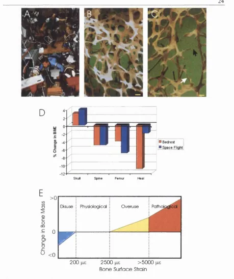
Since then, there have been many in vivo experiments confirming these original
observations. Using strain gauges fixed onto the long bones o f live animals, Lanyon and
Smith (1969) and Rubin and Lanyon (1984a) reported the peak tissue-strain magnitudes in
a variety o f animal species. Because these peak tissue-strains were extremely close
between species and limbs (varying from 2100 to 3200 pa during strenuous exercise), it
was hypothesised that bone might adapt its structure to maintain a certain level o f tissue
strain. This idea was enticing, in particular because strain is an easily measurable variable.
Frost (1973) and Hert et al. (1971) showed that static strains did not influence bone mass in
vivo. Lanyon and Rubin (1984b) mechanically isolated the ulna o f live turkey and
submitted these, via external pins, to a controlled number o f daily loading events o f a given
strain. To assess bone formation, they injected the animals with fluorescent markers at two
different time points. Only four daily loading cycles o f 0.1% strain (1000 ps) were needed
to maintain bone mass. If a higher strain magnitude was applied for the same number o f
cycles or a similar strain magnitude for a larger number o f cycles, bone mass was
increased.
Based on these observations, a law relating the applied mechanical strain to the evolution
o f bone mass was proposed by Frost in 1987 (the mechanostat theory. Fig 2D) (Frost,
1987). Duncan and Turner (1995) reported a fourfold increase in bone formation when the
loading frequency was 2 MHz as compared to static loading. Turner et al. (1995) reported
that the amount o f bone formation is directly proportional to the rate o f strain in the bone
tissue. Mosley and Lanyon (1998) showed that higher strain rates elicited a higher
osteogenic response in rat ulnae. Chow et al. (1998a) showed that a single 5 minute strain
episode could reactivate bone lining cells without extra recruitment o f osteoblasts. Using a
novel approach in which a bone defect is created within a dog tibia and mechanically
isolated in a porous titanium chamber implanted in the tibia, Guldberg et al.(1997) showed
that dynamic mechanical loading enhances the amount o f bone formed as well as the
connectivity o f the newly formed trabecular structure.
It should be noted that all o f the strains reported from in vivo measurements are tissue level
strains (i.e. whole bone strains) measured with strain gauges. Because bone is a composite
24
D
m
Bedrest S p ace Flight
Skul Spine Femur Heal
>0
0 0 P o
^ <0
Disuse Physiological Overuse J o f h o l p ^ ^
y
200 jiG 2500 )ie >5000 jie
Bone Surface Strain
Figure 1-2. Bone adaptation to mechanical strain.
A, Long term space flight or colonisation o f planets with different gravities than earth (Lucas, 1977) will acutely pose the problem o f bone loss during micro-gravity on return to the home planet.
B, Scanning electron microscopy picture o f a normal trabecular bone network (courtesy o f Alan Boyde). Bar=100 pm.
C, Scanning electron microscopy picture o f osteoporotic trabecular bone. Many trabeculae have been lost and the network is less dense. The remaining trabeculae are thinner (white arrow) and some have been perforated and cut by osteoclastic resorption (black arrow). Bar=100 pm.
D, Changes in bone mineral density in the skull and lower limb bones resulting from a long period o f bedrest or space flight. Data taken from (Leblanc et al., 1990; McCarthy et al., 2000).
^
Volkm ann’s canals) and a complex micro-architecture, strain magnitude within bone
would not be expected to be the same as on the bone surface. Indeed, strains around
osteocyte lacunae have been reported to be up to one order o f magnitude larger than tissue
level strains (Hollister et ah, 1994). Therefore, the widely cited physiological strain levels
o f 1000-3000 ps are truly physiological only on the bone surface (Donahue et al., 1995;
Mosley and Lanyon, 1998; Rubin and Lanyon, 1984b).
Bone loss due to disuse has been studied with much attention in animals (using the rat tail
suspension assay; Morey-Holton and Globus, 1998) and in humans (either after long-term
space flights or in bedrest studies). Most interest stemmed from the fact that, very early in
the space flight programs o f the USA and the USSR, an increase in urinary calcium was
noticed after as little as 3 days in flight indicating a loss o f skeletal calcium (Vico et al.,
1998) (Fig 1-2A). In studies o f the effect o f bed rest on bone loss, bone resorption
increased both in the cancellous and the cortical bone and surfaces o f osteoblast activation
were reduced after twelve weeks (Vico et al., 1998). Interestingly, in bedrest experiments
or long-term space flights, bone loss seems to vary from location to location in the skeleton
with the load-bearing bones being more affected (Leblanc et al., 1990; McCarthy et al.,
2000; Vico et al., 1998) (Fig 1-2C).
1.4.2 Bone Remodelling Cycle
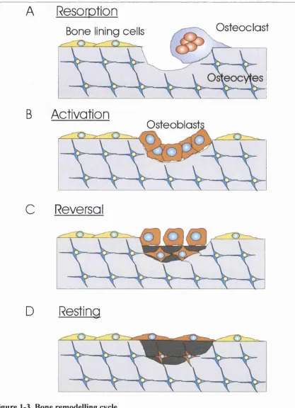
Remodelling allows the skeleton to locally adapt bone mass to applied mechanical stimuli
and to repair damage to the bone matrix (Fig 1-3). Initiation o f the remodelling cycle in a
particular location can either be the result o f a stochastic process or cellular signalling (Ott
SM, 2001).
In normal physiology, the first step o f the remodelling process is the recruitment o f
osteoclasts-precursors to the bone remodelling unit (BMU) and the subsequent formation
o f osteoclasts in the region, presumably through increased expression o f RANKL on bone
lining cell membrane (see paragraph 1.2.3). The newly formed osteoclasts resorb the
existing bone matrix (resorption phase. Fig 1-3 A) before apoptosing after approximately
12 days (Ott SM, 2001). Different processes o f resorption exist for cortical and trabecular
bone. In cortical bone, the osteoclasts tunnel through the bone matrix creating what is
known as a ‘cutting cone’. Cutting cones are 100 to 200 pm wide and up to 4 mm long
(Parfitt, 2001). In trabecular bone, osteoclasts excavate a shallow lacuna known as
^
derived either from the existing bone lining cells (Chow et ah, 1998a), or from newly
recruited osteoblast precursors, follow the osteoclasts and invade the resorption pit once
they have left the area (activation phase. Fig 1-3B), The osteoblasts then secrete osteoid
(demineralised bone matrix) to replace the resorbed matrix (Reversal phase. Fig 1-3C).
During the process some osteoblasts are trapped within the bone matrix (Fig 1-3C) and
mature into osteocytes (For nice pictures o f this, see: Kusuzaki et ah (2000) and Marotti et
ah (1995)). After 15 days, the osteoid begins to mineralise via the action o f alkaline
phosphatase secreted by the osteoblasts (Ott SM, 2001).
In normal physiology, the amount o f bone mass secreted during the reversal phase is
exactly equal to that resorbed. However, during bone adaptation to enhanced mechanical
strain, the amount o f bone secreted exceeds the amount o f resorbed matrix (Fig 1-4B). In
contrast, post-menopause or in cases o f disuse, bone resorption exceeds bone secretion,
leading to global loss o f bone mass (Fig 1-4C). Once the reversal phase is complete, the
osteoblasts become quiescent and acquire the flattened morphology o f bone-lining cells
(Fig 1-3D).
Recently, it has been shown that resorption in trabecular bone may take place in
specialised compartments separated from the marrow by a bone-lining cell monolayer that
entirely covers the resorption pit during excavation and synthesis o f bone (Hauge et al.,
2001). How pit depth, area or resorption duration are regulated is, as o f yet, unknown.
However, detection o f mechanical strain gradients by osteoclasts could play a role in
directing the resorption direction (Martin and Burr, 1989; Smit and Burger, 2000). In
certain cases, such as mechanically induced bone formation, the prior resorption o f matrix
does not seem necessary for appositional bone deposition to proceed (Fig 1-4B). Indeed, a
single period o f mechanical stimulation can give rise to an osteogenic stimulus (Chambers
et al., 1993) and this is unaffected if resorption is blocked by administration o f a large dose
o f bisphosphonates (Jagger et al., 1995).
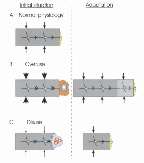
1.5 Detection o f Strain Is a Cell-driven Process
As early as 1881, Roux (1881) suggested that the formation and adaptation o f trabecular
bone to its function was a process regulated by the cells within bone. Over the past decade,
there has been a growing consensus that osteocytes or bone-lining cells act as the sensing
system in bone (Fig 1-4) (Cowin et al., 1991; Huiskes, 2000; Lanyon, 1993; Martin, 2002;
^
types have been shown to be mechanically sensitive in vitro and have a wide distribution
throughout bone. Osteocytes have been favoured as they are ideally situated to sense
strains applied onto the bone matrix and serve no other obvious purpose in bone biology.
Furthermore, osteocytes are linked to each othei and to bone lining cells through
gap-junction lined cellular extensions thereby forming a cellular network that extends from the
matrix to the bone surface (Figs 1-3 and 1-4) (Kusuzaki et al., 2000; Palumbo et al., 1990;
Weinbaum et al., 1994; Yellowley et al., 2000). Bone lining cells cover more than 80% o f
the trabecular and endocortical surfaces o f adult bone (Parfitt, 1983) and can be reactivated
by a single loading event (Chow et al., 1998a).
Computer simulations o f bone adaptation to mechanical strain have provided many
insights into the cell types involved in the detection of, and adaptation to, mechanical
stimuli. Mullender et al.(1997) showed that remodelling directed by the osteocytic network
yielded structures that resisted the same applied loads with less bone mass than those
resulting from remodelling initiated solely by bone-lining cells. Smith et al.(1997) showed
that both osteocytes and bone-lining cells could direct trabecular bone adaptation. Koontz
et al.(2001) showed that strain detection and adaptation by bone-lining
cells could create a well-connected trabecular network from a random distribution o f
unconnected bone nodules. The simulated networks compared favourably to biopsies o f
newly formed trabecular bone submitted to unidirectional cyclic compressive strain in a
bone chamber (Guldberg et al., 1997).
1.6 What Stimulus Do Bone Cells Sense?
There have been many hypotheses as to the nature o f the mechanical stimulus that bone
cells detect including membrane stretch, fluid shear, streaming potentials, and matrix strain
(Fig 1-5A).
Bone cells have been proposed to directly sense the deformations applied onto the bone
matrix through the resulting changes in cell length (Frost, 1987; Lanyon, 1996). In vitro
experiments on osteoblastic cells cultured on .stretchable substrates have shown that
osteoblasts are responsive to deformations applied to their matrix (Fermor et al., 1998;
28
A
Resorption
Bone lining ceils
Osteoclast
B
Activation
Osteoblast
C
Reversai
D
Resting
Figure 1-3. Bone remodelling cycle.
A, Osteoclasts are recruited to the site o f resorption and activated. They form a tight seal between the bone surface and the cell membrane forming a micro-environment favourable to bone degradation and start resorbing the bone matrix.
B, Cells o f the osteoblastic lineage invade the resorption cavity once the osteoclast has left it and acquire the typical cuboidal morphology o f matrix secreting osteoblasts.
C, N ew unmineralised osteoid is synthesised in the bone resorption cavity. During synthesis, some osteoblasts become trapped within the newly formed matrix and start connecting with pre-existing osteocytes.
29
Initial situation
A Normal physiology
B
C
Overuse
Disuse
Adoptotion
Figure 1-4. Bone modelling.
A, In normal physiology, osteocytes detect mechanical strain applied onto the bone matrix and send signals to bone lining cells. B, If higher strains than normal are applied, the osteocytic signals cause the bone lining cells to revert to the matrix synthesising osteoblast phenotype. New bone matrix is deposited until the level o f strains sensed by osteocytes returns to physiological values. After synthesis has ended, the osteoblasts become quiescent bone lining cells.
C, In case o f disuse, the strains applied onto the bone matrix are too small to be sensed by the osteocytes and these do not send any signals to the bone lining cells. Osteoclastic resorption ensues until matrix strains are returned to physiological values.
^
Deformation o f the bony tissue causes the circulation o f extra-vascular fluids through the
bone (Knothe Tate et ah, 2000), which can give rise to shear stresses up to 3 Pa (30
dyn.cm'^) around the osteocytes and osteocytic processes (Weinbaum et ah, 1994). Hence,
shear stress has also been proposed to be the stimulus to which bone cells are responsive.
Furthermore, in vitro experiments on both osteocytes and osteoblasts have shown that both
cell types are responsive to the level o f shear stress predicted (Ajubi et ah, 1999; Smalt et
ah, 1997; You et ah, 2000a).
A consequence o f load-induced fluid flow is the displacement o f ionic charges through the
bone matrix. This creates streaming potentials that have been proposed as a potential
stimulus for the detection o f bone deformation (Harrigan and Hamilton, 1993). However,
this theory has been seriously discredited by two recent experiments (Bakker et ah, 2001;
Hung et ah, 1996a) in which the streaming potential was varied without varying the shear
stress. No changes in the cellular reactions were observed, hence invalidating the theory.
Finally, micro-cracks, or fatigue micro-damage, in the bone matrix have been proposed as
a remodelling stimulus based on the spatial proximity between micro-cracks and apoptotic
osteocytes and bone remodelling units (Martin, 2002; Reilly, 2000; Verborgt et ah, 2000).
1.7 Strain Sensing by Ceiis
Virtually all cell types have been reported to adapt to their mechanical environment
(Donahue et ah, 1995). Direct sensing o f strain by cells can be mediated by a variety o f
means that, as a first step, involve either stretch-activated cation channels, integrin
transmembrane receptors, G-proteins or tyrosine kinases (Fig 1-5A) (Sachs and Morris,
1998; Gudi et ah, 1998; Banes et ah, 1995; Malek and Izumo, 1996). All o f these have
been reported in osteoblasts.
Stretch-activated (or mechano-sensitive) ion channels are ion channels that open in
response to stretch applied onto the cell membrane were originally discovered by Guharay
and Sachs (1984). They have since been identified in a variety o f cells through their
functional properties using patch-clamp electrophysiology (Sachs and Morris, 1998), and
mechano-sensitive channel openings can give rise to whole cell intracellular calcium
31
M em brane
Stretch
Fluid Shear
G-Protein
a ac tin in N u C l e U S i Vlnojlin
Podllln — Tciln
Û
Substrate
Stretch
Side view
c
Closed intermediate Open
Top view
m
m
mFigure 1-5. Cellular Mechanosensory Pathways, Crystal Structure of a
Stretch-Activated Ion Channel, and Tensegrity Architecture.
A, Mechanosensory pathways in cells. Stretch applied onto the cell membrane can be sensed through ion channels that open in response to membrane tension. Stretch applied onto the cell substrate can be sensed via deformations o f the cytoskeleton relayed by the integrin transmembrane receptors. Fluid shear stresses resulting from viscous fluid flow over the cells can be detected either through stretch-activated ion channels, integrin receptors, or through local changes in membrane fluidity detected by G-proteins.
B, Crystal structure o f a prokaryotic mechanosensitive ion channel viewed from the side and from the top. As more tension is applied onto the cell membrane, the channel proteins rearrange and the pore size increases from 1 to 5-6 nm in diameter. From Sukharev et al. (2001a).
^
The amino-acid sequence and crystal structure o f several prokaryotic stretch-activated
channels have been elucidated (Fig 1-5B) (Chang et ah, 1998; Sukharev et ah, 2001a) and
a eukaryotic equivalent was discovered in yeast (Kanzaki et ah, 1999). Stretch-activated
channels have been reported as functional entities in osteoblastic cells (Davidson et ah,
1990; Duncan and Misler, 1989) and implicated in the transduction o f mechanical stimuli
in bone cells (Glogauer et ah, 1997; Hung et ah, 1996b; Rawlinson et ah, 1996; Xia and
Perrier, 1992).
Integrins are transmembrane proteins that link the extracellular matrix to the internal
cytoskeleton and therefore are prime candidates for the detection o f mechanical stimuli
applied onto the extracellular matrix (Banes et ah, 1995; Ingber, 1997b). They have been
reported to be involved in the detection o f mechanical stimulation by osteoblasts by several
groups (Salter et ah, 1997; Schmidt et ah, 1998; Wozniak et ah, 2000).
Recently, it has been shown that G-proteins reconstituted within phospholipid vesicles
increased their GTP-ase activity in response to fluid shear (Gudi et ah, 1998). GTP-ase
activity also increased with increasing vesicle membrane fluidity. The experimentally
determined cell membrane fluidity in living cells increased with the onset o f fluid shear in
the upstream cellular region (Butler et ah, 2001). This would suggest that fluid shear
indirectly activates G-protein activity via an increase in membrane fluidity caused by the
shearing. Finally, non-receptor tyrosine kinases have been implicated in the response o f
cells to fluid shear (Malek and Izumo, 1996), substrate stretch (Carvalho et ah, 2002) and
microbead pulling (Glogauer et ah, 1997).
The pathway involved in the detection o f strain in bone cells remains unknown. However,
it is possible that several pathways participate and mediate the responses o f bone cells to
mechanical strain under different circumstances.
1.8 Cell Mechanics and the Cytoskeleton
The cell cytoskeleton is a complex mechanical structure composed o f an interwoven
network o f actin, tubulin and intermediate-filaments (Fig 1-6B). These networks are
interconnected via a variety cytoskeletal crosslinking proteins (MAP2c, B PA G l, coronin,
etc). The cytoskeleton plays an important role in maintaining the cellular shape and
transducing mechanical signals to the cell nucleus (Forgacs, 1995; Ingber, 1993; Maniotis
_____________________________________________________________________________________ 3 ^
spectrin, filamin A and a variety o f other proteins. Because the cytoskeleton is essential in
resisting mechanical forces, it plays a crucial role in modulating the cellular sensitivity to
mechanical strains by adapting its structure in response to long periods o f mechanical
stimulation (Janmey, 1998; Ko and McCulloch, 2000). The integrity o f the cytoskeletal
components has been shown to be important for the detection and transduction o f
mechanical strain. Malek and Izumo (1996) showed that disruption o f the intermediate
filaments did not affect the cellular realignment in response to fluid flow, whereas
disruption o f the microtubular network did. Toma et al. (1997) showed that disruption o f
the actin microfilaments blocked the up-regulation o f osteopontin in response to substrate
stretch, whereas disruption o f the microtubules did not.
The cytoskeleton is a major determinant o f the material properties o f cells. Disruption o f
the actin network with either cytochalasin B or D or jasplakinolide reduces the elastic
modulus o f cells three-fold when measured by atomic force microscopy (Rotsch and
Radmacher, 2000). Using torque applied through the rotation o f magnetic microbeads,
Wang (1998 and 2001) examined the relative contribution o f each cytoskeletal component
to twist resistance. Depolymerisation o f the F-actin network with cytochalasin D reduced
resistance to torque by 50%, depolymerisation o f the microtubular network reduced it by
2 0%, and acrylamide, a disrupter o f the intermediate filaments, reduced torque resistance by 15%. In contrast, stabilising microtubules with taxol increased stiffness by 10%.
Diamide treatment, which disrupts spectrin, reduces the axial stiffness o f the outer hair
cells o f the cochlea (Adachi and Iwasa, 1997). In summary, the integrity o f the
cytoskeleton is crucial for the maintenance o f cellular elasticity. By modulating its material
properties through cytoskeletal adaptation, the cell can adapt to prolonged mechanical
stimuli and fine tune its sensitivity to mechanical forces (Ko and McCulloch, 2000).
To date, most studies o f cell mechanics have treated the cell as a homogenous continuum
(Boulbitch et al., 2000; Discher et al., 1998; Drury and Dembo, 1999; Evans, 1983; Theret
et al., 1988). However, if the cytoskeleton is considered, this is clearly not true (Fig 1-6B).
The predictions o f continuum theories fit the experimental data well (Drury and Dembo,
2001; Merkel et al., 2000). Although, such simplifying continuum approaches are well-
suited for the prediction o f global cellular deformations, they cannot predict the internal
rearrangement o f the cytoskeleton. Predicting internal reorganisation o f the cytoskeleton
may be o f great importance in the understanding o f detection o f mechanical stimuli