Review
1
What Is the Heart? Anatomy, Form, Function and
2
Misconceptions.
3
Gerald D. Buckberg, M.D., D.Sc.1, Navin C. Nanda, M.D.2 Christopher Nguyen, Ph.D.3 and
4
Mladen J. Kocica M.D. Ph.D.4
5
1 Department of Cardiothoracic Surgery, David Geffen School of Medicine at UCLA, 10833 Le Conte Avenue,
6
Room 62-258 CHS, P.O. Box 951741, Los Angeles, CA 90095-1741, USA; [email protected]
7
2 Division of Cardiovascular Diseases, University of Alabama at Birmingham, 1900 University Boulevard
8
Birmingham, AL 35233, USA; [email protected]
9
3 Cedars-Sinai Medical Center, 8700 Beverly Blvd. PACT, Suite 800, Los Angeles, CA 90048, USA;
10
11
4 UC Clinical Centre of Serbia, Clinic for Cardiac Surgery, 8th Kosta Todorovic St., 11000 Belgrade, Serbia;
12
13
* Correspondence: [email protected]; Tel.: + 310 206-1027; Fax: +310-825-5895; [email protected];
14
Tel.:+381(69)3670-609
15
16
Abstract: Cardiac dynamics are traditionally linked to a left ventricle, right ventricle and septum
17
morphology, a topography that differs from the heart’s 5-century-old anatomic description of
18
containing a helix and circumferential wrap architectural configuration. Torrent Guasp’s helical
19
ventricular myocardial band (HVMB) defines this anatomy and its structure, explains why the
20
heart’s 6 dynamic actions of narrowing, shortening, lengthening, widening, twisting and uncoiling
21
happen.
22
This database raises questions about deductions behind “accepted cardiac mechanics”, and its
23
functional aspects will challenge and overturn them. These suppositions include the LV, RV, and
24
septum description, timing of mitral valve opening, isovolumic relaxation period, reasons for
25
torsion/twisting, untwisting, reasons for longitudinal and circumferential strain, echocardiographic
26
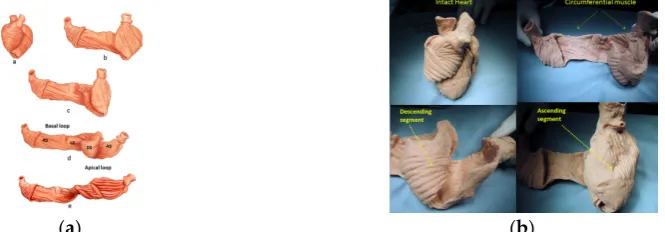
sub segmentation, resynchronization, RV function dynamics, and diastolic dysfunction’s cause and
27
unrecognized septum impairment.
28
Torrent Guasp’s revolutionary contributions may alter future understanding of the diagnosis and
29
treatment of cardiac disease.
30
Keywords: conventional heart anatomy; helical ventricular myocardial band; mitral valve opening;
31
isovolumic relaxation time; RV function; diastolic dysfunction
32
33
1. Introduction
34
35
The current approach to understanding cardiac dynamics relies upon movements that adhere
36
to the conventional topographical separation of cardiac muscle into the left ventricle, right ventricle
37
and septum. Functional analyses have addressed them independently, and this approach has
38
resulted in many suppositions that this report will define and question.
39
Alternatively, cardiac muscle mass is formed by the helix and surrounding circumferential
40
wrap described by Lower in the 1600’s [1] Senac in the 1700’s [2], Krehl in the 1800’s [3], Mall in the
41
1900’s [4] and more recently by Torrent Guasp [5]. The integrated function of this wrap and helical
42
architectural configuration explains the heart’s mechanical actions. [6-8]
43
For example, the left ventricular free wall and septum are usually discussed separately, yet both
44
are formed by the same muscle(Figure 1) and their function cannot be separated unless isolated focal
45
lesions exist. For this reason, the anterior descending and posterior descending coronary arteries are
46
simply vascular highways perched upon the top or bottom of the helical muscle forming the septum
47
and its adjacent LV free wall.
48
49
(a) (b)
Figure 1. (a) Helical Ventricular Myocardial Band unfolding. Upper left – intact heart. Upper
50
right - circumferential or basal loop unfolding its right segment. Second layer – further
51
circumferential or basal loop unfolding of its left segment and showing inner helix. Third layer- helix
52
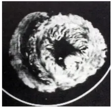
unfolding to display descending segment (DS) after ascending segment (AS) is separated. The entire
53
basal loop (containing RS and LS) is also shown. Bottom layer - HMVB unraveled to display its
54
rope-like model appearance. Longitudinal fibers only exist within the two papillary muscles.; (b)
55
Unfolding of HVMB model. Upper left - intact heart. Upper right- circumferential wrap or basal
56
loop with transverse fibers and the inner helix. Lower right - Unfolded helix showing the oblique
57
fibers of the inner descending helical arm that is separated from the outer ascending helical arm .
58
Lower right - outer ascending helical arm. Marker arrow points to the left anterior descending artery
59
pathway that bisects the helical muscles forming septum and left ventricular free wall.
60
61
Treating disease requires restoration of normality, so decisions must be based upon an
62
understanding of anatomic normality. The heart’s functional counterpart involves only six
63
movements; narrowing, shortening, lengthening, widening, twisting and uncoiling. The Helical
64
Ventricular Myocardial Band model of Torrent Guasp [5] appears in the two classical anatomy texts
65
of Clemente [9], and Moore and Dally [10,11] and its mechanics explain each motion [6,8,12].
66
This knowledge answers the query “what is the heart?”, because the only valid definition of
67
heart structure is an architecture that mechanically accounts for all dynamic movements.
68
Misconceptions happen without it. For example, rather than focusing upon cardiac compression, the
69
pivotal role of twisting for mechanical proficiency must be understood [13]. Twisting occurs because
70
the helical cardiac design allows the natural 15% muscle fiber shortening to create a 60% ejection
71
fraction [13], but this truth is not generally appreciated.
72
The background behind this misunderstanding started 2200 years ago when Erasistratus (280
73
BC), then Galen (180 AD) and subsequently Borelli (1600’s) [14] described twisting, which causes
74
blood to have an ebb and flow motion. But William Harvey, father of the circulation, introduced a
75
compression and dilating action (mimicking a clenched and open fist) [15] that changed Galen’s
76
hypothesis. His supporters deemed Galen’s concept incorrect, and Harvey’s approach has prevailed
77
for 400 years - gaining further support from 2-dimensional imaging (ventriculogram and echo)
78
studies. The newer 3-dimensional imaging tools (MRI and speckle tracking) provide the spatial
79
resolution that allowed re-emergence of the clockwise and counterclockwise twisting rotations
80
Erasistratus described 2200 years ago.
81
Understanding the dynamics of the surrounding wrap and helix is different than relying upon
82
deductions behind many “accepted cardiac mechanical relationships”; these “state of the art”
83
concepts will be questioned. They include heart anatomy as LV, RV, and septum, timing of mitral
84
valve opening, the isovolumic relaxation period, structural reasons for torsion / twisting, the term
85
sub segmentation, resynchronization, RV function dynamics, and diastolic dysfunction’s cause and
87
its unrecognized septum involvement.
88
2. Topographical versus Structural Heart:
89
A heart with two ventricles, separated by a midline muscular septum defines its classic
90
morphologic description. Despite correct topography, no functional insight is provided to define
91
what these 3 structures do. A different structural guideline has existed for 500 years, whereby heart
92
architecture contains a helix formed by right and left handed fibers and a surrounding
93
circumferential wrap [1-3,16,17]. This anatomy preceded Torrent Guasp’s identifying the
94
interweaving architecture by unraveling its muscle bundle formation to solve the Gordian Knot of
95
anatomy. [17] (Figure 2). His novel description of a helical ventricular myocardial band (HVMB)[18]
96
identifies a vortex at the tip of the apex, which is formed by the overlapping of coiled helical arms.
97
Yet when uncoiled, the entire heart’s rope-like configuration becomes revealed. (Video S1)
98
99
Figure 2.Apical view of heart muscle showing the fibers clockwise and counterclockwise
100
spiral formation. Images display common anatomy from Lower in 1669, (left) and Torrent Guasp in
101
1970 (right).
102
103
Torrent Guasp’s macroscopic anatomic pattern (Figure 1), whereby the transverse muscle at the
104
base of the heart (circumferential wrap) turns downward to form an inner descending arm of the
105
helix, which creates an apex by turning upward to form an outer ascending helical arm that ends at
106
the aortic root. Grant [19], Lev and Simkins [20], and Anderson [21] questioned his macroscopic
107
findings because of concerns about the validity of his dissection planes in cadaver tissue. Our
108
analysis of the 1971 functional studies by Armour and Randall’s [22] also questioned the proper site
109
for posterior papillary muscle, but their correct location is now displayed in subsequent HVMB
110
dissections. The only basic element needed to uncover the mechanical reasons for the living heart
111
motions - is a valid structure/ function relationship.
112
Macroscopic analysis has the limitations of not addressing the microscopic display of the nests
113
of layers and lamellae within individual myocytes that interact with cardiac ejection and filling [23]
114
[24].LeGrice found sarcomeres clustered in 1-12 groups within their connective tissue netting [23]
115
[24]. While microscopic relationships are correct, they must also coordinate with the heart’s macroscopic
116
geometric form to fulfill Sallin’s [13] fundamental form/function relationship. This need is emphasized
117
in clinical heart failure where the normal conical heart shape develops a spherical configuration [25].
118
This geometric change transforms the fiber direction of helical arms into a more transverse
119
architectural fiber orientation which impairs LV systolic and diastolic function [13].
120
The conceptual understanding of macroscopic cardiac structure is straight forward because nly
121
3 components need be known. There is a circumferential wrap of transverse fibers that surrounds
122
both ventricles – called a basal loop - which compresses and rotates the global heart, and
123
predominantly forms the RV free wall [26]. The second is the muscular helix that resides within an
124
apical loop nestled within its surrounding circumferential wrap [27]. It’s two oblique fiber arms cross
125
each other at 60° angles [28], with the inner helical coil descending from base to apex, and outer
126
helical coil ascending from apex to base. The same helix forms the septum and part of the LV free
127
Simplicity underlies the solution to the structure function relationship, since its 3 structural
129
components (the wrap and 2 helical arms) produce the heart’s 6 readily apparent dynamic
130
movements of narrowing, shortening, lengthening, widening, twisting and uncoiling. [6-8] This
131
interaction precisely follows Keith’s 1918 Harvean Lecture comment “no theory of function is true
132
unless it explains each detail of structure”. [29]
133
Functional balance exists between the structural wrap and helix, as the wrap causes
134
counterclockwise motion before ejection, the helix produces twisting, and the wrap triggers clockwise
135
recoil during post ejection interval that includes the isovolumic period. [6]
136
State-of-the-art imaging reports do not recognize this dynamic balance because they do not
137
address the fundamental role of the surrounding wrap [30-32]. This gap keynotes the hiatus between
138
deduction and anatomic knowledge, especially since this large circumferential wrap muscle mass
139
highlights the framework structural contributions of William Harvey [15], Krehl [3], Mall [4], Robb
140
[16] and Torrent Guasp [17].
141
Echo motion recordings amplify the circumferential basal loop’s presence by documenting
142
cardiac lengthening during the pre- ejection isovolumic interval [6], while MRI imaging
143
authenticates its global counterclockwise rotation [33,34]. The circumferential wrap’s large muscle
144
mass predominates, and thus overcomes the shortening and clockwise rotation expected from the
145
ongoing subendocardial muscle contraction. A common analogy explains such domination, as one
146
could imagine a train heading south at 60 miles per hour, while its first car houses a runner who
147
speeds northward toward the back car at 6 miles per hour; the power of the southern train always
148
wins, as does the wrap over the helix.
149
The anatomy documenting the location of the outer circumferential wrap becomes evident from
150
echo studies showing the smooth reciprocal movement (in different directions) between the
151
contracting inner and outer helical arms. (Video S2). Interposing a transverse band of compressing
152
muscle between these gliding surfaces would interrupt this motion, as well as impair the smooth
153
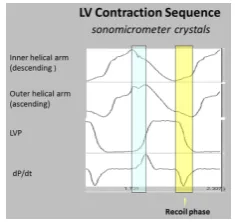
positive and negative septum strain displacement on longitudinal strain recordings (Figure 3).
154
155
Figure 3. Doppler longitudinal strain imaging of septum (right and left sides) in apical
156
4-chamber view. Longitudinal strain marked by red (right) and green (left) circles, showing
157
deformation in opposite directions on right and left septum sides - relative to baseline zero value.
158
Timing shows LV first and RV second. SR, Strain rate; AVC, aortic valve closure; RV, right ventricle;
159
LV, left ventricle.
160
161
Macroscopic interlocking of structure and function is aided by the comparison of their dynamic
162
clockwise and counterclockwise helical motions in Video S2, with their anatomic configuration in
163
post mortem CT images (Figure 4).
Figure 4. Computerized Tomography of cardiac short axis, at mid ventricle level, following air
165
insufflation to separate collagen scaffold netting. Plane between the 2 septum muscle mass rims
166
reflects the echogenic line in septum, and Video S4 records motion between these post mortem rims.
167
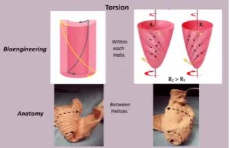
168
The plane identifying the mid ventricular overlapping between helical arms (Video S2) extends
169
from apex to base and is called the hyper-echogenic zone. It is 1 cm wide in low fidelity tracings,
170
narrows to 3mm in higher fidelity recordings, and shrinks to 100 μm [35] following higher fidelity
171
echo examination. [36-38] (Figure 5). The smooth and efficient muscle movements, on either side of
172
it, suggests this zone reflects is a glide path for helical arm motions. The working heart is a precursor
173
for this zone’s presence, since it disappears when cardioplegia arrests the heart. [35]
174
175
(a) (b)
Figure 5. (a) Mid-septal hyperechogenic line shown in low and high-resolution
176
echocardiogram; (b) Septum high-resolution ultrasound image at transducer frequency (12 MHz),
177
displaying its bilayer line with inner dimension structure of 100 to 150 μm. Working septum muscle
178
fibers display different directionality on either side of septum line. This disappears, as does the
179
echogenic line - when cardioplegia (cardiac arrest) stops contractile function.
180
181
These dynamic findings differ from conclusions made from post mortem diffusion Tensor MRI
182
observations that suggest circumferential fibers exist within the septum mid myocardium due to
183
observations of zero helix angles [39]. Yet physiologic testing exposes uncertainty about the
184
anatomic validity of DT MRI findings, because the 100 micron thick echogenic zone disappears
185
following cardioplegia, while dead heart septum MRI studies identify a circumferential muscle
186
occupying up to half its muscle mass. [40,41]
187
3. How Three Structural Components cause Normal Functional Dynamics:
188
The most powerful or dominant muscle amongst these 3 architectural components governs the
189
direction of movement, and their contraction takes place at different timing intervals [42]. For
190
example, the wrap narrows and stretches the ventricle during the pre-ejection isovolumic phase, as it
191
overcomes the simultaneously contracting inner descending helix that should make the ventricle
192
shorten [6,7].
193
Conversely, shortening during ejection emphasizes the dominance of the inner descending
194
helical arm. The ventricle twists as the base rotates clockwise and apex turns counterclockwise from
195
the torque of the outer ascending helical arm’s longer lever radius[43]. The outer helical coilcannot
196
Despite this, both helical arms exert dominance during twisting because the inner descending coil
198
causes shortening (generating twice the longitudinal strain) [44] while the outer ascending helical
199
coil makes the apex turn counterclockwise [43].Our imaging tools provide insight into this action
200
via the echogenic septal line (Figure 5) as the echo beam suggests their fiber orientation pathways by
201
passing along or across the ascending and descending muscle masses.[35]
202
The wrap [6,7] causes clockwise global recoil during the post ejection isovolumic phase, as it
203
springs backward to reverse its pre-ejection counterclockwise motion [34,45,46]. The helix is not
204
involved in this component of recoil because its outer ascending arm is still contracting - but the
205
dominant circumferential wrap’s clockwise motion overturns its counterclockwise motion.
206
Simultaneously, the ventricle lengthens from straightening of the contracting outer ascending helical
207
coil that starts when the dominant inner descending helix contraction stops. (Figure 6) [7].
208
209
Figure 6. Upper left shows intact heart containing a basal loop (circumferential wrap with right
210
(RS) and left (LS) segments) and helix (dark color) with descending (DS) and ascending (AS)
211
segments. Lower left - diastole without contraction. Lower middle - displays shows torsion
212
(twisting) with stronger contraction of descending helical arm (tighter coils), and stretching of
213
ascending helical arm. Lower right - recoil with global clockwise rotation; note lengthening due to
214
ongoing contraction of ascending helical arm. Upper drawing - cobra shows similar elongation from
215
its contracting muscle.
216
217
Global longitudinalstrain measurements match those recorded by sonomicrometry [6] and the
218
observed septum motion in opposite directions reflects the reciprocal fiber orientation of its helical
219
muscles (Figure 3).The outer ascending helical arm starts contracting ~60 ms later than the earlier
220
inner descending helical arm, followed by a ~90 ms timing gap that earmarks the difference between
221
the end of shortening of the inner (descending) arm, followed by the end of outer (ascending) helical
222
arm shortening [42]. This timing hiatus between the end of contraction in the inner and outer helical
223
arms provides a mechanical contradiction (i.e. presence of contraction) to suggestions that
224
repolarization starts earlier in ascending than descending helical arms [47].
225
4. Mitral Valve Opening:
226
Mitral valve opening (MVO) is a term reflecting the Doppler based echocardiographic
227
recording of initial transmitral inflow into the left ventricle. This passage coincides with left
228
ventricular pressure falling below atrial pressure, and the most rapid anterior mitral leaflet motion
229
[48]. Its designation is universal and MVO appears in text books and journal reports (Figure 7). Yet
230
physical separation of the mitral leaflets is the only valid MVO requirement. Lee in 1990 [49] called
231
MVO “the mitral valve artifact that correlates with the E point in the mitral echogram, but is
232
unrelated to actual mitral valve opening”, and others have also questioned its validity[50,51].
233
Figure 7. Physiologic observations in all texts and medical journals, showing Mitral Valve
235
Opening (MVO) when LV pressure falls below left atrial pressure, and that the isovolumic relaxation
236
phase exists between aortic valve closure and MVO.
237
238
MRI recordings quantify leaflet separation, and Video S3 documents that mitral valve leaflets
239
detach from each other at the end of systole - with an open aortic valve. Abrupt loss of leaflet
240
coaptation begins when recoil starts, ~27 ms before aortic valve closure [52], coinciding with
241
negative dP/dt or deceleration of left ventricular pressure. Such MVO documentation exposes an
242
enormous discrepancy between reality versus what has been deduced. The uncoiling process causes
243
MVO, and introduces the muscular action that may trigger diastolic dysfunction.
244
The functional dynamics of the wrap and helix establishes the mechanical insight to explain
245
why MVO occurs during systole. This geometric process focuses upon the architectural interaction
246
between the mobile ventricle that uncoils --- and the mitral annulus that is fixed. The circumferential
247
wrap causes clockwise recoiling, which rotates the helix that contains the papillary muscles that
248
connect with the mitral leaflets and annulus. Their counterclockwise rotation during ejection allows
249
them to close the fixed annulus by a leaflet flap, and this “trap door” re-opens - as they spring
250
backward during recoil to produce MVO. (Video S2). The dynamics of papillary motion are shown
251
in Video S4. Figure 8a and b anatomically displays them at rest and during motion. The 1911 report
252
of Mall [4], a renowned anatomist, demonstrates their clockwise and counterclockwise ventricular
253
movements during twisting and uncoiling.
254
255
(a) (b)
Figure 8. (a) Mitral valve apparatus composed of fixed annulus and mobile leaflets, chordea
256
tendineii, papillary muscles and ventricular wall. (b) Mall’s 1911 report showing how apical
257
counterclockwise rotation of the apex shuts the valve during torsion- by bringing spiral papillary
258
muscles together. Mitral valve inflow area opens during from its clockwise rotation during recoil.
259
260
MVO can only happen when helix’s inner descending contraction has ended, an action that
261
permits initiation of outer ascending helix arm lengthening (Video S3) as quantified by
262
Karwatowski [53]. These cohesive actions of recoiling (by the wrap) and elongation (by the outer
263
helix arm) reinforce why mechanical factors play such a vital role in mitral leaflet separation.
264
Our understanding of diastolic dysfunction is enhanced by a fuller understanding of MVO -
265
since uncoiling cannot begin while torsion is ongoing. Ventricular recoil begins at the inception of
266
negative dP/dt, so that this measurement may become a signpost to determine when unwinding
267
contraction is ~90 ms - and its disruption will impair uncoiling [6,7,54]. Prolonged torsion narrows
269
this gap, so that thinking about the mechanical reasons for MVO may spawn some of the new
270
treatments that will be subsequently considered [55].
271
Finally, the traditional identification of MVO is incorrect, so that the physiological term “mitral
272
valve inflow” (MVI) should replace it. MVO is due to anatomic changes that carry vast physiologic
273
implications --- as we suspect recoiling will become the centerpiece behind understanding diastolic
274
dysfunction.
275
5. Isovolumic Relaxation Time:
276
Isovolumic relaxation time (IVRT) joins MVO in being a universally accepted term (Figure 7). It
277
signifies that part of the cardiac cycle between aortic valve closure and MVO, where the ventricle
278
relaxes (diastole) without lengthening, and ventricular volume is unaltered. Wiggers described it in
279
1923 [56,57], as does today’sStedman Medical Dictionary [57].
280
The ventricle is isovolumic, yet the other 3 components reflect deductions that are incorrect.
281
First, the MVO during clockwise recoil is caused by the wrap – initiating when LV negative dP/pt
282
starts, rather than following a LV pressure fall below left atrial pressure. Second, the entire ventricle
283
does not relax - ongoing strain measurements and sonomicrometer crystal recordings confirm outer
284
ascending helical arm contraction [6]. Finally, it produces lengthening that is quantified by MRI
285
(Video S3), 2-dimensional echo [6] and longitudinal strain recordings [58]. This elongation
286
movement may mirror how a cobra develops an erectile stance before striking [18].
287
Rademakers described the impact of the post ejection isovolumic interval on ventricular filling
288
when he defined a dissociation between untwisting and filling, whereby 50-60 % of recoil occurs
289
during this isovolumic interval [59]. He reasoned that this motion may promote the suction that
290
explains the explosive LV filling that follows the falling LV pressure below left atrial pressure, and
291
wondered if unwinding released the potential energy stored in elastic elements during prior systolic
292
deformation [59].
293
The interdependence of torsion and recoiling is a vital interaction because torsion must stop before
294
unwinding can start. The post ejection isovolumic time frame interval is ~90ms (Figure 9), but
295
diastolic dysfunction may develop when protracted inner descending helix contraction widens this
296
timing gap to >100 ms as seen during aortic stenosis, hypertension and ischemia [54,60-62].
297
298
Figure 9. Left ventricular contraction sequence by anterior LV sonomicrometer crystals. Blue
299
shading shows pre ejection isovolumic interval, yellow shading shows post ejection recoil . Note a)
300
ascending helical arm does not contract during pre ejection, but contracts during recoil, b) negative
301
dP /dt is yardstick for starting recoil, and marks when inner helical arm stops contracting.
302
303
Current thoughts about recoil during isovolumic relaxation imply that the epicardium governs
304
the clockwise rotation of the LV apex, suggesting that it reverses torsion’s apical counterclockwise
305
rotation [59,63]. Functional anatomy challenges this conclusion, since the epicardium (outer
306
ascending helical muscle) is still contracting. (Figure 9) Instead, the circumferential wrap causes
307
clockwise rotation, as outlined previously for mitral valve opening. The term ”isovolumic relaxation
308
interval” is incorrect because the ascending helix is always contracting … it should be renamed the
309
This revision has historic precedence, as Wiggers can now join Harvey (who described
311
compression versus twisting) and Vesalius (who described apex moving toward the base versus its
312
natural base to apex motion) as being astounding titans … that were not perfect.
313
6. Twisting or Torsion:
314
There is agreement that the spiral architecture of LV muscle fibers develops differential rotation
315
of the LV base (clockwise) and apex (counterclockwise) to produce LV systolic wringing or twisting
316
during ejection - torsion quantifies its rotational angle along the LV longitudinal axis. The
317
responsible muscular mechanisms have important functional implications, yet they have been
318
deduced.
319
Taber’s bioengineering model has been accepted [64], as it introduces a helical architecture
320
containing a single layer composed of obliquely aligned muscle fibers embedded in an isotropic
321
matrix [63]. Torsion is described as being developed “within each helix arm” - epicardial fiber
322
contraction rotates the apex counterclockwise and base clockwise, whereas subendocardial fiber
323
contraction rotates them in the opposite directions. Geometric mechanisms differ, as torsion
324
develops “between the helix arms” with the entire inner descending arm rotating the base clockwise,
325
and the entire outer ascending arm rotating the apex counterclockwise. (Figure 10)
326
327
Figure 10. Torsion. The bioengineering approach (upper left) shows it develops “within each
328
helix”; epicardial (outer) muscle has counterclockwise apex and clockwise base rotation, while
329
endocardial (inner) muscle has clockwise apex and counterclockwise base rotation to reflect these
330
reciprocal actions in each helical arm. The right image shows the suggested inner and outer cones
331
occupied by the inner and outer helix. The lower images are anatomic: torsion develops “between
332
helices” as the entire inner descending helix rotates clockwise, and entire outer ascending helix
333
rotates counterclockwise.
334
335
This structural mechanism is supported by architectural and imaging studies. The anatomically
336
unfolded heart reflects an uncoiled rope (Figure 1) that, when re-folded, contains a helix composed
337
of the 60° overlapping of inner and outer spiral coils that aim at either the apex (inner helical arm) or
338
base (outer helical arm). Septum strain recordings confirm deformation in different longitudinal
339
directions [35,58], and high frequency echo tracings record the smooth transition of functionally
340
overlapping fibers (Video S2) that pass along or across its mid septal echogenic line [35].
341
Downward shortening of both helical coils cannot cause torsionbecause the systolic septum
342
longitudinal strain shows the positive deformation (elongation) of the outer ascending helical arm
343
(Figure 3) - a motion that continues during recoil (Figure 9) and by MRI evaluation (Video S3).
344
Interactions between torsion and recoil are independent of torsion’s peak value, since prolonged
345
torsion introduces a twist-based retardation of the unwinding required to develop suction.
346
Torsion and recoil affect ventricular performance in a way that relates to the contractile patterns
347
of the inner descending and outer ascending helical arms. Torsion ends when the inner descending
348
helical arm stops contracting - since uncoiling cannot begin until that happens. This recoil interval
349
includes the post ejection isovolumic phase and first third of diastole.[65] Prolonged torsion delays
350
interdependence between torsion and recoil emphasizes why diastolic dysfunction cannot be
352
thought to exist in a patient whose ejection fraction is considered normal and healthy.
353
7. Untwisting:
354
Untwisting during recoil represents the antonym for twisting during ejection. This traditional
355
term is used to describe the clockwise rotation of the apex that counteracts its counter-clockwise
356
motion during twisting for ejection [52]. Yet the physical designations of these actions differ;
357
“twisting” mirrors rotating the upper fist clockwise and the lower one counterclockwise, to confirm
358
reciprocal left and right handed helical muscle rotations shown by MRI and echocardiogram. But the
359
MRI does not record the expected “untwisting counterpoint” of apical clockwise and basal
360
counterclockwise rotation. Instead, a global clockwise rotation exists during recoil [45]. It is caused by a
361
dominant circumferential wrap that overpowers the outer ascending helical arm’s ongoing systolic
362
counterclockwise motion.
363
Each muscle must return to its starting point, so these anatomic reasons do not counteract the
364
recoil that helix and wrap muscles must develop. Instead, functional aspects the HVMB explain why
365
recoil’s expected global clockwise and counterclockwise “untwisting” cannot occur. The term
366
“untwisting” should be changed into “uncoiling, recoiling or unwinding” to capture recoil’s
367
inception point since it may evolve future diastolic dysfunction treatments.
368
8. Longitudinal and Circumferential Strain:
369
Strain measurements define muscle deformation relative to original length by addressing
370
“shortening in circumferential or longitudinal dimensions or thickenening in radial dimensions”,
371
but they do not define why deformation happens or what it means. For example, 2/3 of strain is
372
circumferential and only 1/3 longitudinal, so that it is considered a more robust tool [36,66],
373
especially because some believe the predominant muscle fiber orientation is followed [67].
374
The functional importance of deformation is linked to its muscular cause. Longitudinal strain
375
reflects coiling downward of the oblique helical spiral fibers to generate the normal ~60% ejection
376
fraction [13]. Conversely, short axis shortening arises from predominantly circumferential fiber
377
deformation, but only a ~30% ejection fraction is yielded. [13].These fiber angulation changes have
378
powerful implications, because impaired systolic contractile strength develops when the ventricle
379
becomes spherical and the helical muscle fibers develop a more transverse orientation [13].
380
Longitudinal strain quantifies ventricular shortening, and was thought to reflect how the apical
381
part pulls the ventricular base downward [68]. The helix and wrap architectural configuration
382
application defines a different sequence, since longitudinal strain reflects how the spiral coil of the
383
inner descending helical arm sequentially shortens due to its base to apex contraction – a trajectory
384
that follows the human excitation studies showing that upper septum activation precedes apical
385
stimulation. [69-71]. Longitudinal muscles “for pulling” do not exist in the ventricle (Figure 1)
386
(except for thin papillary muscles), so that “ pulling down“ deductions contradict natural motion
387
dynamics.[68].
388
Two factors explain why circumferential deformation produces ventricular compression or
389
cardiac narrowing. The first is short axis shortening of the wrap or basal loop’s transverse fibers. The
390
second is transverse shortening produced by thickening of the contracting inner helix’s descending
391
arm. Their individual contributions cannot be determined because they are superimposed within the
392
LV free wall. Conversely, the septum does not contain a circumferential wrap so that its longitudinal
393
strain measurement is possible. (Figure 3).
394
Longitudinal strain reflects deformation of helical spiral fibers, and is measured by recording
395
mitral annulus excursion toward the apex (or MAPSE mitral annular plane systolic excursion) [72].
396
Figure 6 shows the dominance of the spiral inner descending helical segment in producing
397
longitudinal strain during systole.
398
Impaired longitudinal strain is an early finding in dilated hearts [73] and develops when
399
ventricular shape becomes spherical. This geometric change makes the natural oblique fibers
400
develop a more transverse configuration [74]-[55] [75] that disrupts their twisting capacity (Figure
11). This concept’s functional validity is confirmed by the consistent return the cardiac twisting
402
when the failing heart’s spherical shape becomes rebuilt into its natural elliptical shape [76].
403
404
Figure 11. Helical fiber orientation (yellow arrows) in normal ventricle (above) with reciprocal
405
60˚ angulation and a conical shape. Spherical heart (below) shows a more transverse pattern with 45˚
406
or less angulation, mirroring the failing dilated heart.
407
408
The surrounding basal loop fibers provide a short axis supporting restraint [77]that follows
409
ventricular longitudinal strain limitation. For example, circumferential strain is augmented when
410
intraoperative septum damage causes RV longitudinal systolic dysfunction [78]; the LV follows a
411
similar sequence [67]. Conversely, increased ventricular sphericity follows loss of this
412
circumferential restraint [77].
413
Worsened longitudinal systolic strain exists when relaxation is altered during diastolic
414
dysfunction [79]. The query ”is it only diastolic dysfunction?” arose after this coincidence was
415
observed in hypertensive patients with diastolic dysfunction [80]. The differential function of the
416
inner descending and outer ascending helical arms answers this mechanical question [54], as
417
described in the upcoming section on diastolic dysfunction.
418
9. Regional Function versus HVMB:
419
Regional echo based structure function analysis is linked to perfusion related changes [81].
420
Regional deformation (grading shortening and thickening) is aligned with the perfusion territories
421
of 17 echo segments that are based within 3 circular rings of the basal, mid and apical ventricle [81].
422
This arbitrary selection of transmural muscle (with comparable mass) into echo based LV
423
sub-segmentation provides useful information about regional perfusion, yet except for segment 17
424
that shows absent apical deformation, this topographical approach does not record the performance
425
dynamics forthcoming from HVMB analysis [42].
426
A similar arbitrary concept guides how readers are taught to understand the architecture of the
427
helical muscles in the LV elliptical shape. They are considered to exist within overlapping cones,
428
with “state of the art” reports [63] showing the inner cone describes endocardial muscle, and outer
429
cone identifying epicardial muscle [63,82].
430
This designation is incorrect because the outer ascending helical arm forms different parts of
431
the endocardium . An example of this unsuitability is that helical muscle overlap is absent in the
432
septum muscle endocardium below the aortic valve. [7] Velocity vector imaging studies confirm this
433
during the pre-ejection isovolumic phase, since the upper septum bulges like an aneurysm because
434
contraction of outer ascending helix has not yet started [7,35] (Figure12). The outer ascending helix
435
also forms the endocardium in the posterior LV wall where helical overlap is absent. Validity of
436
descriptions of the heart’s architecture is only possible by following the sheaths within muscle planes of
437
(a) (b)
Figure 12. (a) Pre wall ejection motion from velocity vector imaging (VVI) is correlated with
439
cardiac anatomy. The upper septum bulges into the right ventricle (upper right), (b) The helical
440
architecture demonstrates absence of helical overlap in region beneath the aortic valve. Its
441
endocardium is formed by the outer, ascending left handed helical arm that does not contract during
442
this interval.
443
10. Resynchronization:
444
Excitation contraction coupling provides the infrastructure behind how twist develops during
445
each HVMB contraction. This process requires the natural flow of electrical impulse from the
446
conduction system into the responsible myocytes. Insight into the HVMB anatomy and
447
interventricular conduction relationship is gained by reviewing the evolution of clinical
448
“resynchronization” therapy (CRT). “Synchrony” is the traditional term used to characterize the
449
coordinated contraction of the LV, RV and septum - observed from 2-dimensional echo or
450
ventriculography. They record harmonized “all at once“ movement – mimicking the making of a fist
451
with all fingers squeezing in unison in patients with a normal narrow QRS interval. Conversely, a
452
wide QRS interval defines delayed regional electrical activation, and is associated with
453
uncoordinated contraction that may produce septum bulging or billowing, mitral regurgitation and
454
raise LV volume.
455
“Resynchronization” is achieved by simultaneously pacing the LV and RV with the aim of
456
restoring “synchrony”, but its portended treatment goals are only sometimes met [83]. For example,
457
~30% of patients do not respond [83], LV volume is not reduced in 40% [84], recovery of twisting is
458
inconsistent [82,85], and late survival [86] increases only 0.85 years in comparison to optimal
459
pharmacologic therapy. A suggested way to solve this dilemma is to search for a better site for LV
460
pacing [84], but the validity of the “resynchronization concept” must also be examined.
461
This pecking order is based upon the fact that normal cardiac motion is not “synchronous”.
462
Instead it is a “sequential” motion that evolves when the electrical impulse traverses the natural
463
conduction system and its collagenous cardiac netting matrix to stimulate individual muscle fibers.
464
The clenched fist that is traditionally used to define contractility does not reflect an “all at once”
465
motion. Instead it represents the “chronological closing” of the little finger, ring finger, middle and
466
then index fingers, as they create the whirling motion which mirrors the normal heart’s natural
467
twisting movement. Such coordinated torsion develops because the electrical propagation velocity is
468
10x faster (at 3m/sec) through the natural His-Purkinje fiber conduction system, than via direct
469
ventricular muscle stimulation (at 0.3 m/sec) [87]. Wiggers in 1925 described the functional dilemma
470
of asynergic ventricular muscle motions that follow synchronous single electrical excitation [88]. His
471
observation had no impact, because this form of stimulation persists to remain the unchanged
472
stalwart of conventional pacing.
473
Recent HVMB studies demonstrate that inconsistent twisting follows either isolated (direct)
474
ventricular, or biventricular stimulation (CRT) [89]. Conversely, pacing of the His-Purkinje system
475
[90] returns the sequential activation responsible for twisting to restore natural torsion. The clash
476
between these pacing avenues highlights that a limitation of CRT is that it reflects the 2-dimensional
477
approach of isolated site excitation which can only gain compression. In contrast, natural conduction
478
excites the inbuilt conduction circuits which unfold the 3-dimensional approach that returns cardiac
479
CRT provides mechanical, but not physiological improvements. For example, a wide QRS
481
interval delays septum activation, so that the earlier LV free wall contraction will make it billow or
482
bulge. The resultant ventricular stretching will tether the posterior papillary muscle (adjacent to
483
septum) connected to the mitral leaflet, and produce mitral regurgitation from poor leaflet
484
coaptation. CRT returns the septum to its midline position to offset papillary muscle tethering and
485
remedy mitral regurgitation - but it does not consistently restore cardiac twisting. Conversely,
486
His-Purkinje pacing rebuilds normal conduction by shortening the QRS interval. [91]. It generates a
487
sequential heart beat that permits the natural twisting motion to resume. [92]. Awareness of these
488
differences may hasten interest in evolving Bundle of His pacing approaches.
489
11. The Right Ventricular Function:
490
Right ventricular failure is difficult to manage because its underlying mechanisms are
491
uncertain. Decision dilemmas follow such incomplete functional knowledge. For example, the RV is
492
considered a passive chamber because early functional recovery follows its exclusion by the Glenn
493
(superior vena cava to pulmonary artery) or Fontan (pulmonary artery to right atrium) procedures
494
[93,94] Yet this conclusion is contradicted by the ~40% mortality that develops in patients that
495
occlude a right coronary artery containing large septal branches [95].
496
RV cardiac function is determined by myocardial fiber orientation (Figure 13). A thin
497
circumferential wrap containing predominantly transverse muscle fibers forms its free wall. Their
498
contraction produces compression via a bellows action that accounts for 20-30% of ejection fraction
499
[13,96]. Conversely, only helical fibers construct its thicker midwall septum, and these oblique fibers
500
[97] generate the twisting action responsible for 80% of RV global function, making the septum “the
501
functional lion of the RV” [26,98].
502
503
504
Figure 13. Right ventricular fiber pattern and HVMB, where the circumferential wrap or basal
505
loop causes compression and narrowing, and the underlying helix with oblique fibers at 60˚ angles
506
causes shortening and lengthening .
507
508
Investigative studies document the dynamics of RV’s compression and twisting movements.
509
For example, unaltered RV function follows RV free wall exclusion by cauterization, patch
510
replacement [99,100] orits regional ventricular fibrillation [101] if the septum is intact. Yet RV failure
511
follows septal injury when pulmonary hypertension co-exists [102]. This functional distinction
512
between helix and wrap has not influenced the guidelines of the American Society of
513
Echocardiography and European Association of CV Imaging that recommend measuring 2D STE-
514
RV free wall strain, but do not identify that it accounts for only 30% of RV performance [81]. This
515
functional discrepancy explains outcomes following a report that limited RV free wall
516
ventriculotomy, aiming to avoid late RV failure [103]. It was not successful because of RV free wall’s
517
minor effect on right heart performance.
518
The interplay becomes especially clear following cardiac surgery, where postoperative RV
519
performance remains unimpaired, despite almost 50% of patients developing paradoxical septum
520
motion [104]. But this compensatory circumferential wrap compression can only be effective when
521
is increased [102]. This explains why the Glenn and Fontan procedures are contraindicated if PVR is
523
high, since they exclude the septum, whose role is to the generate the twisting needed to counteract
524
high afterload.
525
Tricuspid annular plane systolic excursion (TAPSE) quantifies the extent and rate of helically
526
induced longitudinal strain [26] by documenting how ventricular shortening by the coiling helix will
527
bring the base closer to the apex. The RV and LV chambers are thought to be topographically
528
separate, but they share a common HVMB architecture, so that the twisting behind TAPSE in the RV
529
reflects MAPSE in the LV. Vascular resistance is the counterforce to twisting and influences
530
decision-making when right heart failure is caused by impaired septum function. If pulmonary
531
pressure is high, treatment with vasodilator drugs (amrinone, milrinone) may be preferable to
532
vasoconstrictor agents (epinephrine, dopamine) that would accentuate the afterload confronting the
533
poorly contracting septum.
534
The interface between RV helical and circumferential muscle interaction is further clarified
535
from RV function examination following either conventional or catheter based aortic valve
536
replacement (AVR). Longitudinal and circumferential strain is unaltered in catheter based AVR that
537
does not use cardioplegia. In contrast, the use of cardioplegia during surgical SVR results in the
538
commonplace finding of septal damage [104]. This injury had an associated 50% reduction in TAPSE
539
(the helix), which resulted in a 60% increase of compensatory circumferential strain (the wrap) [78].
540
RV failure sometimes develops in heart failure patients with high PVR following implantation
541
of a left ventricular assist device (LVAD). Ventricular decompression by LVAD changes cardiac
542
geometry by collapsing the LV. This maneuver bows the septum toward the left side, making its
543
helical fibers more transverse and dysfunctional [55]. RV failure ensues, but can be quickly resolved
544
by diminishing the extent LV decompression in order to mechanically restore the septum’s midline
545
position as it re-establishes its natural geometry [105].
546
12. Diastolic Dysfunction:
547
Diastolic dysfunction is characterized by how impaired ventricular relaxation and increased
548
wall stiffness can limit LV filling in ~50% of patients with heart failure, despite their having normal
549
ejection fraction [106]. Nishimura’s [65] classic 1997 overview emphasized empirical treatment,
550
because of absent clear-cut pathophysiologic concepts - a limitation that still prevails. Historian
551
David McCullough observed a similar conundrum in politics, stating “it is like trying to repair an
552
engine that you do not know how to take apart”.
553
Normal ventricles receive 70% of their filling during the first third of diastole, but uncertainty
554
exists about why this happens. Uncoiling via apical clockwise rotation influences early filling, but
555
this motion begins in the last half of systole and extends to the first third of diastole [65]. Despite its
556
acknowledgment [65,107], calling this interval “passive” rapid ventricular filling introduces a vis a
557
tergo mechanism with reliance upon an intraventricular pressure gradient (IVPG) that promotes
558
blood movement toward the apex [107,108]
559
The alternate mechanism is “active” ventricular suction to aspirate atrial blood, using peak
560
negative dP/dt and the time constant of relaxation (tau) as measurement indexes [65].
561
Understanding the energy conversion process needed for either active recoil [109] or via a muscular
562
mechanism [108,110] has been its missing component. The HVMB structure function relationship
563
identifies this answer.
564
There is clear-cut understanding of how ventricular pressure increases during filling of a stiff
565
ventricular wall, yet this “compliance mechanism” is absent during normal rapid ventricular filling,
566
where ventricular pressure falls as volume increases [111]. “Diastolic suction” explains this process,
567
as it draws ventricular blood from the atrial reservoir [112-115] to create a negative diastolic
568
pressure. Brecher documented this negativity from recordings of the heart withdrawing blood from
569
a lower reservoir. [113]
570
The current term “passive rapid ventricular filling” parallels the prior controversy between
571
twisting or compression for ejection. Background exists, as Galen, in 180 AD described “the
572
1921, rejected a vis a fronte ventricular filling [116], and Roberts, in 1979, described “suction” by
574
finding the left atrium had invaginated into the left ventricle through the mitral valve [117] during a
575
post mortem exam after terminal hypovolemic hemorrhage.
576
Concepts about elastic recoil or systolic ventricular filling are best tested by evaluating the
577
integration of form and performance. Van Dalen’s elegant series of observations [118-122] clarified
578
the echo based dynamics of “untwisting”, but he ascribed these motions to oblique muscles. He
579
reasoned that repolarized epicardial fibers (outer helical arm) actively untwisted to move clockwise,
580
since endocardial fibers (inner helical arm) were reported to be still depolarized [47]. His focus upon
581
“untwisting” as a new diastolic dysfunction treatment goal is an important consideration (as
582
described below), but his suggested mechanism cannot occur. The helix is not involved in post
583
ejection isovolumic phase recoil, because unwinding is caused by the basal loop whose contraction
584
has stopped [6]. Conversely, the ascending segment (epicardium) is still contracting (depolarized)
585
during the isovolumic interval, so that its persistent counter- clockwise motion would oppose the
586
prevailing clockwise movement. (Figure 9) [6,30,38]
587
Torrent Guasp’s geometric contribution’s are central to our structure/function approach, but his
588
“systolic ventricular filling” concept [108] uses only form to deduce function. He ascribes it to
589
ongoing contraction of the ascending (outer helical) arm. HVMB dynamic analysis certifies that its
590
post ejection isovolumic phase shortening has stopped before rapid filling begins (Figure 9).
591
Consequently, its elastic recoil causes the explosive ventricular filling that follows its springing back
592
into its starting position. As stated, uncoiling may reflect how stored potential energy during
593
ejection [59] is released during this unwinding process that creates a centrifugal force for aspiration
594
of atrial blood.
595
The interdependence of torsion and recoil are fundamental concepts behind understanding
596
how HVMB clarifies why diastolic dysfunction develops. The central theme is that recoil cannot start
597
until torsion ends. The recoil process causes the predominance of ventricular filling in the healthy
598
heart, yet 50% of this unwinding exists during the pre filling phase, reaching 60% after
599
catecholamine infusion [59,123]. The remainder of recoil happens during rapid filling and is due to
600
uncoiling of the outer ascending helical arm [6].
601
The time-frame between the end of contraction of the inner descending and then, of the outer
602
ascending helix arms, creates a “temporal hiatus” (Figure 9), and this “time gap” becomes the
603
centerpiece for understanding the HVMB muscular actions that are responsible for diastolic
604
dysfunction. Curtailment of recoil will restrict suction and by narrowing of this temporal hiatus.
605
Consequently, a vis a tergo mechanism must be used to cause ventricular filling by raising the
606
compensatory factor of atrial pressure. Pressure related enhancement of filling is normal after atrial
607
contraction at the end of diastole, but lung congestion may follow its presence during early diastole.
608
Disruption of the HVMB dynamics responsible for the interface between torsion and recoil
609
creates diastolic dysfunction in several ways, because prolonged systole during torsion will
610
encroach upon the aforementioned “temporal hiatus” during aortic stenosis [60], hypertrophic
611
cardiomyopathy, ischemia [62], impaired sarcolemmal calcium flux efficiency [124], age-related
612
calcium turnover [124]. Its genesis is that unwinding cannot start until the prolonged inner
613
descending helix arm contraction has ended.
614
Most importantly, thoughts of diastolic dysfunction that focus upon an isolated relaxation
615
disorder must be reexamined, since each patient shows combined systolic and diastolic
616
abnormalities [125]. These alterations involve impaired ventricular twisting and longitudinal
617
deformation (strain) patterns [80] that delay untwisting to reduce suction and impair early diastolic
618
filling [126]. Restricted helical systolic function is evident by reduced longitudinal strain, despite
619
normal ejection fraction. This corresponds to how the prolonged torsion in patients with aortic
620
stenosis can compromise their uncoiling process. [60].
621
Mechanism related treatments can reverse diastolic dysfunction, since aortic valve replacement
622
allows regression of LV hypertrophy with resulting return of twisting and recoil to normal [127].
623
Post ischemic diastolic dysfunction is reversed when sodium hydrogen ion inhibitors limit calcium
624
accumulation within the inner helix [54]. Their avoidance of prolonged contraction restores the
natural time gap between the end of inner and then outer helix shortening to allow the recovery of
626
the suction that accentuates ventricular filling [128].
627
The LV free wall and septum are formed by the same HVMB muscle mass (Figure 1 and 14 )
628
and this association confirms why diastolic dysfunction after routine cardiac surgery is clinically
629
important. Diastolic and septum dysfunction are similar but this conclusion has not yet been
630
appreciated. It is well known that septum dysfunction is commonplace after cardiac surgery, as 43%
631
of 3292 patients [104] develop septal paradoxical motion (lesser damage was not reported). Impaired
632
myocardial protection causes this injury, but this damage can be avoided [129], as undamaged
633
hearts show improved longitudinal helical deformation [130].
634
635
Figure 14. Figure 1 anatomy modification with “simulated left anterior descending artery” that
636
is a vascular highway that bisects the helix, which constructs the septum and the LV free wall.
637
Anatomy (above), and how (below) LV free wall is on vessel’s left side, and septum (which is 3
638
dimensionally deeper) is on right side.
639
640
Diastolic dysfunction, its sequel, develops in 44% to 75% of patients undergoing coronary
641
grafting or aortic valve procedures [131-133]. This incidence mirrors the frequency of septum
642
damage [104] and both entities may disappear in 6-12 months [134,135]. Conventional consideration
643
has not recognized the similarity between septum damage and diastolic dysfunction, because heart
644
anatomy had been viewed topographically (LV, RV, septum), yet their coincidence is apparent and
645
becomes predictable when the HVMB is used to recognize their commonality.
646
13. Conclusions:
647
This rethinking of the core cardiology values started after learning of the contributions of
648
Francisco Torrent Guasp. His studies exposed the simple design of cardiac architecture by showing
649
that the HVMB contains a helix and a circumferential wrap. These two structures create the
650
functional mechanics behind cardiac motion and thus define ”what is the heart”. Application of this
651
foothold knowledge reveals that many “accepted” cardiac events are based upon deductions drawn
652
from presumed myocardial structure, rather than from its natural configuration. Torrent Guasp’s
653
revolutionary contribution opens the door toward an exciting future for the understanding,
654
diagnosis and treatment of cardiac disease.
655
Supplementary Materials: The following are available online, Video S1: Francisco Torrent Guasp doing
656
cardiac dissection to unravel the HVMB; Video S2: MRI of Mitral Valve Opening. Note MVO occurs while the
657
aortic valve is open, and its inception is just as the septum begins to elongate. Recoil begins as the mitral valve
658
opens, an event that accompanies the beginning of clockwise ventricular rotation evident on next MRI video;
659
Video S3: Short axis apex MRI that displays cardiac rotation and papillary muscle motion. Note the
660
counterclockwise rotation during torsion, and closeness of papillary muscles. Clockwise motion is associated
661
with a widening or the distance between papillary muscles. They attach to the mitral leaflets and leaflet
662
separation (mitral opening) accompanies the clockwise ventricular motion during recoil; Video S4: Short axis
663
high resolution echocardiographic view of torsion and recoil in normal heart. The inner and outer mobile arms
664
correspond to how they are portrayed in the Figure 4 post mortem MRI images. Note a) the smoothness of these
665
counterclockwise during torsion development and b) how the inner and outer coils spring back to their starting
667
position during recoil.
668
Author Contributions: G.B. wrote the paper; G.B. and M.K. made substantial contributions to conception,
669
drafting and revising the article, giving final approval of the version to be submitted; N.N. and C.N. made
670
contributions to drafting and revising the article, giving final approval of the version to be submitted.
671
Conflicts of Interest: Dr. Buckberg consults with Helical Heart Company LLC (www.helicalheart.com), which
672
makes a spatial heart model of helical ventricular myocardial band anatomic configuration. The other authors
673
declare no conflict of interest.
674
675
References:
676
1. Lower, R. Tractus de corde 1669. In Early science in oxford, Gunther, R., Ed. Sawsons: Oxford, 1932.
677
2. Senac, J.B. Traite de la structure du coeur. Vincent: Paris, 1749.
678
3. Krehl, L. Kenntniss der fallung und entleerung des herzens. Abhandl Math Phys 1891, 29, 341-362.
679
4. Mall, F.P. On the muscular architecture of the ventricles of the human heart. Am J Anat 1911, 11,
680
211-278.
681
5. Torrent-Guasp, F.; Buckberg, G.D.; Clemente, C.; Cox, J.L.; Coghlan, H.C.; Gharib, M. The structure
682
and function of the helical heart and its buttress wrapping. I. The normal macroscopic structure of the
683
heart. Semin.Thorac.Cardiovasc.Surg. 2001, 13, 301-319.
684
6. Buckberg, G.; Hoffman, J.I.; Mahajan, A.; Saleh, S.; Coghlan, C. Cardiac mechanics revisited: The
685
relationship of cardiac architecture to ventricular function. Circulation 2008, 118, 2571-2587.
686
7. Buckberg, G.; Hoffman, J.I.; Nanda, N.C.; Coghlan, C.; Saleh, S.; Athanasuleas, C. Ventricular torsion
687
and untwisting: Further insights into mechanics and timing interdependence: A viewpoint.
688
Echocardiography. 2011, 28, 782-804.
689
8. Buckberg, G.D.; Hoffman, J.I.; Coghlan, H.C.; Nanda, N.C. Ventricular structure-function relations in
690
health and disease: Part i. The normal heart. Eur J Cardiothorac Surg 2015, 47, 587-601.
691
9. Clemente, C. Anatomy: A regional atlas of the human body - fifth edition. Fifth ed.; Lippincott, Williams
692
and Wilkins: 2006.
693
10. Clemente, C. Anatomy: A regional atlas of the human body. 4 ed.; 1997.
694
11. Moore, K.L.; Dalley, A.F. Clinically oriented anatomy: Fifth edition. Lippincott & Wilkins: 2005.
695
12. Buckberg, G.D. Echogenic zone in mid-septum: Its structure/function relationship. Echocardiography.
696
2016, 33, 1450-1456.
697
13. Sallin, E.A. Fiber orientation and ejection fraction in the human ventricle. Biophys J 1969, 9, 954-964.
698
14. Borelli, G.A. History of cardiology. Medical Life Press: New York, 1927.
699
15. Harvey, W. De motu cordis. 1628.
700
16. Robb, J.S.; Robb, R.C. The normal heart: Anatomy and physiology of the structural units. Am Heart J
701
1942, 23, 455-467.
702
17. Torrent-Guasp, F.; Buckberg, G.D.; Clemente, C.; Cox, J.L.; Coghlan, C.; Gharib, M. The structure and
703
function of the helical heart and its buttress wrapping i--the normal macroscopic structure of the heart.
704
Sem Thorac & Cardiovasc Surg 2001, 13, 301-319.
705
18. Buckberg, G.D. Basic science review: The helix and the heart. J.Thorac.Cardiovasc.Surg. 2002, 124,
706
863-883.
707
19. Grant, R.P. Notes on the muscular architecture of the left ventricle. Circulation 1965, 32, 301-308.
708
20. Lev, M.; Simkins, C.S. Architecture of the human ventricular myocardium, technique for study using a