MP-10.01
Anticholinergic use in children: Persistence and patterns of therapy Blais, Anne-Sophie1; Bergeron, Michelle1; Nadeau, Geneviève1; Bolduc,
Stephane1
1Division of Urology, Centre hospitalier universitaire de Québec, Quebec,
QC, Canada
Introduction and Objectives: Overactive bladder symptoms (OAB) are
complex and generally require long-term therapy. Despite the impact of these symptoms on patients’ well-being, persistence rates of antimuscarin-ics have been shown to be low in adults, but it has never been studied in children. Better understanding of the treatment patterns of children treated with antimuscarinics could help to improve the quality of drug management and outcomes. Our objective was to evaluate treatment patterns of patients <18 years of age with on antimuscarinic therapy over a 4-year period.
Methods: Pediatric patients receiving a first antimuscarinic prescription
between April 2007 and March 2008 were identified using IMS Brogan’s Public and Private Drug Plans Database. Canadian drug claims data from Private Drug Plans, Régie de l’Assurance Maladie du Québec and Ontario Public Drug Plans were analyzed retrospectively. Patients were followed for 4 years to assess the prescribed drugs, the lines of treatment and the average duration on each treatment.
Results: Data were available for 374 patients. The most prescribed drug as
a first line therapy was Oxybutynin (326 patients), followed by Tolterodine ER (22). Patients refilled their index prescriptions for an average of 429 days. Solifenacin had the highest mean duration of index therapy (765 days) fol-lowed by Oxybutynin and Tolterodine ER. During the 4-year follow-up, 324 patients (86,6%) had only one line of therapy. At the end of the follow-up, 44 patients (11,8%) who initiated medication were still on therapy.
Conclusion: Overall discontinuation rates of antimuscarinic therapy in
children (88% at 4 years) seems comparable to what has been reported in adult patients with OAB (65-89% at 12 months). However, children seem to persist on the medication for a longer duration before adherence rates start declining. The low rate of persistence highlights the importance of continuing to identify alternatives to antimuscarinics.
MP-10.02
Prospective pilot study of mirabegron in pediatric patients with overactive bladder
Blais, Anne-Sophie1; Nadeau, Geneviève 1; Moore, Katherine1; Genois,
Lucie1; Bolduc, Stephane1
1Urology, Centre hospitalier universitaire de Québec, Quebec, QC, Canada Introduction and Objectives: Antimuscarinics are the current
pharmaco-logic mainstay for management of overactive bladder (OAB) but bother-some side effects (S/E) limit their clinical use. Recently, a new molecule with a distinct mechanism of action has entered the market. Mirabegron, a χ3-adrenoreceptor agonist, is currently approved in Canada as monotherapy for idiopathic OAB in adults. Several studies have shown promising results. Mirabegron has not been studied yet in the pediatric population. The objec-tive was to evaluate the efficacy and safety of mirabegron to treat urinary incontinence in children with idiopathic OAB that were refractory and/or intolerant to antimuscarinics.
Methods: A prospective off-label study using adjusted-dose regimens of
mirabegron was conducted. Pediatric patients without symptom improve-ment under intensive behavioural and medical therapies and/or significant S/E with at least 2 different antimuscarinic agents were recruited. Efficacy and tolerability were assessed with: voiding diaries, post-void residuals,
urine cultures, EKG and vital signs. Families were questioned for conti-nence, S/E, compliance, and patient perception of bladder condition (PPBC) questionnaire.
Results: Forty-four patients were recruited. Mean age at initiation was 10.7
year-old and patients were on Mirabegron for a mean of 5.1 months. Mean bladder capacity improved from 176ml to 242 ml (p<0.0001). Continence improved in all patients but 2, with 8 being completely dry. Under treat-ment, post-void residuals were not significant (<20ml) for all patients but 1. Mean PPBC improved from 4.6 to 2.2 (p<0.0001). Seven patients reported mild or moderate S/E: nasal congestion, abdominal cramps, constipation and nausea. Two of these patients withdrew from the protocol.
Conclusions: Mirabegron, a novel first-in-class therapy as a selective
χ3-agonist, appears as a safe and effective alternative for children with idiopathic overactive bladder refractory to antimuscarinics.
MP-10.03
Caregiver impressions of uncorrected hypospadias
Keays, Melise A.1; Snodgrass, Warren2; Starke, Nathan3; Bernstein, Ira3; Lee,
Simon Craddock3; Corbin Bush, Nicol2
1Children’s Hospital of Eastern Ontario, University of Ottawa, Ottawa,
ON, Canada; 2Pediatric Urology, PARC Urology, Frisco, TX, United States; 3University of Texas, Southwestern, Dallas, TX, United States
Introduction and Objectives: Although surgeons have discussed impact of
uncorrected hypospadias, we are not aware of any study reporting caregiver perceptions about the condition. We administered a patient/parent-reported outcomes measure (PROM) and report caregiver opinions of their child’s hypospadias prior to surgical consultation.
Methods: We developed a PROM and administered it to consecutive
care-givers of prepubertal patients presenting with non-operated hypospadias prior to meeting a urologist. Survey items were generated based on a critical literature review and semistructured
interviews with patients, parents and lay experts. Cosmesis, urinary func-tion, overall satisfacfunc-tion, and social functioning/body image were assessed. A modified International Prostate Symptom Score (I-PSS) was included for toilet-trained patients.
Results: 104 surveys were completed by caregivers of patients with a mean
age 18 months (0-82) who had nonoperated distal (n=83), midshaft (n=4) or proximal (n=16) hypospadias. The overall appearance was rated abnormal but acceptable, dissatisfying, and very dissatisfying in 15%, 14%, and 19%, respectively. 41% expressed concern about what others would think of their son’s penis, and 46% of caregivers reported they had been teased about the appearance. 46% and 93% of parents of distal and proximal patients, respectively, responded they would be unhappy or very unhappy if their child were to spend the rest of his life voiding the way he currently was. Among a subset of 25 toilet-trained patients with distal hypospadias, caregivers reported urinary symptoms ranging from mild to severe in 48% based on I-PSS classification.
Conclusions: A significant portion of caregivers of boys undergoing
pre-senting for evaluation of uncorrected hypospadias report abnormalities in cosmesis, urinary function, penile satisfaction, and body image. These data from a population of patients with mostly distal hypospadias lend support to the current practice of routinely recommending surgical correc-tion. However, additional inclusion of patient/parent reported outcomes will help establish if these caregiver-reported parameters improve with successful surgical correction.
MP-10.04
Pediatric urology fellows survey: Mentor-based vs. evidence based learning
Keays, Melise A.1; Corbin Bush, Nicol2; Snodgrass, Warren2
1Children’s Hospital of Eastern Ontario, University of Ottawa, Ottawa,
ON, Canada; 2Pediatric Urology, Pediatric and Adult Reconstructive
Center for Urology, Frisco, TX, United States
Introduction and Objectives: A major goal in pediatric urology fellowship
training is learning hypospadias repair. We surveyed recently graduated and current fellows regarding their learning preferences, surgical expo-sure and participation in operations, and their expected future practice.
Methods: We developed a 27 question Red Cap survey emailed to
pedi-atric urology fellows in their last 2 years of fellowship.
Results: 69 surveys were emailed with 29 (42%) returning responses, of
which 24 indicated they had ≥6 months clinical fellowship training and comprised the study group. To learn operative techniques, 52% consulted surgical atlases, 35% textbooks, 7% reviewed original articles, and 6% watched videos. For distal repairs, 59% were taught TIP, while 38% were taught multiple procedures depending upon anatomic factors. Techniques learned for proximal repair with <30° curvature were TIP (48%), onlay flap (21%) and a variety of other procedures including Byar’s flaps (10%) and staged grafts (10%). For curvature >30⁰, respondents reported learning staged grafts (62%), Byar’s flaps (14%), transverse island prepucial flaps (7%) and TIP (7%). 74% reported they performed >50% of these repairs. Respondents indicated their knowledge of outcomes following these pro-cedures primary came from original articles (41%), their mentors (31%), or textbooks (21%). When asked regarding their future decision-making, 83% stated they would follow their mentor’s preferences.
Conclusions: Our survey found fellows preferred atlases and textbooks
to learn hypospadias procedures. They reported wide variation in deci-sion-making for primary hypospadias repair. Less than half stated their knowledge of expected outcomes derived from original articles, while a third relied primarily on their mentor’s teaching. In an era of evidence-based medicine, these results suggest continued reliance on traditional mentor-based learning, which they reported would continue to form the basis for hypospadias decision-making in practice.
MP-10.05
Presentation and treatment of late ureteric obstruction after endoscopic Dextranomer/Hayluronic Acid Copolymer (Deflux) injection for vesicoureteral reflux in children
Sami, Samir1; Dhaliwal, Navraj1; Fermin Risso, Carolina1; Cook, Anthony
J.1; Weber, Bryce A.1
1Pediatric Urology, University of Calgary, Calgary, AB, Canada Introduction and Objectives: Subtrigonal endoscopic injection
dextrano-mer/hyaluronic acid copolymer (Dx/Ha) is firmly established in treating vesicoureteric reflux (VUR). Post operative obstruction is an extremely rare complication leaving a limited awareness of risk factors and a paucity of literature describing management strategies.
Methods: A total of 742 children (1078 ureteral units) underwent
endo-scopic injection between 2002-2012, and have been regularly monitored since. Patients presenting with significant ureteral obstruction were pro-spectively captured. Data consisting of grade of VUR, volume of Dx/Ha injected, timing of obstructive signs, type of management and outcome were collected by chart review.
Results: Six patients (8 ureters) developed significant ureteral obstruction.
Initial Dx/Ha injection was performed at a median age of 2.4 years (range 7 months to 4 years). The median grade of VUR in the ureter at the time of injection was 4 (range 3-5). The average amount of Dx/Ha injected was 1.8ml/ureter. Delayed onset ureteral obstruction occurred at a median time of 1.58 years (range 1.25-8 years) post endoscopic Dx/Ha injection. Two patients required an additional Dx/HA procedure for persistent VUR prior to obstruction. All six patients presenting with obstruction initially underwent endoscopic Holmum/YAG laser endoureterotomy. Amongst these 2 patients failed and required surgical re-implantation of the ureter.
Conclusions: Risk factors for delayed onset of ureteral obstruction
fol-Endoscopic management of post Dx/Ha obstruction is effective and rep-resents the least invasive option prior to considering the need for ureteral re-implantation. We conclude that while delayed-ureteral obstruction is rare it is a serious complication that can occur years after Dx/Ha injection and consequently warrants long-term follow-up.
MP-10.06
Investigation and management of blunt genital urinary injuries in children: A review of a metropolitan city with winter sports facilities
Sami, Samir1; Frusescu, Adrian1; Fermin Risso, Carolina1; Cook, Anthony
J.1; Weber, Bryce A.1
1Pediatric Urology, University of Calgary, Calgary, AB, Canada Introduction: Trauma remains the single greatest cause of morbidity and
mortality among children. Renal trauma specifically is more common, secondary to reduced chest wall rigidity, less peri-renal fat, and weaker paraspinal and abdominal muscles. We evaluated all pediatric blunt renal trauma admitted to Alberta Children’s Hospital during the past 6 years in order to determine incidence, mechanism of injury, severity, management as well as the practicality of CT imaging.
Methods: We searched the medical records at Alberta Children’s Hospital
from 2008 to 2014 and identified 86 patients up to 18 years old with objective data on blunt genitourinary trauma. We analyzed these cases, including mechanism of trauma, associated injuries, management, days of hospitalization and the injury severity score (ISS).
Results: 86 patients were identified, median age was 11 years, ranging
from 2 to 18 years. Renal injuries where involved in 90% (77/86) of cases with a 2:1 male to female preponderance. All patients had initial CT scans; 24 of 77 renal injuries where classified as high grade (AAST grade 4 or 5) with significantly more of these occurring secondary to bike or ski hill injuries (p=0.04) versus MVA. The most common accompany-ing injuries were; spleen (44%), liver (17%) and spine/head (7%). Bike and Ski hill injuries also had significantly more associated abdominal injuries (P=0.47). Patients presenting after MVA had significantly higher ISS scores (P<0.02) yet only 4 of these patients had high grade renal injuries. Only 7 patients required interventions (6 ureteric stents, one embolization) and just 3 renal units where lost due to devascularization at time of trauma (no deaths).
Conclusion: The results of our study have shown that activities like winter
sports and biking play an important role during renal trauma injuries in the pediatric population, more so than injuries secondary to MVA. CT scan was useful in diagnosis of grade of renal injury and determination of associated injuries.
MP-10.07
Introduction of robotic-assisted pediatric urology procedures in the Canadian set up: Assessment of early learning curve, results and operative times
Huynh, Melissa1; Dave, Sumit1; Parikh, Nishi1
1Surgery, Division of Urology, Western University, London Health Sciences
Centre, London, ON, Canada
Introduction and Objectives: The introduction of robotic assisted
pedi-atric urologic procedures in our healthcare system brings forth a discus-sion on the impact of increased operative times on resources. This first Canadian study analyzes the trend in operative times and early results of robotic assisted pyeloplasty (RAP) and ureteral reimplantation (RAR).
Methods: Twenty- six patients underwent RAP (13) and RAR (13) since the
initiation of our robotic program. All procedures were digitally recorded to allow calculation of operative times divided into distinct steps. Indication for surgery, imaging findings and follow up was documented prospec-tively. Success in the RAP group was defined as resolution of Dietl’s crisis and/or decrease in hydronephrosis with/without improved post–op MAG 3 T half time when performed (n=5). Success in the RAR group was defined as cessation of UTI’s off antibiotic prophylaxis, absence of hydronephrosis and a negative VCUG (n=6) when performed for persisting UTI’s.
and 7 mo (1-17) for the RAR group. In the RAP group, there were no intra or post- op complications. The mean OR time decreased from 222 min. for the first 5 cases to 192 min. for the last 5 cases, largely due to a reduction in suturing time (114.4 to 86.6 min.), despite resident console time in the latter half of this series. In the RAR group, one girl developed a delayed urinoma possibly due to ureteral electrothermal injury and required stenting. The mean OR time in the RAR group decreased from 221.7 min in the first 4 cases to 180.5 min in the last 4, again, primarily as a result of a decrease in the detrusor tunnel suturing time from 46.9 min to 32.3 min. Post-op VCUG performed in 6 showed resolution of VUR in all, while the rest stayed infection free. There was no significant difference in the dissection, spatulation or detrusor tunnel creation times.
Conclusions: This study suggests a comparable outcome of RAP and RAR
to open or laparoscopic approaches. Console times for RAP and RAR decrease early in the learning curve, primarily in the suturing compo-nent, with a more consistent trend in the RAR group. Early results suggest an easy transition to a robotic approach and a greater opportunity for resident teaching.
MP-10.08
Contemporary practice patterns in voiding cystourethrogram (VCUG) use: The impact of evidence-based guidelines
Lee, Linda1; Lorenzo, Armando1; Odeh, Rakan1; Bowlin, Paul1; Traubici,
Jeffrey2; Koyle, Martin1
1Division of Urology, Department of Surgery, Hospital for Sick Children,
Toronto, ON, Canada; 2Diagnostic Imaging, Hospital for Sick Children,
Toronto, ON, Canada
Introduction and Objectives: While VCUG is a widely accepted test, it
is invasive and associated with radiation exposure. Most cases of VUR are low-grade and unlikely to be associated with acquired renal scarring. In an effort to select patients at greatest risk, the American Academy of Pediatrics (AAP) published revised guidelines on urinary tract infections in children ages 2 to 24 months in 2011. In this project, we examine contemporary practice patterns and indications for VCUG, in the context of newer evidence-based guidelines. We hypothesize that the rate of VCUGs has declined over time, which may be geared towards detection of more clinically significant VUR.
Methods: All VCUGs performed at our institution from 2008 to present
were identified. Follow-up VCUGs were excluded. Further data collection was performed for patients whom had a VCUG in the first six months of 2009 and 2014, in order to obtain representative data before and after publication of the 2011 AAP guidelines. Medical records for these patients were retrospectively reviewed for baseline patient characteristics, indica-tion for VCUG, type of ordering physician, prior history of febrile UTIs, renal and bladder ultrasound and renal scan findings.
Results: From January 2008 to August 2014, a total of 8214 VCUGs were
performed at our institution. The annual number of VCUGs has declined over time. We then compared the 6-month periods from January to June (inclusive) in 2009 and 2014, which identified 634 and 292 VCUGs, respectively. There were no statistically significant differences in mean age or gender between both groups. Although there was a decline in VCUGs in 2014, the rate of VCUGs performed for UTIs remained the same (65% in 2009 and 64% in 2014, p=0.88). In both groups, pediatric urologists comprised the minority of ordering physicians (14% in 2009 and 16% in 2014, p=0.48). While there is no statistically significant difference in detection rate of VUR from 2009 to 2014 (31% vs. 37%, p=0.07), there has been a three-fold increase in diagnosis of high grade (IV-V) VUR in 2014, compared to 2009 (10.2% vs. 3.2%, p=0.0001).
Conclusions: There has been an overall trend towards fewer VCUGS being
performed at our institution, even prior to the 2011 AAP guidelines. While the majority of VUR cases detected remain low-grade, there has been a higher detection rate of high-grade (IV-V) VUR in 2014, compared to 2009. This may be a reflection of changing practice patterns of ordering physicians.
MP-10.09
Intra-operative 3D ultrasonography to improve success rates for endoscopic correction of vesicoureteric reflux: Creation of a “good mound” lies in the eye of the beholder!
Dave, Sumit1; Wang, Peter1; Romagnoli, Cesare2; Fenster, Aaron2 1Surgery, Division of Urology, Western University, London Health Sciences
Centre, London, ON, Canada; 2Diagnostic Radiology, Robarts Research
Institute, London, ON, Canada
Introduction: The success of endoscopic vesicoureteric reflux (VUR)
cor-rection depends on creation of an adequate mound at the ureterovesical junction. Our previous pilot study using intra-operative trans-rectal 3D ultrasonography (3DUS) showed the ability to assess this mound in 3 dimensions. This study assesses the impact of using this technology to improve success of endoscopic VUR correction, while simultaneously attempting to decrease the injected volume of dextranomer-hyaluronic acid (DxHA).
Methods: Ten patients underwent an initial pilot project to standardize
the intra-op 3D US and allow the study radiologist to interpret the studies. Subsequently, 17 consecutive patients underwent a prospective trial of DxHA injection for VUR correction with intra-op 3DUS performed by the same radiologist, blinded to the initial injection. The initial injection was performed with the minimal volume required to achieve a good mound as assessed by the surgeon. The radiologist then assessed the mound and either concurred with the surgeon or suggested a repeat injection, if the mound morphology was unfavorable. VUR grade was a mean of 2.7 in the first 10 patients and 2.8 in the second group. Success was defined as resolution of VUR on postoperative VCUG at 3 months. Repeat trans- abdominal 3DUS was performed at 3 and 6 months to calculate the mound volume. The results (success rate and injected volume) were compared between the 2 groups.
Results: Age, gender, VUR grade, indication for surgery, DMSA results,
incidence of BBD and follow up duration were similar in both groups. The success rate in the first 10 patients was 80% with a mean injected volume of 0.85 ml/ureter. Two patients in the second group underwent a repeat DxHA injection for persistent VUR. The success in the second group of patients with intra-op radiologic input was 88.2 % (p=<0.05) and the mean injected volume was lower (0.7 ml/ureter, p=>0.05). In 4 cases of the second group, radiologic interpretation after initial injection suggested a poor mound, necessitating further injection and VUR resolved in all. There were no episodes of UVJ obstruction in either group.
Conclusions: 3DUS can serve as a valuable adjunct to improve success
rates of endoscopic injection for VUR and may allow the use of lower injection volumes, while maintaining baseline success rates.
MP-10.10
Compliance to antimuscaninics for children with overactive bladder
Bolduc, Stephane2; Fortin, Alexandra1; Morin, Valérie 1; Gervais, Pascale1 1Pediatrics, Centre mère-enfant Soleil, Quebec, QC, Canada; 2Urology,
Centre mère-enfant Soleil, Quebec, QC, Canada
Introduction and Objectives: Overactive bladder is a common disorder
characterized by urinary urgency symptoms, sometimes accompanied by urinary incontinence. Treatment is oriented toward symptoms and consists on using a long-term antimuscarinic medication. It is known that compliance rates in adults are low. However, too little data exists for the paediatric population, and we don’t know the factors that can influence it. Non-compliance can lead to unnecessary escalation of therapy. The objective of this study was to report the compliance in children treated for overactive bladder with antimuscarinic medication.
Methods: Patients suffering of overactive bladder, aged from 0 to 18
two refills, was calculated. These data were grouped by three months periods to obtain a more accurate picture of the compliance over time. A good compliance was established as a MPR of 80% or more on every 3 months period. This number was compared to the compliance reported on a questionnaire filled by patients/parents.
Results: Seventy-one patients were recruited at a mean age of 9.7±2.8
years. They have used the antimuscarinic medication for a mean of 20±17 months and a total of 2565 periods of prescription were available for evaluation. If we group the prescription periods by 3, 6 or 12 months, a MPR of 80% or more was found in 50.7%, 63.4% and 74.6% of patients respectively. Solifenacin was the medication mostly used by the partici-pants (81.7%). No difference in compliance was found between differ-ent antimuscarinic medications. When patidiffer-ents/pardiffer-ents were asked to estimate their rate of compliance and compared it to the MPR pharmacy reality, there is a 5 to 10% difference, therefore patients overestimated their compliance.
Conclusion: Medication compliance is also an important problem in
the pediatric population suffering from overactive bladder but seemed significantly better in our cohort. It has to be addressed and considered in the follow-up of the patients.
MP-10.11
Prepubertal yolk sac tumor: Lessons learned from the SEER data-base
Daugherty, Michael R.1; Bratslavsky, Gennady1; Riddell, Jonathan1 1Urology, SUNY Upstate Medical University, Syracuse, NY, United States Introduction and Objectives: Pediatric germ cell tumors are infrequent
in children, occurring at a rate of about 2-3 cases per million. The most common malignant germ cell tumor subtype in boys is yolk sac tumor, with the majority occurring in those less than 2 years of age. While previous research demonstrates that localized disease is associated with a favorable outcome, the timing and risk for recurrence and mortality are not well known. We evaluate the outcomes of all prepubertal patients registered with yolk sac tumor in the SEER database.
Methods: SEER-18 Registries were queried for all pediatric testicular
tumors identified between 1973 and 2011 in patients ≤10 years of age. A total of 366 patients were found to have primary testicular tumors and of these 200 were identified with primary yolk sac tumor. Assessment included demographics (Table 1), tumor stage at diagnosis, follow up duration and time to cancer specific death.
Results: Five cancer specific deaths occurred at a median of 29 months
(range 12 – 58 months) after initial diagnosis. All deaths occurred in patients that presented with localized or regional disease (four and one patient, respectively). None of the patients initially diagnosed with dis-tant disease died. Neither age at presentation nor race tended to impact survival.
Conclusions: Pre-pubertal patients found to have yolk sac tumor exhibit
excellent long term cancer-specific survival of 97.5%. As shown in this series, no deaths occurred in patients presenting with distant disease which indicates the relative efficacy of chemotherapy, but localized dis-ease can still prove lethal. Our findings reinforce the need for early and aggressive surveillance even after localized disease. Given the small number of deaths, further collaboration is necessary to determine the optimal surveillance protocol in terms of alpha-fetoprotein, chest x-ray and cross sectional imaging.
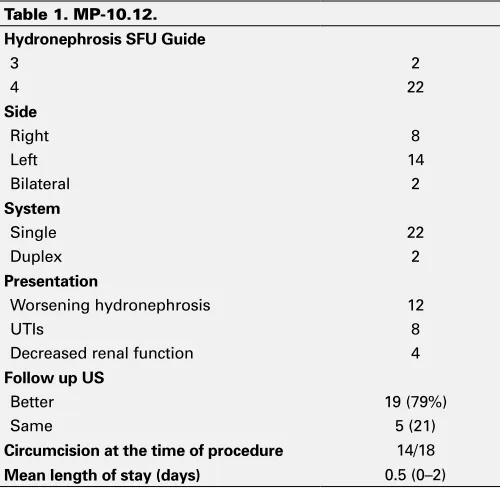
MP-10.12
Side-to-side ureterocystotomy: Keeping an “intact” UVJ, simpli-fying the Kaefer technique or obstructed megaureters in children Alyami, Fahad1; Bowlin, Paul1; Lee, Linda1; Braga, Luis H.2; Koyle, Martin1;
Lorenzo, Armando1
1Department of Surgery, Division of Urology, Hospital for Sick Children,
University of Toronto, Toronto, ON, Canada; 2Department of Surgery,
Division of Urology, McMaster University, McMaster Children’s Hospital, Hamilton, ON, Canada
Introduction and Objectives: An obstructed non-refluxing megaureter
(OM) is a relatively common diagnosis in neonates with antenatal hydro-nephrosis (ANH). Although conservative management is indicated in most cases of primary OM, surgery is still considered when associated with urinary tract infections (UTIs), worsening hydronephrosis or worsening renal function. Recently Kaefer described his technique of end to side refluxing ureteric reimplantion for OM, as a temporizing strategy. Herein we describe our experience with a modified non-dismembered side-to-side refluxing ureterocystotomy (UC) as a simple method for internal diversion in cases of OM.
Methods: Between February 2012 and August 2014, 24 consecutive
side-to-side refluxing UC were performed at two large academic tertiary pedi-atric referral centers in Canada. Demographics, surgical indications and follow up results were obtained
from a prospectively maintained database. The procedure was performed through a small inguinal incision, with a generous refluxing side-to-side anastomosis between the distal ureter and ipsilateral bladder wall.
Results: Mean patient age was 7.4 (Range 0-38) months at time of surgery;
18 (75%) were males. All patients were initially detected with ANH and followed with US every 3 months and renal scans accordingly. Unilateral procedures were done in 22 patients. The procedure was condcuted for primary OM in 23 patients and as salvage procedure for obstruction post common sheath reimplant in one child with a duplex system. Most children stayed overnight except for 7 patients. The average follow up was9.2 months (1-28). At time of last follow-up, most children experienced improvement in dilation (19/24, 79%) or stable findings (5/24, 21%).
There were no major surgical complications except for one patient that developed stenosis of the anastomosic site, which required a secondary procedure to create a loop cutaneous ureterostomy (Table 1).
Conclusions: Our preliminary results show that side-to-side refluxing UC
is a simple, feasible, safe minimally invasive procedure for primary OM, either as a temporizing or definitive intervention, which can be added to the surgical armamentarium of this condition. Clearly, close follow up is critical to document the long-term results of the procedure.
Table 1. MP-10.11. Patient demographics and tumour characteristics
Race
White 154 (77)
Black 7 (3.5)
Other 39 (19.5)
Stage
Localized 180 (90)
Regional 10 (5)
Distant 7 (3.5)
Unstaged 3 (1.5)
Mean age 1.28 years
Median Age 1 year
UP-10.01
Trends and outcomes of infant pyeloplasty for ureteropelvic junction obstruction in the first 6 months of life: Single institu-tion analysis over 14 years
Alyami, Fahad1; Alqarni, Naif1; Odeh, Rakan1; Penna, Frank J.1; Lee,
Linda1; Dos Santos, Joana1; Koyle, Martin1; Lorenzo, Armando1; Farhat,
Walid A.1
1Department of Surgery, Division of Urology, Hospital for Sick Children,
University of Toronto, Toronto, ON, Canada
Introduction and Objectives: Although ureteropelvic junction obstruction
(UPJO) occurs in all age groups, there tends to be clustering in early life due to evaluation for antenatal hydronephrosis (ANH). Definitive treat-ment of presumed UPJO remains surgical correction. Over the last two decades, management has shifted to a more conservative approach. As a result, a decrease in the number of infant pyeloplasties performed over time has been observed by some. Herein, we present our experience with children <6 months following pyeloplasty, and report trends in surgical management over the last 14 years.
Methods: Retrospective chart review on all infants who underwent
pyelo-plasty between 2000 to 2014. The trend and the outcomes of infant pyeloplasty in patients < 6 months of age were assessed. Data collected included: patient age at diagnosis and surgery, year of surgery, indication for surgery, first and last follow up ultrasound (US) findings and the need for further surgeries.
Results: At our institution, 2240 patients were referred for ANH, with a
mean of 160 referrals per year (136-487). Over the study period, a total of 400 open pyeloplasties were performed, with 169 (42%) being done in the first 6 months of life. Male patients predominated: 128 (76%). The mean age at surgery was 107 days (6-187) and average follow up was 49 months (2-162). Indications for surgery were worsening ANH in 162(96%), UTIs in 4 (2.2%) and decrease renal function in 3 (1.8%). Hydronephrosis degree at last follow up showed improvement in 133 (79%), stable dilation in 20 (12%), and worsening in 10 (6%). Six children were lost to follow-up. Redo surgery was successfully performed in 12 (7%) patients. Over the period of analysis, the overall and <3 months of age pyeloplasty rate remained the same but decreased for children > 3 month of age.
Conclusions: Pyeloplasty in the first 6 months of life is technically feasible,
safe, and effective. Our success rate is comparable to the published
suc-cess rate for the older children. Over the last 14 years in our institution the trend of infant pyeloplasty in children < 3 months of age has not changed significantly but had declined for children 3-6 month of age. This finding may be attributed to number of ANH referrals per year, reflecting a referral pattern or surgical bias.
UP-10.02
Endoscopic botulinum toxin injection for neuropathic bladder dysfunction: Is intra-detrusor delivery truly happening? Alyami, Fahad2; Bowlin, Paul2; Lee, Linda2; Gleason, Joseph M.2; Braga,
Luis H.1; Bagli, Darius J.2; Koyle, Martin2; Lorenzo, Armando2
1Department of Surgery, Division of Urology, Hospital for Sick Children,
University of Toronto, Toronto, ON, Canada; 2Department of Surgery,
Division of Urology, McMaster University, McMaster Children’s Hospital, Hamilton, ON, Canada
Introduction and Objectives: Botulinum neurotoxin A (BoNT/A) treatment
has become a viable option for patients with neuropathic bladder dysfunc-tion (NPBD) who fail medical management. Bladder BoNT/A injecdysfunc-tion mechanism of action is thought to be mediated by chemodenervation of the bladder muscle. For that reason, intra-detrusor (ID) injection is often favored in cases of NPBD. A submucosal (SM) injection technique has also been described, with the goal of targeting sensory pathways. By creating a SM bleb, visual confirmation of insertion depth provides assurance, in contrast to the ID delivery, which relies solely on
estimated depth of injection. In the present study we describe concurrent ultrasound (US) assessment at the time of BoNT/A bladder injection and correlate sonographic with endoscopic findings.
Methods: Between January and May 2014 we performed cystoscopy and
BoNT/A injection with US surveillance in a total of 8 patients with NPBD who were on maintenance BoNT/A program. All procedures were done under general anesthesia, by one surgeon, and trans-abdominal US was performed. When feasible, we employed the US images to determine depth and position of the injection, segregated based on the surgeon’s impression of a SM and the ID delivery by endoscopy (blinded to findings on US). We only included data for injection sites with visualization on both modalities.
Results: We included 8 patients with an age range of 5-12 years. There
were no reported complications and all patients reported stable response on follow-up. Data contrasted depth of injection by endoscopic and US guidance, segregated by SM and ID injection techniques. We confirmed the injections site accurately with US in 39/40 (97%) and 5/23 (22%) in the SM and ID space respectively. No cases of intra-peritoneal delivery were encountered.
Conclusions: This study demonstrates that trans-abdominal US-guided
bladder BoNT/A injection may provide additional information regarding depth. By US evaluation, it appears that SM delivery is reliable based on visual cues, whilst ID attempts often result in delivery outside of the detrusor muscle. If accurate ID delivery is preferred, intra-operative US provides a novel, non-invasive way of obtaining real-time feedback.
UP-10.03
Histopathological examination of the foreskin after circumci-sion for clinically suspected BXO in children: Is it a waste of resources?
Alyami, Fahad1; Odeh, Raken1; Heidari Bateni, Zhoobin1; Farhat, Walid
A.1; Koyle, Martin1
1Department of Surgery, Hospital for Sick Children, Toronto, ON, Canada Introduction and Objectives: Circumcision is one of the most widely
practiced procedures in the world. It is primarily for religious and cul-tural reasons. One of the unique medical indications for circumcision is Balanitis xerotica obliterans (BXO). The natural history of the disease in adults is well documented; progression of the disease can cause meatal stenosis and urethral stricture. In paediatric patients the natural history and the disease progression is not well documented and it is based on few reports in the literature. The appearance of the meatus at circum-cision for BXO has been used by surgeons to predict the pathological diagnosis. Our aim is to assess the concordance between the clinical and Table 1. MP-10.12.
Hydronephrosis SFU Guide
3 2
4 22
Side
Right 8
Left 14
Bilateral 2
System
Single 22
Duplex 2
Presentation
Worsening hydronephrosis 12
UTIs 8
Decreased renal function 4
Follow up US
Better 19 (79%)
Same 5 (21)
Circumcision at the time of procedure 14/18
pathological diagnosis and to assess the need for sending the foreskin for pathological examination.
Methods: A retrospective analysis of the medical records and
histopatho-logical findings of 70/420 boys who underwent circumcision with the foreskin sent for pathology due to the clinical suspicion of BXO from June 2005 to June 2013. The clinic visit notes and operative notes were reviewed for the age, presenting symptoms, previous medical treatments, intraoperative and postoperative management, subsequent outcome and follow up.
Results: The median (range) age of children was 9.4 (3-20) years. All the
children who had circumcision for presumed BXO diagnosis were symp-tomatic, 48 (68.5%) boys had spraying, 32 (46%) boys had straining, 49 (70%) had ballooning of the foreskin, 11 (16%) had urinary retention, 10 (14%) had history of UTIs and 23 (33%) had recurrent balanoposthitis. Balanitis xerotica obliterans (BXO) was confirmed in 53 out of 70 fore-skins (76%). In 13 and 6 of the 53 BXO, the histopathological examina-tion revealed focal BXO and early BXO respectively. There was suspicion of BXO in the physical examination in 47 (88%) out of the 53 patients. Chronic inflammation was reported in 15 (21%) patients and normal foreskin was found in 2 (3) patients. Follow up date will be updated.
Conclusions: In our series in the majority of the cases the clinical
diag-nosis correlated with the pathological diagdiag-nosis, which questions the need for sending the foreskin for pathological assessment. Knowing the diagnosis of BXO is important and can effect the management and follow up and can help counseling the patients and their parents. If we can sus-pect the diagnosis of BXO clinically we should follow the patient closely without the need to send the foreskin for pathological assessment and this can reduce the overall cost on the health care system.
UP-10.04
Asymptomatic urachal cysts in pediatric patients: A systematic review and meta-analysis
Nguyen, Laura N.2; Rediger, Christopher2; Leonard, Michael1; Guerra,
Luis1
1Pediatric Urology, Children’s Hospital of Eastern Ontario, Ottawa,
ON, Canada; 2Division of Urology, Department of Surgery, The Ottawa
Hospital and University of Ottawa, Ottawa, ON, Canada
Introduction and Objectives: Asymptomatic urachal remnants often are
found in the pediatric population on imaging that has been performed for other reasons. In these cases management is unclear, and the risk of future symptoms, complications or malignant transformation is difficult to quantify. A systematic review was performed to determine the prevalence of urachal remnants and likelihood of symptom development to guide management recommendations.
Methods: A systematic literature search of Medline, Embase, and Central
databases was performed for publications up to 2013. Studies were included if they reported on pediatric patients diagnosed with urachal cysts who were asymptomatic at diagnosis. One reviewer abstracted data.
Results: 578 abstracts were identified, then 569 were excluded for
irrel-evance, duplication, adult population, non-original research, non-Eng-lish language, because all patients were symptomatic or asymptomatic patients data could not be extracted. 9 studies remained. 6 of the studies included patients who did not undergo surgery (n=438). Of these, 3 were cross-sectional studies examining the prevalence of asymptomatic urachal cysts, 2 were observational studies that followed patients for between 3-30 months for development of symptoms or for radiological resolu-tion of the cysts, and 1 study reported both prevalence and follow-up. Reported prevalence ranged from 50- 99%, and from 0-100% in sub-group of infants. No patients developed of symptoms; rates of involution ranged from 0-100%. The remaining 3 studies were case reports or series including a total of 12 asymptomatic patients with urachal cysts (1-9 patients per study) who underwent surgical remnant excision. While 1 patient had neuroblastoma on pathology, no patients had an unexpected malignant diagnosis.
Conclusions: Asymptomatic urachal cysts are not uncommon. Patients
who are observed have not been reported to develop symptoms. The
entirely, with reports of a 100% involution rate. When remnants are surgi-cally excised they have not been reported to unexpectedly show evidence of malignancy. These remnants when asymptomatic and incidentally found in children may be safe to be observed without further intervention.
UP-10.05
Is the incidence of hypospadias really increasing?
Wehbi, Elias1; Dorgalli, Crystal1; Hidas, Guy1; McLorie, Gordon1; McAleer,
Irene1; Selby, Blake1; Pribish, Maryellen1; Khoury, Antoine E.1
1Pediatric Urology, University of California Irvine and The Children’s
Hospital of Orange County, Orange, CA, United States
Introduction: Literature on trends in hypospadias has been inconclusive.
To date, there has been no convincing data to support the theory that hypospadias rates are on the rise. Here we use population-based data from a large California registry to shed light on this topic.
Methods: We reviewed data obtained from the California Office of
Statewide Health Planning and Development (OSHPD), which includes data on all newborns discharged form all licensed California hospitals. Data was obtained form 1995-2010 and examined for trends in rates of hypospadias across Californian counties.
Results: Complete data was obtained on 49 counties. There were
4,590,611 births recorded during the study time period with 18,847 new-borns diagnosed with hypospadias, representing a mean rate of 0.41% (range=0.39-43%, 95%CI 0.40-0.42%).
Analysis of variance (ANOVA) showed no significant change in the annual rates of hypospadias in 49 counties across the state of California over 16 years (F (15,768) = 0.738, p = 0.75).
Of the counties with annual births exceeding 10,000, San Mateo and Trinity counties had highest and lowest mean annual rates respec-tively [0.62% (95%CI 0.58-0.67%) versus 0.33% (95%CI 0.29-0.36%), p<0.001]. Interestingly, there was also variation between other major metropolitan centers.
Conclusion: Although annual rates of hypospadias in California have
remained constant over the 16-year time period of this study, there appears to be statistically significant regional variation. This may intimate a role for population diversity as a cause, or more likely, an unknown environ-mental stressor. Further studies are needed to explain this phenomenon.
UP-10.06
Does increased pesticide use impact the prevalence of hypo-spadias?
Wehbi, Elias1; Dorgalli, Crystal1; Hidas, Guy1; McLorie, Gordon1; McAleer,
Irene1; Selby, Blake1; Pribish, Maryellen1; Khoury, Antoine E.1
1Pediatric Urology, University of California Irvine and The Children’s
Hospital of Orange County, Orange, CA, United States
Introduction: There appears to be variation in the rates of hypospadias
across major counties in California. The cause of this variation is not yet clear and although there is some literature on residential proximity to pesticide application and incidence of hypospadias, no conclusive data exists. Herein we use data from a large California registry to look for links between overall usage of pesticides and incidence of hypospadias across counties.
Methods: Data from the California Department of Pesticide Regulation,
which tracks mandatory reporting of total county usage of pesticides, was obtained between 1995 and 2010. The California Office of Statewide Health Planning and Development was queried for rates of hypospadias among newborns discharged from all licensed hospitals in California over the same time period.
Results: We evaluated data form 49 counties across California. There were
To adjust for the possible temporal effect of pesticide usage and fluctua-tions in hypospadias rates in the subsequent year, analysis was repeated for pesticide usage in a given year and hypospadias rates in the subse-quent year. There was no correlation observed using this model as well (R values for correlations -0.03 to -0.23, all corresponding p values > 0.01). Correlations were repeated among counties with highest and lowest rates of hypospadias and pesticide usage, with no associated link identified.
Conclusion: Although there is wide variation in rates of hypospadias and
usage of pesticides across counties in California, there does not appear to be an increased risk of developing hypospadias with increased use of pesticides. Looking to other environmental causes for regional variations in hypospadias should be undertaken.
UP-10.07
Bladder diameter ratio: A measure of bladder elongation and correlation to bladder trabeculation in children with spina bifida Wehbi, Elias1; Walia, Arman1; Dorgalli, Crystal1; Hidas, Guy1; McLorie,
Gordon1; McAleer, Irene1; Selby, Blake1; Pribish, Maryellen1; Khoury,
Antoine E.1
1Pediatric Urology, University of California Irvine and The Children’s
Hospital of Orange County, Orange, CA, United States
Introduction: To date, there is no widely accepted objective measure to
help quantify the shape of the normal urinary bladder in the literature. Patients with spina bifida are a potentially complex population who can present with a spectrum of upper and lower urinary tract derangements. Better understanding of the link between bladder shape in patients with normal bladders and those with spina bifida would greatly add to this understanding. Herein I attempt to shed like on this subject and evalu-ate a new measure, bladder diameter ratio (BDR), used to quantify the shape of bladders in children with and without evidence of vesicular neurologic pathology.
Methods: A small retrospective pilot study evaluated all voiding
cysto-urethrograms (VCUGs) that were performed at our institution in 2010. Those that were performed in children without known bladder pathology or underlying neurologic condition, and read as normal, were included. Patients with spina bifida with and without were also evaluated. A BDR was calculated in a standard fashion and defined as the ratio of maximal bladder length to width on cystography at cystometric capacity. Mean difference was compared using an independent samples t-test.
A larger retrospective study was performed to confirm the findings in a larger cohort of patients with spina bifida and to include ultrasounds, as a possible non-invasive imaging modality to measure BDR and follow patients. A similar analysis was performed which confirmed the similari-ties with regards to mean BDR between the 3 groups both on both x-ray contrast studies and ultrasound.
Results: Seventy-five children with normal bladders and 63 patients with
SB were included with mean ages at time of VCUG of 6.2(0.3-17.6) and 7.2(0.1-21) years respectively, (p=0.2). Children with normal bladders had a BDR of 1.03(95%CI 0.99-1.07). Patients with spina bifida and no trabeculation had no difference in BDR compared to those with normal bladders (BDR=1.04(95%CI 0.96-1.12;p=0.78)), unlike patients with spina bifida and trabeculation (BDR=1.44(95%CI 1.33-1.55;p<0.001). In the larger retrospective study, there was also a positive correlation between the presence of an elevated BDR and upper tract changes on ultrasound in both studies.
Conclusion: BDR, as a measure of bladder shape in children with normal
bladders, can be a useful tool. It provides the first objective measure of normal bladder shape. It can be used as a marker of disease progression. Further studies are needed to correlate BDR with hostile bladder pressures in a prospective fashion.
UP-10.08
Standardization of surgical instrument trays for pediatric hernia repair using tools of quality of improvement
Alqarni, Naif1; Odeh, Rakan 1; Butt, Hissan1; Alkahtani, Mohammed 1;
Konstant, Louis 1; Pandergast, Lisa 1; D’Arpino, Tania1; Koyle, Martin1 1Division of Urology, Department of Surgery, Hospital for Sick Children,
University of Toronto, Toronto, ON, Canada
Introduction: Pediatric hernia repair (PHR) is the most common elective
operation performed in children. As each surgeon performing PHR may “demand” his/her own set of instruments to perform the same opera-tion. This lack of standardization creates variability with inherent waste, increased cost and potential miscommunication.
Herein we try to establish a strategy to reduce and standardize instruments used for PHR, using PDSA (Plan, Do Study, Act), Quality improvement (QI) tools.
Materials and Methods: After approval by our hospital’s QI committee, we
prospectively evaluated each PHR over a 2 month period and analyzed OR time and instruments used from the standard PHR tray, by the surgical services (Urology(U) vs. Surgery(S).
Results: 56 cases were evaluated, 44 by S and 12 by U (‘Do’ D phase of
PDSA). In studying this process (‘Study’ S phase of PDSA), for S, 16 items (/51) were used in >50% of the cases, 18 used in <50% and 17 never used. In U, 16 items (/96) were used in >50% of the cases, 11 used in <50% and 69 were never used. OR times were similar in both services.
Conclusions: The majority of instruments in PHR tray were unused,
regardless of U or S. This translates into huge waste, especially in cen-tral processing. So, there is potential for improving value by appropriate standardization. Further PDSA cycles to act (‘Act’ A phase of PDSA) - in order to reduce and standardize instruments for both S and U into a single set and to study the results using PDSA cycles and to ensure that outcome measures are met without negative balancing measures occur-ring- are needed.
UP-10.09
Histopathological analysis following pediatric circumcision: Relevant?
Pelletier, Joanie1; Bolduc, Stephane1; Moore, Katherine1
1Departement of Surgery, Division of Urology, Centre hospitalier
univer-sitaire de Québec, Université Laval, Quebec, QC, Canada
Introduction and Objectives: The neonate circumcision is a marginal
practice in Quebec even with the most recent statement of the American Academy of Pediatrics. Two to 5% of uncircumcised boys will suffer from persistent phimosis and lichen sclerosus (LS) is one of its causes. LS is a risk factor for penile carcinoma, meatal stricture and urethral stricture. Indications for circumcision in our centre include persisting phimosis despite topical treatment, paraphimosis and less frequently because of parent’s desire. For this study, we compared LS signs obtain from the clinical exam and the pathology report to validate the relevance of this analysis. Our second end-point was to evaluate the incidence of LS in the pediatric population as the prevalence varies greatly between stud-ies in this age group and may have been underestimated for children younger than 5 yo.
Methods: We retrospectively reviewed 146 pediatric circumcision
per-formed between 2012 and 2014. Were excluded, modified circumcision as part of a minor hypospadias correction (n=4) and incomplete patho-logical report (n=6). We recorded the delay from consultation to surgery, LS clinical signs, duration of topical treatment, pathological analysis and follow-up.
Results: Complete analysis of 136 charts was performed after exclusion
(mean age 8 yo). White atrophic lesions were observed preoperatively in 28. The microscopy reported 72 normal prepuces, 30 nonspecific inflam-mation and LS in 34 for a prevalence of LS of 25%. Four of the 34 (11.8%) with LS were younger than 4 yo. The clinical assessment sensitivity is 56% with a specificity of 91%. Its positive predictive value is 67.9% and its negative predictive value is 86.1%.
Conclusions: The prevalence of LS is relatively small but the pediatric
UP-10.10
Effectiveness of peristeen anal irrigation system on fecal conti-nence in patients with spinal dysraphism
Wilkes, Courtney G.1; Thibodeau, Betty Ann1; Metcalfe, Peter D.1 1Divison of Urology, University of Alberta, Edmonton, AB, Canada Introduction and Objectives: The Peristeen Anal Irrigation (PAI) system
has been shown to be a successful treatment for bowel management in the adult spinal cord injury population (Christensen et al, J Spinal Cord Med 2008; 31:560-7)(Popolo et al, Spinal Cord 2008; 46:517-22)( Christensen et al, Gastroenterology 2006; 131:738-47). However, minimal evidence exists regarding its effectiveness in the pediatric and adult spina bifida population. We analyzed the effectiveness of the PAI system in this population with respect to fecal continence.
Methods: 10 patients from our spina bifida clinics (9 pediatric, 1 adult;
mean age=9.1years) whose bowel management had not been successfully treated with conservative measures were selected to use the PAI system as an alternative treatment. These patients were required to undergo the daily enemas for 3 months and be able to perform the regime independently or have an available caregiver. The patients completed a validated 10-item Neurogenic Bowel Dysfunction (NBD) questionnaire(Krogh et al, Spinal Cord 2006; 44:625-31), which clinically assessed their bowel dysfunction before and after using the PAI system. The NBD score falls into one of four categories: very minor (0-6), minor (7-9), moderate (10-13), or severe (≥14). Pre- and post-treatment NBD scores were analyzed to measure the effectiveness of the PAI system for treating bowel dysfunction. Each question was additionally analyzed to observe where the most benefit was gained. Student t test was used to determine significance.
Results: Follow-up data is available on 6 patients. 2 patients were lost to
follow-up, 1 patient withdrew due to increased flatus and digital evacu-ation, and 1 patient withdrew due to the time-consuming process (30-45 minutes). The 6 patients who completed the 3-month trial demonstrated a statistically significant improvement in their overall NBD score (p = .003). Specifically, fecal continence (p = .01) and perianal skin problems (p = .04) demonstrated statistical significance. Every patient remained in the same clinical category from pre-treatment to post-treatment.
Conclusions: Although the data was statistically significant, the PAI system
demonstrated moderate clinical improvement in quality of life. It pro-vided the most benefit for treating perianal skin problems and fecal incon-tinence. Therefore, in a carefully selected, motivated patient and family, the PAI system may provide some benefit for overall bowel management.
UP-10.11
Clinical presentation of urolithiasis in children Baltazar, Juan Antonio J.1
1Pediatric Urology, Instituto Mexicano del Seguro Social, Guadalajara,,
Mexico
Introduction: Urolithiasis en pediatrics has different characteristics than
in adults, mainly in the clinical presentation and treatment. Its presenta-tion in children is rare, but recent informapresenta-tion suggests an increasing incidence.
Objective: To evaluate the clinical manifestations on arrival to the
emer-gency department according to age and known associated factors.
Material and Methods: Through a retrospective study, we made a chart
review of those patients diagnosed with urolithiasis (kidney, ureter, blad-der, and urethra) attending the pediatric emergency department from June 2012 to June 2014. The data obtained were symptoms referred as reason for admission to the emergency room and possible associated risk factor for urolithiasis.
Results: We evaluated 38 cases of urolithiasis; the mean age at
presenta-tion was 9 years-old (1-15 years). Ureteral colic was the most common presentation with 15 cases (39%), followed by symptoms related with urinary tract infection with 11 cases (28%), hematuria in 9 cases (23%), nonspecific abdominal pain in 7 cases (18%), hydronephrosis in 5 cases (13%), fever in 5 cases (13%) and obstructive symptoms in only 2 cases (5%). In 23 urolithiasis cases (60%) no known risk factor was found, however there were 15 with associated factors, 5 patients (33%) with a
patient with Lesch-Nyhan syndrome (6%) and one patient with history of antiretroviral therapy (6%) as a possible associated risk factor.
Conclusions: In our pediatric population ureteral colic is the most
com-mon form of presentation, predominating in children older than 9 years-old, below this age the usually features varied from nonspecific abdominal pain sometimes accompanied with fever, hematuria or symptoms related to urinary tract infection. In infant and preschool children we considered an asymptomatic presentation because only hematuria or hydronephrosis was identified during the initial evaluation. The younger the patient non-specific the symptoms at presentation can be expected.
UP-10.12
The impact of photographic email correspondence on the rate of emergency room visits post pediatric urological procedures D’Cruz, Jennifer1; Kim, Soojin 1; Rickard, Mandy1; DeMaria, Jorge E.1;
Braga, Luis H.1
1Department of Surgery, McMaster University, Hamilton, ON, Canada Introduction and Objective: Concerns post urological procedures are
usually addressed by traditional telephone communication (TTC) with nurse practitioners or, more recently, by reviewing digital photographs of postoperative sites. Our objective was to determine the impact of photo-graphic email correspondence (PEC) on ER visits and hospital admission rates following pediatric urological interventions.
Methods: A chart audit of 2393 consecutive pts was carried out at a
ter-tiary children’s hospital between 2011-2014. Number of ER visits within a 30-day postoperative period and hospital admissions were recorded. Comparative analyses of ER visits and hospital admission rates before (Jan 2011–May 2013) and after institution of PEC (Jun 2013–Nov 2014) were carried out using chi-square test, at significance level of p<0.05.
Results: Of 2393 pts, 1726 (72%) were seen Jan 2011-May 2013 and 667
(28%) from Jun 2013-Nov 2014. A total of 133 (6%) visits were made to the ER for postoperative urology concerns. Mean age at presentation was 4±4.5 years, with visits on post-operative day 9 being the most com-mon. ER visits occurred more often on weekdays [93/133] compared to weekends [40/133] (70% vs. 30%, p<0.05), and during days [87/133] compared to overnight [46/133] (65% vs. 35% p<0.05). Most common reasons for seeking postoperative care were catheter concerns (24%); incision related issues such as surgical site bleeding, hematomas and redness (20%); surgical site pain (15%); and postoperative fever (12%). ER visits were a result of primarily hypospadias repairs (26%), followed by hernias and orchidopexies combined (16%), ureteric reimplantation/ ureteroureterostomy (15%) and circumcisions (12%). Significantly fewer ER visits were made by those followed by PEC [23/667] compared to TTC only [110/1726] (3.4% vs. 6.4%, p<0.01). Likewise, after the initiation of PEC hospital admissions rates [3/23] were lower than the pre-PEC period [25/110] (13% vs. 23%, p<0.01).
Conclusion: Initiation of PEC was associated with a 50% decrease in ER
visit and a 43% reduction in hospital admissions relative to the TTC only period. Nearly 80% of pts returning to the ER after pediatric urological procedures were not admitted to hospital. Most common reasons for returning to the ER were catheter and dressing related issues post hypo-spadias repair.
UP-10.13
Radiation exposure to children with posterior urethral valves in the first year of life: A necessity or an avoidable oversight? Penna, Frank J.1; Odeh, Raken1; Butt, Hissan1; Lorenzo, Armando1; Farhat,
Walid A.1; Bagli, Darius J.1; Koyle, Martin1
1Division of Paediatric Urology, Hospital for Sick Children, Toronto, ON,
Canada
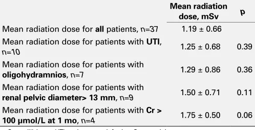
Introduction and Objectives: Antenatal detection of posterior urethral
exposure risk, may be avoidable. The long-term radiation exposure risk is unknown, but it has been estimated that exposure to a Sievert (Sv) of radiation carries a 5.5% long-term risk of malignancy.
Methods: A retrospective review was performed of all children born with
a diagnosis of PUV over a 10-year period, from 2003-2013 at our institu-tion. Various parameters for each patient were examined; the mean total radiation exposure from all imaging studies performed during the first year of life was estimated. The patients were sub-categorized by risk factor and radiation dose was calculated for each sub-category.
Results: Thirty-four children were included in the study. The mean
ges-tational age was 36.6 ± 1.9 weeks and the mean birth weight was 3.0 ± 0.3 kg. All of the children except for 3 had a documented history of antenatal hydronephrosis, with 7 having a history of oligohydramnios. The mean creatinine at 1 month and 1 year was 62.7 ± 53.1 μmol/L and 52.0 ± 40.1 μmol/L, respectively. The majority of the children were treated with valve ablation; only three had vesicostomy placement. Ten of the children had a urinary tract infection (UTI) in the first year of life. The mean number of renal/bladder ultrasound and VCUG studies performed during the first year of life was 3.9 ± 1.8 and 1.6 ± 0.66, respectively. The mean radiation from VCUG and DMSA was 0.80 ± 0.36 millisieverts and 1.09 ± 0.30 millisieverts, respectively (Table 1).
Conclusions: Several risk factors were associated with higher radiation
exposure including a history of UTI, oligohydramnios, renal pelvic diam-eter >13 mm, and creatinine > 100 μmol/L at 1 month of life, with only creatinine approaching statistical significance. Attempts should be made at preferentially employing studies that use non-ionizing radiation such as ultrasound, thereby minimizing the total ionizing radiation exposure to children at such a critical and early developmental period.
UP-10.14
Outpatient pediatric open pyeloplasty: A novel clinical pathway for same-day hospital discharge
Lee, Linda1; Gleason, Joseph M.1; Bowlin, Paul1; Alyami, Fahad1; Penna,
Frank J.1; Braga, Luis H.2; Lorenzo, Armando1
1Urology, Hospital for Sick Children, Toronto, ON, Canada; 2Urology,
McMaster University, Hamilton, ON, Canada
Introduction and Objectives: To assess the feasibility and safety of
out-patient pediatric open pyeloplasty, we present a novel clinical pathway to select children undergoing open pyeloplasty who can to be discharged on the same day of surgery.
Methods: Over a 1 year period, 12 out of 14 children undergoing
con-secutive open unilateral pyeloplasties performed by a single surgeon met inclusion criteria for same-day discharge: informed parental consent, nor-mal contralateral kidney, clearance by anesthesia team, uncomplicated operation conducted as first case of the day, optimal post-operative pain control with oral analgesics, adequate PO intake, and voiding prior to discharge. All patients underwent an open dismembered pyeloplasty, following an extra-peritoneal approach, through small, muscle-splitting, ultrasound-guided incision, with an externalized ureteropyelostomy (EUP) stent intra-operatively placed through the renal pelvis. No retrograde pyelograms were performed. In all cases, the EUP stent was clamped and the Foley catheter removed at the end of the procedure.
Results: Mean age at time of surgery was 7.9 ± 5.8 months, with 50%
(n=6) patients under the age of 6 months. The majority (92%, n=11) of day-surgery patients had a history of antenatal hydronephrosis. Mean operative time was 1 hour and 42 minutes. Two patients (16.7%) returned to the Emergency Department for stent-related issues: one had a dislodged stent, and another fever that improved by unclamping the EUP tube (urine culture was subsequently negative). Both were managed conservatively without complications. All patients had an uneventful removal of their stents in 8-15 days in clinic without need for a second anesthetic. All patients have experience improvement in dilation and remain asymptom-atic at 3-month follow-up.
Conclusions: We present our initial series of outpatient pediatric open
pyeloplasty, following a novel clinical pathway and strict inclusion cri-teria. This appears to be a safe and feasible practice in an appropriately selected patient population, maximizing adequate use of inpatient hos-pital resources.
Table 1. UP-10.13. Patient Radiation Exposure Parameters During the First Year of Life
Mean radiation dose, mSv p
Mean radiation dose for all patients, n=37 1.19 ± 0.66 Mean radiation dose for patients with UTI,
n=10 1.25 ± 0.68 0.39
Mean radiation dose for patients with
oligohydramnios, n=7 1.29 ± 0.86 0.36 Mean radiation dose for patients with
renal pelvic diameter> 13 mm, n=9 1.50 ± 0.71 0.11 Mean radiation dose for patients with Cr >