Role of polycomb proteins Ring1A and Ring1B
in the epigenetic regulation of gene expression
MIGUEL VIDAL*
Department of Developmental and Cell Biology, Centro de Investigaciones Biológicas, CSIC, Madrid, Spain
ABSTRACT Generation of cell diversity depends on epigenetic regulatory mechanisms. Polycomb group (PcG) proteins are central components of epigenetic regulation in metazoans. The system, initially associated with transcriptional program stability during development, is also involved in the regulation of other processes, such as maintenance of stem cell pluripotency and cell proliferation. PcG regulation involves chromatin modifications through covalent histone modifi-cations. One of these modifications, the monoubiquitylation of the H2A histone, depends on Ring1 proteins, which are essential for development in insects and mammals. In murine embryonic stem cells, Ring1A and Ring1B-dependent ubiquitylation of H2A is linked to repression of transcrip-tional initiation. Studies in mammalian cells have found a multiplicity of protein complexes containing Ring1A and Ring1B, suggesting an expanded regulatory role for Ring1A, Ring1B proteins in the epigenetic regulation of gene expression.
KEY WORDS: Ring1A, Ring1B, polycomb, H2A ubiquitylation, epigenetic regulation
Ring1A/Ring1 and its paralog Ring1B/Rnf2 are core components of the mammalian Polycomb system, which is one of the epigenetic regulators of cell diversity generation during embryonic development and tissue maintenance during adult life (Rajasekhar, 2007; Sparmann, 2006). Although initially identified during genetic studies of developmental regulation of the fruit fly D. melanogaster, the Polycomb group (PcG) of genes is represented throughout all metazoans (Ringrose, 2004; Schuettengruber, 2007; Whitcomb, 2007). PcG was named after the founding member, Polycomb (Pc), a term that refers to the extra number of sexual combs found in Pc mutant fly males. During this genetic analysis of Drosophila development, another group of genes, the trithorax group (trxG) was identified because of their ability to counteract the activity of PcG genes (Ringrose, 2004;Grimaud, 2006; Schwartz, 2007).
PcG genes encode subunits of multiprotein complexes with a role as transcriptional repressors (Table 1). In mammals, devel-opmental processes controlled by PcG genes include specifica-tion of the antero-posterior axis, monoallelic expression of im-printed genes and self-renewal of embryonic and somatic stem cells. In addition, deregulation of the Polycomb system is often associated to neoplastic events and tumor formation (Valk-Lingbeek, 2004; Sparmann, 2006;Rajasekhar, 2007).
Polycomb complexes act, at least in part, through chemical
BIOLOGY
www.intjdevbiol.com*Address correspondence to: Dr. Miguel Vidal. Department of Developmental and Cell Biology. Centro de Investigaciones Biológicas (CSIC). Ramiro de Maeztu, 9. 28040 Madrid, Spain. Fax: +34-91-536-0432. e-mail: [email protected]
Supplementary Material for this paper (a multiple sequence alignment) is available at: http://dx.doi.org/10.1387/ijdb.082690mv
Published online: 29 April 2009.
ISSN: Online 1696-3547, Print 0214-6282 © 2009 UBC Press
Printed in Spain
Abbreviations used in this paper: PcG, polycomb group; PRC, polycomb repressive complex; trxG, trithorax group.
view of how the Polycomb system works (Sparmann, 2006; Schuettengruber, 2007).
Recently three lines of investigation are paving the ground for elucidating PcG function most of which, up to now, has come from genetic analysis: (i) characterization of the enzymatic activities of PcG products (ii) proteomic analysis of PcG complexes and (iii) identification of their target genes. In this Review, I will consider the biochemical intricacies of complexes that contain Ring1A/ Ring1 and Ring1B/Rnf2 proteins, thereafter referred to as Ring1A and Ring1B. I will then describe their activities as monoubiquitin H2A ligases and the mechanisms of transcriptional control af-fected by this histone modification. I will conclude by summarizing the major biological processes on which the mammalian Ring1 proteins have been found to participate, including their role in stem cell maintenance and tissue homeostasis.
Ring1A and Ring1B are RING finger transcriptional
repressors
Ring1A and Ring1B were found in searches for components of mammalian Polycomb complexes using the yeast two-hybrid system to identify interacting partners of vertebrate homologs of Drosophila PC. Murine Ring1A and Ring1B were identified using the transcriptionally repressive domain of M33/Cbx2 (Schoorlemmer, 1997). Human Ring1A was similarly isolated using a full length form of a Pc homolog in Xenopus, Cbx4 (Satijn, 1997). The Ring1A cDNAs were found to be identical to the product of one of a set of “Really INteresting Genes” previously identified during a search for transcripts from the major histocom-patibility complex (Hanson, 1991; Lovering, 1993; Saurin, 1996). Ring1B cDNA encodes a related, but distinct protein (Schoorlemmer et al., 1997), and subsequently was also identi-fied in similar molecular screens that used a vertebrate homolog of Drosophila PSC as a bait (Suzuki, 2002).
Paradoxically, at the time of Ring1A and Ring1B isolation there was no molecularly characterized Drosophila PcG gene encoding a Ring1 ortholog. However, loss of function of both genes in the mouse showed alterations in the axial skeleton similar to those seen in other PcG mutant mouse lines (del Mar Lorente, 2000; Suzuki, 2002). Eventually, Sex combs extra (Sce), a PcG gene
identified in a genetic screen in Drosophila, was found to encode the ortholog of vertebrate Ring1 products (Fritsch, 2003; Gorfinkiel, 2004).
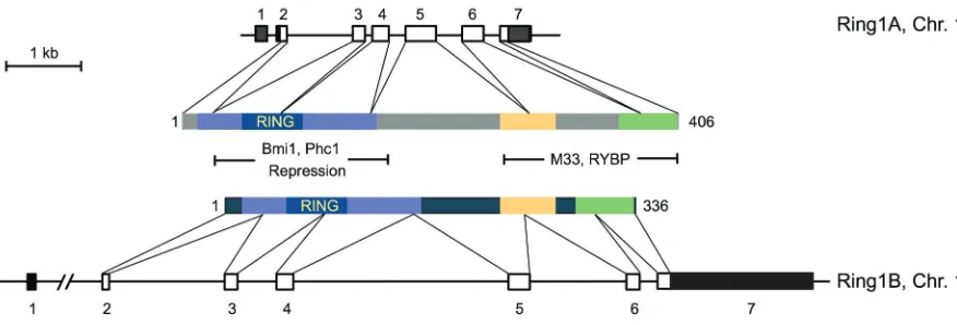
The initially isolated Ring1A cDNAs encoded for a 377 amino acid protein (Lovering, 1993; Schoorlemmer, 1997; Satijn, 1997). Subsequent cloning of 5’ ends of Ring1A cDNA and comparison of their sequences with that of genomic clones (chromosome 16 in humans, chromosome 17 in mouse) provided evidence for a full length protein of 406 amino acids (Schoorlemmer, 1997). Se-quence analysis of Ring1A identified a N-terminal Cys-rich do-main conserved among many other proteins that defined a motif termed RING finger (Lovering, 1993). This new protein motif also contains histidine and is organized as a three-stranded antiparal-lel β-sheet, two Zn2+ binding loops, and an α-helix following the second β-strand. Zn2+ binding is needed for the proper folding of the RING motif (Borden, 2000). The cross-braced arrangement of Zn2+ binding by the conserved cysteine and histidine residues (either in the C3HC4 or the C3H2C3 form), sets this domain apart from other cysteine-histidine zinc binding motifs, such as PHD (Plant homeodomain) or LIM (Lisl1/Isl-1/Mec3) domains (Capili, 2001). Thus, the first and third pair of Zn ligands share Zn 1 and the second and fourth pair of Zn ligands share Zn 2. The RING fingers of Ring1A and Ring1B are of the C3HC4 type, and threedimensional structures have recently been derived for that of Ring1B (Li, 2006). The RING motif is also found in other PcG products, namely the family of Polycomb group RING finger (Pcgf) proteins, the homologs of Drosophila PSC (Table 1).
The murine or human Ring1B genes (both located on chromo-some 1) encode a 336 amino acid protein. The seven exon transcriptional unit, however, is much less compact than that of the Ring1A genes, mostly because the transcription start site is separated by a large intervening sequence from the first coding exon (Fig. 1). Comparison of Ring1A and Ring1B protein se-quences identifies three conserved regions (Fig. 1). The largest, at the N-terminal end of the proteins spans the RING finger, and is separated from other two regions by a nonconserved se-quence. Biophysical analysis of a fragment of Ring1B/Rnf2 en-compassing the two C-terminal conserved regions (amino acids 222-336) shows that it is a well formed, globular, structure with a high content of α-helix and β-sheets that forms homodimers
through the docking of preformed monomers (Czypionka, 2007). Conservation of these C-terminal regions may be imposed by a given folding needed for specific contacts with chromodomain-containing proteins [for instance, M33/Cbx2, (Schoorlemmer, 1997)] or the Ring1 and YY1 binding protein (García, 1999; Neira et al., 2009) that occur through these regions of Ring1 proteins. When fused to a DNA binding domain such as that of the yeast GAL4 transcription factor, both Ring1A and Ring1B acts as transcriptional reppressors of reporter constructs that have GAL4 operators (Schoorlemmer, 1997; Wong, 2007). These studies, however, show contrasting results about the role of the RING
fingers, which appear essential for Ring1A repression but dis-pensable for Ring1B repression.
Evolutionary conservation of Ring1 proteins
Searches for homologs of Ring1A and Ring1B proteins among multicellular organisms by using a sequence-similarity method between full length sequences, show single copies of the Ring1 proteins in invertebrates and two copies only in vertebrates. This contrasts with a multiplicity of paralogs (Table 1) identified in vertebrates and plants for several other PcG proteins. For
in-Drosophila Humans Domains, activities
PRC2 (initiation complex)
Esc EED WD40
E(z) EZH2, EZH1 SET, H3K27 methyltransferase
Su(z)12 SUZ12 FCS-type Zn finger, RNA binding
PRC1 (maintenance complex)
Polycomb, PC M33/CBX2†, PC2/CBX4, PC3/CBX8, CBX7, CBX6* Chromodomain, binding to H3K27me3
Polyhomeotic, PH PH1/PHC1, PH2/PHC2, PH3/PHC3 SAM, oligomerization; FCS-type Zn finger, RNA binding
Posterior sex combs, PSC BM1/PCGF4, MEL18/PCGF2, NSPC1/PCGF1, MBLR/PCGF6, PCGF5*, PCGF3* RING finger
Sex combs extra, Sce/dRING RING1A/RING1, RING1B/RNF2 RING finger, E3 ubiquitin ligase for H2A
Sex combs on midleg, SCM SCMH1, SCMH2* MBT, histone binding; SAM, oligomerization
Other
Pleiohomeotic, PHO YY1 Zn finger, DNA binding
Pleiohomeotic-like, PHO-L YY1 Zn finger, DNA binding
TABLE 1
POLYCOMB PROTEINS
†In protein names given in pairs, the latter, after the slash, corresponds to the official nomenclature.
*Homologs without evidence for a Polycomb function.
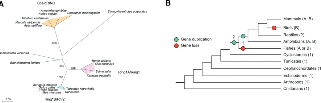
Fig. 2. Conservation of Ring1 proteins during evolution. (A) Phylogenetic tree of full length Ring1 proteins showing bootstrap confidence levels of the reliability of grouping in the various branches. The scale bar indicates distances calculated with JTT substitution model implemented in the PHYLIP package. The tree does not include all known mammalian sequences (because of extreme similarity) and only those of human and mouse are plotted for simplicity. (B) Canonical tree of life (http://tolweb.org/tree/phylogeny.html) indicating the number and type of Ring1 proteins (A and B denote Ring1A type and Ring1B type, respectively) among animal groups. A number 1 indicates only one protein that is not assigned to either Ring1A or Ring1B type. Green and red dots indicate the positions at which gene duplication and loss, respectively, may have occurred during evolution. The species considered for each of the animal groups shown are: Homo sapiens and Mus musculus (Mammals), Gallus gallus (Birds), Xenopus tropicales (Amphibians), Tetraodon nigroviridis, Danio rerio and Salmo salar (Fishes), Branchiostoma floridae (Cephalocordates), Apis melifera, Nasonia vitripennis, Aedes aegypti, Anopheles gambiae and Tribolium castaneum (Arthropods), Strongylocentrus purpuratus (Echinoderms) and Nematostella vectensis (Cnidarians).
stance, the family of PC paralogs has five members in mammals, M33/Cbx2, Pc2/Cbx4, Cbx6, Cbx7 and Pc3/Cbx8, whereas that of PSC has six, Nspc1/Pcgf1, Mel18/Pcgf2, Pcgf3, Bmi1/Pcgf4, Pcgf5 and MBLR/Pcgf6. Sequence similarity analysis of Ring1 paralogs groups most species around three major categories of Ring1 proteins: insects, which express the Drosophila Sce/dRING type, and vertebrates that express either Ring1A or Ring1B-type proteins or both (Fig. 2). No Ring1 homologs have been found in plants or in worms, although sequences encoding RING finger domains related to those of Ring1 proteins have been identified (not shown and Karakuzu, 2009).
The high degree of conservation among Ring1 orthologs can be seen in the phylogenetic tree constructed with the full length Ring1 proteins sequences (Fig. 2A) and is much higher than that found for the orthologs of any other PcG protein (Whitcomb, 2007). Alignment of Ring1 protein sequences allows to easily identify the conserved N-terminal and the two C-terminal regions (supplementary Fig. 1). In insects, the Ring1 proteins show a distinctive long linker sequence separating the N-terminal region spanning the RING finger motif from the C-terminal conserved regions. In agreement with the high degree of structural conser-vation, the phenotype of a Sce/dRing mutation in Drosophila larvae is substantially rescued by the ectopic expression of the murine Ring1A protein (Gorfinkiel, 2004). Protochordates (Branchiostoma) and Teleostei fishes express only one Ring1 protein. In the case of fishes it is either of the Ring1B type (Tetraodon, Danio) or the Ring1A type (Salmo). If Salmo only has a Ring1 protein (its genome is not fully sequenced yet) and it is an ortholog of the Ring1 protein of other Teleostei it would imply that a Ring1 gene was duplicated after the appearance of fishes and was selectively lost at Birds (Gallus) or monotreme mammals (Ornithorhynchus) which have a single Ring1protein, of the Ring1B-type. Only eutherian mammals and amphibia (Xenopus) have Ring1 proteins of both types. A tree of life showing the possible duplication and loss of Ring1 genes during evolution is shown in Fig. 2B.
Ring1A, Ring1B-containing complexes
Genetic analysis in Drosophila provided evidence for synergis-tic effects in compound PcG mutants, which suggested that PcG products may act as part of multiprotein complexes. On this basis, initial characterization of PcG complexes relied on the use of yeast-two hybrid interaction assays and coimmunoprecipitation assays that involved known PcG subunits. However, Ring1A and Ring1B have also been isolated as interactors in yeast-two hybrid screens that used non-PcG proteins as baits. The interactor pairs thus identified, are usually proteins that associate directly to each other, as validated in pull down assays that use recombinant forms of the proteins. Table 2 summarizes vertebrate proteins that associate directly to either or both of the Ring1 proteins. Many other proteins associate indirectly, as subunits of Ring1 proteins-containing complexes, and are described below. Most direct interactors, including Ring1 proteins themselves (Satijn, 1999), are PcG products (M33, Pc2, Pc3, Bmi1, Ph2, etc (Satijn, 1997; Schoorlemmer et al., 1997; Cao, 2005; Hemenway, 1998; Satijn, 1999; Hemenway, 2000)] or their homologs [NsPc1, MBLR (Akasaka, 2002; Gearhart, 2006)]. Others are transcriptional repressors such as RYBP (García, 1999), or proteins that interact
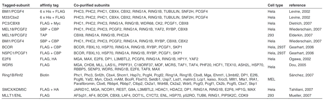
with transcription factors, such as KyoT2 (Qin, 2004) or DNA binding proteins as the ubiquitously expressed transcriptional CP-2/Tcfpcf2 (Tuckfield, 2002). Other interactors are involved in ubiquitylation reactions [the E2 protein ligase Hip2/Ube2k (Lee, 2001)] or belong to the regulatory subunit of the proteasome [TBP-1/PSMC3 (Lee, 2005)]. However, not all of these interactors have appeared among the subunits of purified Ring1A-Ring1B-containing complexes (see Table 3), isolated in tag-based ap-proaches. In these, a tagged form of the protein of interest is expressed in tissue culture cells or fly embryos to assist in the efficient affinity-based isolation (using antibodies or interacting proteins coupled to a solid phase) of complexes that contain the tagged protein. The subunits in these complexes mixtures are identified by mass spectrometry analysis. Table 3 summarizes the subunits that have been identified in these tag-based isolation approaches that contain Ring1A, Ring1B or both.
Ring1 proteins are core subunits of Polycomb
Repres-sive Complex 1
The isolation of the first Ring1-containing complex used ex-tracts from early D. melanogaster embryos expressing tagged-PcG subunits (FLAG-tagged PH or FLAG-tagged PSC). The complex thus isolated was termed Polycomb repressive complex 1 (PRC1) (Shao, 1999; Saurin, 2001), and in addition to PH and PSC contained the PcG products PC and Sce/dRING. Many other polypeptides not known as PcG products, copurified with FLAG-PH or FLAG-PSC, including some components of the transcrip-tion initiatranscrip-tion machinery (Saurin, 2001). Complexes isolated us-ing similar approaches with human tissue culture cells, however, contained fewer subunits, most of them PcG products (Levine, 2002). Thus, human PRC1 subunits include homologs of Pc (M33/CBX2 and PC3/CBX8), of PSC (BMI1/PCGF4), Polyhomeotic (HPH1/PHC1, HPH2/PHC2 and HPH3/PHC3), and of Sce (RING1A and RING1B) together with substoichiometric amounts of another PcG product, the homolog of SMC (SCMH1) and of a few non PcG subunits (Levine, 2002).
Purified Drosophila PRC1 interferes with the activity of SWI/ Ring1 protein Interactor Reference
Ring1A/Ring1 M33/Cbx2 Schoorlemmer, 1997
Pc2/Cbx4 Satijn, 1997
BMI1/PCGF4 Satijn and Otte, 1999
RYBP García, 1999
KyoT2 Qin, 2002
NSPC1/PCGF1 Gearhart, 2006
PH2/PHC2 Cao, 2005
PC3/CBX8 Hemenway, 2000
RING1A/RING1 Satijn and Otte, 1999
Ring1B/Rnf2 BMI1/PCGF4 Hemenway,1998
Mblr/Pcgf6 Akasaka, 2002
Mel18/Pcgf2 Suzuki, 2002
PH2/PHC2 Hemenway, 1998
PC3/CBX8 Hemenway, 2000
CP-2/Tcfpcf2 Tuckfield, 2002
Fbxl10/Jhdm1B Sánchez, 2007
PHB Choi, 2007
RYBP Gearhart, 2006
NSPC1/PCGF1 Gearhart, 2006
RING1A/RING1 Gearhart, 2006
HIP2/UBE2K Lee, 2003
TBP-1/PSMC3 Lee, 2005
TABLE 2
SNF chromatin remodelers in a plasmid supercoiling assay (Shao, 1999). The smallest stable PRC1 subcomplex that still blocks SWI/SNF contains the PcG subunits PC, PH, PSC and Sce/ dRING (Francis, 2001b), all of which have homologs (Table 1) in human PRC1 (Levine, 2002). Therefore, these subunits are considered the core components of PRC1. Similar PRC1 com-plexes, differing in the core subunit homologs composition, per-haps a reflection of their relative abundance in the cell type used, have been isolated using tagged MEL18/PCGF2, BMI1/PCGF4 or CBX8/Pc3 subunits (Wiederschain, 2007; Dietrich, 2007). In every case, the mammalian version of PRC1 lacked most of non-PcG subunits found in Drosophila PRC1. One puzzling observa-tion about the mammalian PRC1 complex is the absence of DNA-binding proteins that may assist on specific recruitment to targets. Up to now, only histone-binding motifs, such as the chromodomain present in PC homologs (Table 1), are known to be involved in recruiting, although some evidence points at alternative mecha-nisms (see below).
Ring1 subunits play a role in the stability of PRC1 complexes. For instance, the Drosophila core PRC1 complex is only stable if Sce/dRING1 is present (Francis, 2001b). In line with this obser-vation, the inactivation of Ring1B in murine embryonic stem ES cells is associated to a dramatic decrease of the stationary levels of core PRC1 subunits (Leeb, 2007; Endoh, 2008). It is possible that the ability of Ring1 proteins to directly contact each of the other core subunits (Hemenway, 1998; Satijn, 1999; Cao, 2005; Wei, 2006) may be the basis for their stable assembly in a complex.
Other Ring1A-Ring1B containing complexes
Ring1A and Ring1B have also been found in molecular assem-blies distinct from PRC1, together with many other polypeptdies with a role in transcriptional regulation (Table 3). The complexes, identified by proteomic approaches, have been isolated by pulling a variety of tagged subunits such as E2F6 (Ogawa, 2002), a member of the E2F family of transcription factors (Trimarchi, 2002), the histone demethylase SMCX/KDM5CA (Tahiliani, 2007)), the corepressor BCOR (Gearhart, 2006), the histone-binding WD40-repeat protein WDR5 (Dou, 2005) or ENL (Mueller, 2007),
a protein fused to the histone H3K4 methyltransferase Mll in a number of leukemias (Krivtsov, 2007). When tagged-PcG sub-units were used, complexes more related to PRC1 have been isolated. For instance, those purified through BMI1 (Levine, 2002; Wiederschain, 2007), MEL18 (Wiederschain, 2007; Elderkin, 2007), M33 (Levine, 2002) or PC3 (Dietrich, 2007). However, Ring1A, Ring1B and associated components isolated by pulling out NSPC1 (Gearhart, 2006) revealed a complex different from PRC1. Together, these observations suggest that Ring1A and Ring1B are represented more frequently than any other PcG component in complexes involved in transcriptional control. This notion is confirmed by the variety of transcriptional regulators found to copurify with Ring1B (Sánchez, 2007), that includes most other interactors identified in previous purification schemes and some additional ones. It is important to note that the proteins thus identified are often part of several complexes that share the tagged subunit. Mass spectrometry analysis of Ring1B-associ-ated polypeptides in a murine erythroleukemic cell line (Sánchez, 2007) suggests that PRC1 subunits are abundantly represented, but clearly they are not alone among the most abundant ones. Thus, together with PRC1, one or more complexes containing subunits identified in a E2F6 complex, and in a BcoR-Fbxl10 complex are equally abundant. Other enriched subunits belong to assembles not fully characterized. It is likely that similar purifica-tion schemes that use other cell types will render similar sets of proteins, but it is also conceivable that new sets of Ring1A-Ring1B containing complexes are uncovered.
Among the subunities found to copurify with Ring1 proteins some have known/suspected enzymatic activities as chromatin modifiers: for instance, histone demethylases (Lsd1/Kdm1, Fbxl10/ Kdm2b, SCMX/KDM5B), histone methyltransferases (the heterodimeric GLP/EHMT- G9a/EHMT2 and DOT1L), protein kinases (regulatory and catalytic subunits of casein kinase 2) or histone deacetylases (HDAC1, HDAC2). Also found are DNA-binding proteins such as the heterodimers E2F6/DP1 and MAX/ MGA; other subunits are corepressors, such as BCOR and NCOR1, that associate to to the DNA binding proteins BCL6 and REST1, respectively. Histone binding motifs are present in a number of subunits: chromobox in HP1 (in addition to Polycomb paralogs), MBT domains in L3mbt2, WD40 repeats in Wdr5,
Tagged-subunit affintiy tag Co-purified subunits Cell type reference
BMI1/PCGF4 6 x His + FLAG PHC3, PHC2, PHC1, CBX4, CBX2, RING1A, RING1B, TUBULIN, SNF2H, PCGF4 Hela Levine, 2002
M33/Cbx2 6 x His + FLAG PHC3, PHC2, PHC1, CBX4, CBX2, RING1A, RING1B, TUBULIN, SNF2H, PCGF4 Hela Levine, 2002
PC3/CBX8 FLAG + Myc PHC1, PHC2, PHC3, RING1A, RING1B, WDR68, CK2, PCGF1, CBX8 Hela Dietrich, 2007
MEL18/PCGF2 SBP + CBP PHC1, PHC2, PHC3, PCGF2, RING1A, RING1B, YAF2, RYBP, CBX8 Hela Wiederschain, 2007
MEL18/PCGF2 TAP CBX8, RING1A, RING1B, PHC2A 293 Elderkin, 2007
BMI1/PCGF4 SBP + CBP PHC1, PHC2, PHC3, PCGF2, RING1A, RING1B, RYBP, CBX8, CBX2 Hela Wiederschain, 2007
BCOR FLAG + CBP BCOR, FBXL10, HSP70, RING1A, RING1B, RYBP, PCGF1, SKP1 Hela, 293T Gearhart, 2006
NSPC1/PCGF1 FLAG + CBP BCOR, FBXL10, HSP70, RING1A, RING1B, RYBP, PCGF1, SKP1 Hela, 293T Gearhart, 2006
E2F6 FLAG, HA MGA, MAX, E2F6, DP1, L3MBTL2, PCGF6, RING1A, RING1B, HP1Y, YAF2 Hela Ogawa, 2002
WDR5 FLAG MGA, CHD8, MLL, LAS1L, PRPF31, C18ORF37, MOF, MCRS, TAF1, TAF4, PHF20, HCF1, TEX10, ASH2L, HSP70,
RBBP5, SENP3, WDR5, RING1B, E2F6, TAF9, MAX Hela
Dou, 2005
Ring1B/Rnf2 Biotin Phc1, Phc3, Snf2h, Cbx4, Shcm1, Hsp7c, Pcgf4, Pcgf2, Ring1A, Ring1B, Cbx8, Mga, Ehmt1, L3mbtl2, DP1, E2f6, Pcgf6, Yaf2, Myn, Cbx3, mAM, BcoR, Fbxl10, Setdb1, Usp7, Lsd1, matrin3, Lcp1, kaiso, Xrcc5, Mllt1, Mta1, IRA1, Parafibromin, Cbx6, Rbbp4, Rbbp7, Ctbp2, Ck2a1, Wdr68, Ck2a2, Wdr5, Pcgf3, Pcgf1, Ck2b, Pcgf5, Cbx7, Skp1
MEL
Sánchez, 2007
SMCX/KDM5C FLAG + HA JARID1C, MGA, NCOR1, REST, G9A, L3MBTL2, HDAC1, HDAC2, DP1, RING1A, RING1B, E2F6, HP1G, MAX Hela Tahiliani, 2007 MLLT1/ENL FLAG AF5q31, AF4, BCOR, CBX8, LAF4, DOT1L, CYCT2, ENL, HSP70, p52RO, TUB6, RING1, PIP5K2C, CDK9 293 Mueller, 2007
TABLE 3
Rbbp, Rbbp7 and other Wdr proteins (Sánchez, 2007).
One of the complexes found to contain Ring1B, isolated through the purification of Wdr5-associated proteins (Dou, 2005), has a clear role in transcriptional activation because the H3K4 methyltransferase Mll1and MOF, a H4K16 histone acetyl trans-ferase (HAT), are found among its subunits. In another complex (Mueller, 2007), RING1A is associated to subunits such as DOT1L, a histone methyl transferase for H3K79, a mark found on coding parts of transcriptional units, and to pTEFb, a kinase for RNA polymerase II (RNA pol II), that are involved in transcription elongation. These observations suggest that roles of Ring1 pro-teins in transcriptional regulation may not necessarily be re-stricted to silencing functions.
Ring1 protein-dependent monoubiquitylation of
his-tone H2A
About 5-15% of all histone H2A in mammalian cells is modified by monoubiquitylation (Matsui, 1979; Goldknopf, 1980) at lysine 119, in the C terminal end of the protein (Nickel, 1989). This bulky modification occurs at the proximity of the binding site of linker histones, facilitating the association of histone H1 to nucleo-somes (Jason, 2005). During the isolation of a complex (es) responsible for H2A monoubiquitilation, a PRC1-like (PRC1l) complex, consisting of RING1A, RING1B, BMI1/PCGF4 and PH2/PHC2 was identified (Wang, 2004). H2A ubiquitylation was determined in an‘in vitro assay with added ubiquitin and E1 (ubiquitin-activating) and E2 (activated ubiquitin transfer) ligases and nucleosomes as a substrate. In the reaction, ubiquitin is activated, in an ATP-dependent reaction by the E1component and subsequently conjugated to the E2 component. Finally, transference of ubiquitin to a lysine residue on the substrate is mediated by the E3 ubiquitin ligase (Kerscher, 2006). Ring1B is the only PRC1l component with E3 ubiquitin ligase activity (Wang, 2004). Mass spectrometry analysis demonstrates that in vitro ubiquitylation of H2A occurs at lysine 119, just as the in vivo modified histone H2A. Mutagenic analysis of Ring1B demon-strates that the ubiquitin E3 ligase activity depends on an intact RING finger (Wang, 2004). In agreement with this, recombinant Drosophila Sce/dRING was found to be active in this assay, whereas the form encoded by the Sce32M allele, a variant protein
with a mutation in the RING finger [R65C, (Fritsch, 2003)], was not. Recently, at least two other histone H2A monoubiquitin E3 ligases have been identified, Rnf8 (Mailand, 2007) and hRUL138 (Zhou, 2008). However, Ring1B appears as the major E3 ubiquitin ligase for H2A in mammalian cells, as deduced from the magni-tude of the decrease of total H2AUb in cells with reduced levels of Ring1B (de Napoles, 2004; Wang, 2004). Moreover, loss of H2AK119Ub1 marks at regulatory regions of a Hox gene and its derepression in cells with reduced Ring1B levels, links Ring1B-dependent H2A monoubiquitylation to transcriptional repression (Wang, 2004). Additional correlative evidence for a role of H2AUb in gene repression comes from its accumulation at inactive X (Xi) chromosome of mammalian cells, a modification which is also dependent on the presence of Ring1B (de Napoles, 2004; Fang, 2004). It is worth noting that histone H2AUb is detected in all multicellular organisms in which Ring1 orthologs have been identified (de Napoles, 2004). Thus plants or slime molds (Dictyostelium) show undetectable levels of H2AUb, whereas the
very low levels detected in Caenorhabditis elegans may result from the activity of some RING finger E3 ligase not related to Ring1 proteins.
Recently, it has been found that H2A.Z, a variant histone that replaces H2A at nucleosomes in active genes and in facultative heterochromatin of mammalian cells (Farris, 2005; Barski, 2007; Gévry, 2007; Sarcinella, 2007) can also be monoubiquitylated in a Ring1B-dependent manner (Sarcinella, 2007). H2A.Z monoubiquitylation, like in H2A, occurs at the C-terminal end. H2A.ZUb1 has been found on the Xi of female cells. Unfortu-nately, the antibodies available do not allow to determine whether H2A.ZUb1is found at other locations.
Coactivators for Ring1-dependent histone H2A
ubiquitylation
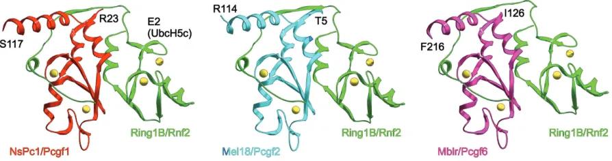
In vitro ubiquitylation of nucleosomal histone H2A by puri-fied, recombinant, RING1B is much less efficient than that of PRC1l or reconstituted complexes containing RING1A, RING1B, BMI1 and PC3 (Wang, 2004; Cao, 2005). This suggested that other PRC1 subunits may act as positive cofactors in the monoubiquitylation of histone H2A. Indeed, Bmi1 was found to stimulate the ubiquitylating activity of Ring1B, either as full length proteins (Cao, 2005) or just as N-terminal fragments of both proteins (Buchwald, 2006) providing that their RING fin-gers are intact (Li, 2006). This is not unexpected, considering that the stable association of Ring1B and Bmi1 depends on their RING fingers (Hemenway, 1998) and it is fully explained by the multiplicity of contacts between the RING fingers of the two proteins, as shown by the crystallographyc analysis of Ring1B-Bmi1 dimers (Buchwald, 2006; Li, 2006). Thus, inacti-vation of Bmi1 in tissue culture cells results in a decrease of global H2AUb levels and in a reduction of H2AUb marks on nucleosomes at target genes such as Hoxc5 (Wei, 2006).
with this notion, the mutation of a residue in this region of Ring1B, I50 abolishes its E3 ligase activity, eventhough the overall RING finger structure is not compromised (Buchwald, 2006).
If Ring1B (and likely Ring1A) provide docking surfaces for the E2 ligase, which is the mechanism for the activating role of Bmi1? In vitro evidence suggests that Ring1B, which is de-graded by an unknown E3 ubiquitin ligase (Ben-Saadon, 2006), is stabilized by Bmi1. At the same time, Bmi1 modulates Ring1B self-ubiquitylation (Buchwald, 2006), an activity which is needed for efficient H2A ubiquitylation (Ben-Saadon, 2006). It has also been proposed that Bmi1 facilitates association to the sub-strate, nucleosomal histone H2A, although no direct experi-mental evidence supports this suggestion (Buchwald, 2006; Li, 2006).
In summary, structural and functional analysis of the Ring1B-Bmi1 pair explains the essential role of RING fingers in H2A ubiquitylation in at least three aspects: (i) they are needed for the mutual stabilization and association of RING finger proteins to each other, (ii) the docking of the E2 ligase, and (iii) modu-lation of the ubiquitymodu-lation process itself. An additional interest-ing observation deduced from these studies is the wrappinterest-ing of Bmi1 by N-terminal sequences of Ring1B. Together with the fact that amino acids 92-190, C-terminal to the RING finger, contain the lysines involved in Ring1B polyubiquitylation (Ben-Saadon, 2006), it may explain the high sequence conservation of the N-terminal domain of Ring1 proteins. It appears as if a very specific module, that includes the RING finger and adyacent sequences, has been selected during evolution to ensure regulated histone H2A ubiquitylation.
Heterogeneity of Ring1-Pcgf H2A ubiquitylating
com-plexes
Bmi1 is one of the members of the so-called Polycomb group ring finger family (Pcgf), a term that may need to be reviewed since two other Polycomb RING finger proteins, Ring1A and Ring1B do not belong into that family. Because of structural similarity, it may have been expected that, like Bmi1, Pcgf proteins might also act as coactivators of Ring1A or Ring1B
mediated H2A ubiquitylation. In fact, like Bmi1, the other Pcgf proteins, Mel18/Pcgf2, MBLR/Pcgf6, Nspc1/Pcgf1, Pcgf5 and Pcgf3 have all been found in complexes that contain Ring1A and Ring1B (Table 3). Of these, the BCOR-FBXL10 complex, which contains NSPC1/PCGF1, has been found to stimulate in vitro ubiquitylation of nucleosomal H2A (Gearhart, 2006). Since Nspc1/Pcgf1 enhances Ring1B ability to ubiquitylate H2A in vivo (Sánchez, 2007) it is likely that it acts as a coactivator similar to Bmi1. A first study on Mel18/Pcgf2 reported its inability to cooperate with Ring1B in H2A modification (Cao, 2005). However, a contrasting report indicates that phosphory-lated, but not unphosphoryphosphory-lated, Mel18/Pcgf2 stimulates Ring1B E3 ligase activity on nucleosomal substrates (Elderkin, 2007). In agreement with this observation, ectopic expression of Mel18 in Bmi1-deficient ES cells represses some of the targets upregulated in the absence of Bmi1 (Elderkin, 2007), suggest-ing a functional redundancy between these Pcgf proteins. As phosphorylated Mel18 associates preferentially to the chroma-tin fraction of tissue culture cells it has been suggested that recruitment of the ubiquitylating complex to nucleosomes is regulated through a phosphorylation dependent event on the E3 ligase cofactor (Elderkin, 2007). However, a more direct approach, perhaps by using cells that express a non-phosphorylatable form of Mel18, is needed to support this regulatory switch.
Remarkably, the available evidence suggests that Ring1B and Ring1A are paired with only one of the various Pcgfs in each of the complexes identified. For instance, BCOR-FBXL10 complexes contain only NSPC1/PCGF1 (Gearhart, 2006; Sánchez, 2007), and E2F6 complexes contain only MBLR/ PCGF6 (Ogawa, 2002). Also, the isolation of complexes using a tag approach with BMI1 or MEL18 does not result in the copurification of BMI1 and MEL18 or any other PCGF member (Wiederschain, 2007). Fig. 3 shows a modelization of predicted Ring1B-Pcgf pairs, based on the Ring1B-Bmi1 structure. Given the high sequence conservation between Pcgf RING fingers, it is not unexpected that the predicted structures for these Ring1B-Pcgf pairs overlaps that of Ring1B-Bmi1. Identical structures can also be modelled for Ring1A-Pcgf pairs (not shown). However, Pcgf protein sequences outside the RING finger and
adjacent sequences diverge considerably, making it possible that the different Ring1-Pcgf pairs are functionally diverse.
Recruiting Ring1A, Ring1B-containing complexes to
their gene targets
Drosophila Hox genes were the first known targets of PcG products. Localization studies on Drosophila polytene chromo-somes showed binding of PcG proteins, including Sce/dRING, to >100 cytological bands (Zink, 1989; Gorfinkiel, 2004), suggesting that the number of targets was larger than anticipated. Recent improvements in methods for the analysis of immunoprecipitated chromatin on a genome-wide scale have confirmed this sugges-tion both in mammalian and Drosophila cells (Boyer, 2006; Bracken, 2006; Lee, 2006; Nègre, 2006; Schwartz, 2006; Tolhuis, 2006).
Ring1B genome-wide binding sites are only known for murine and human ES cells (Boyer, 2006; Endoh, 2008; Ku, 2008). The analysis, both by ChIP-chip and ChIP-seq (immunoprecipitaded DNA identified by hibrydization to tiled arrays of oligonucleotides or by sequencing, respectively) identifies between 1000-1300 sites corresponding to annotated promoters. Most of these sites are also bound by other PcG proteins [eed, Suz12, Ezh2 and Phc1 (Boyer, 2006; Pasini, 2007; Ku, 2008)].Ring1B binding sites correlate with a repressive function on loci that for the most part encode transcription factors and signaling molecules with known roles in major developmental processes. Similar results were obtained for CBX8, EZH2 and SUZ12 in human embryonic fibroblasts (Bracken, 2006). Similar mapping analysis of H3K27me3 marks shows co-occupancy with most sites bound by PcG proteins (Boyer, 2006; Bracken, 2006). Interestingly, however, Ring1B binding sites in ES cells corresponds only to half of H3K27me3 domains, characterized by their large size and when on CpG islands, these are among the longest (Ku, 2008). These genomic domains are conserved between mouse and human ES cells. On the other hand, H2AK119Ub1 marks, are known only for a handful of PcG targets (Stock, 2007). It is worth noting that the study of DNA sequences bound or modified by mammalian PcG proteins, has failed to identify regions similar to the so-called Polycomb response elements (PRE) of Drosophila. PREs are DNA sequences with multiple binding sites for DNA binding proteins able to recruit PcG complexes (Ringrose, 2007). In contrast to PcG proteins, none of these DNA binding proteins, except for YY1, the homolog of Pho and Phol, have vertebrate homologs (Müller, 2006). Whether this implies a fundamental difference between insects and vertebrates in PcG complex(es) recruiting to their targets is not known.
Expression analysis of Ring1B-deficient ES cells shows that most of the Ring1B targets identified in ChIP experiments are upregulated (Leeb, 2007; Endoh, 2008). Inactivation of PRC2 components eed or Suz12 in ES cells leads to a similar observa-tion (Boyer, 2006; Lee, 2006; Pasini, 2007). These results are consistent with the current paradigm for PRC1 recruiting to targets, initially based on the observation that in Drosophila the occupancy of H2AK119Ub1-containing nucleosomes at PcG gene targets was dependent on the activity of PRC2 component E (Z) (Wang, 2004). In this paradigm, chromodomain-containing sub-units in PRC1 complexes (for instance, M33/Cbx2, Pc3/Cbx8, Pc2/Cbx4, etc), which are known to associate preferentially to H3
histone tail peptides trimethylated at the lysine 27 (Fischle, 2003; Min, 2003; Bernstein, 2006b) would act as “readers” of H3K27me3 modified nucleosomes, and contribute to its stable association to regulatory regions. In line with this prediction, an enhancement of H3K27me3 levels at promoters of selected Hox genes, after inactivation of the H3K27 demethylase UTX/KDM6A, is accom-panied by increases in RING1A and BMI1 occupancy and also of increased H2AK119Ub1 marks (Lee, 2007). However, this model of chromatin modification needs to accommodate evidence for a reciprocal activity, by which PRC2 recruitment is facilitated or stabilizated by PRC1, as suggested by the decrease in nucleoso-mal occupancy and H3K27me3 marks at PcG targets in Ring1A and Ring1B-deficient ES cells (Endoh, 2008).
Despite the prevalence of the histone H3K27me3-directed recruitment of Ring1A-Ring1B complexes, there are examples in which their recruiting is independent of such a histone-reading mechanism. For instance, Ring1B association to the Xi chromo-some in female ES cells that are deficient in eed, occurs in the absence of H3K27me3 marks (Schoeftner, 2006). Another ex-ample is the localization of maternal Ring1B and other PRC1 subunits to paternal heterochromatin of preimplantation Ezh2
-/-mouse embryos which lack H3K27me3 marks (Puschendorf, 2008). Whereas these heterochromatic regions may represent specific situations, they show the feasibility of recruiting mecha-nisms other than H3K27me3 binding. One of the possibilities is through binding to RNA, as described for a Zn2+-binding domain in Polyhomeotic PRC1 subunits, a motif initially identified in the product of SOP-2, a Hox gene regulator in C. elegans (Zhang, 2004).
Mechanisms of transcriptional repression of Ring1A,
Ring1B targets
PcG-mediated represses transcription through one or more mechanisms, possibly in a coordinated manner, still poorly char-acterized [reviewed in (Francis, 2001a; Schuettengruber, 2007)]. Three major categories of mechanisms appear involved: (i) nuclear organization, (ii) chromatin remodeling and (iii) modulation of the transcriptional machinery.
SWI/SNF chromatin-remodeling complex, leads to derepression coupled to eviction of PcG complexes from repressed loci (Kia, 2008). As for the transcriptional machinery itself, studies on PcG-mediated repression of a gene model in Drosophila have shown that in the repressed state, RNA pol II cannot initiate transcription (Dellino, 2004). Additional correlative evidence for a role of PcG in transcription initiation is the occupancy of silenced promoters by the TATA binding protein (Breiling, 2001) and the appearance of subunits of the transcription initiation machinery in PRC1 complexes from Drosophila embryos (Saurin, 2001). Recently, histone Ring1-dependent H2A monoubiquitylation has been linked to the regulation of RNA pol II at transcription initiation sites (Stock, 2007).
H2A monoubiquitylation and transcriptional
repres-sion
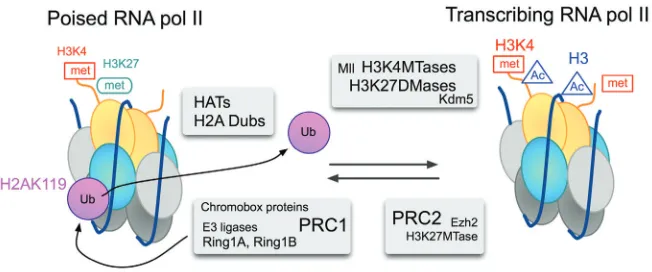
RNA Pol II associates to most promoters, regardless of the levels of transcriptional output (Guenther, 2007). In ES cells, promoters of PcG-repressed targets have a form of RNA Pol II that is phosphorylated at Serine 5 [Ser5P (Stock, 2007)]. Another feature of promoter proximal regions of PcG targets in ES cells is their co-occupancy by both H3K4me3 and H3K27me3 marks (Azuara, 2006; Bernstein, 2006a). Chromatin regions that contain these functionally opposed histone marks are termed bivalent domains and were first thought of as specialized chromatin structures associated to the maintenance of silent genes in a transcription-ready state. Indeed, many of these bivalent struc-tures are resolved during differentiation of ES cells, so that active genes keep the H3K4me3 marks and silent ones keep H3K27me3 or lose both marks (Mikkelsen, 2007; Mohn, 2008; Cui, 2009). Recent genome-wide chromatin immunoprecipitation (ChIP) stud-ies in Drosophila, show that RNA pol II binds at the transcription initiation site of a large number of silent, developmentally regu-lated genes (Muse, 2007; Zeitlinger, 2007), suggesting that stalling RNA Pol II is an important regulatory step for genes that have to accommodate dynamic transcriptional responses to de-velopmental cues.
In this context it is highly relevant that Ring1 proteins play a decisive role in maintaining stalled RNA pol II at promoters of bivalent domains of ES cells (Stock, 2007). Thus, the inactivation of Ring1A and Ring1B results in the sequential loss of H2AK119Ub1 at promoters, the release of poised RNA pol II and gene derepres-sion. The mechanistic link between H2A modification and tran-scription initiation is not known, but conformational changes in RNA pol II upon loss of Ring1A-Ring1B (Stock, 2007) suggests postranslational modification (s) or reorganization of protein com-plex (es). In the converse situation, the stabilization of H2AK119Ub1 marks resulting from the inactivation of a H2A ubiquitin protease, concurs with a decrease in RNA pol II Ser5P occupancy at the promoter and a reduction of two transcription elongation marks, RNA pol II Ser2P and H3K36me3 (Zhou, 2008). Additional evidence supporting a role for H2A monoubiquitylation in transcription initiation comes from in vitro studies in which transcription initiation, but not elongation, occurs when the nucleosomal templates are assembled with H2AK119Ub1 (Nakagawa, 2008). These experiments also showed that H2AK119Ub1 interferes with the dimethylation and trimethylation of histone H3K4, an observation that is in
agree-ment with the enhanced occupancy of H3K4me3 marks at pro-moters of Ring1A and Ring1B-deficient ES cells (Endoh, 2008). The mechanism of transcriptional repression through H2AK119Ub1, however, may differ depending on the E3 ligase involved or the regulatory context. Thus, repression in macroph-ages of a chemokine gene mediated by the E3 ligase 2A-HUB/ hRUL138 occurs with inhibition of transcriptional elongation, perhaps because the E3 ligase is recruited as part of a N-CoR/ HDAC1/3 complex (Zhou, 2008). It would be interesting to inves-tigate whether the repression of the bivalent domains identified in differentiated cells (Azuara, 2006; Barski, 2007; Mikkelsen, 2007; Pan, 2007; Roh, 2006; Zhao, 2007) depends on Ring1 proteins or on another E3 ligase (s). The fact that linker histone H1 associa-tion is facilitated by H2AK119Ub1 (Jason, 2005) may also contrib-ute to transcriptional repression, and one of the consequences of H2A deubiquitylation is the phosphorylation and release of his-tone H1 from nucleosomes (Zhu, 2007).
Reversal of histone H2A monoubiquitylation and gene
activation
A regulatory step in transcriptional control through chromatin structure is the reversal of postranslational marks. In general, histone modifications can be reverted either by nucleosome eviction or through the action of enzymes that remove the cova-lent modification. Which mechanism is used may determine either a long-term or short-term outcome for a given modification. Up to now, five proteases have been shown to remove ubiquitin moi-eties from histone H2A. Most, USP3 (Nicassio, 2007), USP16 (Joo, 2007), USP21 (Nakagawa, 2008) and USP22 (Zhang, 2008; Zhao, 2008), are members of the cysteine family of proteases, which use a carboxyl-terminal hydrolase-like (UCH) zinc finger as a catalytic domain. The remaining ubiquitinase, 2A-DUB/MYSM1 (Zhu, 2007), belongs to the family of metalloproteases that have a JAMM/MPN+ motif as catalytic domain. USP3 and USP22 are not H2A-specific and also deubiquitylate H2B. Since mammalian genomes encode a large number of deubiqutinases (Nijman, 2005), it is possible that additional H2A deubiquitinases will be found. It is not clear, however, whether all of them will be involved in gene expression control. For instance, whereas USP16 inacti-vation leads to upregulation of some Hox genes (Joo, 2007), it also causes defective progression through the cell cycle of mutant cells (Joo et al., 2007). It is known that H2AUb (and H2BUb) loses its ubiquitin moiety during mitotic chromatin condensation, fol-lowed by subsequent reubiquitylation in early anaphase (Matsui, 1979; Goldknopf, 1980). This exchange of ubiquitin on H2A is required for appropriate cell cycle progression because in the absence of deubiquitylation, Aurora B, the kinase that phospho-rylates Serine 10 of histone H3 and that is required for mitotic chromatin condensation, does not associate to nucleosomes compromising adequate mitotic condensation (Joo, 2007). An-other reason for cell cycle delays associated to H2A deubiquitylation is the activation of the DNA damage response that occurs during the inactivation of USP3 (Nicassio, 2007).
development and in adult life. Ring1 proteins, as part of PRC1 or of other complexes, contribute to the execution of genetic pro-grams that determine the maintenance of stem cells and the ordered differentiation of their progeny. This includes not only the regulation of developmentally relevant genes, but also repression of heterochromatic structures and control of cell proliferation. Perhaps not surprisingly, some cancer cells show deregulated expression of Ring1 proteins.
During development
Constitutive inactivation of Ring1B in mice leads to embryo death in uterus, around gastrulation time (Voncken, 2003). The mechanisms underlying this embryonic lethality are not known. In contrast, mice deficient in Ring1A are fertile and without an overt phenotype (del Mar Lorente, 2000). Why would Ring1B compen-sate for the loss of Ring1A and not the other way around? It is possible that the two paralogs are functionally distinct, despite of their structural similarity; alternatively, their expression patterns may not be overlapping. For instance, if Ring1A levels at early stages of development were too low, it would not complement a critical function carried out by Ring1B.
Loss of function of either of the two proteins alters the axial skeleton of mutant mice (del Mar Lorente, 2000; Suzuki, 2002). The reason why Ring1A-deficient fetuses show anterior homeotic transformations instead of posterior transformations usually ob-served in other PcG mutant mice [with the exception of YY1, (Lorente, 2006)] is not known. The minor alterations of Hox gene expression observed in midgestation Ring1A-deficient embryos are unlikely to explain the skeletal alterations (del Mar Lorente, 2000), although the effect of transient deregulation of Hox gene expression during earlier embryogenesis cannot be ruled out. The impact of Ring1B on antero-posterior axis specification has been deduced from the genetic interaction between a Ring1B hypomorphic allele and a null allele for Mel18 (Suzuki, 2002), whereby double mutant mice showed higher penetrance and expressivity of Mel18-/- induced posterior transformations.
Ge-Fig. 4. Model for the reversible switch of transcriptional states at a bivalent promoter.
In the inactive state, the E3 ubiquitin ligase acitivity of Ring1A and Ring1B in PRC1 is needed for the maintenance of RNA pol II in a stalled, non-elongating, state. H3K27me3 marks, that depend on PRC2 contribute to stabilize PRC1 association. Recruitment of transcription activating complexes, perhaps through transcription factors bound to DNA sites uncovered by chromatin remodelers, involve coordinated histone modifying activities. Histone acetylation and H2A deubiquitylation, may be trigger modifications of RNA pol II. Demethylation of H3K27 and further methylation of H3K4 would help recruiting TFIID (Vermeulen, 2007) and other componentes of the transcription initiation machinery, thus sustaining transcription. transferase (Zhu, 2007). Treatment of tissue culture cells with HAT
inhibitors (which results in hyperacetylated nucleosomes) en-hances the deubiquitinating activity of 2A-DUB/MYSM1. The find-ing that androgen and estrogen-dependent gene targets, but not those dependent on retinoic acid or thyroid hormone, are affected by the downregulation of 2A-DUB/MYSM suggests that derepres-sion through H2A deubiquitylation is a process regulated by specific recruiting of HAT-containing complexes (Zhu, 2007). Fig-ure 4 shows a simplified view of reversible modifications at nucleo-somes of bivalent genes. Here, recruitment of a hypothetical complex containing a H2A ubiquitin protease(s) and a HAT would cooperate with another complex involved in H3K27 demethylation (Cho, 2007; De Santa, 2007; Lee, 2007). The activity of SAGA complexes, modular assemblies involved in the regulated access of general transcription factors to DNA mediated by histone acety-lation, provide additional support for gene activation through coor-dinated H2A deubiquitylation, by USP22, and histone acetylation (Zhao, 2008). Conversely recruiting of PRC2, which may bring in a HDAC, through its Eed component (van der Vlag, 1999), would facilitate PRC1 association and RNA pol II stalling. In this respect, dRAF, a Drosophila complex related to the mammalian Ring1-Fbxl10 complex (Gearhardt, 2006; Sánchez, 2007) has been shown to promote H2A ubiquitylation and H3K36me2 demethylation in a coordinated fashion (Lagarou, 2008).
Whether any of these proteases is specifically involved in the removal of Ring1-mediated H2AUb is not known. Ring1A-Ring1B are responsible for most H2AUb in the genome. However, it is possible that only a subset of the nucleosomes thus modified localize to promoters and other cis-acting control regions. For instance, Ring1B-dependent H2AUb has been found to occur also during the DNA damage response induced by UV radiation (Bergink, 2006).
Functional activity of Ring1 proteins
Being general transcriptional regulators, Ring1 proteins activity impacts on a multiplicity of processes both during embryonic
netic interactions have also been observed in Bmi1-deficient mice that are also heterozygous mice for a null Ring1B allele (Voncken, 2003). An example of the wide range of processes affected by PcG products is illustrated by the abnormalities in the anterior eye of Ring1A-deficient mice, a defect enhanced by a reduc-tion of YY1 dosage (Lorente, 2006).
Maintenance of stem cells
PcG act as regulators of embryonic and adult stem cell maintenance (Valk-Lingbeek, 2004). For instance, in ES cells, Ring1 proteins act as a repressors of a large number of developmental regulators (Leeb, 2007; Endoh, 2008).
Mar Lorente, 2000). Whereas these observations suggest func-tional differences between Ring1 proteins, double mutant Ring1A and Ring1B ES cells show enhanced gene derepression and eventually cessation of cell proliferation (Endoh, 2008), indicating a convergence of regulatory pathways. In ES cells, Ring1 proteins and other PcG products, play their repressive functions under the control of the selfmaintained network of transcriptional factors that are mainly orchestrated by Oct3/4, Nanog (Lee, 2006; Endoh, 2008). When, in response to extracellular signals, the program for pluripotency gives in to new transcriptional programs, a reorder-ing of epigenetic marks takes place. For instance, upregulation of GATA6, an effector of differentiation towards extraembryonic endoderm, results in dissociation of Ring1B, and other PcG products, from their former targets at undifferentiated ES cells (Endoh, 2008). Therefore, at least in ES cells, Ring1 proteins act as a part of a set of instructed epigenetic regulators that prevent inappropriated differentiation, illustrating intertwined cooperation of genetic and epigenetic mechanisms to ensure pluripotency (Niwa, 2007). The premature neuronal differentiation of Ring1B-deficient fetal neural stem cells is further evidence of the contribu-tion of PcG products to stem cell self-renewal by preventing their differentiation. In addition, the enhanced neuronal diffferentiation and reduced gliogenesis of Ring1B mutant mice (Román-Trufero, 2009) suggest a role for PcG in the temporal specification of developing neural progenitors. In contrast, Ring1B inactivation in adult stem cells and primitive progenitors of the hematopoietic compartment leads to proliferative alterations, rather than to differentiation defects (Calés, 2008). Although a detailed study on the stem cell function of Ring1B-deficient HSC is lacking, the results contrast with those derived from the study of Bmi1-deficient mice (Lessard, 2003; Park, 2003). Whether this is a reflection of redundant Ring1B function in HSCs or of differences in regulatory pathways affected by the mutations remains to be established.
Functions in heterochromatin
In mammals, the imbalance of gene dosage for genes on the X chromosome (two in females, one in males) is functionally equalized through the inactivation of one of the two X chromo-somes in females. The inactivation process is triggered by the transcription of the Xist gene on the to-be silenced X chromosome [reviewed in (Heard, 2006)]. Ring1A and Ring1B, together with other subunits from PRC1 and PRC2 complexes accumulate on the Xi chromosome of female mammalian cells. Accumulation of PcG products and colocalization with H2AUb and H3K27me3 marks occurs in both imprinted and randomly inactivated Xi (Cao, 2004; de Napoles, 2004). As expected, the PRC2 component eed is needed for the appearance of H3K27me3 marks and the accumulation of PRC1 subunits Phc1 and Phc2 on the Xi (Plath, 2003; Silva, 2003; Plath, 2004). However, accumulation of Ring1B was found independent of both eed and H3K27me3 (Schoeftner, 2006). Inactivation of Ring1B in undifferentiated ES cells leads to the loss of H2AUb marks on the Xi (de Napoles, 2004; Leeb, 2007). Upon differentiation, Ring1B-deficient cells regain H2AUb marks on the Xi, possibly due to upregulation of Ring1A (Leeb et al., 2007). Together, these observations suggest that PcG in general, and Ring1A and Ring1B in particular, are involved in the initiation of the silencing of the Xi. However, beyond this
correla-tive evidence, the analysis of Ring1B-deficient ES cells has shown that neither initiation of Xist-induced Xi silencing, nor maintenance of gene repression of loci on the Xi depends on Ring1B (Leeb, 2007). It is possible that whereas Ring1B is required for repression of developmental regulators, mainte-nance of a silenced Xi relies on a multiplicity of repressing mechanisms. In contrast, targeting of Ring1B and PRC1 (mater-nally provided) to paternal heterochromatin in the zygote is accompanied by active repression, as suggested by the en-hanced transcription of pericentric major satellite sequences in zygotes depleted from both embryonic and maternal Ring1B (Puschendorf, 2008). Since parentally defined formation of het-erochromatic structures is over at the end of the 8-cell stage (Puschendorf, 2008), and heterozygous Ring1B mothers provide Ring1B to the zygote, it appears unlikely that a role as repressor of paternal heterochromatin has much of an impact on the developmental arrest of gastrulating Ring1B null embryos.
Cell homeostasis and cancer
The involvement of PcG products in cancer may occur both at the phase of the metastatic expansion of tumor cells and, per-haps, at the early stages of the generation of tumor stem cells. A central mechanism for cell expansion is the inactivation of the Ink4a locus, a well known tumor suppressor that encodes two powerful inhibitors of cell cycle progression, p16Ink4a and also p19Arf, a negative regulator of p53, a major promoter of apoptosis (Gil, 2006). Both products derive from transcripts initiated at independent promoters, although they share exons that are translated in different reading frames. The fact that Ring1B specifically represses p16Ink4a (Voncken, 2003; Calés, 2008) may explain, at least in part, the finding of Ring1B among a reduced set of 11 genes (expression signature) that identifies human tumors with a bad prognosis (Glinsky, 2005). Certainly, Ring1B is found overexpressed in germinal-centre derived lymphomas (large B-cell lymphoma, Hodgkin’s lymphoma) and in gastric and colon tumors (Sánchez-Beato, 2006). Ring1A is also overexpressed in lymphomas (Dukers, 2004; Raaphorst, 2004; Sánchez-Beato, 2004) and in malignant prostate epithelia (van Leenders, 2006). Of course, such an enhanced expression in tumor cells does not allow to distinguish between a causal role in cell transformation or in the selection of Ring1-expressing cells during tumor expan-sion. In this regard, it is of interest to note that the ectopic expression of Ring1A in rodent fibroblasts results in transformed cells that propagate when transplanted in immunocompromised recipient mice (Satijn, 1999).
(Ishikawa, 2007), although whether they play a causal role in their malignant transformation is not known.
Upregulation of PcG products in cancer cells has been usually associated to their role as positive regulators of cell proliferation, of which the repression of the cell cycle inhibitor, p16Ink4a is an example. Recently, however, it has been reported that, in the murine hematopoietic system, Ring1B acts both as promoter and as an inhibitor of cell proliferation, depending on the cell compart-ment. This dual activity is a consequence of the repression of positive and negative regulator of the cell cycle machinery, namely cyclin D2 and its inhibitor p16Ink4a (Calés, 2008). Cyclin D2 is upregulated in all Ring1B-deficient cells, whereas p16Ink4a is only upregulated in the more mature precursors. In this way, Ring1B contributes to hematopoietic cell homeostasis through restraining expansion of immature progenitor cells, whose num-bers are increased by two to three fold in the absence of Ring1B, and concomitantly ensuring the appropriate expansion of their maturing progeny, which is decreased in Ring1B-deficient cells. Thus, lymphomas and leukemia normally appearing in Ink4a null mice, undergo accelerated onset in compound Ring1B, Ink4a mutant mice (Calés, 2008). Ring1B-deficient progenitors show an enhanced proliferation rate, without alterations in apoptotic levels or differentiative potential. Thus, the control of expanding popu-lations of progenitor cells links Ring1B function to the regulation of cell homeostasis. Whether this observation is specific of the hematopoietic cell compartment or a general feature of other regenerating tissues remains to be determined.
Perspectives
The E3 protein ligase activity contributing to the maintenance of stalled RNA pol II at repressed promoters is the best molecu-larly characterized function of Ring1proteins. However, much remain to be learned about the specific role of this histone modification and tits dependence on Ring1A and Ring1B pro-teins: for instance, whether H2A ubiquitylation serving transcrip-tional control can be separated from global genomic ubiquitylation. The elucidation of targeting mechanisms that recruit Ring1A-Ring1B-containing complexes is also important. This may shed light on differential use of Ring1A-Ring1B or of other E3 ubiquitin ligases to achieve H2A ubiquitylation, perhaps dependent on the stage of cell differentiation. Obtaining genomic maps of H2AUb marks, and their comparison with those of other histone marks would fill a considerable gap in the current understanding of the relationship between diverse histone modifications and would provide clues about the recruiting and differential use of E3 ligases at specific loci. Regulation of the ubiquitylation process itself, in a physiological context, has been hardly explored. Fi-nally, further research on the link between H2AUb and RNA pol II stalling, together with the removal of H2AUb marks would help deciphering the molecular mechanisms for Ring1A-Ring1B role in transcription initiation.
Whether Ring1A-Ring1B always act as E3 ubiquitin ligases for H2A in the various complexes they belong to is not known. Ring1A-Ring1B proteins may also participate in other poorly characterized functions. For instance, it is worth exploring the possibility that Ring1A-Ring1B participate in the assembly and dynamics of multiprotein complexes, as illustrated by the depen-dence of PRC1 stability on Ring1B. In this regard, a remaining
uncertainty is that of the possible functional differences between Ring1A and Ring1B.
The study of PcG gene function has come a long way from their initial association to the control of developmental processes regulated by Hox proteins. Recent advances show that PcG gene products are involved in a wide range of regulatory settings. Answers to the above questions should help us to obtain a better understanding of the contribution of the Polycomb system of chromatin modifiers to epigenetic regulation of gene expression.
Acknowledgements
I thank Atsushi Hijikata (RIKEN RCAI, Yokohama), Salvatore D’Aniello and Jordi García-Fernández (Universidad de Barcelona) for help with protein evolutive studies; Mario García (CIB) for molecular modeling, and Carmela Calés and members of the lab are acknowledged for comments on the manuscript. Work in my lab is funded by a grant from the Education Ministry (SAF2007-06952-CO2-01) and the OncoCycle program from the Comunidad de Madrid.
References
AKASAKA, T., TAKAHASHI, N., SUZUKI, M., KOSEKI, H., BODMER, R. and KOGA H. (2002). MBLR, a new RING finger protein resembling mammalian Polycomb gene products, is regulated by cell cycle-dependent phosphorylation. Genes Cells 7: 835-850.
AZUARA, V., PERRY, P., SAUER, S., SPIVAKOV, M., JØRGENSEN, H.F., JOHN, R.M., GOUTI, M., CASANOVA, M., WARNES, G., MERKENSCHLAGER, M., et al. (2006) Chromatin signatures of pluripotent cell lines. Nat.Cell Biol. 8: 532-538.
BANTIGNIES, F., GRIMAUD, C., LAVROV, S., GABUT, M. and CAVALLI, G. (2003). Inheritance of Polycomb-dependent chromosomal interactions in Droso-phila. Genes Dev. 17: 2406-2420.
BARSKI, A., CUDDAPAH, S., CUI, K., ROH, T.Y., SCHONES, D.E., WANG, Z., WEI, G., CHEPELEV, I. and ZHAO, K. (2007). High-resolution profiling of histone methylations in the human genome Cell. 129: 823-837.
BEN-SAADON, R., ZAAROOR, D., ZIV, T. and CIECHANOVER, A. (2006). The Polycomb protein Ring1B generates self atypical mixed ubiquitin chains re-quired for its in vitro histone H2A ligase activity. Mol. Cell 24: 701-711. BERGINK, S., SALOMONS, F.A., HOOGSTRATEN, D., GROOTHUIS, T.A., DE
WAARD, H., WU, J., YUAN, L., CITTERIO, E., HOUTSMULLER, A.B., NEEFJES, J., et al. (2006). DNA damage triggers nucleotide excision repair-dependent monoubiquitylation of histone H2A. Genes Dev. 20: 1343-1352.
BERNSTEIN, B.E., MIKKELSEN, T.S., XIE, X., KAMAL, M., HUEBERT, D.J., CUFF, J., FRY, B., MEISSNER, A., WERNIG, M., PLATH, K., et al. (2006a). A bivalent chromatin structure marks key developmental genes in embryonic stem cells. Cell. 125: 315-326.
BERNSTEIN, E., DUNCAN, E.M., MASUI, O., GIL, J., HEARD, E. and ALLIS, C.D. (2006b). Mouse polycomb proteins bind differentially to methylated histone H3 and RNA and are enriched in facultative heterochromatin. Mol. Cell. Biol. 26: 2560-2569.
BORDEN, K.L. (2000). RING domains: master builders of molecular scaffolds? J. Mol. Biol. 295: 1103-1112.
BOYER, L.A., PLATH, K., ZEITLINGER, J., BRAMBRINK, T., MEDEIROS, L.A., LEE, T.I., LEVINE, S.S., WERNIG, M., TAJONAR, A., RAY, M.K., et al. (2006). Polycomb complexes repress developmental regulators in murine embryonic stem cells. Nature. 441: 349-353.
BRACKEN, A.P., DIETRICH, N., PASINI, D., HANSEN, K.H. and HELIN, K. (2006). Genome-wide mapping of Polycomb target genes unravels their roles in cell fate transitions. Genes Dev. 20: 1123-1136.
BREILING, A., TURNER, B.M., BIANCHI, M.E. and ORLANDO, V. (2001). General transcription factors bind promoters repressed by Polycomb group proteins.
Nature 412: 651-655.
of the Ring-Ring complex of Polycomb proteins Bmi1 and Ring1b. EMBO. J. 25: 2465-2474.
CALÉS, C., ROMÁN-TRUFERO, M., PAVÓN, L., SERRANO, I., MELGAR, T., ENDOH, M., PÉREZ, C., KOSEKI, H. and VIDAL, M. (2008). Molecular and cellular biology inactivation of the Polycomb group protein Ring1B unveils an antiproliferative role in hematopoietic cell expansion and cooperation with tumorigenesis associated to Ink4a deletion. Mol. Cell. Biol. 28: 1018-1028. CAO, R. and ZHANG, Y. (2004). The functions of E (Z)/EZH2-mediated methylation
of lysine 27 in histone H3. Curr. Opin. Genet. Dev. 14: 155-164.
CAO, R., TSUKADA, Y.I. and ZHANG, Y. (2005). Role of Bmi-1 and Ring1A in H2A Ubiquitylation and Hox Gene Silencing. Mol. Cell 20: 845-854.
CAPILI, A.D., SCHULTZ, D.C., RAUSCHERIII, F.J. and BORDEN, K.L. (2001). Solution structure of the PHD domain from the KAP-1 corepressor: structural determinants for PHD, RING and LIM zinc-binding domains. EMBO. J. 20: 165-177.
CHAMBEYRON, S. and BICKMORE, W.A. (2004). Chromatin decondensation and nuclear reorganization of the HoxB locus upon induction of transcription. Genes Dev. 18: 1119-1130.
CHEN, D., DUNDR, M., WANG, C., LEUNG, A., LAMOND, A., MISTELI, T. and HUANG, S. (2005). Condensed mitotic chromatin is accessible to transcription factors and chromatin structural proteins. J. Cell Biol. 168: 41-54.
CHO, Y., HONG, T., HONG, S., GUO,H., YU, H., KIM, D., GUSZCZYNSKI, T., DRESSLER, G.R., COPELAND, T.D., KALKUM, M., et al. (2007). PTIP asso-ciates with MLL3- and MLL4-containing histone H3 lysine 4 methyltransferase complex. J. Biol. Chem. 282: 20395-20406.
CUI K, ZANG C, ROH TY, SCHONES DE, CHILDS RW, PENG W and ZHAO, K. (2009) Chromatin signatures in multipotent human hematopoietic stem cells indicate the fate of bivalent genes during differentiation. Cell Stem Cell 4:80-93. CZYPIONKA, A., RUIZ DE LOS PAÑOS, O.R., MATEU, M.G., BARRERA, F.N., HURTADO-GÓMEZ, E., GÓMEZ, J., VIDAL, M. and NEIRA, J.L. (2007). Biochemistry the isolated C-terminal domain of Ring1B is a dimer made of stable, well-structured monomers. Biochemistry. 46: 12764-12776.
DE NAPOLES, M., MERMOUD, J.E., WAKAO, R., TANG, Y.A., ENDOH, M., APPANAH, R., NESTEROVA, T.B., SILVA, J., OTTE, A.P., VIDAL, M., ET AL.
(2004). Polycomb group proteins Ring1A/B link ubiquitylation of histone H2A to heritable gene silencing and X inactivation. Dev. Cell 7: 663-676.
DE SANTA, F., TOTARO, M.G., PROSPERINI, E., NOTARBARTOLO, S., TESTA, G. and NATOLI, G. (2007) The histone H3 lysine-27 demethylase Jmjd3 links inflammation to inhibition of Polycomb-mediated gene silencing. Cell 130: 1083-1094.
DEL MAR LORENTE, M., MARCOS-GUTIÉRREZ, C., PÉREZ, C., SCHOORLEMMER, J., RAMÍREZ, A., MAGIN, T. and VIDAL, M. (2000). Loss-and gain-of-function mutations show a polycomb group function for Ring1A in mice. Development 127: 5093-5100.
DELLINO, G.I., SCHWARTZ, Y.B., FARKAS, G., MCCABE, D., ELGIN, S.C. and PIRROTTA, V. (2004). Polycomb silencing blocks transcription initiation. Mol. Cell 13: 887-893.
DIETRICH, N., BRACKEN, A.P., TRINH, E., SCHJERLING, C.K., KOSEKI, H., RAPPSILBER, J., HELIN, K. and HANSEN, K.H. (2007). Bypass of senescence by the polycomb group protein CBX8 through direct binding to the INK4A-ARF locus. EMBO. J. 26: 1637-1648.
DOU, Y., MILNE, T.A., TACKETT, A.J., SMITH, E.R., FUKUDA, A., WYSOCKA, J., ALLIS, C.D., CHAIT, B.T., HESS, J.L. and ROEDER, R.G. (2005). Physical association and coordinate function of the H3 K4 methyltransferase MLL1 and the H4 K16 acetyltransferase MOF. Cell 121: 873-885.
DUKERS, D.F., VAN GALEN, J.C., GIROTH, C., JANSEN, P., SEWALT, R.G., OTTE, A.P., KLUIN-NELEMANS, H.C., MEIJER, C.J. and RAAPHORST, F.M. (2004). Unique polycomb gene expression pattern in Hodgkin’s lymphoma and Hodgkin’s lymphoma-derived cell lines. Am. J. Pathol. 164: 873-881. ELDERKIN, S., MAERTENS, G.N., ENDOH, M., MALLERY, D.L., MORRICE, N.,
KOSEKI, H., PETERS, G., BROCKDORFF, N. and HIOM, K. (2007). A
phospho-rylated form of Mel-18 targets the Ring1B histone H2A ubiquitin ligase to chromatin. Mol. Cell 28: 107-120.
ENDOH, M., ENDO, T.A., ENDOH, T., FUJIMURA, Y., OHARA, O., TOYODA, T., OTTE, A.P., OKANO, M., BROCKDORFF, N., VIDAL, M., et al. (2008). Polycomb group proteins Ring1A/B arefunctionally linked to the core transcriptional
regulatory circuitry to maintain ES cell identity. Development 135: 1513-1524. FANG, J., CHEN, T., CHADWICK, B., LI, E. and ZHANG, Y. (2004). Ring1b-mediated H2A ubiquitination associates with inactive X chromosomes and is involved in Initiation of X-inactivation. J. Biol. Chem. 279: 52812-52815. FARRIS, S.D., RUBIO, E.D., MOON, J.J., GOMBERT, W.M., NELSON, B.H. and
KRUMM, A. (2005) Transcription-induced chromatin remodeling at the c-myc gene involves the local exchange of histone H2A.Z. J. Biol. Chem. 280: 25298-25303.
FISCHLE, W., WANG, Y., JACOBS, S.A., KIM, Y., ALLIS, C.D. and KHORASANIZADEH, S. (2003). Molecular basis for the discrimination of repressive methyl-lysine marks in histone H3 by Polycomb and HP1 chromodomains. Genes Dev. 17: 1870-1881.
FRANCIS, N.J. and KINGSTON, R.E. (2001a). Mechanisms of transcriptional memory. Nat. Rev. Mol. Cell. Biol. 2: 409-421.
FRANCIS, N.J., SAURIN, A.J., SHAO, Z. and KINGSTON, R.E. (2001b).
Reconstitu-tion of a funcReconstitu-tional core polycomb repressive complex. Mol. Cell 8: 545-556. FRANCIS, N.J., KINGSTON, R.E. and WOODCOCK, C.L. (2004). Chromatin
compaction by a polycomb group protein complex. Science. 306: 1574-1577. FRITSCH, C., BEUCHLE, D. and MÜLLER, J. (2003). Molecular and genetic analysis of the Polycomb group gene Sex combs extra/Ring in Drosophila.
Mech. Dev. 120: 949-954.
GARCÍA, E., MARCOS-GUTIÉRREZ, C., DEL MAR LORENTE, M., MORENO, J.C. and VIDAL, M. (1999). RYBP, a new repressor protein that interacts with components of the mammalian Polycomb complex, and with the transcription factor YY1. EMBO. J. 18: 3404-3418.
GEARHART,M.D., CORCORAN, C.M., WAMSTAD, J.A. and BARDWELL, V.J. (2006). Polycomb Group and SCF Ubiquitin Ligases Are Found in a Novel BCOR Complex That Is Recruited to BCL6 Targets. Mol. Cell. Biol. 26: 6880-6889.
GÉVRY, N., CHAN, H.M., LAFLAMME, L., LIVINGSTON, D.M. and GAUDREAU, L. (2007). p21 transcription is regulated by differential localization of histone H2A.Z. Genes Dev. 21: 1869-1881.
GIL, J. and PETERS, G. (2006). Regulation of the INK4b-ARF-INK4a tumour suppressor locus: all for one or one for all. Nat. Rev. Mol. Cell. Biol. 7: 667-677. GLINSKY, G.V., BEREZOVSKA, O. and GLINSKII, A.B. (2005). Microarray analy-sis identifies a death-from-cancer signature predicting therapy failure in patients with multiple types of cancer. J. Clin. Invest. 115: 1503-1521.
GOLDKNOPF, I.L., SUDHAKAR, S., ROSENBAUM, F. and BUSCH, H. (1980). Biochemical and biophysical research communications timing of ubiquitin synthesis and conjugation into protein A24 during the HeLa cell cycle. Biochem. Biophys. Res. Commun. 95: 1253-1260.
GORFINKIEL, N., FANTI, L., MELGAR, T., GARCÍA, E., PIMPINELLI, S., GUERRERO, I. and VIDAL, M. (2004). The Drosophila Polycomb group gene Sex combs extra encodes the ortholog of mammalian Ring1 proteins. Mech. Dev. 121: 449-462.
GRIMAUD, C., NÈGRE, N. and CAVALLI, G. (2006). From genetics to epigenetics: the tale of Polycomb group and trithorax group genes. Chromosome Res. 14: 363-375.
GUENTHER, M.G., LEVINE, S.S., BOYER, L.A., JAENISCH, R. and YOUNG, R.A. (2007). A chromatin landmark and transcription initiation at most promoters in human cells. Cell 130: 77-88.
HANSON, I.M., POUSTKA, A. and TROWSDALE, J. (1991). Genomics New genes in the class II region of the human major histocompatibility complex. Genomics
10: 417-424.
HEARD, E. and DISTECHE, C.M. (2006). Dosage compensation in mammals: fine-tuning the expression of the X chromosome. Genes Dev. 20: 1848-1867. HEMENWAY, C.S., HALLIGAN, B.W. and LEVY, L.S. (1998). The Bmi-1 oncoprotein
interacts with dinG and MPh2: the role of RING finger domains. Oncogene 16: 2541-2547.
HEMENWAY, C.S., HALLIGAN, B.W., GOULD, G.C. and LEVY, L.S. (2000). Identification and analysis of a third mouse Polycomb gene, MPc3. Gene. 242: 31-40.