Fatty acid binding proteins
in brain development and disease
RONG-ZONG LIU, RAJA MITA, MICHAEL BEAULIEU, ZHIHUA GAO and ROSELINE GODBOUT*
Department of Oncology, University of Alberta, Edmonton, Canada
ABSTRACT Long chain polyunsaturated fatty acids (PUFAs) are critical structural components of the brain and essential for normal brain development. The cellular transportation and physiologi-cal actions of PUFAs are mediated by fatty acid binding proteins (FABPs) which are encoded by the intracellular lipid-binding protein gene family. Three of the ten mammalian FABPs identified to date (FABP3, FABP5, FABP7) are expressed in the brain. These three FABPs, along with their fatty acid ligands, have distinct and dynamic spatio-temporal expression profiles that correlate with specific developmental stages and processes in the brain. Functional studies have revealed a variety of roles for FABPs in brain development including the generation of neuronal and/or glial cells, differentiation, neuronal cell migration and axis patterning. A number of transcription factors have been shown to be involved in the developmental regulation of FABP gene expression in the brain. Furthermore, FABPs appear to be major downstream effectors of signaling pathways such as Reelin-Dab1/Notch which mediate neuron-glia crosstalk during brain development. As PUFAs and FABPs play critical roles in brain development, considerable effort has been placed in elucidating their function in the pathogenesis and progression of brain cancers and neuropsychi-atric disorders.
KEY WORDS:
fatty acid-binding protein, fatty acid, radial glia, brain development, glioblastoma
Introduction
Long chain polyunsaturated fatty acids (PUFAs) are enriched in developing brain and are essential for normal development of the central nervous system (reviewed in Neuringer et al. 1988; Wainwright 2002). PUFAs play important roles in the develop-ment of visual, cognitive, attentional, and learning functions (Neuringer et al. 1988; Carlson and Neuringer 1999; Wainwright 2002; McCann and Ames 2005; Carlson 2009). PUFA deficiency in the brain is implicated in various neuropsychiatric/ neurodegenerative disorders, such as schizophrenia, depres-sion, attention deficit hyperactivity disorder, Parkinson’s disease and Alzheimer’s disease (Freeman 2000; Richardson and Puri 2000; Salvati et al. 2006).
There are two major types of PUFAs: omega-3 (ω-3 or n-3) and omega-6 (ω-6 or n-6). Docosahexaenoic acid (DHA) and arachi-donic acid (AA) represent the most abundant ω-3 and ω-6 PUFAs in brain, respectively. Important roles for PUFAs during brain development include: (i) integration into the phospholipids of cell membranes resulting from continuous membrane remodelling, which in turn modulates signal transduction, neurotransmission,
BIOLOGY
www.intjdevbiol.com*Address correspondence to: Roseline Godbout. Department of Oncology, Cross Cancer Institute, 11560 University Avenue, Edmonton, Alberta, Canada T6G 1Z2. Fax: +1-780-432-8892. e-mail: rgodbout@ualberta.ca
Accepted: 15 December 2009. Final author corrected PDF published online: Published online: 14 May 2010.
ISSN: Online 1696-3547, Print 0214-6282
© 2010 UBC Press Printed in Spain
Abbreviations used in this paper: AA, arachidonic acid; ANS, 1-anilino-naphthalene-8-sulfonate; DAB1, Disabled 1; DHA, docosahexaenoic acid; DS, Down syndrome; Kd, dissociation constant; ED, embryonic day; EPA, eicosapentaenoic acid; FABP, fatty acid-binding protein; LOA, linoleic acid; MUFA, monounsaturated fatty acid; ITC, isothermal titration calorimetry; NF1, nuclear factor I; P, postnatal day; PA, palmitic acid; PPAR, peroxisome proliferator-activating receptor; PUFA, polyunsaturated fatty acid; RXR, retinoid X receptor; SA, stearic acid.
cell motility, and the composition and formation of lipid rafts (Stillwell et al. 2005; Grossfield et al. 2006); (ii) regulation of the expression of genes implicated in genesis, proliferation, differen-tiation and migration of neural cells through binding and activation of nuclear receptors such as peroxisome proliferator activating receptors (PPARs) and/or retinoid X receptors (RXRs) (Kitajka et al. 2004; Schroeder et al. 2008); and (iii) serving as precursors for eicosanoids which are key messengers involved in inter- and intracellular signaling cascades in brain (Purasiri et al. 1997; Jump 2002; Venkatraman and Meksawan 2002).
aqueous cytoplasm is facilitated by intracellular protein vehicles, called fatty acid-binding proteins (FABPs). FABPs belong to the intracellular lipid binding protein (iLBP) family which binds fatty acids, retinoids and other hydrophobic compounds and mediates their physiological functions (reviewed in Storch and Corsico 2008). FABPs exhibit tissue-specific expression patterns and distinct ligand preferences, although they all share a conserved 3-dimensional structure consisting of two orthogonal β-sheets and an α-helical cap (Hanhoff et al. 2002; Haunerland and Spener 2004).
Ten FABPs have been identified in mammals to date, with three phylogenetically-related FABPs, FABP3, FABP5 and FABP7, described in the developing and/or adult brain (Fig. 1) (Schoentgen et al. 1989; Godbout 1993; Bennett et al. 1994; Feng et al. 1994; Kurtz et al. 1994; Owada et al. 1996; Liu et al. 1997; Denovan-Wright et al. 2000; Liu et al. 2004; Liu et al. 2007). Each of these three FABPs shows distinct preference for specific fatty acids. The main purpose of this review is to summarize current knowl-edge regarding the cellular/subcellular distribution, transcrip-tional regulation and function of these three FABPs in the normal and diseased brain of humans, as well as in the developing brain of model organisms.
Developmental expression patterns of FABPs in the
brain
The spatio-temporal expression patterns of FABP3, FABP5 and FABP7 in the developing rodent brain have been thoroughly investigated, with similar results obtained in mouse and rat (Kuhar et al. 1993; Feng et al. 1994; Kurtz et al. 1994; Owada et al. 1996). FABP7 levels increase as a function of embryonic mouse brain development, reaching a peak at embryonic day (ED)14, and undergoing significant decreases from post-natal day (P)1 to P14
(Kuhar et al. 1993; Feng et al. 1994; Owada et al. 1996). Fabp7 mRNA is present at low levels and in restricted regions of the adult olfactory bulb, hippocampus and cerebellum (Kuhar et al. 1993; Feng et al. 1994; Owada et al. 1996; Owada and Kondo 2003). In contrast, Fabp3 mRNA is barely detectable in brain during embry-onic development and the levels gradually increase after birth until adulthood (Owada et al. 1996). Fabp5 mRNA is detected in mid-term embryonic rat brain, reaches its peak at birth, and gradually decreases from P1 to P21 (Owada et al. 1996). Like Fabp7, Fabp5 is only weakly expressed in adult rat brain (Owada et al. 1996). The temporal pattern of Fabp7 RNA expression parallels neurogenesis, whereas that of Fabp3 correlates with synaptogenesis and myelinogenesis (Fig. 2A). The spatial distri-bution of FABPs in the developing rodent brain has been previ-ously described by Owada et al. (1996) and is summarized in Table 1.
Several conclusions can be drawn from the spatial and tempo-ral expression patterns of FABP3, FABP5 and FABP7. First, dynamic changes in expression are observed for all three Fabp genes during brain development, with the patterns of expression correlating with specific developmental processes such as estab-lishment of the radial glial fiber system (Fabp7), neuronal cell differentiation and migration (Fabp7, Fabp5), and neurite forma-tion and synapse maturaforma-tion (Fabp3) (Sellner et al. 1995; Pu et al. 1999; Liu et al. 2000; Owada and Kondo 2003; Owada et al. 2006). Second, localization of different Fabps in the developing brain is either redundant or complementary. For instance, at P7,
Fig. 1. Phylogenetic tree of human fatty acid binding proteins. The bootstrap neighbor-joining phylogenetic tree was constructed using CLUSTALX. The human lipoclalin 1 protein sequence (LCN1, GenBank accession number NP_002288) was used as an outgroup. The bootstrap values (based on number per 1000 replicates) are indicated on each node. The three phylogenetically-related FABPs with brain functions are high-lighted. Amino acid sequences of human FABPs were retrieved from the NCBI website (http://www.ncbi.nlm.nih.gov/).
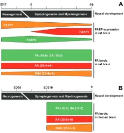
Fig. 2. Temporal correlation of dynamic changes in FABP expression and fatty acid levels in developing rat (A) and human (B) brain.
Temporal expression patterns of Fabp3, Fabp5 and Fabp7 in rat brain during fetal and postnatal development are based on Northern blot data (Owada et al. 1996). Dynamic changes in the levels of DHA (22:6n-3), AA (22:4n-6) and PA (18:0) in the developing rat brain and human brain are based on Green and Yavin (1996) and Martinez et al. (1992), respectively.
A
both Fabp7 and Fabp5 mRNA are present in the Bergmann glia of the cerebellum, while Fabp3 and Fabp5 mRNA co-localize in the neurons of the olfactory bulb. However, for the most part Fabps show complementary distribution patterns, with transcripts of different Fabps expressed in specific brain regions, cell layers, cell types and developmental stages (Owada and Kondo 2003; Owada et al. 2006). Third, transcripts and proteins of all three Fabps localize to both the cytoplasm and nucleus of either glia or neurons (Liu et al. 2000; Owada and Kondo 2003). The relative intensity of Fabp mRNA and protein in cytoplasm and nucleus changes during development (Liu et al. 2000). The presence of FABPs in the nucleus suggests a role in the modulation of gene expression, presumably by controlling the availability of the fatty
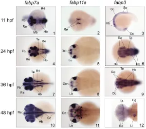
developing zebrafish brain (Fig. 3). Of all fabps expressed in zebrafish brain, fabp7a transcripts were the most abundant and showed the widest distribution in the developing forebrain, mid-brain, hindmid-brain, spinal cord as well as the retina. Similar to mammalian FABP7, zebrafish fabp7a is also the earliest gene expressed during brain development, although fabp7a mRNA is not spatially restricted when it is first detected at the early segmentation stage (Liu et al. 2004). The levels of zebrafish fabp3 transcripts decrease as the brain develops, with barely detectable levels observed 48 hours post-fertilization (hpf). In contrast, fabp7a and fabp11a transcripts are still abundant at this stage. Thus, the relative temporal expression patterns of the fish fabp3 and fabp7 in the developing brain are different from their mamma-Gene Main occurrence Cell type Subcellular distribution References
Fabp7 Cerebral cortex, olfactory bulb, hippocampus, thalamus, hypothalamus, corpus callosum, cerebellum
Radial glia cells, immature astrocytes
Cytoplasm, nucleus Kurtz et al., 1994; Feng and Heintz, 1994; Owada, 1996
Fabp5 Cerebral cortex, olfactory bulb, hippocampus, thalamus, hypothalamus, corpus callosum, cerebellum, caudate putamen, retina, lens.
Neuron, glia cells Cell body/soma, processes, nucleus
Liu et al., 2000; Owada et al., 1996
Fabp3 Olfactory bulb, hippocampus, thalamus, hypothalamus, cerebellum, caudate putamen
Neuron Cytoplasm, nucleus Sellner et al., 1995; Owada et al., 1996 TABLE 1
SPATIAL DISTRIBUTION OF FABPs IN DEVELOPING RODENT BRAIN
Fig. 3. Comparative expression patterns of FABPs in the developing brain of zebrafish (Danio rerio). fabp7a, fabp11a and fabp3transcriptsin zebrafish embryos at 11, 24, 36 and 48 hours post-fertilization (hpf) were detected by whole mount in situ hybridization using specific DIG-dUTP-labeled antisense RNA probes. Images are dorsal views of the zebrafish embryos except for III-3, which is a lateral view. The head is on the left. Abbreviations: Fb, forebrain; Te, tectum; Re, retina; Hb, hindbrain; R4, rhombomere 4; Dc, diencephalon; Le, lens; Sc, spinal cord; So, somites; Li, liver; Cg, Cranial ganglion. Arrowheads indicate brain vasculature. The data shown in this figure were compiled from our previous publications (Liu et al. 2004; Liu et al. 2007) with permission from the editor.
acid ligands required for nuclear receptor PPAR and/ or RXR activity (Kitajka et al. 2004; Schroeder et al. 2008).
The first avian FABP7 (also known as R-FABP) was identified by Godbout (1993) by screening a cDNA library prepared from embryonic chick retina at ED3.5. Northern blot analysis revealed elevated levels of FABP7 mRNA in the ED3.5 retina, with a 50 to 100-fold decrease in transcript levels observed from ED3.5 to ED19. In contrast, FABP7 mRNA levels increased 30 to 40-fold in the differentiating brain from ED3.5 to ED19 (Godbout 1993). Godbout et al. (1995) later analyzed the cellular and subcellular distribution of FABP7 mRNA and protein in the developing chick retina. At early stages of development, from ED3 to ED7, FABP7 was found throughout the retina, with accumulation in the neurites of ganglion cells. By ED11, FABP7 was mainly found in the inner nuclear layer, inner plexiform layer, optic nerve fiber layer and non-pigmented ciliary epithelium (Godbout et al. 1995). In contrast to mammalian brain FABP7 which is prima-rily found in radial glial cells (Kurtz et al. 1994; Feng and Heintz 1995), chicken FABP7 localizes to retinal neurons (Sellner 1993). The spatio-temporal expres-sion pattern of FABP7 and other FABPs in the devel-oping avian brain has yet to be described.
lian counterparts (Owada and Kondo 2003; Liu et al. 2004). The zebrafish fabp11a (also named fabp4) shows restricted expres-sion in the developing diencephalon, lens and brain vasculature (Agulleiro et al. 2007; Liu et al. 2007). As fabp11a is the only FABP gene other than fabp7 and fabp3 that is expressed in the devel-oping brain, this suggests a possible orthologous relationship with the mammalian Fabp5. Of note, both the zebrafish fabp11a and the mammalian Fabp5 are also expressed in lens (Wen et al. 1995; Liu et al. 2007).
Fatty acid ligand preference for FABPs in developing
brain
Commonly used approaches for measuring binding of FABPs to their fatty acid ligands include: Isothermal Titration Calorimetry (ITC), ANS Fluorometry and Lipidex 1000. Despite method-dependent and species-method-dependent variation in absolute dissocia-tion constant (Kd) values, FABP3, FABP5 and FABP7 each demonstrate preferences for specific classes of fatty acids (Table 2). For example, ITC analysis of human FABP3 in the presence of ω-3 PUFA (DHA, EPA), ω-6 PUFA (LOA) and monounsaturated fatty acid (MUFA) [oleic acid (OA)] yields Kd values in the range of 3-4 μM, 1 μM and 0.8 μM, respectively (Balendiran et al. 2000). In support of a preference for ω-6 PUFA, Fabp3 gene-ablated mice show impairment of AA incorporation into the brain (Murphy et al. 2005). These in vivo data suggest a requirement for FABP3 binding to ω-6 PUFA that is not compensated by expression of other FABPs in the brain (FABP5 and FABP7).
Rat and human FABP5 both show a preference for the more
saturated fatty acids, generating Kd values of 0.168 μM (ANS Fluorometry) and 0.290 μM (Lipidex) for stearic acid (SA) com-pared to Kd values ≥0.4 μM for ω-3 and ω-6 PUFAs (Liu et al. 2008; Hohoff et al. 1999). In comparison to FABP3 and FABP5, human FABP7 has the highest affinity for ω-3 PUFA, with a Kd of 0.053 μM when bound to DHA. This is in contrast to Kd values of 0.207 μM for FABP7-AA and 7.1 μM for FABP7-stearic acid (SA) (Balendiran et al. 2000). Although not numerically consistent with the Kd of 0.010 μM reported for murine FABP7-DHA using Lipidex (Xu et al. 1996), both ITC and Lipidex demonstrate that FABP7 preferentially binds to ω-3 PUFA.
Green et al. (1996) and Martinez et al. (1992) have examined the fatty acid composition of the developing rat and human brain, respectively. In rat, the levels of saturated fatty acids (16:0; 18:0) and AA (ω-6 PUFA) remain stable throughout neural develop-ment. In contrast, the levels of DHA in rat brain are relatively low during neurogenesis (~50% of AA levels and 25% of PA and SA levels) and increase during synaptogenesis (to ~50% of PA and SA levels and close to 100% of AA levels by P8) (Fig. 2A). A similar trend is observed in the developing human brain, with stable levels of saturated fatty acids from 30 weeks gestation to birth. A slight decrease in AA levels is observed during this period of time, whereas synaptogenesis and myelinogenesis is accompanied by an ~20% increase in DHA levels (Fig. 2B). As there is no data on the fatty acid composition of the human brain during neurogenesis, one can only postulate that, similar to what has been observed in rat brain, the transition from neurogenesis to synaptogenesis is accompanied by a significant increase in DHA in the developing human brain.
FABP Fatty Acid Classification Fatty Acid Kd (nM) Method Reference
Hs-FABP3 ω-3 PUFA DHA (22:6) 4100 ± 6 ITC (Balendiran et al., 2000)
EPA (20:5) 3300 ± 10 ITC (Balendiran et al., 2000)
ω-6 PUFA LOA (18:2) 970 ± 8 ITC (Balendiran et al., 2000)
AA (20:4) 370 ± 20 Lipidex (Veerkamp et al., 1999)
MUFA OA (18:1) 820 ± 10 ITC (Balendiran et al., 2000)
OA (18:1) 440 ± 50 Lipidex (Veerkamp et al., 1999)
Saturated FA PA (16:0) 960 ± 50 Lipidex (Veerkamp et al., 1999)
Rn-FABP5 ω-3 PUFA DHA (22:6) 422.2 ± 58.1 ANS Fluorometry (Liu et al., 2008)
EPA (20:5) 598 ± 100.5 ANS Fluorometry (Liu et al., 2008)
ω-6 PUFA AA (20:4) 390.9 ± 54.2 ANS Fluorometry (Liu et al., 2008)
LOA (18:2) 512.0 ± 34.5 ANS Fluorometry (Liu et al., 2008)
MUFA OA (18:1) 154.6 ± 35.3 ANS Fluorometry (Liu et al., 2008)
Saturated FA SA (18:0) 168.1 ± 38.1 ANS Fluorometry (Liu et al., 2008)
Hs-FABP5 ω-6 PUFA AA (20:4) 1730 ± 250 Lipidex (Hohoff et al., 1999)
MUFA OA (18:1) 1600 ± 200 Lipidex (Hohoff et al., 1999)
Saturated FA SA (18:0) 290 ± 60 Lipidex (Hohoff et al., 1999)
Hs-FABP7 ω-3 PUFA DHA (22:6) 53.4 ± 4.1 ITC (Balendiran et al., 2000)
EPA (20:5) 48.1 ± 21 ITC (Balendiran et al., 2000)
ω-6 PUFA AA (20:4) 207 ± 19 ITC (Balendiran et al., 2000)
LOA (18:2) 115 ± 19 ITC (Balendiran et al., 2000)
MUFA OA (18:1) 46.7 ± 1.4 ITC (Balendiran et al., 2000)
Saturated FA SA (18:0) 7100 Lipidex (Balendiran et al., 2000)
PA (16:0) 13500 Lipidex (Balendiran et al., 2000)
LA (12:0) 443 ± 55 ITC (Balendiran et al., 2000)
Mm-FABP7 ω-3 PUFA DHA (22:6) 10 ± 2 Lipidex (Xu et al., 1996)
ω-6 PUFA AA (20:4) 250 ± 34 Lipidex (Xu et al., 1996)
MUFA OA (18:1) 440 ± 27 Lipidex (Xu et al., 1996)
TABLE 2
Although there is some overlap in the temporal expression pattern and fatty acid binding affinities of FABP3, FABP5 and FABP7, it is clear from in vitro binding studies that these FABPs have specific fatty acid ligand preferences (i.e. ω-6 PUFA for FABP3, saturated fatty acids for FABP5 and ω-3 PUFA for FABP7). The temporal expression patterns of FABP3, FABP5 and FABP7 during brain development, combined with the tempo-ral availability of saturated fatty acids, ω-6 PUFA and ω-3 PUFA, suggest that different FABPs and their fatty acid ligands play specialized roles during neurogenesis and synaptogenesis.
Emerging functions for FABPs in brain development
FABP7 and neuronal cell migration
During brain development, neuronal precursor cells proliferate in the ventricular zone of the developing neocortex. Neurons then travel from their origin or “birth place” using radial glial fibers that serve as a scaffold for migrating neuronal cells. Each subsequent wave of migrating cells travel past their predecessors, forming layers in an inside-out manner, i.e. the youngest neurons are the closest to the cortical surface (Rakic 1972; Nadarajah and Parnavelas 2002). It has been estimated that as much as 90% of neuronal migration in the cortex is mediated by radial glial cells (Sidman and Rakic 1973; Hatten 1999).
The mammalian FABP7 is specifically expressed in radial glial cells during brain development (Feng et al. 1994; Kurtz et al. 1994; Hartfuss et al. 2001). Feng et al. (1994) used an in vitro neuron-glia co-culture system to investigate whether FABP7 plays a role in the establishment of a glial fiber system and neuronal migration. When purified anti-FABP7 antibody was added to the neuron-glia culture, both glial cell processes exten-sion and neuronal migration were blocked, while cell proliferation and adhesion were not affected (Feng et al. 1994). Subsequent investigations further demonstrated that expression of FABP7 in radial glial cells is induced by the presence of migrating neurons (Feng and Heintz 1995) and that this induction is mediated by the Notch signalling pathway (Anthony et al. 2005).
Pax6 and GGF/neuregulin also mediate migrating neuron-radial glia interactions. Both these molecules control FABP7 expression in the radial glial cells of the developing rat brain (Anton et al. 1997; Arai et al. 2005). These studies suggest that FABP7 is a critical molecule in radial glial cells, on one hand functioning as a sensor for neuronal signals, and on the other hand modulating radial glia differentiation and neuronal migra-tion.
FABP7-expressing cells as neural progenitors
As mentioned above, FABP7 is present at very early develop-mental stages in both mammalian and fish brains. Mammalian FABP7 is specifically expressed in the neuroepithelial cells and radial glial cells of the ventricular and subventricular zones, the hot spots of neurogenesis (Feng et al. 1994; Kurtz et al. 1994; Owada et al. 2006). In addition to their role in neuronal cell guidance, radial glial cells have been shown to act as neural stem/ progenitor cells during brain development (Malatesta et al. 2000; Miyata et al. 2001; Noctor et al. 2001; Malatesta et al. 2003; Anthony et al. 2004).
Anthony et al. (2004) used a 1.6 kb FABP7 promoter-driven Cre/Rosa26 Cre reporter double transgenic mice for fate mapping
analysis. They found that all FABP7-expressing radial glial cells, regardless of location within the brain, go through a neuron generating stage. These authors concluded that FABP7-express-ing radial glial cells give rise to most of the neurons in the brain. Furthermore, analysis of Fabp7-/- mice revealed dramatic de-creases in the number of astrocytes, neural stem cells and early progenitor cells in the developing brain (Watanabe et al. 2007). Fabp7-/- mice also showed decreased numbers of late progenitor cells and attenuated neurogenesis in the developing hippocam-pus compared to wild-type mice (Watanabe et al. 2007). Knock-down of FABP7 in cultured rat neuroepithelial cells results in decreased proliferation, loss of processes and induction of pre-mature neurogenesis, suggesting a role for FABP7 in the mainte-nance of proliferation (Arai et al. 2005). FABP7 may therefore play a key role in neurogenesis, by maintaining the pool of neural stem/ progenitor cells (Watanabe et al. 2007).
Role of FABP3 and FABP5 in neuronal differentiation During brain development, specific biochemical, physiological and morphological properties must be acquired by neuroprogenitor cells to allow differentiation along the neuronal cell lineage. The coordinated regulation of gene expression is fundamental to the control of neuronal cell differentiation. FAPB5 is specifically found in the differentiating neurons of the developing rat cerebral cortex and retina. FABP5 mRNA and protein levels are markedly higher in the prenatal and early postnatal neurons compared to adult neurons, suggesting a function for this FABP in neuronal differen-tiation (Liu et al. 1997; Liu et al. 2000). Other evidence in support of a role for FABP5 in neuronal cell differentiation is the observa-tion that FABP5 is induced in injured and regenerating neurons (De Leon et al. 1996; Owada et al. 1997). FABP5 is also upregulated with NGF-induced neurite growth in rat pheochromocytoma cells (PC12), a model system for neuronal differentiation (Allen et al. 2000). Depletion of FABP5 in PC12 using an antisense Fabp5 construct significantly reduces NGF-mediated neurite growth, suggesting a role for FABP5 in axonal development and regen-eration (Allen et al. 2000). Of note, no brain phenotype has been reported thus far in Fabp5 knockout mice, suggesting that other FABPs may compensate for loss of FABP5 function (Owada et al. 2002).
FABP3 is expressed in mouse brain at relatively late develop-mental stages compared to Fabp7 and Fabp5, with barely detect-able levels until ED17-19 (Sellner et al. 1995; Owada et al. 1996). Based on its spatio-temporal expression pattern in mouse brain, Sellner et al. (1995) proposed that FABP3 is involved in neurite formation and synapse maturation. As Fabp3-null mouse brains show a reduction in both the incorporation of AA and the propor-tion of total ω-6 fatty acids in the major phospolipid classes, it was concluded that FABP3 may be involved in the uptake and metabo-lism of ω-6 fatty acids in the developing brain (Murphy et al. 2005).
early developing vertebrate brain are involved in local patterning of axis formation, including the midbrain-hindbrain boundary (reviewed by Rhinn and Brand 2001; Wurst and Bally-Cuif 2001), the forebrain organizer (Shimamura and Rubenstein 1997; Houart et al. 1998) and the hindbrain-spinal cord border (Maden 2002). However, the signaling mechanisms from organizing centers involved in neuronal and glial cell differentiation are not well understood. Isolating genes with restricted patterns of expression in organizing centers combined with mutant screening using model organisms would be a powerful approach toward under-standing the signaling pathways governing axis patterning in vertebrate brain.
As FABP7 is expressed in the early developing brain and has cell signaling properties (Feng et al. 1994), it is logical to postulate that FABP7 may play a role in neural axis patterning. For instance, the vertebrate hindbrain consists of several subdivided
seg-the same mechanism is not known.
The Notch signaling pathway
Notch proteins are single-pass transmembrane receptors which include four family members, NOTCH1, NOTCH2, NOTCH3 and NOTCH4. Notch proteins play a role in a variety of developmental processes by controlling cell fate decisions (reviewed by Lewis 1998; Gaiano and Fishell 2002; Rodriguez-Rivera et al. 2009). Both neural stem cells and intermediate neural progenitors re-spond to Notch receptor activation, but through differential action of the downstream effector C-promoter binding factor 1 (CBF1) (Mizutani et al. 2007). A single binding site for CBF1 (5’-GTTCCCAGGC-3’, conserved nucleotides underlined) has been mapped within the proximal promoter region of mouse Fabp7 and is required for Fabp7 expression in radial glial cells (Anthony et al. 2005). Furthermore, FABP7 protein levels are significantly
down-Marginal zone
Dab1 Notch1 Dab1 Notch1
Radial glia
Formation and maintenance of the radial glia scaffold
Synaptogenesis Neurite outgrowth
Neuron
Neuronal migration guided by radial glial scaffold
Subplate cells Cortical plate
Ventricular zone Marginal zone
Pia
Pia Cajal-Retzius cells
FABP7 FABP5
Reelin
?
FABP3
Mature cortical neurons Cortical plate Radial
glial fiber
Fig. 4. Schematic illustration of a proposed Reelin-Dab1/Notch/FABP signal-ing pathway involved in neural development. FABP7 expressed in radial glial cells is a major target of Reelin-Dab1/Notch signaling, which coordinates interaction between migrating neurons and radial glia during cortical neural development. We propose a model whereby FABP5 and FABP3 could function as downstream effectors of Reelin-Dab1/Notch signaling in neuronal cells.
ments, called rhombomeres, each of which has distinct cellular and molecular characteristics (reviewed by Lumsden and Krumlauf 1996). Rhombomere 4 (r4) is the first one to form in the developing vertebrate hindbrain and acts as a signaling center in hindbrain patterning (Graham and Lumsden 1993; Graham and Lumsden 1996; Maves et al. 2002). In zebrafish, fabp7a transcripts specifically localize to a restricted region of r4 during the middle and late segmentation phase, suggesting an important role for the fabp7a product in patterning of the hindbrain (Liu et al. 2004). Recent data indicate that Pbx homeodomain proteins may play a role in the patterning of zebrafish retina and optic tectum, and that one of the major downstream effectors of these transcription fac-tors is FABP7 (French et al. 2007). Indeed, zebrafish fabp7a mRNA is abundantly expressed in the developing retina and tectum from early embryonic stages (Liu et al. 2004) and its levels are markedly reduced in the pbx2/4-null embryos compared with the wild-type (French et al. 2007).
Regulation of FABP expression in developing
brain
Nuclear factor I (NF1)
regulated in the developing forebrain of mice lacking Notch1 and Notch3, indicating an essential role for Notch signaling in modu-lating Fabp7 expression in developing mouse brain (Anthony et al. 2005).
The Reelin-Dab1 pathway
Reelin is a large secreted glycoprotein which controls cell-cell interactions critical for cell positioning and neuronal migration during brain development (D’Arcangelo et al. 1995). Disabled-1 (DAB1) serves as the intracellular adaptor for Reelin and has also been shown to be involved in neuronal cell positioning in the developing brain (Howell et al. 1997; Howell et al. 1999). DAB1 is tyrosine phosphorylated when Reelin binds to the lipoprotein receptors Very Low Density Lipoprotein Receptor (VLDLR) and Apolipoprotein E Receptor 2 (APOER2) on the cell membrane (Huang et al. 2005). In mutant mice deficient for Reelin, DAB1, or Reelin receptors (VLDLR and APOER2), radial glial morphology is altered and the radial glial scaffold is impaired in the developing brain (Forster et al. 2002; Hartfuss et al. 2003; Weiss et al. 2003). Reelin signalling affects FABP7 expression in radial glial cells both in vitro and in vivo (Hartfuss et al. 2003; Keilani and Sugaya 2008) and DAB1 is required for Reelin-mediated FABP7 expres-sion (Hartfuss et al. 2003).
A recent study of the role of Reelin in FABP7 induction in human neural progenitor cells revealed cross-talk between the Reelin and Notch signaling pathways (Keilani and Sugaya 2008). Reelin treatment of these cells increased the levels of both the NOTCH1 intracellular domain (NICD) and FABP7, and DAB1 co-immunoprecipitated with NICD, indicating physical interaction between these molecules (Keilani and Sugaya 2008). Interaction between Reelin-Dab1, Notch and FABP7 (BLBP) has also been demonstrated using the radial glial cells of Reeler mice (Sibbe et al. 2009). The inter-relationship between these major signaling molecules may be of significance in elucidating the links between the neuronal-radial glial signaling networks which regulate glial cell development, neuronal generation and migration.
Reelin is primarily secreted by neurons termed Cajal–Retzius cells, which establish early neuronal circuitry in the developing brain (Aguilo et al. 1999). Secreted Reelin promotes the differen-tiation of progenitor cells into radial glia and affects the orientation of radial glia fibers, which serve as guides for migrating neurons (Fig. 4). Interaction between Reelin and Notch signaling path-ways has also been demonstrated in the migrating and differen-tiating neurons of the developing cerebral cortex (Hashimoto-Torii et al. 2008). Reelin may therefore serve as a general signaling messenger to integrate the events associated with radial glia fiber formation and neuronal cell migration (Fig. 4). Thus, Reelin-Dab1/Notch/FABP7 may represent a major and essential signaling pathway mediating neuronal-glial interaction during the early development of the brain.
As FABP3 and FABP5 are expressed in the neurons of devel-oping brain and their expression patterns correlate with neuronal cell differentiation and/or migration (Sellner et al. 1995; Liu et al. 2000), it will be interesting to know whether the neuronal Reelin-Dab1/Notch signaling pathway also functions through a neuronal FABP (FABP3 or FABP5) downstream effector, as it does for FABP7 in radial glial cells (Anthony et al. 2005; Keilani and Sugaya 2008; Sibbe et al. 2009).
PAX6
The transcription factor PAX6 affects cell fate, cell proliferation and patterning during brain development in various species (Haubst et al. 2004). In Pax6-deficient mice, the morphology, quantity and cell cycle distribution of radial glial cells in the developing cortex are altered (Gotz et al. 1998). In the embryonic brains of rats, there is a strong correlation in the distribution patterns of PAX6 and FABP7. Pax6 mutation in rat embryonic brains results in dramatically reduced FABP7 whereas over-expression of PAX6 induces FABP7 over-expression, suggesting a role for PAX6 in the regulation of Fabp7 (Arai et al. 2005). Although a putative cis-binding site for PAX6 was identified in the mouse Fabp7 promoter (Feng and Heintz 1995), it is not known whether PAX6 regulates Fabp7 transcription by directly interact-ing with this cis element or through other downstream effectors. It is noteworthy that FABP7 levels are not significantly affected by depletion of PAX6 in mouse brain (Gotz et al. 1998; Arai et al. 2005), suggesting that PAX6-mediated regulation of FABP7 expression is species-dependent.
POU-domain proteins
POU-domain proteins are widely expressed in developing brain with distinct temporal and spatial patterns (He et al. 1989; Alvarez-Bolado et al. 1995). Using an in vivo reporter transgenic mouse model, Josephson et al. (1998) tested the ability of serial deletions of the proximal 5’ upstream region of Fabp7 (Feng and Heintz 1995) to drive developmentally-regulated Fabp7 expres-sion in the embryonic CNS. A POU-domain protein PBX-1 and BRN-1 binding element was subsequently identified in the Fabp7 promoter (Josephson et al. 1998). Mutant mice with germ-line deletion of the 9 bp POU-domain binding element lacked Fabp7 expression in the developing forebrain and midbrain, suggesting an essential role for POU-domain proteins in Fabp7 regulation (Josephson et al. 1998).
In zebrafish, the levels of the fabp7a transcripts are dramati-cally reduced in the embryonic brain and retina when the POU-domain protein is knocked-down by morpholino transfection (French et al. 2007), directly implicating this POU-domain protein in the regulation of fabp7 expression.
FABPs and brain disease
models and exploration of the mechanism of action of FABP7 in the nucleus will shed light on its role in human glioblastoma.
Mental retardation is a typical phenotypic feature of Down syndrome (DS) patients. As FABPs are involved in development, establishment and maintenance of the central nervous system, it is reasonable to propose that FABPs may be implicated in DS pathogenesis. FABP7 has been found to be overexpressed in DS adult (Cheon et al. 2003) and fetal brains (Sanchez-Font et al. 2003), whereas FABP3 is significantly decreased in DS adult brains (Cheon et al. 2003). Furthermore, FABP7 upregulation in DS brains correlates with PKNOX1 gene-dosage imbalance. As PKNOX1 is a POU domain protein, it may directly control FABP7 expression by interacting with the Pbx/POU binding element in the FABP7 promoter (Sanchez-Font et al. 2003).
Different lines of investigation directly associate FABP7 with schizophrenia, a psychiatric illness characterized by abnormali-ties in the perception or expression of reality (Watanabe et al. 2007). Firstly, a quantitative trait locus (QTL) screening in a reference mice population demonstrates that Fabp7 is a promis-ing candidate gene responsible for deficits in prepulse inhibition (PPI), a biological marker for schizophenia. Secondly, Fabp7-deficient mice show decreased PPI and impaired hippocampus neurogenesis. Thirdly, human FABP7 mRNA levels are signifi-cantly upregulated in the postmortum brains of schizophrenia patients compared with normal controls. Fourthly, there is a correlation between a single nucleotide polymorphism (SNP) variant within the second exon of human FABP7 and schizophre-nia pathology (Watanabe et al. 2007). As PPI impairment has been demonstrated in many other neuropsychiatric disorders (e.g. Alzheimer’s disease, autism, bipolar disorders, Tourette syndrome, etc.) in addition to schizophrenia (Braff et al. 2001; Hejl et al. 2004), the association between FABP7 and PPI status suggest that this gene is involved in the pathology of a wide range of neuropsychiatric and/or neurodegenerative diseases. Rela-tionships between FABP3 and FABP5, and neurodegenerative diseases have also been reported (Gearhart et al. 2002; Steinacker et al. 2004).
Summary
FABP3, FABP5 and FABP7 are expressed in specific spatio-temporal patterns in the developing brain, with each showing preferential binding to different categories of long chain fatty acid ligands. Although the precise roles of these three FABPs in brain development awaits more in-depth studies, investigations using animal models have revealed a number of possible functions in neurogenesis, neuronal migration and differentiation, and axis patterning. Data to date indicate that FABP3, FABP5 and FABP7 are multi-functional proteins regulated by complex signaling net-works and transcription factors. In particular, FABP7 in radial glial cells may be a major downstream effector of the Reelin-Dab1/ Notch signal pathway which plays a key role in the formation and maintenance of the radial glia scaffold and neuronal cell migration in the developing brain. FABP7 may serve as a mediator for the signaling crosstalk required between migrating neurons and radial glial cells. So far, FABP expression, its cellular distribution and genetic variation have been implicated in a number of brain diseases including human glioblastoma and neuropsychiatric disorders. Future investigations will shed additional light on the
role of FABPs and their mechanisms of action in the development and health of the brain.
Acknowledgment
This work is supported by the Alberta Cancer Research Insti-tute. RZL and ZG are supported by an Alberta Heritage Founda-tion for Medical Research fellowship and studentship, respec-tively. RM is the recipient of an Alberta Cancer Research Institute studentship. MB has a Canadian Institutes of Health Research studentship.
References
AGUILO A, SCHWARTZ TH, KUMAR VS, PETERLIN ZA, TSIOLA A, SORIANO E, YUSTE R (1999). Involvement of cajal-retzius neurons in spontaneous corre-lated activity of embryonic and postnatal layer 1 from wild-type and reeler mice.
J Neurosci 19: 10856-10868.
AGULLEIRO MJ, ANDRE M, MORAIS S, CERDA J, BABIN PJ (2007). High transcript level of fatty acid-binding protein 11 but not of very low-density lipoprotein receptor is correlated to ovarian follicle atresia in a teleost fish (Solea senegalensis). Biol Reprod 77: 504-516.
ALLEN GW, LIU JW, DE LEON M (2000). Depletion of a fatty acid-binding protein impairs neurite outgrowth in PC12 cells. Brain Res Mol Brain Res 76: 315-324.
ALVAREZ-BOLADO G, ROSENFELD MG, SWANSON LW (1995). Model of forebrain regionalization based on spatiotemporal patterns of POU-III homeobox gene expression, birthdates, and morphological features. J Comp Neurol 355:
237-295.
ANTHONY TE, KLEIN C, FISHELL G, HEINTZ N (2004). Radial glia serve as neuronal progenitors in all regions of the central nervous system. Neuron 41:
881-890.
ANTHONY TE, MASON HA, GRIDLEY T, FISHELL G, HEINTZ N (2005). Brain lipid-binding protein is a direct target of Notch signaling in radial glial cells.
Genes Dev 19: 1028-1033.
ANTON ES, MARCHIONNI MA, LEE KF, RAKIC P (1997). Role of GGF/neuregulin signaling in interactions between migrating neurons and radial glia in the developing cerebral cortex. Development 124: 3501-3510.
ARAI Y, FUNATSU N, NUMAYAMA-TSURUTA K, NOMURA T, NAKAMURA S, OSUMI N (2005). Role of Fabp7, a downstream gene of Pax6, in the mainte-nance of neuroepithelial cells during early embryonic development of the rat cortex. J Neurosci 25: 9752-9761.
BALENDIRAN GK, SCHNUTGEN F, SCAPIN G, BORCHERS T, XHONG N, LIM K, GODBOUT R, SPENER F, SACCHETTINI JC (2000). Crystal structure and thermodynamic analysis of human brain fatty acid-binding protein. J Biol Chem
275: 27045-27054.
BENNETT E, STENVERS KL, LUND PK, POPKO B (1994). Cloning and character-ization of a cDNA encoding a novel fatty acid binding protein from rat brain. J Neurochem 63: 1616-1624.
BISGROVE DA, MONCKTON EA, PACKER M, GODBOUT R (2000). Regulation of brain fatty acid-binding protein expression by differential phosphorylation of nuclear factor I in malignant glioma cell lines. J Biol Chem 275: 30668-30676.
BRAFF DL, GEYER MA, LIGHT GA, SPROCK J, PERRY W, CADENHEAD KS, SWERDLOW NR (2001). Impact of prepulse characteristics on the detection of sensorimotor gating deficits in schizophrenia. Schizophr Res 49: 171-178.
BRUN M, COLES JE, MONCKTON EA, GLUBRECHT DD, BISGROVE D, GODBOUT R (2009). Nuclear Factor I Regulates Brain Fatty Acid-Binding Protein and Glial Fibrillary Acidic Protein Gene Expression in Malignant Glioma Cell Lines. J Mol Biol. 391: 282-300.
CARLSON SE (2009). Docosahexaenoic acid supplementation in pregnancy and lactation. Am J Clin Nutr 89: 678S-684S.
CARLSON SE, NEURINGER M (1999). Polyunsaturated fatty acid status and neurodevelopment: a summary and critical analysis of the literature. Lipids 34:
171-178.
syn-drome and Alzheimer’s disease. J Neural Transm Suppl: 225-234.
D’ARCANGELO G, MIAO GG, CHEN SC, SOARES HD, MORGAN JI, CURRAN T (1995). A protein related to extracellular matrix proteins deleted in the mouse mutant reeler. Nature 374: 719-723.
DAS NEVES L, DUCHALA CS, TOLENTINO-SILVA F, HAXHIU MA, COLMENARES C, MACKLIN WB, CAMPBELL CE, BUTZ KG, GRONOSTAJSKI RM (1999). Disruption of the murine nuclear factor I-A gene (Nfia) results in perinatal lethality, hydrocephalus, and agenesis of the corpus callosum. Proc Natl Acad Sci USA 96: 11946-11951.
DE LEON M, WELCHER AA, NAHIN RH, LIU Y, RUDA MA, SHOOTER EM, MOLINA CA (1996). Fatty acid binding protein is induced in neurons of the dorsal root ganglia after peripheral nerve injury. J Neurosci Res 44: 283-292.
DENOVAN-WRIGHT EM, PIERCE M, WRIGHT JM (2000). Nucleotide sequence of cDNA clones coding for a brain-type fatty acid binding protein and its tissue-specific expression in adult zebrafish (Danio rerio). Biochim Biophys Acta 1492:
221-226.
FENG L, HATTEN ME, HEINTZ N (1994). Brain lipid-binding protein (BLBP): a novel signaling system in the developing mammalian CNS. Neuron 12:
895-908.
FENG L, HEINTZ N (1995). Differentiating neurons activate transcription of the brain lipid-binding protein gene in radial glia through a novel regulatory element.
Development 121: 1719-1730.
FORSTER E, TIELSCH A, SAUM B, WEISS KH, JOHANSSEN C, GRAUS-PORTA D, MULLER U, FROTSCHER M (2002). Reelin, Disabled 1, and beta 1 integrins are required for the formation of the radial glial scaffold in the hippocampus.
Proc Natl Acad Sci USA 99: 13178-13183.
FREEMAN MP (2000). Omega-3 fatty acids in psychiatry: a review. Ann Clin Psychiatry 12: 159-165.
FRENCH CR, ERICKSON T, CALLANDER D, BERRY KM, KOSS R, HAGEY DW, STOUT J, WUENNENBERG-STAPLETON K, NGAI J, MOENS CB, WASKIEWICZ AJ (2007). Pbx homeodomain proteins pattern both the zebrafish retina and tectum. BMC Dev Biol 7: 85.
GAIANO N, FISHELL G (2002). The role of notch in promoting glial and neural stem cell fates. Annu Rev Neurosci 25: 471-490.
GEARHART DA, TOOLE PF, WARREN BEACH J (2002). Identification of brain proteins that interact with 2-methylnorharman. An analog of the parkinsonian-inducing toxin, MPP+. Neurosci Res 44: 255-265.
GODBOUT R (1993). Identification and characterization of transcripts present at elevated levels in the undifferentiated chick retina. Exp Eye Res 56: 95-106.
GODBOUT R, BISGROVE DA, SHKOLNY D, DAY RS, 3RD (1998). Correlation of B-FABP and GFAP expression in malignant glioma. Oncogene 16: 1955-1962.
GODBOUT R, MARUSYK H, BISGROVE D, DABBAGH L, POPPEMA S (1995). Localization of a fatty acid binding protein and its transcript in the developing chick retina. Exp Eye Res 60: 645-657.
GOTZ M, STOYKOVA A, GRUSS P (1998). Pax6 controls radial glia differentiation in the cerebral cortex. Neuron 21: 1031-1044.
GRAHAM A, LUMSDEN A (1993). The role of segmentation in the development of the branchial region of higher vertebrate embryos. Birth Defects Orig Artic Ser
29: 103-112.
GRAHAM A, LUMSDEN A (1996). Patterning the cranial neural crest. Biochem Soc Symp 62: 77-83.
GREEN P, YAVIN E (1996). Fatty acid composition of late embryonic and early postnatal rat brain. Lipids 31: 859-865.
GRONOSTAJSKI RM (2000). Roles of the NFI/CTF gene family in transcription and development. Gene 249: 31-45.
GROSSFIELD A, FELLER SE, PITMAN MC (2006). A role for direct interactions in the modulation of rhodopsin by omega-3 polyunsaturated lipids. Proc Natl Acad Sci USA 103: 4888-4893.
HANHOFF T, LUCKE C, SPENER F (2002). Insights into binding of fatty acids by fatty acid binding proteins. Mol Cell Biochem 239: 45-54.
HARTFUSS E, FORSTER E, BOCK HH, HACK MA, LEPRINCE P, LUQUE JM, HERZ J, FROTSCHER M and GOTZ M (2003). Reelin signaling directly affects radial glia morphology and biochemical maturation. Development 130:
4597-4609.
HARTFUSS E, GALLI R, HEINS N, GOTZ M (2001). Characterization of CNS
precursor subtypes and radial glia. Dev Biol 229: 15-30.
HASHIMOTO-TORII K, TORII M, SARKISIAN MR, BARTLEY CM, SHEN J, RADTKE F, GRIDLEY T, SESTAN N, RAKIC P (2008). Interaction between Reelin and Notch signaling regulates neuronal migration in the cerebral cortex.
Neuron 60: 273-284.
HATTEN ME (1999). Central nervous system neuronal migration. Annu Rev Neurosci 22: 511-539.
HAUBST N, BERGER J, RADJENDIRANE V, GRAW J, FAVOR J, SAUNDERS GF, STOYKOVA A, GOTZ M (2004). Molecular dissection of Pax6 function: the specific roles of the paired domain and homeodomain in brain development.
Development 131: 6131-6140.
HAUNERLAND, NH, SPENER F (2004). Fatty acid-binding proteins—insights from genetic manipulations. Prog Lipid Res 43: 328-349.
HE X, TREACY MN, SIMMONS DM, INGRAHAM HA, SWANSON LW, ROSENFELD MG (1989). Expression of a large family of POU-domain regulatory genes in mammalian brain development. Nature 340: 35-41.
HEJL AM, GLENTHOJ B, MACKEPRANG T, HEMMINGSEN R, WALDEMAR G (2004). Prepulse inhibition in patients with Alzheimer’s disease. Neurobiol Aging 25: 1045-1050.
HOHOFF C, BÖRCHERS T, RÜSTOW B, SPENER F, VAN TILBEURGH H (1999). Expression, purification, and crystal structure determination of recombinant human epidermal-type fatty acid binding protein. Biochemistry 38:
12229-12239.
HOUART C, WESTERFIELD M, WILSON SW (1998). A small population of anterior cells patterns the forebrain during zebrafish gastrulation. Nature 391: 788-792.
HOWELL BW, GERTLER FB, COOPER JA (1997). Mouse disabled (mDab1): a Src binding protein implicated in neuronal development. EMBO J 16: 121-132.
HOWELL BW, HERRICK TM, COOPER JA (1999). Reelin-induced tyrosine [cor-rected] phosphorylation of disabled 1 during neuronal positioning. Genes Dev
13: 643-648.
HUANG Y, SHAH V, LIU T, KESHVARA L (2005). Signaling through Disabled 1 requires phosphoinositide binding. Biochem Biophys Res Commun 331:
1460-1468.
JOSEPHSON R, MULLER T, PICKEL J, OKABE S, REYNOLDS K, TURNER PA, ZIMMER A, MCKAY RD (1998). POU transcription factors control expression of CNS stem cell-specific genes. Development 125: 3087-3100.
JUMP DB (2002). The biochemistry of n-3 polyunsaturated fatty acids. J Biol Chem
277: 8755-8758.
KALOSHI G, MOKHTARI K, CARPENTIER C, TAILLIBERT S, LEJEUNE J, MARIE Y, DELATTRE JY, GODBOUT R, SANSON M (2007). FABP7 expression in glioblastomas: relation to prognosis, invasion and EGFR status. J Neurooncol
84: 245-248.
KARANTH S, DENOVAN-WRIGHT EM, THISSE C, THISSE B, WRIGHT JM (2008). The evolutionary relationship between the duplicated copies of the zebrafish fabp11 gene and the tetrapod FABP4, FABP5, FABP8 and FABP9 genes. Febs J 275: 3031-3040.
KEILANI S, SUGAYA K (2008). Reelin induces a radial glial phenotype in human neural progenitor cells by activation of Notch-1. BMC Dev Biol 8: 69.
KITAJKA K, SINCLAIR AJ, WEISINGER RS, WEISINGER HS, MATHAI M, JAYASOORIYA AP, HALVER JE, PUSKAS LG (2004). Effects of dietary omega-3 polyunsaturated fatty acids on brain gene expression. Proc Natl Acad Sci USA 101: 10931-10936.
KUHAR SG, FENG L, VIDAN S, ROSS ME, HATTEN ME, HEINTZ N (1993). Changing patterns of gene expression define four stages of cerebellar granule neuron differentiation. Development 117: 97-104.
KURTZ A, ZIMMER A, SCHNUTGEN F, BRUNING G, SPENER F, MULLER T (1994). The expression pattern of a novel gene encoding brain-fatty acid binding protein correlates with neuronal and glial cell development. Development 120:
2637-2649.
LEWIS J (1998). Notch signalling and the control of cell fate choices in vertebrates.
Semin Cell Dev Biol 9: 583-589.
LIANG Y, BOLLEN AW, ALDAPE KD, GUPTA N (2006). Nuclear FABP7 immunore-activity is preferentially expressed in infiltrative glioma and is associated with poor prognosis in EGFR-overexpressing glioblastoma. BMC Cancer 6: 97.
LAMBORN KR, BERGER MS, BOTSTEIN D, BROWN PO, ISRAEL MA (2005). Gene expression profiling reveals molecularly and clinically distinct subtypes of glioblastoma multiforme. Proc Natl Acad Sci USA 102: 5814-5819.
LIU JW, ALMAGUEL FG, BU L, DE LEON DD, DE LEON M (2008) Expression of E-FABP in PC12 cells increases neurite extension during differentiation: in-volvement of n-3 and n-6 fatty acids. J Neurochem 106: 2015-2029.
LIU RZ, DENOVAN-WRIGHT EM, DEGRAVE A, THISSE C, THISSE B, WRIGHT JM (2004). Differential expression of duplicated genes for brain-type fatty acid-binding proteins (fabp7a and fabp7b) during early development of the CNS in zebrafish (Danio rerio). Gene Expr Patterns 4: 379-387.
LIU RZ, DENOVAN-WRIGHT EM, WRIGHT JM (2003a). Structure, linkage map-ping and expression of the heart-type fatty acid-binding protein gene (fabp3) from zebrafish (Danio rerio). Eur J Biochem 270: 3223-3234.
LIU RZ, DENOVAN-WRIGHT EM, WRIGHT JM (2003b). Structure, mRNA expres-sion and linkage mapping of the brain-type fatty acid-binding protein gene (FABP7) from zebrafish (Danio rerio). Eur J Biochem 270: 715-725.
LIU RZ, SAXENA V, SHARMA MK, THISSE C, THISSE B, DENOVAN-WRIGHT EM, WRIGHT JM (2007). The fabp4 gene of zebrafish (Danio rerio) genomic homology with the mammalian FABP4 and divergence from the zebrafish fabp3 in developmental expression. Febs J 274: 1621-1633.
LIU Y, LONGO LD, DE LEON M (2000). In situ and immunocytochemical
localiza-tion of E-FABP mRNA and protein during neuronal migralocaliza-tion and differentialocaliza-tion in the rat brain. Brain Res 852: 16-27.
LIU Y, MOLINA CA, WELCHER AA, LONGO LD, DE LEON M (1997). Expression of DA11, a neuronal-injury-induced fatty acid binding protein, coincides with axon growth and neuronal differentiation during central nervous system devel-opment. J Neurosci Res 48: 551-562.
LUMSDEN A, KRUMLAUF R (1996). Patterning the vertebrate neuraxis. Science
274: 1109-1115.
MADEN M (2002). Positional information: knowing where you are in a limb. Curr Biol
12: R773-5.
MALATESTA P, HACK MA, HARTFUSS E, KETTENMANN H, KLINKERT W, KIRCHHOFF F, GOTZ M (2003). Neuronal or glial progeny: regional differences in radial glia fate. Neuron 37: 751-764.
MALATESTA P, HARTFUSS E, GOTZ M (2000). Isolation of radial glial cells by fluorescent-activated cell sorting reveals a neuronal lineage. Development 127:
5253-5263.
MARTINEZ M (1992). Tissue levels of polyunsaturated fatty acids during early human development. J Pediatr 120: S129-138.
MAVES L, JACKMAN W, KIMMEL CB (2002). FGF3 and FGF8 mediate a rhombomere 4 signaling activity in the zebrafish hindbrain. Development 129:
3825-3837.
MCCANN JC, AMES BN (2005). Is docosahexaenoic acid, an n-3 long-chain polyunsaturated fatty acid, required for development of normal brain function? An overview of evidence from cognitive and behavioral tests in humans and animals. Am J Clin Nutr 82: 281-295.
MITA R, COLES JE, GLUBRECHT DD, SUNG R, SUN X, GODBOUT R (2007). B-FABP-expressing radial glial cells: the malignant glioma cell of origin? Neopla-sia 9: 734-744.
MIYATA T, KAWAGUCHI A, OKANO H, OGAWA M (2001). Asymmetric inheritance of radial glial fibers by cortical neurons. Neuron 31: 727-741.
MIZUTANI K, YOON K, DANG L, TOKUNAGA A., GAIANO N. (2007). Differential Notch signalling distinguishes neural stem cells from intermediate progenitors.
Nature 449: 351-355.
MURPHY EJ, OWADA Y, KITANAKA N, KONDO H, GLATZ JF (2005). Brain arachidonic acid incorporation is decreased in heart fatty acid binding protein gene-ablated mice. Biochemistry 44: 6350-6360.
NADARAJAH B, PARNAVELAS JG (2002). Modes of neuronal migration in the developing cerebral cortex. Nat Rev Neurosci 3: 423-432.
NEURINGER M, ANDERSON GJ, CONNOR WE (1988). The essentiality of n-3 fatty acids for the development and function of the retina and brain. Annu Rev Nutr 8: 517-541.
NOCTOR SC, FLINT AC, WEISSMAN TA, DAMMERMAN RS, KRIEGSTEIN AR (2001). Neurons derived from radial glial cells establish radial units in neocortex.
Nature 409: 714-720.
OWADA Y, ABDELWAHAB SA, KITANAKA N, SAKAGAMI H, TAKANO H, SUGITANI Y, SUGAWARA M, KAWASHIMA H, KISO Y, MOBARAKEH JI, YANAI K, KANEKO K, SASAKI H, KATO H, SAINO-SAITO S, MATSUMOTO N, AKAIKE N, NODA T, KONDO H (2006). Altered emotional behavioral responses in mice lacking brain-type fatty acid-binding protein gene. Eur J Neurosci 24: 175-187.
OWADA, Y. and KONDO, H. (2003). Fatty acid binding proteins of the brain. In
Cellular proteins and their fatty acids in health and disease (Eds. A.K. Duttaroy
and F. Spener), Wiley-VCH Verlag GmbH & Co. KGaA, Weinheim. pp. 253-265.
OWADA Y, SUZUKI I, NODA T, KONDO H (2002). Analysis on the phenotype of E-FABP-gene knockout mice. Mol Cell Biochem 239: 83-86.
OWADA Y, UTSUNOMIYA A, YOSHIMOTO T, KONDO H (1997). Changes in gene expression for skin-type fatty acid binding protein in hypoglossal motor neurons following nerve crush. Neurosci Lett 223: 25-28.
OWADA Y, YOSHIMOTO T, KONDO H (1996). Spatio-temporally differential expression of genes for three members of fatty acid binding proteins in developing and mature rat brains. J Chem Neuroanat 12: 113-122.
PU L, ANNAN RS, CARR SA, FROLOV A, WOOD WG, SPENER F, SCHROEDER F (1999). Isolation and identification of a mouse brain protein recognized by antisera to heart fatty acid-binding protein. Lipids 34: 363-373.
PURASIRI P, MCKECHNIE A, HEYS SD, EREMIN O (1997). Modulation in vitro of
human natural cytotoxicity, lymphocyte proliferative response to mitogens and cytokine production by essential fatty acids. Immunology 92: 166-172.
RAKIC P (1972). Mode of cell migration to the superficial layers of fetal monkey neocortex. J Comp Neurol 145: 61-83.
RHINN M, BRAND M (2001). The midbrain—hindbrain boundary organizer. Curr Opin Neurobiol 11: 34-42.
RICHARDSON AJ, PURI BK (2000). The potential role of fatty acids in attention-deficit/hyperactivity disorder. Prostaglandins Leukot Essent Fatty Acids 63:
79-87.
RODRIQUEZ-RIVERA NS, MOLINA-HERNANDEZ A, SANCHEZ-CRUZ E, ESCALANTE-ALCALDE D, VELASCO I (2009). Activated Notch1 is a stronger astrocytic stimulus than leukemia inhibitory factor for rat neural stem cells. Int J Dev Biol 53: 947-953.
SALVATI S, ATTORRI L, DI BENEDETTO R, DI BIASE A, LEONARDI F (2006). Polyunsaturated fatty acids and neurological diseases. Mini Rev Med Chem 6:
1201-1211.
SANCHEZ-FONT MF, BOSCH-COMAS A, GONZALEZ-DUARTE R, MARFANY G (2003). Overexpression of FABP7 in Down syndrome fetal brains is associated with PKNOX1 gene-dosage imbalance. Nucleic Acids Res 31: 2769-2777.
SCHOENTGEN F, PIGNEDE G, BONANNO LM, JOLLES P (1989). Fatty-acid-binding protein from bovine brain. Amino acid sequence and some properties.
Eur J Biochem 185: 35-40.
SCHROEDER F, PETRESCU AD, HUANG H, ATSHAVES BP, MCINTOSH AL, MARTIN GG, HOSTETLER HA, VESPA A, LANDROCK D, LANDROCK KK, PAYNE HR, KIER AB (2008). Role of fatty acid binding proteins and long chain fatty acids in modulating nuclear receptors and gene transcription. Lipids 43:
1-17.
SELLNER PA (1993). Retinal FABP principally localizes to neurons and not to glial cells. Mol Cell Biochem 123: 121-127.
SELLNER PA, CHU W, GLATZ JF, BERMAN NE (1995). Developmental role of fatty acid-binding proteins in mouse brain. Brain Res Dev Brain Res 89: 33-46.
SHIMAMURA K, RUBENSTEIN JL (1997). Inductive interactions direct early regionalization of the mouse forebrain. Development 124: 2709-2718.
SHU T, BUTZ KG, PLACHEZ C, GRONOSTAJSKI RM, RICHARDS LJ (2003). Abnormal development of forebrain midline glia and commissural projections in Nfia knock-out mice. J Neurosci 23: 203-212.
SIBBE M, FORSTER E, BASAK O, TAYLOR V, FROTSCHER M (2009). Reelin and Notch1 cooperate in the development of the dentate gyrus. J Neurosci 29:
8578-8585.
SIDMAN RL, RAKIC P (1973). Neuronal migration, with special reference to developing human brain: a review. Brain Res 62: 1-35
36-39.
STILLWELL W, SHAIKH SR, ZEROUGA M, SIDDIQUI R, WASSALL SR (2005). Docosahexaenoic acid affects cell signaling by altering lipid rafts. Reprod Nutr Dev 45: 559-579.
STORCH J, CORSICO B (2008). The emerging functions and mechanisms of mammalian fatty acid-binding proteins. Annu Rev Nutr 28: 73-95.
VEERKAMP JH, VAN MOERKERK HT, PRINSEN CF, VAN KUPPEVELT TH (1999). Structural and functional studies on different human FABP types. Mol Cell Biochem 192: 137-142.
VENKATRAMAN J, MEKSAWAN K (2002). Effects of dietary omega3 and omega6 lipids and vitamin E on chemokine levels in autoimmune-prone MRL/MpJ-lpr/lpr mice. J Nutr Biochem 13: 479-486.
WAINWRIGHT PE (2002). Dietary essential fatty acids and brain function: a developmental perspective on mechanisms. Proc Nutr Soc 61: 61-69.
WATANABE A, TOYOTA T, OWADA Y, HAYASHI T, IWAYAMA Y, MATSUMATA M, ISHITSUKA Y, NAKAYA A, MAEKAWA M, OHNISHI T, ARAI R, SAKURAI K, YAMADA K, KONDO H, HASHIMOTO K, OSUMI N, YOSHIKAWA T (2007).
Fabp7 maps to a quantitative trait locus for a schizophrenia endophenotype.
PLoS Biol 5: e297.
WEISS KH, JOHANSSEN C, TIELSCH A, HERZ J, DELLER T, FROTSCHER M, FORSTER E (2003). Malformation of the radial glial scaffold in the dentate gyrus of reeler mice, scrambler mice, and ApoER2/VLDLR-deficient mice. J Comp Neurol 460: 56-65.
WEN Y, LI GW, CHEN P, WONG E, BEKHOR I (1995). Lens epithelial cell mRNA, II. Expression of a mRNA encoding a lipid-binding protein in rat lens epithelial cells. Gene 158: 269-274.
WONG YW, SCHULZE C, STREICHERT T, GRONOSTAJSKI RM, SCHACHNER M, TILLING T (2007). Gene expression analysis of nuclear factor I-A deficient mice indicates delayed brain maturation. Genome Biol 8: R72.
WURST W, BALLY-CUIF L (2001). Neural plate patterning: upstream and down-stream of the isthmic organizer. Nat Rev Neurosci 2: 99-108.
XU LZ, SÁNCHEZ R, SALI A, HEINTZ N (1996). Ligand specificity of brain lipid-binding protein. J Biol Chem 271: 24711-24719.
Further Related Reading, published previously in the Int. J. Dev. Biol.
See our recent Special Issue Placenta edited by Joan S. Hunt and Kent L. Thornburg at: http://www.ijdb.ehu.es/web/contents.php?vol=54&issue=2-3
Molecular mechanisms controlling brain development: an overview of neuroepithelial secondary organizers
Claudia Vieira, Ana Pombero, Raquel García–Lopez, Lourdes Gimeno, Diego Echevarria and Salvador Martínez
Int. J. Dev. Biol. (2010) 54: 7-20
Activated Notch1 is a stronger astrocytic stimulus than leukemia inhibitory factor for rat neural stem cells
Nidia S. Rodríguez-Rivera, Anayansi Molina-Hernández, Erika Sánchez-Cruz, Diana Escalante-Alcalde and Iván Velasco
Int. J. Dev. Biol. (2009) 53: 947-953
Implications of peroxisome proliferator-activated receptors (PPARS) in development, cell life status and disease
J M Keller, P Collet, A Bianchi, C Huin, P Bouillaud-Kremarik, P Becuwe, H Schohn, L Domenjoud and M Dauça
Int. J. Dev. Biol. (2000) 44: 429-442
Cerebellar histogenesis as seen in identified cells of normal-reeler mouse chimeras A Yoshiki and M Kusakabe
Int. J. Dev. Biol. (1998) 42: 695-700
Retinoic acid receptors and nuclear orphan receptors in the development of Xenopus
laevis
C Dreyer and H Ellinger-Ziegelbauer Int. J. Dev. Biol. (1996) 40: 255-262