© 2015, IRJET.NET- All Rights Reserved
Page 1899
Case Study on Implant Material
A.G. Pawar
1, K.B.Bansode
21
Lecturer, Department of Mechanical Engineering, AITP, Vita, Maharashtra, India
2
Assistant Professor, Department of Mechanical Engineering, AITRC, Vita,Maharashtra, India
---***---Abstract -
Implants has great importance in medicalfield, their use, material selection, development are the very critical works. As implant materials and their application is related to human life, new research and development is emerging in this field. Implant material field has its research areas like new material development, corrosion resistant, new development methods etc. This paper represents a case study on application of implant material and its use in human body.
Key Words:
Implant material, Medical, Organ implant
Introduction
Cranioplasty is defined as a neurosurgical procedure to cover an injured bone in the skull. Such injuries can be caused by congenital defects, diseases, accidents, infections or tumors. This procedure is carried out in order to protect and restore intracranial structures and to restore the appearance and psychological stability of the patient. The success of reconstructive skull surgery depends on the preoperative evaluation of the defect, the design and manufacturing of the implant and the execution of the surgical procedure. Advances in medical imaging, such as MRI and CT, have allowed the 3D reconstruction of anatomical structures for several medical applications, including the design of custom made implants. In the case of cranial implants, several studies have reported the advantages of using different computer aided design and advance manufacturing platforms (CAD/CAM). These advantages are reflected in the better fit of the implant, a decrease in the surgery time and better esthetic results compared with manual methods that require longer design and manufacturing times, the Throughout history, different materials have been used in the manufacture of cranial
implants among the most common are (i) acrylics such as Polymethylmethacrylate (PMMA) (ii) Implants designed from bone grafts (iii)ceramic materials such as hydroxyapatite. Furthermore in the group of biocompatible metals, titanium alloys are highlighted. In this study, a plate of biocompatible titanium alloy (Ti6Al4V) was used for the construction of the implant. With the methodology used, the prosthesis was successfully implanted. The surgical time decreased by 85% and the appearance of the patient was restored, allowing the patient to safely perform daily activities.
Materials And Methods
Clinical description of the patient:
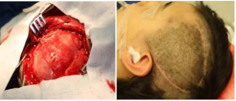
The patient was 13 year old male who suffered a fall from a height of approximately 1.6 m. As a result, he experienced trauma that generated a left acute epidural hematoma. The mortality rate for this type of injury is about 40%. As the hematoma grows, it moves the brain away from the brain stem, causing severe headache, a decrease in the contra lateral force, confusion or coma, dilated pupils, respiratory rhythm disturbances and ultimately, death. For this reason, it was necessary to perform a craniotomy in order to remove or drain the hematoma and aid the clogging of the arteries that were broken to prevent bleeding. The patient was referred to neurosurgery. The bone flap was removed to allow expansion of the brain in case cerebral edema had developed. The portion of skull removed was approximately 68.7 cm2.
© 2015, IRJET.NET- All Rights Reserved
Page 1900
(Ti6Al4V) was used to manufacture the implant; this material was chosen because of its high degree of biocompatibility and mechanical resistance.
3D Reconstruction:
For the 3D reconstruction of the injured area, a CT scan of the head and neck was used. The scan consisted of 751 images with a distance between cuts of 0.3 mm. This procedure was conducted in Rapid. As a first step, a 3Dreconstruction of the skull was performed, with the purpose of observing the injury. Subsequently, a 3D
reconstruction of the skin was performed to clearly observe the asymmetry caused by the injury, which compromised the appearance of the patient.
Geometric modeling of the implant:
Once a 3D reconstruction of the relevant anatomy was obtained, a symmetry plane that coincided with the sagittal plane of the patient was created. From the sagittal plane, the skull geometry was divided in two halves. The injury was located in the left half (Fig. 4).
Fig1.1Brain protected by the meninges and the scalp
© 2015, IRJET.NET- All Rights Reserved
Page 1901
Fig. 1.3: Asymmetry caused by the injury
Fig. 1.4 Skull geometry divided in two halves
© 2015, IRJET.NET- All Rights Reserved
Page 1902
Fig. 1.6 Geometric modeling of the implant
Fig. 1.7: Dimensional and functional validation of the implant
Thereafter, using the sagittal plane as the symmetry plane, a mirror operation was performed on the healthy half of the skull, which was used as the basis of the geometric modeling of the implant. On the skull surface, adjacent to the periphery of the injury, five areas of support were defined and these areas were used as assembly regions between the implant and the skull using 10 mini screws.
Dimensional and functional validation of the implant:
In a Rapid Prototyping (RP) machine, using Fused Deposition Modeling technology (FDM), model of the implant and a portion of the injury were manufactured. To reduce the cost and manufacturing time, only a small portion of the skull injury was fabricated (Fig. 1.7).
Manufacturing of the implant:
Using a 1.2 mm thick titanium alloy (Ti6Al4V) plate, the implant was fabricated. The manufacturing process was carried out by applying pressure on the titanium plate, which was exerted by two pieces of Duraluminium in which the outer 3D surface of the implant had been machined. Finally, the holes for inserting the mini screws were machined.
1.
Results
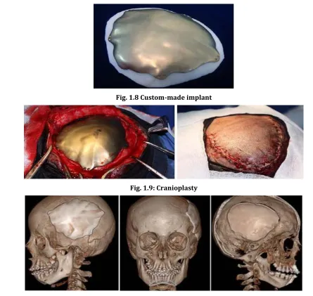
A custom-made, titanium alloy (Ti6Al4V) implant was obtained. Fig. 8 shows the final appearance of the implant and its dimensional and functional verification using the portion of the skull previously fabricated in RP. The weight of the implant was 66.33 g. The approximate volume of the portion of skull that was removed was 19.7 cm3. Assuming that the density of bone is ρ = 210 kg /m3 , an
© 2015, IRJET.NET- All Rights Reserved
Page 1903
weight of the implant was 1.6 times greater than the portion of bone removed.
1 Implantation process:
As a first step, the implant was sterilized with ethylene oxide. After sterilization, the implant was successfully placed on the skull. Modifications to the implant, such as folds or removal of material, were not needed. Once the implant was in place, the holes to insert the mini screws were made. Finally, the patient was sutured. The entire implantation process lasted about 45 min the surgery time was reduced by 85%
compared with surgeries in which commercial implants and titanium meshes were used.
2 Patient monitoring:
After four months, a control follow-up was performed. The patient was very satisfied with the cosmetic result and the security offered by the implant. The surgical scar was healthy and no pain, inflammation or rejection of the material was observed. A new CT scan demonstrated that the implant was in an excellent position and no further complications were. Excepte
Fig. 1.8 Custom-made implant
Fig. 1.9: Cranioplasty
© 2015, IRJET.NET- All Rights Reserved
Page 1904
2.
Conclusion
A custom-made cranial implant was designed, fabricated and placed into a patient whose brain was only protected by the meanings and the scalp after a 1.6 m fall. This situation represented a high risk for his health. After surgery, the appearance of the patient was restored, allowing the patient to safely perform daily activities. Typical cranioplasty procedures, in which standard implants and titanium meshes are used, last approximately 4-5 h due to the trial and error procedures that are implemented during the surgery in order to achieve a good fit of the implant. With our methodology, a decrease in the surgery time of approximately 85% was obtained. This reduction in time also reduces the risk for the patient and reduces surgery costs. The use of 3D reconstruction techniques from medical images reduces the possibility of errors during surgery, improves fit and provides better implant stability after fixation with mini screws. The dim functional verification of the implant using 3D models designed in RP proved to be an effective practice in the design process because during the surgery modifications of the implant or the skull region around the injury were not necessary. The models were also an effective communication tool for the neurosurgeon, the patient and the family of the patient when discussing the surgical procedure. The Titanium Alloy (Ti6Al4V) used to manufacture the implant proved to be a suitable material for this type of application due to its biocompatibility and good mechanical properties. This is demonstrated by the absence of postoperative infections and the proper healing and restoration of normal activities of the patient without any medical complications. The process used to manufacture the implant was less expensive and faster than other high-cost technologies, such as Electron Beam Melting (EBM) and traditional machining methods using Computer Numerical Control Machines (CNC), in which large amounts of titanium are wasted. If a casting process had been used, it would have been necessary to make a mold and have access to a furnace that reached temperatures up to 1600°C . An implant was constructed with a weight increase of 57% relative to the weight of the portion of the skull removed. In future research, the weight of the implant could be made closer to the weight of the portion of skull using thinner plates. According to Joffeet al., implants made from 0.72 mm thick plates were successful. Once the CT scan had been completed, the design and manufacturing process was carried out in 10 days. This situation makes this procedure highly competitive with other commercial alternatives due to the time and cost required to import an implant into Colombia and Latin America.
REFERENCES
1. J. Bruce, D.J. Effeney and M.W. Laniganet al., 1998. Stereolithographic (SL) biomodelling in craniofacial surgery. British J. Plastic Surg., 51: 522-530.
2. V.L. Nocchi and S.A. Zanini, 1979 Frozen skull supplemented with fresh iliac corticocancellous bone. Neurosurgery, 15: 846-851. PMID: 6514157Psillaki
3. M., L. Ferrante, F.S. Pastore, E.O. Ramundoand D. Cantare lliet al., 2003. Bone auto grafting of the calvaria and craniofacial skeleton
4. C. A. Mosse and M. Harris, 1992. Computer-generated titanium cranioplasty: Report of a new technique for repairing skull defects. Br. J. Neurosurg, 6: 343-350.Joffe,
5. T. Koyama and S. Kobayashi et al., 1990. Cranioplasty with a frozen and autoclaved bone flap.