Computed tomography identified factors that preclude living kidney donation
Katerina Mastrocostas1; Christina M. Chingkoe1; Kenneth T. Pace2; Joseph J. Barfett1; Anish Kirpalani1; Gevork N. Mnatzakanian1; Paraskevi A. Vlachou1; Errol Colak1
1Department of Medical Imaging; 2Division of Urology, Department of Surgery;
St. Michael’s Hospital, University of Toronto, Toronto, ON, Canada
Cite as: Can Urol Assoc J 2018 April 6; Epub ahead of print. http://dx.doi.org/10.5489/cuaj.4909
Published online April 6, 2018
***
Abstract
Introduction: The purpose of this study was to determine the variety and prevalence of renal and non-renal abnormalities detected on multi-detector computed tomography (MDCT) that precluded patients from donating a kidney.
Methods: Institutional review board approval was obtained and the requirement for informed consent was waived. A retrospective, single-centre review of 701 patients (444 female, 257 male; age range 18–86 years; mean age 43.2±11.9 years) that underwent renal donor protocol MDCT was conducted. A systematic review of the CT report, records from multidisciplinary renal transplantation rounds, and electronic medical records was performed to determine which patients were approved or
declined as live renal donors. If declined as a donor, CT-identified reasons were categorized as abnormalities of renal vasculature, renal parenchyma, collecting system, or extra-renal.
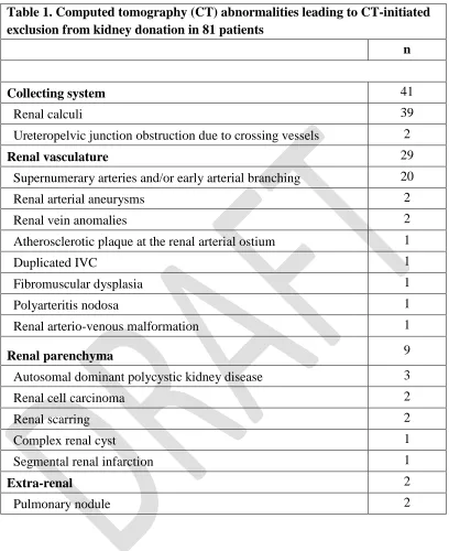
Results: A total of 81 patients were excluded as renal donors on the basis of CT findings. Abnormalities of the collecting system accounted for the most frequent cause of exclusion (n=41), with asymptomatic renal calculi being detected in 39 patients. Complex vascular anatomy and vascular abnormalities resulted in the exclusion of 29 patients. Supernumerary arteries and early arterial branching resulted in the exclusion of 20 patients, while renal vein anomalies leading to exclusion were uncommon (n=2). Abnormalities of renal parenchyma resulted in the exclusion of nine patients. Three patients were diagnosed with autosomal dominant polycystic kidney disease, two patients had renal cell carcinoma, and two patients had areas of cortical scarring. A complex cystic lesion requiring surveillance imaging was encountered in one patient and a large area of renal infarction related to prior
Conclusions: MDCT plays a critical role in the preoperative assessment of potential
renal donors by identifying contraindications to donor nephrectomy and providing accurate vascular mapping. This study is anticipated to be informative for those involved in the workup of potential living renal donors by quantifying the incidence and reasons for donor exclusion identified on CT.
Introduction
Chronic kidney disease (CKD) is a common condition across the developed world1 with a prevalence of 12.5% in the adult Canadian population.2 The prevalence of CKD continues to rise as a result of increasing rates of diabetes, hypertension,
obesity, and an aging population.3 Kidney transplantation has been recognized as the preferred long term treatment for end-stage kidney disease (ESKD). When compared to dialysis, kidney transplantation results in improved quality of life,4 prolonged patient survival,5 and long-term health system cost savings .6 With a growing ESKD population and limited supply of cadaveric kidneys, living donor kidney
transplantation plays an increasingly important role in the management of these patients by potentially increasing the pool of available kidneys. Apart from improved graft survival and recipient outcomes,7 the overall outcomes for donors are similar to the general population in terms of survival and the risk of developing CKD.8 The benefits of living kidney donation have led to it becoming the preferred surgical option in many centers.
Laparoscopic donor nephrectomy (LDN) is the preferred surgical procedure for kidney procurement due to less post-operative pain, faster recovery times, and improved quality of life when compared to open nephrectomy.9 Living donor transplantation is a high-stakes procedure that requires careful patient and kidney selection and meticulous pre-operative planning. Multi-detector computed tomography (MDCT) is well established as an accurate imaging modality for the anatomic assessment of renal vasculature, parenchyma, and collecting system.10 In addition to assessing the kidneys, MDCT is able to detect other contraindications to pursuing donor nephrectomy. The purpose of this study was to determine the variety and prevalence of renal and non-renal abnormalities detected on MDCT that
ultimately precluded patients from donating a kidney.
Methods
This retrospective single center study was approved by our Institutional Review Board with a waiver for informed consent. Our radiology information system (syngo, Siemens Medical Solutions USA, Inc., Malvern, PA) was searched using Montage Search and Analytics (Montage Healthcare Solutions, Philadelphia, PA) for renal donor protocol CT examinations performed between January 1, 2005 and December 31, 2014.
reasons. CT-identified reasons were categorized as abnormalities of renal vasculature,
renal parenchyma, collecting system, or extra-renal.
MDCT Technique and Image Analysis
All renal donor CT examinations were performed on a 64 slice MDCT scanner (LightSpeed 64 and Optima 64, General Electric Medical Systems, Milwaukee, WI). Our protocol includes an unenhanced phase, late arterial phase, and delayed scout topogram. The unenhanced and late arterial phases of imaging were acquired from the level of the dome of the liver to the pelvic brim. The following parameters were used for the unenhanced phase: 120 kVP, 100 – 500 mA, 0.5 second rotation time, pitch of 1.375:1, noise index of 26, and reconstructed images with a standard soft tissue algorithm at 2.5 mm. The late arterial phase was acquired using an intravenous injection of 80 cc Visipaque 320 (GE Healthcare, Waukesha, WI) at a rate of 4 cc/second with a power injector. Smart Prep bolus tracking (General Electric Medical Systems, Milwaukee, WI) was used with the region of interest positioned over the abdominal aorta at the level of the kidneys. The following parameters were used for the late arterial phase: 140 kVP, 200 – 750 mA, 0.5 second rotation time, pitch of 0.969:1, noise index of 21, and reconstructed images with a standard soft tissue algorithm at 1.25 mm. A delayed scout topogram was acquired supine at a 6 minute delay and following the patient sitting upright for 1 minute to encourage contrast passage into the ureters. Coronal and oblique maximum intensity projection (MIP) reconstructions were generated from source data.
Each examination was reported by a subspecialty trained board certified radiologist on a picture archiving and communication system (PACS) workstation (eFilm, Merge eFilm Inc., Chicago, IL or Carestream PACS, Carestream Health, Rochester, New York). Special attention was made to describing renal size, arterial and venous anatomy, nephrolithiasis, cortical scarring, focal renal lesions, collecting system configuration, and significant extra-renal findings. Every CT examination was reviewed a second time during our multidisciplinary renal transplantation rounds that included radiologists, urologists, transplant nephrologists, allied health disciplines, and transplant team coordinators. The workup of potential renal donors at our institution has been reported previously.11
Results
declined for donation. CT findings resulted in the exclusion of 81 patients and 67
patients were excluded due to medical reasons that were discovered after the CT was performed. Donor nephrectomy was ultimately performed in 388 of the 553 approved patients.
Abnormalities of the collecting system accounted for the most frequent cause of CT-initiated donor exclusion (n = 41). Asymptomatic renal calculi were detected in 39 of these patients. Medullary sponge kidney was present in 5 patients, 5 patients had bilateral nephrolithiasis, and 20 patients had unilateral nephrolithiasis. Of the 20 patients with unilateral nephrolithiasis, 18 had a solitary calculus (mean size 0.3 ± 0.3 cm, median 0.2 cm, range 0.1 - 1.3 cm), 6 patients had two calculi, and 3 patients had three or more calculi. Unilateral ureteropelvic junction obstruction due to crossing vessels was demonstrated in 2 patients.
Complex vascular anatomy and vascular abnormalities were the second most frequent cause of CT-initiated donor exclusion (n = 29). Two hundred fifty-five patients had at least one kidney with two or more arteries. Supernumerary arteries and/or early arterial branching resulted in the exclusion of 20 patients. Ten patients were deemed to require difficult vascular reconstruction during our multi-disciplinary rounds due to the number of arteries, close origin of the arteries, and early branching. Five patients had left-sided supernumerary arteries where the accessory artery was distant from the main artery. A further 4 patients had bilateral supernumerary arteries where the accessory arteries were distant from the main arteries on both sides.
Another patient was excluded due to 4 right and 3 left renal arteries. Renal vein anomalies leading to exclusion were uncommon (n = 2). Less frequent causes of exclusion included arterial aneurysms (n = 2), fibromuscular dysplasia (n = 1),
atherosclerotic plaque at renal arterial ostium (n = 1), multiple renal artery aneurysms suggesting polyarteritis nodosa (n = 1), arterio-venous malformation (n = 1), and duplicated inferior vena cava (n = 1).
Abnormalities of renal parenchyma were the third most frequent cause of CT-initiated donor exclusion (n = 9). Three patients were diagnosed with autosomal dominant polycystic kidney disease (ADPCKD), 2 patients had solid lesions that were excised and confirmed to represent clear cell renal cell carcinoma, and 2 patients had areas of significant cortical scarring. A complex cystic lesion requiring surveillance imaging was encountered in 1 patient and a large area of renal infarction related to prior adrenalectomy was demonstrated in 1 patient.
Extra-renal abnormalities leading to exclusion were limited to 2 patients with incidental pulmonary nodules that required surveillance imaging.
Discussion
Imaging plays a critical role in helping select appropriate patients for donor
an accurate depiction of vascular anatomy and is able to depict contraindications to
donor nephrectomy. Imaging findings resulted in the exclusion of 11.6% of our donors which is comparable to 4.1 – 16% in prior studies.12 – 15
Incidental nephrolithiasis was the most common cause of renal donor exclusion in our study. We found the prevalence of nephrolithiasis in our study population was similar to that encountered at other institutions.12 - 13 Nephrolithiasis has traditionally been considered a contraindication to renal donation due to the risk of stone recurrence in the donor and complications of “gifted calculi” in recipients. However, the shortage of available kidneys and plateauing rates of kidney donation16 has led some centers to reconsider nephrolithiasis as an absolute contradiction.17 If incidental nephrolithiasis without an associated significant metabolic abnormality becomes widely accepted under extended criteria, it is logical to expect
nephrolithiasis to become a less frequent reason of potential donor exclusion in the future.
Variations of renal vascular anatomy were the second most common cause of donor exclusion. Despite accessory renal arteries13 and prehilar branching18 being relatively common, they represented the cause of exclusion in only 2.9% of patients. Multi-disciplinary rounds deemed these patients as requiring more extensive surgical reconstruction, higher operative risks, or necessitating sacrifice of accessory vessels that could compromise graft function. Less frequent causes of donor exclusion on the basis of vascular abnormalities included arterial aneurysms, atherosclerosis, and an arterio-venous malformation. All patients with fibromuscular dysplasia were excluded as donors. Patients with bilateral ostial calcification that resulted in luminal narrowing were excluded as donors. However, patients with ostial calcification only on the side of the proposed donor nephrectomy were considered as potential donors. Patients were excluded if there was ostial calcification affecting what would become a solitary kidney following donor nephrectomy.
Solid renal masses were noted in 0.03% patients which is lower than the 0.6% prevalence of incidental renal masses seen in trauma patients.19 Three patients were newly diagnosed with autosomal dominant polycystic kidney disease following CT. Congenital abnormalities such as a horseshoe kidney, pelvic kidney, and cross-fused renal ectopia were not encountered in our study population.
to donor nephrectomy have evolved over this 10-year period. Third, there may be
References
1. Coresh J, Selvin E, Stevens LA, et al. Prevalence of chronic kidney disease in the United States. JAMA 2007;298:2038-47.
2. Arora P, Vasa P, Brenner D, et al. Prevalence estimates of chronic kidney disease in Canada: results of a nationally representative survey. Can. Med. Assoc. J
2013;185:E417-23.
3. Fox CS, Larson MG, Leip EP, et al. Predictors of new-onset kidney disease in a community-based population. JAMA. 2004;291:844-50.
4. Russell JD, Beecroft ML, Ludwin D, et al. The quality of life in renal transplantation--a prospective study. Transplantation 1992;54:656-60.
5. Schnuelle P, Lorenz D, Trede M, et al. Impact of renal cadaveric transplantation on survival in end-stage renal failure: evidence for reduced mortality risk
compared with hemodialysis during long-term follow-up. J. Am. Soc. Nephro.
1998;9:2135-41.
6. Laupacis A, Keown P, Pus N, et al. A study of the quality of life and cost-utility of renal transplantation. Kidney Int 1996;50:235-42.
7. Cecka JM. The UNOS Scientific Renal Transplant Registry--ten years of kidney transplants. Clin Transpl 1996;4:1-14.
8. Ibrahim HN, Foley R, Tan L, et al. Long-term consequences of kidney donation.
N. Engl. J. Med 2009;360:459-69.
9. Nanidis TG, Antcliffe D, Kokkinos C, et al. Laparoscopic versus open live donor nephrectomy in renal transplantation: a meta-analysis. Ann Surg 2008;247:58-70. 10.Pazniak MA, Balison DJ, Lee FT, et al. CT Angiography of Potential Renal
Transplant Donors, RadioGraphics 1998;18:565-587.
11.Perlis N, Connelly M, John RD, et al. Evaluating potential live-renal donors: Causes for rejection, deferral and planned procedure type, a single-centre experience. Can Urol Assoc J 2013;7:41-5.
12.Strang AM, Lockhart ME, Kenney PJ, et al. Computerized tomographic angiography for renal donor evaluation leads to a higher exclusion rate. J Urol
2007;177:1826-9.
13.Lorenz EC, Vrtiska TJ, Lieske JC, et al. Prevalence of renal artery and kidney abnormalities by computed tomography among healthy adults. Clin J Am Soc
Nephrol 2010;5:431-8.
14.Sebastià C, Peri L, Salvador R, et al. Multidetector CT of Living Renal Donors: Lessons Learned from Surgeons 1. Radiographics 2010;30:1875-90.
15.Chu LC, Sheth S, Segev DL, et al. Role of MDCT angiography in selection and presurgical planning of potential renal donors. AJR Am J Roentgenol
2012;199:1035-41.
recommendations from a consensus conference. Clin J Am Soc Nephrol
2015;10:1687-95.
17.Kim IK, Tan JC, Lapasia J, et al. Incidental kidney stones: a single center experience with kidney donor selection. Clin Transpl 2012;26:558-63.
18.Harrison Jr LH, Flye MW, Seigler HF. Incidence of anatomical variants in renal vasculature in the presence of normal renal function. Ann Surg 1978;188:83. 19.Ekeh AP, Walusimbi M, Brigham E, et al. The prevalence of incidental findings
on abdominal computed tomography scans of trauma patients. J Emerg Med. 2010;38:484-9.
Figures and Tables
Table 1. Computed tomography (CT) abnormalities leading to CT-initiated exclusion from kidney donation in 81 patients
n
Collecting system 41
Renal calculi 39
Ureteropelvic junction obstruction due to crossing vessels 2
Renal vasculature 29
Supernumerary arteries and/or early arterial branching 20
Renal arterial aneurysms 2
Renal vein anomalies 2
Atherosclerotic plaque at the renal arterial ostium 1
Duplicated IVC 1
Fibromuscular dysplasia 1
Polyarteritis nodosa 1
Renal arterio-venous malformation 1
Renal parenchyma 9
Autosomal dominant polycystic kidney disease 3
Renal cell carcinoma 2
Renal scarring 2
Complex renal cyst 1
Segmental renal infarction 1
Extra-renal 2