STRUCTURAL CHARACTERISATION OF 5-HYDROXYTRYPTAMINE
2ARECEPTOR IN HOMO
SAPIENS BY
IN - SILICO
METHOD
SONI SINGH, ALOK JHA*
Department of Biotechnology and Life Sciences, Mangalayatan University, Aligarh, Uttar Pradesh, India. Email: [email protected]
Received: 19 March 2018, Revised and Accepted: 10 July 2018
ABSTRACT
Objective: Structural characterization of 5-hydroxytryptamine (5-HT)2A receptor in homo sapiens using in silico method.
Methods:In silico approach has particularly providing a realistic representation needed to understand the fundamental molecular structure of a serotonin receptor. The structure has been generated using Swiss model, Modeller 9.14, Phyre2, and Geno three-dimensional, which was visualized using PyMol, and validated by Procheck and ERRAT analysis along with the values of different secondary structures mapping to diverse sections of the Ramachandran plot.
Results: We compared all different models. Further structural analysis suggested that the structure of 5-HT2A is a monomer with 18 alpha helices, seven beta sheets, and one disulfide bridge. There is no signal peptide region in the protein sequence. The structure contains mostly polar and aromatic amino acid as suggested by using hydropathy plot. However, in both partitioning systems bilayer to water and water to bilayer, there are some hydropathy predicted segments, which are also transmembrane segments. Finally, the pore features, including diameter profile, size, and shape, were determined by porewalker, and the shape of the pore was found to be UDSD.
Conclusion: This study suggested that 5-HT2A receptor interaction with its natural ligand serotonin and other inhibitor compounds would further additional information about G protein-coupled receptors. The 5-HT2A receptor could be an important target for therapeutics development.
Keywords: 5-Hydroxytryptamine2a receptor, Homology modeling, G Protein-coupled receptors, Transmembrane protein, Model comparison.
INTRODUCTION
Serotonin 5-hydroxytryptamine (5-HT) is one of the neurotransmitters present at synapses of nerve cells. In the central nervous system (CNS), serotonin is mainly involved in the regulation of depression, anxiety, aggression, memory, appetite, cognition, sleep, emotion, perception, and consciousness [1]. The 5-HT receptors have been classified into 5-HT1–5HT7, and again they are divided into 12 different subpopulations. Some agonists and antagonists for subpopulations of 5-HT receptors are being developed by using different approaches. Still as far yet, to design and develop a specific inhibitor for 5-HT receptors with therapeutic potential is a challenge, although many agonists and antagonists have been suggested [2]. All serotonergic receptors come under the category of G-protein-coupled receptor (GPCR) superfamily [3] except 5-HT3 receptor. The 5-HT3 receptor is one of the ligand-gated ion channels and depends on the nicotinic acetylcholine receptor superfamily having cysteine-loop transmitter gate, and constitute of heteropentamers [4,5]. The 5-HT2A receptor is mainly present in the prefrontal cortex of CNS, and they are present in that region of the brain, which is essential for learning and cognition. The activity of this receptor is linked to many neurological disorders and conditions such as schizophrenia and depression. [5]. The 5HT2 receptors are subdivided into 5-HT2A, 5-HT2B, and 5-HT2C receptors. 5-HT2A receptors exhibit high sequence homology with other 5-HT receptors (78%). Phenylalkylamines (like (2,5-dimethyl-4-bromoamphetamine) and (2,5-dimethoxy-4iodoamphetamine)) are well known agonists for 5-HT2A [6]. There are so many drugs available to target this receptor. Although the receptor has been widely studied about multiple functions in the CNS, high level of this receptor has been studied in other parts of the body such as platelets, endothelial cells, and intestine (Fig. 1).
METHODS
Template selection and model building
The protein sequence and information of 5-HT2A were collected from the National Center of Biotechnology (NCBI), in FASTA format, with the accession number NP > AAH96839.1 5. The sequence length reported to be 471 amino acids. To search for homologous sequence of 5-HT2A, BLASTP was performed against the nonredundant database of NCBI. Sequences are selected from the sequence similarity search based on the identity percentage (cutoff 95% identity). The 471 amino acid residue of 5-HT2A receptor was subjected to BLASTP against the Protein Data Bank [7] to identify suitable template for comparative study of the protein structure for modeling.Further, the protein sequence was subjected to comparative homology modeling through Modeler 9.14, SWISS-MODEL Server [8], PHYRE2 server [9], and Geno three-dimensional (3D) server [10]. These modeling servers on the basis of their different algorithm provide 3D structure of 5-HT2A structure for further study.
Validation of generated model
Finally, once the 3D structures were generated, evaluation of the structure and stereochemical analysis was performed using different types of validation tools. A study of backbone conformation of all models was evaluated by the analysis of Ramachandran plot using RAMPAGE program [11]. ERRAT tool finds the overall quality factor of the proteins. ERRAT plot gives statistics of the error in the structure for each residue in the 3D protein structure model. This process was repeated, until when most of the amino acid residues was shown below 95% cutoff [12].
© 2018 The Authors. Published by Innovare Academic Sciences Pvt Ltd. This is an open access article under the CC BY license (http://creativecommons. org/licenses/by/4. 0/) DOI: http://dx.doi.org/10.22159/ajpcr.2018.v11s2.28588
Structural characterization
The secondary structure analysis, sequence motifs, matching folds, nest analysis, and cleft analysis were done by using ProFunc [13]. Hydropathy analysis was done by MPEx protein explorer [14]. Protein identification and characterization of channel in transmembrane were identified by porewalker [15]. SPPIDER predict residues present at the protein surface by analyzing the 3D structure. It also analyses protein-protein complex with 3D structural information and identify those residues that are mainly involved in interchain connection [16].
Refinement of the structure
The 3D refine refinement protocol is mainly performed to make the efficient structural model refining by optimizing the hydrogen-bonding network [17]. The refined model is useful in determining the spatial properties such as backbone position of amino acid residues and the side chain conformation of protein model [18].
RESULTS AND DISCUSSION
Retrieval of protein sequence and modeling analysis
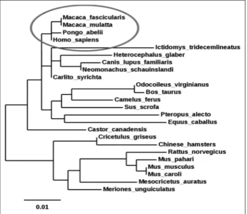
The amino acid sequence of the target protein, 5-HT2A (Homo-sapiens) was downloaded from NCBI. Blast search was performed for comparative sequence analysis. The 5-HT2A receptor of homo sapiens is closely related to that of Macaca fascicularis, Macaca mulatta, and Pongo abelii species with a maximum evolutionary relation is shown in the Fig. 2.
Sequences producing a significant alignment with the protein 5tvn1A were selected by query coverage (90%), percentage (cutoff >95%) of the identity, and E-value (cutoff 0). The 5-HT2A from different species is highly conserved throughout the evolutionary. The phylogenetic tree was represented with an overall mean distance 0.01.
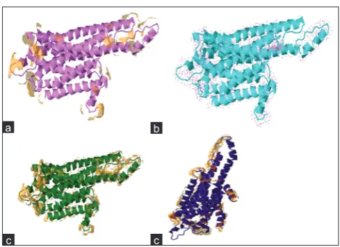
For the building of 3D structures, comparative modeling is one of the best and accurate methods, which provide wide spectrum of applications. The protein sequence was subjected to homology modeling by a Modeler 9.14, SWISS-MODEL Server, shown in Fig. 3. In this study, the best homolog found in Swiss model server with 55.25% of sequence similarity.
After model comparison, we conclude that Swiss model gives better results as compared to among four servers.
Analysis of generated models
On the basis of the results from four different programs SWISS-MODEL, Modeller 9.14, Phyre 2, and Geno3D, it was notified that the model obtained from Swiss model was satisfactory as compared to others. The final protein model was seen in J-MOL and is shown in Fig. 3. Comparison of all the four models generated by RAMPAGE is shown in Table 1. To find the accuracy of predicted models using Ramachandran plot calculations and to check the stereochemical quality PROCHECK program was used. The results obtained from Ramachandran plot
Fig. 1: Mechanism of the serotonin signaling pathway. (a) Serotonin is present in the presynaptic nerve cell, and 5-hydroxytryptamine (5-HT)2A receptor is present on the postsynaptic nerve cell. (b) Serotonin is released from the presynaptic nerve cell, and the
concentration of serotonin is increased in synaptic cleft. (c) Now, the serotonin binds the 5-HT2A receptor on postsynaptic nerve cell. This
leads to the conclusion that high density of 5-HT2A receptor plays an important function in neurological disorder
Fig. 2: Phylogenetic tree analysis of 5-hydroxytryptamine (5-HT)2A
receptor protein sequence on the basis of migration skills assessment of 24 5-HT2A receptor protein sequence from different
species. The tree depicts that the 5-HT2A receptor of homo sapiens
is closely related to that of Macaca fascicularis, Macaca mulatta,
and Pongo abelii species as compared to other 20 species
a b c
Fig. 3: Homology modeling: (a) Modeler 9.14, (b) Swiss model, (c) Phyre2, (d) Geno 3D
a b c d
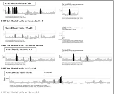
Th principle of ERRAT server is mainly for the analysis of the statistics of nonbonded interactions between different types of atom. It is a protein structure verification algorithm, which is especially designed
for evaluating the crystallographic structure of the model building and the refinement. In Fig. 5 shows the overall quality factor of four models; however, the ERRAT analysis of Swiss model is significant because the overall quality factor is 94.218, which is not beyond their cutoff and a result expected for crystallographic models with resolution >2.5Å.
In this (Table 2) comparative analysis of 5-HT2A receptor (Homo-sapiens) models builds in-silico method shows that Swiss model gives significant results among all the parameters such as QMEAN Score, Errat analysis, and Ramachandran analysis.
Structural analysis
Signal peptide and hydropathy plot
On analysis, we found no signal peptide region in the protein sequence. A hydropathy scale depends on the hydrophobic, and hydrophilic properties of the 20 amino acids are used. The significant role of this plot is to determine spanning segments of membrane-bound proteins and the hydrophobic interior portions of globular proteins. The structure contains mostly polar and aromatic amino acid as suggested by hydropathy plot in Fig. 6.
The validated structure model is further analyzed using ProFunc. The result found eight motifs that were scanned and matched comparatively with PROSUTE, PRINTS, PFam-A, TIGRFAM, PROFILES, and PRODOM motifs. Functionally, important residues of 5-HT2A were predicted by ProFunc through nest analysis. Nest analysis located four nests in the structure. A score above 2.0 is suggestive of the nest being functionally significant, there is only one significant hit in the structure. In the (Fig. 7) secondary structure of 5-HT2A consists of 18 helices, seven beta turns, seven gamma turns, and one disulfide.
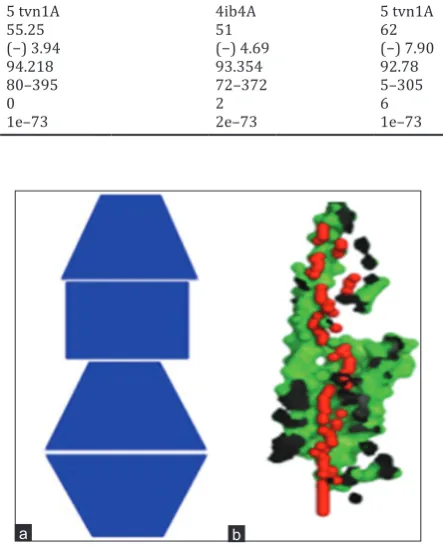
Pore shape and features of the cavity
5-HT2A receptor structure has an identified pore shape UDSD (Fig. 8), D = conical frustum with decreasing diameter, U = conical frustum with increasing diameter, S = cylinder.
Fig. 4: The Ramachandran plot analysis had shown that the 5-hydroxytryptamine2A model build by modeler having only one
residue in the outlier region and the model build by Swiss model with no residue in the outlier region could be accepted for further
analysis and experimentations
Protein interface prediction
This predicts that residues present at the protein surface by analyzing the 3D structure. It is also used to identify residues that are being involved in interchain contact. It also shows the different types of surfaces which are involved in the interactions such as solvent accessibility surface, Dot surface, Vander wall surfaces, and Molecular surfaces (Fig. 9).
Analysis of the refined structure
The MolProbity score relates to the expected resolution of the protein model as compared to the standard experimental structures, and therefore, a lower score of MolProbity means more physically realistic model of the protein. Correct positioning of the Cα atoms is mainly
represented by root-mean-square deviation (RMSD) value. Hence, correlation of RMSD and global distance test (GDT)-total score is week, a lower value of RMSD suggested that the predicted structure is close to its native state of the protein model. The GDT high accuracy value for human (5-HT2A) was 0.9931, Mol. Probability was 1.405, and RMSD was found to be 0.281. This predicts that the refinement of the model was good.
CONCLUSION
The structural model may act as the best template for 5-HT2A characterization. The structured model is in agreement with other GPCRs structures. Model comparison suggested that Swiss model is better among four. The 5-HT2A receptor could be an important target for therapeutics development. Based on this model, the antagonists designing might be a potential therapeutic target for the development of novel therapeutics. The site-specific drug will be more efficient for inhibiting the 5-HT2A receptor for the treatment of neurological disorder. Since the drug will be site specific; it would have less side effects. The new drug molecules could be an addition to the pharmacoinformatics data bank as a newly proposed drug.
Table 1: Comparison of all the four models generated from RAMPAGE
Parameters Modeler 9.14 Swiss-model Phyre2 Geno3D
Number of residues in the favoured region 381 311 317 271
Number of residues in favoured region 2 12 7 47
Number of residues in the favoured region 1 0 2 6
3D: Three-dimensional
Table 2: Comparative analysis of 5-HT2A receptor (Homo-sapiens) models builds in-silico by Modellet9.14, Swiss model, Phyre2, and
Geno3D
Comparative criteria Modeller9.14 Swiss-model Phyre2 Geno3D
Template 4ib4A 5 tvn1A 4ib4A 5 tvn1A
Identity (%) 58 55.25 51 62
QMEAN score (37) (−) 5.00 (−) 3.94 (−) 4.69 (−) 7.90
Errat analysis (%) 81.53 94.218 93.354 92.78
Structure covered 1–400 80–395 72–372 5–305
Ramachandran plot outliers 1 0 2 6
BLAST E value of PDB structures 2e–73 1e–73 2e–73 1e–73
3D: Three-Dimensional, 5-HT: 5-Hydroxytryptamine, PDB: Protein Data Ban
Fig. 6: A hydropathy plot constructed for the 5-hydroxytryptamine 2A (5-hydroxytryptamine2A receptor)
Fig. 7: The secondary structure represents as follows: H1-H18, β, γ and represent helices, beta-turn, gamma turn, and beta-hairpin,
respectively
Fig. 8: (a) Pore shape (Identified shape: UDSD) and (b) Top right: XZ-plane section, Y>0 coordinates only
REFERENCES
1. Remigio GJ. The Neurophysiology and Behavioral Pharmacology of Memory Enhancement and Memory Deficits in the Dentate Gyrus (Doctoral Dissertation, The University of Utah); 2017.
2. Leopoldo M, Lacivita E, Berardi F, Perrone R, Hedlund PB. Serotonin 5-HT7 receptor agents: Structure-activity relationships and potential therapeutic applications in central nervous system disorders. Pharmacol Ther 2011;129:120-48.
3. Katritch V, Cherezov V, Stevens RC. Structure-function of the G protein-coupled receptor superfamily. Annu Rev Pharmacol Toxicol 2013;53:531-56.
4. Peters JA, Kelley SP, Dunlop JI, Kirkness EF, Hales TG, Lambert JJ. The 5-hydroxytryptamine Type 3 (5-HT3) receptor reveals a novel determinant of single-channel conductance. Biochem Soc Trans 2004;32:547-52.
5. Raote I, Bhattacharya A, Panicker MM. Serotonin 2A (5-HT2A) receptor function: Ligand-dependent mechanisms and pathways.
Fig. 9: (a) Solvent accessible surface, (b) Dot surfaces, (c) Vander wall surfaces, (d) Molecular
a b
c c
Serotonin Receptors in Neurobiology. Boca Raton: CRC Press; 2007. p. 1-17.
6. Zhang G, Stackman RW Jr. The role of serotonin 5-HT2A receptors in memory and cognition. Front Pharmacol 2015;6:225.
7. Dhonnchadha BÁ, Ripoll N, Clénet F, Hascoët M, Bourin M. Implication of 5-HT 2 receptor subtypes in the mechanism of action of antidepressants in the four plates test. Psychopharmacology 2005;179:418-29.
8. Biasini M, Bienert S, Waterhouse A, Arnold K, Studer G, Schmidt T, et al. SWISS-MODEL: Modelling protein tertiary and quaternary structure using evolutionary information. Nucleic Acids Res 2014;42:W252-8.
9. Kelley LA, Mezulis S, Yates CM, Wass MN, Sternberg MJ. The Phyre2 web portal for protein modeling, prediction and analysis. Nature Protocols 2015;10:845-58.
10. Combet C, Jambon M, Deleage G, Geourjon C. Geno3D: Automatic comparative molecular modelling of protein. Bioinformatics 2002;18:213-4.
11. Lovell SC, Davis IW, Arendall WB, de Bakker PI, Word JM, Prisant MG, et al. Structure validation by Cα geometry: ϕ, ψ and Cβ deviation.
Proteins Struct Funct Bioinforma 2003;50:437-50.
12. Laskowski RA, MacArthur MW, Moss D, Thornton JM. PROCHECK: A program to check the stereo chemical quality of protein structures. J Appl Cryst 1993;26:283-91.
13. Laskowski RA, Chistyakov VV, Thornton JM. PDBsum more: New summaries and analyses of the known 3D structures of proteins and nucleic acids. Nucleic Acids 2005;33:D266-8.
14. Chen Y, Capponi S, Zhu L, Gellenbeck P, Freites JA, White SH, et al. YidC insertase of Escherichia coli: Water accessibility and membrane shaping. Structure 2017;25:1403-14.
15. Pellegrini-Calace M, Maiwald T, Thornton JM. Pore-walker: A novel tool for the identification and characterization of transmembrane protein channels from their three-dimensional structure. PLoS Comput Biol 2009;5:1-16.
16. Yang J, Roy A, Zhang Y. Protein-ligand binding site recognition using complementary binding-specific substructure comparison and sequence profile alignment. Bioinformatics 2013;29:2588-95.
17. Porollo A, Meller J. Prediction-based fingerprints of protein-protein interactions. Proteins 2007;66:630-45.