DOI: 10.1534/genetics.107.071688
Mutations in Cytochrome c Oxidase Subunit VIa Cause Neurodegeneration
and Motor Dysfunction in Drosophila
Wensheng Liu,*
,1Radhakrishnan Gnanasambandam,
†Jeffery Benjamin,
†Gunisha Kaur,*
,2Patricia B. Getman,* Alan J. Siegel,
†Randall D. Shortridge
†and Satpal Singh*
,3*Department of Pharmacology and Toxicology, State University of New York, Buffalo, New York 14214 and†Department of Biological Sciences, State University of New York, Buffalo, New York 14260
Manuscript received February 6, 2007 Accepted for publication March 29, 2007
ABSTRACT
Mitochondrial dysfunction is involved in many neurodegenerative disorders in humans. Here we report mutations in a gene (designatedlevy) that codes for subunit VIa of cytochrome c oxidase (COX). The mutations were identified by the phenotype of temperature-induced paralysis and showed the additional phenotypes of decreased COX activity, age-dependent bang-induced paralysis, progressive neurode-generation, and reduced life span. Germ-line transformation using the levy1
gene rescued the mutant flies from all phenotypes including neurodegeneration. The data fromlevymutants reveal a COX-mediated pathway in Drosophila, disruption of which leads to mitochondrial encephalomyopathic effects including neurodegeneration, motor dysfunction, and premature death. The data present the first case of a mutation in a nuclear-encoded structural subunit of COX that causes mitochondrial encephalomyopathy rather than lethality, whereas several previous attempts to identify such mutations have not been successful. Thelevymutants provide a genetic model to understand the mechanisms underlying COX-mediated mitochondrial encephalomyopathies and to explore possible therapeutic interventions.
M
ITOCHONDRIA are involved in several neuro-degenerative diseases such as Parkinson’s dis-ease (Abou-Sleiman et al. 2006; Keeney et al. 2006),Alzheimer’s disease (Atamnaand Boyle2006; Esposito
et al. 2006), and Huntington’s disease (Chooet al. 2004;
Benchoua et al. 2006). In addition, several disorders
arise directly from mutations that disrupt mitochon-drial function. These include Leber hereditary optic neuropathy (LHON), mitochondrial encephalopathy with lactic acidosis and stroke-like episodes (MELAS), myoclonic epilepsy with ragged red fibers (MERRF), chronic progressive external ophthalmoplegia and Kearns Sayre syndrome (CPEO/KSS), myoneurogas-trointestinal encephalopathy syndrome (MNGIE), mi-tochondrial DNA depletion syndrome (MDS), Barth syndrome, and Leigh syndrome (LS) (McFarlandet al.
2002; DiMauroand Schon2003).
Complex I (NADH dehydrogenase) genes are re-sponsible for the majority of LHON cases. Reduced activities of complexes I and IV (cytochrome c oxidase) are commonly associated with MELAS, and mutations
in the thymidine phosphorylase gene on chromosome 22 and multiple mtDNA mutations are commonly as-sociated with MERRF (DiMauro 2004; Gropman
2004). Deficiencies in a variety of factors such as pyru-vate dehydrogenase complex (PDHC), cytochrome c oxidase (COX), and ATPase6 give rise to LS, a geneti-cally heterogeneous disorder (Dahl1998).
Mitochon-drial copy number is a central characteristic of MDS
(Gropman2004) and is linked to MNGIE (Giordano
et al. 2006).
Mutations that disrupt molecular components of biochemical pathways allow identification of specific steps in such pathways and let us study the consequences of disrupting individual steps. Genetic tools available in Drosophila make it a useful model system for such studies. Drosophila has contributed significantly to our understanding of pathways involved in neuromuscular excitability and function under normal physiological conditions (Guand Singh1997; Bhattacharyaet al.
1999; Ganetzky2000; Bhattacharyaet al. 2004; Ueda
and Wu2006). In addition, this model system is playing
a significant role in analysis of pathways involved in neuromuscular dysfunction, especially in relation to neurodegenerative disorders (Fortiniand Bonini2000;
Palladino et al. 2002; Bonini and Fortini 2003;
Celottoet al. 2006; Gnereret al. 2006; Pallanckand
Greenamyre2006). Here we report mutations that
dis-rupted COX subunit VIa and resulted in several fea-tures of mitochondrial encephalomyopathies. Analysis
1Present address: Department of Pharmacology and Therapeutics,
Roswell Park Cancer Institute, Elm & Carlton Sts., GCDC 545, Buffalo, NY 14263.
2Present address:Weill Medical College of Cornell University, 445 E. 69th
St., New York, NY 10021.
3Corresponding author:Department of Pharmacology and Toxicology,
102 Farber Hall, State University of New York, Buffalo, NY 14214-3000. E-mail: singhs@buffalo.edu
of these mutations, of the phenotypes produced by them, and of the rescue from these phenotypes shows that COX plays a vital role in a pathway essential in maintaining neural integrity and that disruption of this pathway leads to encephalomyopathic effects including conditional paralysis, neurodegeneration, and early death.
MATERIALS AND METHODS
Fly strains and mutant isolation:Canton-S (CS) was used as the wild-type strain in all experiments. Flies were raised on a standard cornmeal medium at 21°and aged at 21°or 29°as described inresults. Flies carrying compound chromosomes
(Chovnicket al. 1970; Singh1983; Singhet al. 1987) were fed
ethylmethanesulfonate according to the mutagenesis pro-cedure of Lewis and Bacher (Lewis and Bacher 1968;
Ashburner 1989), and the progeny were screened for
pa-ralysis on exposure to 38° for 5 min. This resulted in the identification of the levy1 mutation. Additional levy alleles (levy2,levy3, andlevy4) were identified usingP-element mobi-lization. Briefly, thePelement in Dcp-1k05606was induced to mobilize by providing transposase (D2-3). F1flies with bothP element and transposase were crossed withw*;levy1. The F
2 progeny were screened for temperature-induced paralysis. After screening10,000 F2flies, 3 individual paralytic flies were isolated and the newly generatedP-element mutations were balanced withCyO. Insertion sites forPelements were determined using inverse PCR according to the methods described by the Berkeley Drosophila Genome Project (BDGP) (Bellenet al. 2004).
Behavioral assays:For paralysis test, a clear vial containing flies was submerged in water maintained at 38°. Time was recorded for flies to paralyze at 38°and to recover from pa-ralysis after transfer to room temperature. Wild-type flies did not paralyze for at least 15 min under these conditions. For bang sensitivity, flies were placed in a clear vial and vortexed on a Vortex Genie-2 bench mixer (VWR) at its highest speed for 10 sec, followed by quantification of the time needed for paralyzed flies to stand up. To determine life span, flies were kept at either 29°or 21°. Flies were transferred to fresh food vials three times a week. Dead flies were counted every day and removed from the vials at the time of the next transfer.
Germ-line transformation:A 2-kbCG17280genomic DNA fragment was cloned into thepCaSpeR4vector and injected into early embryos produced by white-eyedlevy1mutant flies (McKay et al. 1995). Transformants were identified on the
basis of red eye color. In the control, the rescue construct was replaced with vectorpCaSpeR4without an added insert.
Measurement of cytochrome c oxidase and citrate synthase activity:Microsomal fractions were prepared by grinding 30 adults in 2 ml of an extraction buffer containing 10 mmHEPES
at pH 7.5, 200 mmmannitol, 70 mmsucrose, 1 mmEGTA,
0.1 mmphenylmethyl-sulfonyl fluoride, 0.25 mmdibucaine, and
1 mmbenzamidine, using five strokes on a motorized
Potter-Elvehjem tissue grinder operating at 200 rpm. Homogenates were microfuged at 6003gfor 5 min at 4°and the resulting supernatants microfuged at 14,000 rpm for 20 min to collect mi-crosomal pellets. Mimi-crosomal pellets were suspended in 200ml of 10 mmHEPES at pH 7.5, 250 mmsucrose, 1 mmATP, 80mm
ADP, 5 mmsodium succinate, 2 mmpotassium phosphate, and
1 mmdithiothreitol and stored at70° until used. Protein
concentrations in microsomal preparations were determined using a bicinchoninic acid (BCA) protein assay kit from Pierce Biochemical. COX activity in microsomal preparations was
determined using a COX assay kit (CYTOC-OX1) from Sigma-Aldrich. Citrate synthase activity in mitochondrial preparations was assayed according to instructions provided by Sigma-Aldrich.
Measurement of ATP levels: Extracts for ATP measure-ments were prepared by vigorously grinding a male and a female adult in 200ml of 6mguanidine hydrochloride using a
small pestle that fits a microfuge tube, followed by collection of the liquid fraction after microfuging homogenates at 14,000 rpm for 20 min. Protein concentrations in extracts were determined using a BCA protein assay kit from Pierce Bio-chemical. ATP concentration was determined by measuring relative luminescence (RLU) of 1:50 dilutions of extracts in a luminometer using an ATP bioluminescence assay kit from Roche Diagnostics.
Characterization of neurodegeneration in levy and trans-formants: Plastic sectioning: Flies were decapitated and the heads fixed overnight in 1% paraformaldehyde and 1% glutaraldehyde. Fixed head samples were embedded in Epon 812 and then cut into 1-mm thick horizontal sections, stained with toluidine blue, and visualized using bright-field microscopy.
Paraffin sectioning: Standard paraffin embedding and sec-tioning techniques were followed on the basis of the protocol established by Crittenden et al. (1998). Briefly, flies were
fixed in Carnoy’s solution (60% vol/vol ethanol, 30% vol/vol chloroform, 10% vol/vol glacial acetic acid) for 4 hr at room temperature. After four 100% ethanol washes of 30 min each, the tissue was immersed in methyl benzoate overnight, trans-ferred to a 1:1 solution of methyl benzoate/paraffin for 1 hr at 56°, and then transferred to a 1:2 solution of methyl benzoate/ paraffin for another hour at 56°. The flies were then immersed in pure paraffin, four times for 1 hr each at 56°, and finally embedded in paraffin at room temperature. The paraffin blocks were cut into 4-mm floating serial sections that were mounted on gelatinized glass slides. Prior to histological exam-inations, the tissue was deparaffinized with a 3-min wash in xylene and rehydrated through an ethanol series ending in either distilled water or Tris-buffered saline as appropriate. To characterize the presence or absence or neurodegeneration in levymutants, rehydrated paraffin sections were stained with a 1% toluidine blue/10% bromophenol blue (BPB) solution for 10 min.
RESULTS
Isolation oflevymutations and identification of the gene: The levy1
mutation was isolated in a screen in which a number of temperature-sensitive paralytic mu-tants were identified in Drosophila (Hegdeet al. 1999;
Singh and Singh 1999; Chopra et al. 2000). The
mutation was induced with EMS using compound chro-mosomes (Singhet al. 1987). Mutant flies paralyzed on
exposure to 38°. The flies paralyzed and recovered in an age-dependent manner as follows: 2-day-old flies took up to 5 min to paralyze and recovered within 30 min, while 30-day-old flies paralyzed within 30 sec and took up to 3 hr to recover.
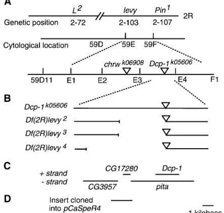
The levy gene was localized to the right arm of chromosome 2 and mapped to 103 map units by recom-bination with visible markers L2
andPin1
the levy mutation were Df(2R)bw-HB132, with break-points at 59D11 and 59F6-8, andDf(2R)egl2
, with break-points at 59E and 60A1. These deficiencies localized the
levy mutation between 59D11 and 59E on polytene chromosomes (Figure 1A), a region encompassing 30 genes (http://flybase.bio.indiana.edu/).
Site-specific recombination mapping in males (Chen
et al. 1998) placed thelevygene distal to the insertion site of thechrwk06908
Pelement and proximal to location of the Dcp-1k05606
P element (Figure 1A). Genetic crosses were conducted to mobilize the Dcp-1k05606 P element into the levy DNA and three new levy mutant alleles (levy2,levy3, andlevy4) were identified. All three alleles produced embryonic lethality in homozygous configu-ration and gave rise to temperature-induced paralysis when heterozygous withlevy1
.
Inverse PCR and sequencing of DNA flanking theP
element inlevy2 ,levy3
, andlevy4
revealed that all three carried small deletions (Figure 1B). The smallest of the three deletions removed a 2.2-kb region that fully
included theCG17280gene and parts of theDcp-1and
pita genes (Figure 1C). The most likely gene among these three to be levy was CG17280 because the P -element insertion inDcp-1k05606complemented thelevy1 mutation in genetic crosses and Dcp-1k05606 flies are known to have the insert within Dcp-1 and pita DNA (Figure 1, B and C).
TheCG17280gene is an ortholog of the human COX subunit VIa precursor (Fabriziet al. 1989). Sequencing
ofCG17280DNA in thelevy1
mutant revealed a G -to-A transition occurring at the 39-splice junction of the single intron (Figure 2A). This base transition would pre-sumably result in missplicing ofCG17280RNA causing the deletion of a nucleotide, in turn leading to a reading frame shift in the second exon. This misspliced RNA would encode an aberrant protein of 54 amino acids, of which the first 32 match those in the nascent wild-type Levy protein of 109 amino acids (Figure 2B). RT–PCR amplification and DNA sequencing showed that a nucleotide was missing at the relevant position in the CG17280-encoded RNA (Figure 2C). The presence of this misspliced RNA suggested thatCG17280is thelevy
gene.
To obtain proof thatCG17280is thelevygene, a 2-kb DNA fragment encompassing the CG17280 gene (see Figure 1D) was amplified from the wild-type (CS) geno-mic DNA and cloned into pCaSpeR4 transformation vector at KpnI and XhoI sites. The recombinant DNA construct was transferred into the genome of levy1 mutants using P-element-mediated germ-line transfor-mation (McKay et al. 1995). As a control,
transforma-tion was performed with the pCaSpeR4 vector without theCG17280gene.levy1transformants carrying the wild-typeCG17280transgene were not paralytic at 38°, show-ing that wild-typeCG17280DNA comprises thelevygene. None of the seven independently generated transform-ants showed paralysis at 38° and all of the five trans-formant controls showed paralysis.
Reduction of cytochrome c oxidase activity in levy
mutants:Subunit VIa plays a regulatory role in yeast and in mammalian COX (Taanman and Capaldi 1993;
Taanmanet al. 1994; Ludwiget al. 2001). To determine
if the levy mutation affected COX activity, enzymatic assays were performed on age- and temperature-controlled flies. As shown in Figure 3 (A and B), levy1 mutants exhibited a reduction in COX activity com-pared to that in wild-type flies and levytransformants. Activity was reduced inlevy1mutant flies to 41% of that in the wild-type flies (Figure 3A). The reduction in COX activity was greater in 7-day-old flies (Figure 3B) than in 1-day-old flies. In this case the activity in levy1 was reduced to 17% of that in the wild type. Transformant flies carrying levy1
transgene showed enzymatic activity similar to that in the wild-type flies, whereas activity levels in the transformant control flies were similar to those in the mutant. These data support the proposal that thelevygene encodes a COX subunit. There was no Figure1.—Molecular identification of thelevygene.
further reduction in COX activity on exposing the flies to 38°for 5 min.
Heterozygous combinations of thelevy1allele with two deletion alleles (levy2,
levy3) were tested for COX activity to test a prediction that these would also exhibit a reduction in COX activity compared to wild-type flies. Indeed,levy1/levy2andlevy1/levy3flies, aged for 7 days, exhibited a reduction in COX activity (to 12% of that in the wild type in each case) that was comparable to the reduction seen inlevy1homozygous flies (to 4% of the activity in the wild type) (Figure 3C). These data further corroborate our conclusion that thelevymutations lead to a reduction in COX activity and are in complete agreement with results showing the rescue of wild-type activity by levy1
transgene.
To test if a reduction in COX activity might be due to a reduction in the number of mitochondria in levy
mutants, fly preparations were examined by assaying for citrate synthase, an enzymatic marker for the mitochon-drial matrix (Ferguson et al. 2005). No measurable
differences were seen in levels of citrate synthase activity between levy mutants and wild-type flies (Figure 3F). This suggests that reduced COX activity observed inlevy
mutants was due to a decrease in COX enzymatic activity rather than to a reduction in the overall number of mitochondria inlevytissues.
Decreased levels of ATP inlevymutants:A defect in oxidative phosphorylation results in reduced levels of ATP in mitochondrial encephalomyopathies such as LS (Dahl1998; Carrozzo et al. 2001). Similarly,levy1
mutants exhibited reduced ATP (Figure 3, D and E).
Compared to the wild type, 7-day-oldlevy1
mutants that were close to dying showed 33% reduction in ATP after being subjected to temperature shock of 38°for 5 min (Figure 3E). ATP levels without the temperature shock were 103 6 4 RLU/mg for the wild type and 956 8 RLU/mg for thelevy1 flies. In flies given temperature shock, the levels were 102 6 8 RLU/mg and 68 6 5 RLU/mg in wild type andlevy1, respectively. ATP levels inlevytransformants were comparable to those in wild-type flies. ATP reduction inlevy1mutants appeared to be age dependent because there was a difference in ATP between 1-day-old and 7-day-old levy1
mutants after temperature shock (Figure 3, D and E) (1-day-oldlevy1
: 9166 RLU/mg; 7-day-oldlevy1
: 6865 RLU/mg).
Reduced life span in levy mutants: levy1
mutants displayed additional phenotypic characteristics that are similar to COX-deficient encephalomyopathies. A major characteristic of such disorders is early death (Leigh1951; Rahmanet al. 1996; Darinet al. 2001). This
is paralleled by premature death oflevy1mutants, which showed a median life span of 8 days at 29°compared to 28 days in wild type and 26 days in transformants (Figure 4A). The median life span of transformant control (9 days) was similar to that of levy1mutants, proving that
CG17280DNA is essential to restore normal life span in
levy1
transformants. Life span oflevy1
mutants was also examined at 21°and had a mean value of 51 days com-pared to 100 days in the wild type, 93 days inlevy trans-formants, and 55 days in transformant control flies.
Bang sensitivity in levy mutants: In another pheno-typic parallel to mitochondrial encephalomyopathies, Figure
levy1
mutants exhibited motor deficiency by displaying age-dependent bang-induced paralysis (Figure 4B) (Ganetzkyand Wu 1982; Pavlidis et al. 1994).
Two-day-oldlevy1
mutants were not paralytic in response to mechanical shock, but there was an increase in the num-ber that showed such paralysis as they aged. In flies aged for 10 days at 21°, 73% oflevy1
and 69% of transformant control flies paralyzed in response to a bang shock, whereas all 20-day-old mutant and transformant control flies exhibited bang-induced paralysis. With the onset and increase in bang-induced paralysis with age, there was also a concomitant increase in the severity of paralysis, prolonging the time required for recovery from paralysis. Ten-day-old paralyzed flies took up to 90 sec to recover from bang-induced paralysis while 20-day-old flies took up to 330 sec. Bang sensitivity was also examined inlevy1
/levy2
andlevy1 /levy3
heterozygous flies and they also showed age-dependent bang-induced paralysis (data not shown). Wild-type and levy trans-formant flies did not show bang-induced paralysis at any of the three ages at which flies were tested.
Characterization of neurodegeneration in levy mu-tants: The phenotype of temperature-induced paralysis has been shown to enrich for mutations that lead to neurodegeneration (Palladino et al. 2002). In
addi-tion, several mitochondrial encephalomyopathies lead to neurodegeneration (DiMauro2004; Gropman2004).
To determine whether or not levy1
mutants exhibited neurodegeneration, tissue sections taken through head capsules of mutant and the wild-type (CS) flies were fixed in Epon and examined microscopically. Neuro-degeneration was not seen in wild-type flies (Figure 5A), but the brain and optic lobes oflevy1mutants showed Swiss cheese-like holes that are reminiscent of spongi-form neurodegeneration (Figure 5B). Neurodegenera-tion was observed to be absent in levy transformants (Figure 5C), demonstrating that the phenomenon was produced by thelevymutation. As in the case of other phenotypes, neurodegeneration appeared to be age dependent. Paraffin sections from 20 flies each from the wild-type and thelevy1
strains, aged for either 1 day or 6 days at 29°, were examined for holes in the brain. None Figure3.—levymutants exhibited
age-depen-dent reduction in COX activity and ATP, but not in a marker for the mitochondrial matrix, cit-rate synthase. COX activity and levels of ATP were measured in adult flies kept at 29°for 1 or 7 days. COX activity is set to 100% in untreated wild-type (CS) flies for comparison purposes. COX activity was reduced inlevy1mutants compared to that in wild-type flies (Student’st-test,P,0.025 for 1-day old andP,0.050 for 7-day old) andlevy trans-formants (P , 0.005 for 1-day old and P , 0.010 for 7-day old) (A and B). Transformant flies carrying levy1
of wild-type flies in either age category, and none of the
levy1 flies aged for 1 day, showed any signs of neuro-degeneration (Figure 5D). On the other hand, 19 of 20
levy1flies aged for 6 days showed neurodegeneration. Possible occurrence of neurodegeneration in addi-tionallevyalleles was examined inlevy1
/levy2
andlevy1 /
levy3
heterozygotes. Heterozygous animals aged at 29°
posteclosion for 6 days were stained using hematoxylin and eosin to determine the extent of vacuolization. The
levy1 /levy2
(Figure 5E) and levy1 /levy3
(Figure 5F) flies exhibited levy1-type neurodegeneration in the lobula and extensive vacuolization within other neural struc-tures. The phenotype of neurodegeneration overlap-ped that seen in the homozygous levy1 mutants. The
extent or the nature of neurodegeneration did not seem to be significantly different among the alleles examined. This is not unexpected sincelevy2andlevy3are deletion mutations and levy1 itself is likely to be a null allele because of a shift in the reading frame after 32 amino acids, followed by a premature stop codon.
DISCUSSION
Mitochondrial function is integral to the healthy function of cells, particularly in cells such as neurons and muscles that are critically dependent on an abun-dant energy supply. A spectrum of myopathic and neu-ropathic symptoms in humans have been correlated to lesions in mitochondrial respiratory chain. Use of genet-ically tractable model systems such as Drosophila can help in identifying individual steps in the pathways leading to such disorders. Animal models are particu-larly useful for diseases such as mitochondrial encepha-lomyopathies that are intransigent to treatment and about which relatively little is known. By generating mutations in a COX subunit, analyzing the resulting phenotypes, and obtaining rescue from these pheno-types by germ-line transformation, we provide direct evidence that disruption of COX VIa results in mito-chondrial encephalomyopathic effects including neuro-degeneration and motor dysfunction.
Mitochondrial encephalomyopathies related to COX are characterized by enzyme deficiency, reduced ATP production, motor difficulty, neurodegeneration, and shortened life span. Phenotypes associated with levy
mutants mimic all of these symptoms. Consistent with the progressive nature of these symptoms in humans, all
levyphenotypes including temperature-induced paraly-sis, bang sensitivity, reduction in COX activity and ATP level, and neurodegeneration show age-dependent in-creases in severity. For example, vacuolization in the brains oflevymutants is not detected in flies aged for 1 day. However, it is widespread in flies aged for 6 days. Importantly, germ-line transformation using wild-type
levygene rescues the mutant flies from all the pheno-types described above. However, the control vector—the same vector used in transformation rescue but without thelevygene—is unable to provide such a rescue. Thus all these phenotypes can be directly linked to the disruption of subunit VIa of COX. These data reveal a COX-mediated pathway in Drosophila, disruption of which leads to mitochondrial encephalomyopathic ef-fects including neurodegeneration, motor dysfunction, and premature death. In addition, the transformation experiments provide direct evidence for a causal link between the disruption of COX and these phenotypes.
Human COX consists of 3 mitochondrial-encoded and 10 nuclear-encoded subunits. Defects in the 3 mitochondrial-encoded subunits COXI (also called MTCO1), COXII (MTCO2), and COXIII (MTCO3) as Figure 4.—Reduced life span and progressive
well as in several COX assembly factors such as COX10, COX15, SURF1, SCO1, and SCO2 have been correlated with encephalomyopathies (Barrientos et al. 2002;
DiMauro and Schon 2003; DiMauro and Hirano
2005). For example, .30 distinct mutations in the SURF1 gene have been associated with COX-deficient LS (Pequignotet al. 2001). In light of this, it has been
noted with intrigue that no mutations in any of the nuclear-encoded structural subunits of COX have been associated with such disorders and that attempts to find such associations have not been fruitful (Shoubridge
2001; DiMauroand Schon2003; Schon2004; Schapira
2006). This strongly suggests that mutations in the nuclear-encoded structural subunits of COX may be lethal (DiMauro and Schon 2003; Schapira 2006).
This view is reinforced by results from an efficient screen conducted for mutations in nuclear-encoded mitochondrial proteins that yielded many such muta-tions in Drosophila (Liao et al. 2006). The only
mutation in this set to target a structural subunit of COX, the tenured mutation in subunit Va, produces lethality. Similarly, a mutation in thecyclopegene, which codes for subunit VIc of COX, leads to lethality in Drosophila (Szuplewski and Terracol 2001). The
levy1
mutation discussed here provides the first case of
such a mutation leading to encephalomyopathic effects rather than lethality. However, the findings presented here raise additional questions. While subunit VIa is a structural component of COX, the role of this subunit is likely to be regulatory in nature (Taanmanand Capaldi
1993; Taanmanet al. 1994; Ludwiget al. 2001). This is
likely to be the reason for the enzyme retaining some activity even with frame-shifted and truncated subunit VIa (Figure 3).
The levy mutant was identified for its temperature-induced paralysis. However, the primary biochemical effect of the mutation—reduction in COX activity—is not temperature dependent. It implies that the COX enzyme itself does not have to be temperature sensitive to produce temperature sensitivity (paralysis) in flies. This is a common feature among temperature-paralytic mutants of Drosophila where sensitivity to high temper-ature arises not from the tempertemper-ature sensitivity of the primary biochemical target but from a constitutive change in a biochemical or a physiological parameter. For example, temperature-induced paralysis in theparats and thenapts
mutants arises from a constitutive decrease in the number of sodium channels and not from chan-nels that become temperature sensitive (Loughney
et al. 1989; Kernanet al. 1991). Similarly, temperature
Figure 5.—levy mutants exhibited
sensitivity of ATP levels in old flies does not derive from temperature sensitivity of COX. Aged mutant flies showed a decrease in ATP levels after they were paralyzed by a temperature shock while COX activity was not affected by this treatment. While the mecha-nisms underlying an effect of temperature shock on ATP are unknown at this stage, it may possibly be related to the seizure activity thatlevyflies go through during temperature-induced paralysis. A reduction in ATP levels after seizure activity is known to occur in rat models of seizures (DeFranceand McCandless1991;
Yageret al. 2002; Darbinet al. 2005).
While the experiments reported here have identified one step in the pathway(s) leading tolevyeffects, and
while further experimentation is needed to identify additional steps, the nature of the observed effects points to some likely mechanisms. Inhibition of COX has been shown to increase free radical generation in many systems including Drosophila (Smith and
Bennett1997; Duranteauet al. 1998; Fergusonet al.
2005). Excessive levels of free radicals can in turn lead to cell death via either apoptosis or necrosis (Beal 2000;
Mattsonand Kroemer2003). There are other aspects
of pathways involved in mitochondrial-mediated neu-rodegeneration. For example, ion channels have been implicated, either in the context of oxidative stress or outside of this context, in several types of neurodegen-eration (Uedaet al. 1997; Liss et al. 2005; Burg et al.
2006; Chinopoulosand Adam-Vizi2006). It remains
to be seen if increased production of free radicals is involved in the effects observed inlevy, whetherlevybrains show apoptotic or necrotic cell death, and if any ion channel dysfunction occurs inlevy(Figure 6). Availability oflevymutations, genetic tractability of Drosophila, and the ease with which questions about oxidative stress (Ferguson et al. 2005; Dias-Santagata et al. 2007) as
well as ion channel function can be explored in this model system (Chopraand Singh1994; Gielowet al.
1995; Kralizand Singh1997; Kralizet al. 1998) provide
an excellent opportunity to address these questions. Availability oflevymutations will particularly help in identifying steps in the pathway(s) leading to mitochon-drial encephalopathy seen in the mutants. For example, it will be helpful to identify interacting genetic compo-nents by screening for suppressors or enhancers of alevy
mutant phenotype. It is relatively easy to identify such modifier mutations in Drosophila, as thousands of mu-tagenized flies can be tested easily for phenotypes such as temperature-induced paralysis. Such modifier muta-tions can provide further leads into the pathways dis-rupted by the original mutations (thelevymutations in this case).
A significant level of our understanding on the structure, function, and regulation of COX has devel-oped from identification and analysis of mutants in yeast (Barrientoset al. 2002). Mutations that lead to
COX-related mitochondrial encephalomyopathies in organ-isms such as Drosophila and mice can similarly help us understand pathways leading to these disorders. Agostinoet al. (2003) have generated aSurf1knockout
mouse model of Leigh syndrome that lacks Surfeit-1, an enzyme involved in COX assembly. Similarly, a Surf1
knockdown model has been generated in Drosophila by post-transcriptional silencing using dsRNA (Zordan
et al. 2006). Studies comparing biochemical and phys-iological characteristics of various models of COX-related mitochondrial encephalomyopathies, including
levymutants andSurf1models in mice and Drosophila, can help in understanding the mechanisms underlying the effects of such disorders. This is particularly true in regard to neurodegeneration due to a fortuitous Figure 6.—From COX deficiency to neuromuscular
difference between thelevyflies and theSurf1models. NeitherSurf1mice norSurf1flies show neurodegenera-tion (Agostinoet al. 2003; Zordanet al. 2006). Thus,
studies of similarities and differences betweenlevy mu-tants andSurf1models may enable us to address ques-tions about what leads to neurodegeneration in one case but not in the other. These studies would lead to a better understanding of neurodegenerative processes in general and their occurrence in mitochondrial encephalomyopathies in particular. This information will also be useful in developing possible therapeutic approaches against such disorders.
This work was supported by grants MCB-0094477 and MCB-0322461 from the National Science Foundation to S.S. and R.D.S.
LITERATURE CITED
Abou-Sleiman, P. M., M. M. Muqit and N. W. Wood, 2006
Ex-panding insights of mitochondrial dysfunction in Parkinson’s disease. Nat. Rev. Neurosci.7:207–219.
Agostino, A., F. Invernizzi, C. Tiveron, G. Fagiolari, A. Prelle
et al., 2003 Constitutive knockout of Surf1 is associated with high embryonic lethality, mitochondrial disease and cytochrome c oxidase deficiency in mice. Hum. Mol. Genet.12:399–413. Ashburner, M., 1989 Drosophila: A Laboratory Handbook. Cold Spring
Harbor Laboratory Press, Cold Spring Harbor, NY.
Atamna, H., and K. Boyle, 2006 Amyloid-beta peptide binds with
heme to form a peroxidase: relationship to the cytopathologies of Alzheimer’s disease. Proc. Natl. Acad. Sci. USA103: 3381– 3386.
Barrientos, A., M. H. Barros, I. Valnot, A. Rotig, P. Rustinet al.,
2002 Cytochrome oxidase in health and disease. Gene286:53–63. Beal, M. F., 2000 Energetics in the pathogenesis of
neurodegener-ative diseases. Trends Neurosci.23:298–304.
Bellen, H. J., R. W. Levis, G. Liao, Y. He, J. W. Carlson et al.,
2004 The BDGP gene disruption project: single transposon in-sertions associated with 40% of Drosophila genes. Genetics167:
761–781.
Benchoua, A., Y. Trioulier, D. Zala, M. C. Gaillard, N. Lefort
et al., 2006 Involvement of mitochondrial complex II defects in neuronal death produced by N-terminus fragment of mutated huntingtin. Mol. Biol. Cell17:1652–1663.
Bhattacharya, A., G. G. Gu and S. Singh, 1999 Modulation of
dihydropyridine-sensitive calcium channels in Drosophila by a cAMP-mediated pathway. J. Neurobiol.39:491–500.
Bhattacharya, A., S. S. Lakhmanand S. Singh, 2004 Modulation
of L-type calcium channels in Drosophila via a pituitary adenylyl cyclase-activating polypeptide (PACAP)-mediated pathway. J. Biol. Chem.279:37291–37297.
Bonini, N. M., and M. E. Fortini, 2003 Human neurodegenerative
disease modeling using Drosophila. Annu. Rev. Neurosci. 26:
627–656.
Burg, E. D., C. V. Remillardand J. X. Yuan, 2006 K1channels in
apoptosis. J. Membr. Biol.209:3–20.
Carrozzo, R., A. Tessa, M. E. Vazquez-Memije, F. Piemonte,
C. Patronoet al., 2001 The T9176G mtDNA mutation severely
affects ATP production and results in Leigh syndrome. Neurol-ogy56:687–690.
Celotto, A. M., A. C. Frank, S. W. McGrath, T. Fergestad, W. A.
VanVoorhieset al., 2006 Mitochondrial encephalomyopathy
in Drosophila. J. Neurosci.26:810–820.
Chen, B., T. Chu, E. Harms, J. P. Gergen and S. Strickland,
1998 Mapping of Drosophila mutations using site-specific male recombination. Genetics149:157–163.
Chinopoulos, C., and V. Adam-Vizi, 2006 Calcium, mitochondria
and oxidative stress in neuronal pathology. Novel aspects of an enduring theme. FEBS J.273:433–450.
Choo, Y. S., G. V. Johnson, M. MacDonald, P. J. Detloff and
M. Lesort, 2004 Mutant huntingtin directly increases
suscep-tibility of mitochondria to the calcium-induced permeability transition and cytochrome c release. Hum. Mol. Genet.13:1407– 1420.
Chopra, M., and S. Singh, 1994 Developmental temperature
selec-tively regulates a voltage-activated potassium current in Drosoph-ila. J. Neurobiol.25:119–126.
Chopra, M., G.-G. Guand S. Singh, 2000 Mutations affecting the
delayed rectifier potassium current inDrosophila. J. Neurogenet.
14:107–123.
Chovnick, A., G. H. Ballantyne, D. L. Baillieand D. G. Holm,
1970 Gene conversion in higher organisms: half-tetrad analysis of recombination within the rosy cistron ofDrosophila melanogaster. Genetics66:315–329.
Crittenden, J. R., E. M. Skoulakis, K. A. Han, D. Kalderonand
R. L. Davis, 1998 Tripartite mushroom body architecture
re-vealed by antigenic markers. Learn. Mem.5:38–51.
Dahl, H. H., 1998 Getting to the nucleus of mitochondrial
disor-ders: identification of respiratory chain-enzyme genes causing Leigh syndrome. Am. J. Hum. Genet.63:1594–1597.
Darbin, O., J. J. Risso, E. Carre, M. Lonjonand D. K. Naritoku,
2005 Metabolic changes in rat striatum following convulsive seizures. Brain Res.1050:124–129.
Darin, N., A. Oldfors, A. R. Moslemi, E. Holmeand M. Tulinius,
2001 The incidence of mitochondrial encephalomyopathies in childhood: clinical features and morphological, biochemical, and DNA anbormalities. Ann. Neurol.49:377–383.
DeFrance, J. F., and D. W. McCandless, 1991 Energy metabolism in
rat hippocampus during and following seizure activity. Metab. Brain Dis.6:83–91.
Dias-Santagata, D., T. A. Fulga, A. Duttaroyand M. B. Feany,
2007 Oxidative stress mediates tau-induced neurodegeneration in Drosophila. J. Clin. Invest.117:236–245.
DiMauro, S., 2004 Mitochondrial diseases. Biochim. Biophys. Acta
1658:80–88.
DiMauro, S., and M. Hirano, 2005 Mitochondrial
encephalomyo-pathies: an update. Neuromuscul. Disord.15:276–286. DiMauro, S., and E. A. Schon, 2003 Mitochondrial
respiratory-chain diseases. N. Engl. J. Med.348:2656–2668.
Duranteau, J., N. S. Chandel, A. Kulisz, Z. Shao and P. T.
Schumacker, 1998 Intracellular signaling by reactive oxygen
species during hypoxia in cardiomyocytes. J. Biol. Chem.273:
11619–11624.
Esposito, L., J. Raber, L. Kekonius, F. Yan, G. Q. Yu et al.,
2006 Reduction in mitochondrial superoxide dismutase modu-lates Alzheimer’s disease-like pathology and accelerates the onset of behavioral changes in human amyloid precursor protein trans-genic mice. J. Neurosci.26:5167–5179.
Fabrizi, G. M., R. Rizzuto, H. Nakase, S. Mita, B. Kadenbachet al.,
1989 Sequence of a cDNA specifying subunit VIa of human cytochrome c oxidase. Nucleic Acids Res.17:6409.
Ferguson, M., R. J. Mockett, Y. Shen, W. C. Orrand R. S. Sohal,
2005 Age-associated decline in mitochondrial respiration and elec-tron transport in Drosophila melanogaster. Biochem. J.390:501–511. Fortini, M. E., and N. M. Bonini, 2000 Modeling human
neurode-generative diseases in Drosophila: on a wing and a prayer. Trends Genet.16:161–167.
Ganetzky, B., 2000 Genetic analysis of ion channel dysfunction in
Drosophila. Kidney Int.57:766–771.
Ganetzky, B., and C. F. Wu, 1982 Indirect suppression involving
be-havioral mutants with altered nerve excitability in Drosophila melanogaster. Genetics100:597–614.
Gielow, M. L., G. G. Guand S. Singh, 1995 Resolution and
phar-macological analysis of the voltage-dependent calcium channels of Drosophila larval muscles. J. Neurosci.15:6085–6093. Giordano, C., M. Sebastiani, G. Plazzi, C. Travaglini, P. Saleet al.,
2006 Mitochondrial neurogastrointestinal encephalomyopathy: evidence of mitochondrial DNA depletion in the small intestine. Gastroenterology130:893–901.
Gnerer, J. P., R. A. Kreberand B. Ganetzky, 2006 wasted away, a
Drosophila mutation in triosephosphate isomerase, causes paral-ysis, neurodegeneration, and early death. Proc. Natl. Acad. Sci. USA103:14987–14993.
Gropman, A. L., 2004 The neurological presentations of childhood
Gu, G. G., and S. Singh, 1997 Modulation of the
dihydropyridine-sensitive calcium channels in Drosophila by a phospholipase C-mediated pathway. J. Neurobiol.33:265–275.
Hegde, P., G. G. Gu, D. Chen, S. J. Freeand S. Singh, 1999
Muta-tional analysis of the Shab-encoded delayed rectifier K1channels
in Drosophila. J. Biol. Chem.274:22109–22113.
Keeney, P. M., J. Xie, R. A. Capaldi and J. P. Bennett, Jr.,
2006 Parkinson’s disease brain mitochondrial complex I has oxidatively damaged subunits and is functionally impaired and misassembled. J. Neurosci.26:5256–5264.
Kernan, M. J., M. I. Kuroda, R. Kreber, B. S. Baker and
B. Ganetzky, 1991 napts, a mutation affecting sodium channel
activity in Drosophila, is an allele of mle, a regulator of X chro-mosome transcription. Cell66:949–959.
Kraliz, D., and S. Singh, 1997 Selective blockade of the delayed
rectifier potassium current by tacrine in Drosophila. J. Neuro-biol.32:1–10.
Kraliz, D., A. Bhattacharyaand S. Singh, 1998 Blockade of the
delayed rectifier potassium current in Drosophila by quinidine and related compounds. J. Neurogenet.12:25–39.
Leigh, D., 1951 Subacute necrotizing encephalomyelopathy in an
infant. J. Neurochem.14:216–221.
Lewis, E. B., and F. Bacher, 1968 Method of feeding
ethylmethane-sulfonate (EMS) to Drosophila males. Drosoph. Inf. Serv.43:193. Liao, T. S., G. B. Call, P. Guptan, A. Cespedes, J. Marshallet al.,
2006 An efficient genetic screen in Drosophila to identify nuclear-encoded genes with mitochondrial function. Genetics174:525–533. Liss, B., O. Haeckel, J. Wildmann, T. Miki, S. Seinoet al., 2005
K-ATP channels promote the differential degeneration of dopami-nergic midbrain neurons. Nat. Neurosci.8:1742–1751. Loughney, K., R. Kreberand B. Ganetzky, 1989 Molecular
anal-ysis of the para locus, a sodium channel gene in Drosophila. Cell
58:1143–1154.
Ludwig, B., E. Bender, S. Arnold, M. Huttemann, I. Leeet al.,
2001 Cytochrome C oxidase and the regulation of oxidative phosphorylation. Chembiochem2:392–403.
Mattson, M. P., and G. Kroemer, 2003 Mitochondria in cell death:
novel targets for neuroprotection and cardioprotection. Trends Mol. Med.9:196–205.
McFarland, R., R. W. Taylorand D. M. Turnbull, 2002 The
neu-rology of mitochondrial DNA disease. Lancet Neurol.1:343–351. McKay, R. R., D. M. Chen, K. Miller, S. Kim, W. S. Starket al.,
1995 Phospholipase C rescues visual defect in norpA mutant of Drosophila melanogaster. J. Biol. Chem.270:13271–13276. Palladino, M. J., T. J. Hadley and B. Ganetzky, 2002
Tem-perature-sensitive paralytic mutants are enriched for those caus-ing neurodegeneration in Drosophila. Genetics161:1197–1208. Pallanck, L., and J. T. Greenamyre, 2006 Neurodegenerative
dis-ease: pink, parkin and the brain. Nature441:1058.
Pavlidis, P., M. Ramaswamiand M. A. Tanouye, 1994 The
Dro-sophila easily shocked gene: a mutation in a phospholipid syn-thetic pathway causes seizure, neuronal failure, and paralysis. Cell79:23–33.
Pequignot, M. O., R. Dey, M. Zeviani, V. Tiranti, C. Godinotet al.,
2001 Mutations in the SURF1 gene associated with Leigh syn-drome and cytochrome C oxidase deficiency. Hum. Mutat.17:
374–381.
Rahman, S., R. B. Blok, H. H. Dahl, D. M. Danks, D. M. Kirbyet al.,
1996 Leigh syndrome: clinical features and biochemical and DNA abnormalities. Ann. Neurol.39:343–351.
Schapira, A. H., 2006 Mitochondrial disease. Lancet368:70–82.
Schon, E. A., 2004 Complements of the house. J. Clin. Invest.114:
760–762.
Shoubridge, E. A., 2001 Cytochrome c oxidase deficiency. Am. J.
Med. Genet.106:46–52.
Singh, S., 1983 A mutagenesis scheme for obtaining autosomal
mu-tations inDrosophila. Indian J. Exp. Biol.21:635–636.
Singh, A., and S. Singh, 1999 Unmasking of a novel potassium current
in Drosophila by a mutation and drugs. J. Neurosci.19:6838–6843. Singh, S., P. Bhandari, M. J. S. Chopraand D. Guha, 1987
Iso-lation of autosomal mutations inDrosophila melanogasterwithout setting up lines. Mol. Gen. Genet.208:226–229.
Smith, T. S., and J. P. Bennett, Jr., 1997 Mitochondrial toxins in
models of neurodegenerative diseases. I:In vivobrain hydroxyl radical production during systemic MPTP treatment or following microdialysis infusion of methylpyridinium or azide ions. Brain Res.765:183–188.
Szuplewski, S., and R. Terracol, 2001 The cyclope gene of
Dro-sophila encodes a cytochrome c oxidase subunit VIc homolog. Genetics158:1629–1643.
Taanman, J. W., and R. A. Capaldi, 1993 Subunit VIa of yeast
cyto-chrome c oxidase is not necessary for assembly of the enzyme complex but modulates the enzyme activity. Isolation and charac-terization of the nuclear-coded gene. J. Biol. Chem.268:18754– 18761.
Taanman, J. W., P. Turinaand R. A. Capaldi, 1994 Regulation of
cytochrome c oxidase by interaction of ATP at two binding sites, one on subunit VIa. Biochemistry33:11833–11841.
Ueda, A., and C. F. Wu, 2006 Distinct frequency-dependent
regula-tion of nerve terminal excitability and synaptic transmission by IA
and IKpotassium channels revealed by Drosophila Shaker and
Shab mutations. J. Neurosci.26:6238–6248.
Ueda, K., S. Shinohara, T. Yagami, K. Asakuraand K. Kawasaki,
1997 Amyloid beta protein potentiates Ca21 influx through
L-type voltage-sensitive Ca21 channels: a possible involvement
of free radicals. J. Neurochem.68:265–271.
Yager, J. Y., E. A. Armstrong, H. Miyashitaand E. C. Wirrell,
2002 Prolonged neonatal seizures exacerbate hypoxic-ischemic brain damage: correlation with cerebral energy metabolism and excitatory amino acid release. Dev. Neurosci.24:367–381. Zordan, M. A., P. Cisotto, C. Benna, A. Agostino, G. Rizzoet al.,
2006 Post-transcriptional silencing and functional characteriza-tion of theDrosophila melanogasterhomolog of human Surf1. Ge-netics172:229–241.