DOI: 10.1534/genetics.104.038018
A Drosophila Model of Multiple Endocrine Neoplasia Type 2
Renee D. Read,* Paul J. Goodfellow,
†,‡Elaine R. Mardis,
‡Nancy Novak,
†Jon R. Armstrong
‡and Ross L. Cagan*
,1*Department of Molecular Biology and Pharmacology,†Department of Surgery and‡Department of Genetics, Washington University School of Medicine, St. Louis, Missouri 63110
Manuscript received October 31, 2004 Accepted for publication June 7, 2005
ABSTRACT
Dominant mutations in theRetreceptor tyrosine kinase lead to the familial cancer syndrome multiple endocrine neoplasia type 2 (MEN2). Mammalian tissue culture studies suggest thatRetMEN2 mutations significantly alter Ret-signaling properties, but the precise mechanisms by which RetMEN2 promotes tumorigenesis remain poorly understood. To determine the signal transduction pathways required for RetMEN2 activity, we analyzed analogous mutations in the Drosophila Ret ortholog dRet. Overexpressed dRetMEN2isoforms targeted to the developing retina led to aberrant cell proliferation, inappropriate cell fate specification, and excessive Ras pathway activation. Genetic analysis indicated that dRetMEN2acts through the Ras-ERK, Src, and Jun kinase pathways. A genetic screen for mutations that dominantly suppress or enhance dRetMEN2 phenotypes identified new genes that are required for the phenotypic outcomes of dRetMEN2activity. Finally, we identified human orthologs for many of these genes and examined their status in human tumors. Two of these loci showed loss of heterozygosity (LOH) within both sporadic and MEN2-associated pheochromocytomas, suggesting that they may contribute to Ret-dependent oncogenesis.
D
URING development, receptor tyrosine kinases (RTKs) integrate extracellular signals to influ-ence cellular processes such as growth and differenti-ation. Signaling through RTKs requires ligand-induced oligomerization to direct tyrosine autophosphoryla-tion; autophosphorylation both stimulates catalytic activity and creates phospho-tyrosine docking sites for cytoplasmic proteins that activate intracellular signal-ing pathways. To date, mutations in more than half of all RTKs have been implicated in human cancer (reviewed in Blume-Jensenand Hunter2001). Thesemutations commonly function by relieving RTK regu-latory constraints, leading to inappropriate kinase ac-tivity and hyperactivation of downstream pathways. These events promote oncogenic transformation by dri-ving aberrant cellular growth, proliferation, and survival. Still, tumorigenesis requires mutations in multiple loci: along with dominant mutations in oncogenes such as RTKs, tumorigenesis also requires loss-of-function muta-tions in tumor suppressors. The relamuta-tionship between oncogenic tyrosine kinases and tumor suppressors, and the extent to which mutations in each cooperate to direct oncogenic growth, is not well understood.
TheRetRTK plays an essential role in both develop-ment and oncogenesis. During embryogenesis, Ret is required for development of the sympathetic and
enteric nervous systems, the neural crest, and the excretory system (Schuchardt et al. 1994; Durbec
et al. 1996; Enomoto et al. 2001). The extracellular
portion of Ret contains cysteine repeats and a cadherin-like domain. The intracellular portion of Ret contains a tyrosine kinase catalytic domain and multiple tyrosine autophosphorylation sites. Four activating ligands have been identified: GDNF, Neurturin, Persephin, and Artemin all activate Ret through the GPI-linked co-receptors GFRa1-GFRa4 ( Jinget al.1996; Kotzbauer
et al.1996; Sanchezet al. 1996; Buj-Belloet al. 1997;
Kleinet al.1997; Balohet al.1998; Enokidoet al.1998;
Enomotoet al. 1998; Milbrandt et al.1998). Ligand
stimulation of Ret signal transduction activates various signaling pathways and effectors, including Ras/Raf/ ERK, PI-3 kinase/AKT, Src, p38-MAPK, Jun kinase ( JNK), PLCg, and ERK5 (van Weering et al. 1995;
Borrelloet al.1996; Chiarielloet al.1998; Melillo
et al. 1999, 2001; Soler et al.1999; Bessetet al. 2000;
Califanoet al.2000; Hayashiet al.2000; Encinaset al.
2001; Kurokawa et al. 2001, 2003). Through these
pathways, Ret signaling directs cell differentiation, pro-liferation, survival, and migration during neural de-velopment and renal organogenesis (Schuchardtet al.
1994; Heuckeroth et al. 1998; Taraviraset al.1999;
Enomotoet al.2001; Hashinoet al.2001; Natarajan
et al.2002; Tanget al.2002).
Dominant, gain-of-function mutations in Ret lead to the familial cancer syndromes multiple endocrine neo-plasia type 2A (MEN2A) and type 2B (MEN2B) and
1Corresponding author:Department of Molecular Biology and
Pharma-cology, Washington University School of Medicine, 660 S. Euclid Ave., St. Louis, MO 63110. E-mail: [email protected]
familial medullary thyroid carcinoma (FMTC). These syndromes are defined by tumors of the endocrine glands arising from neural crest derivatives. MEN2 and FMTC are characterized by the occurrence of medullary thyroid carcinoma (MTC), a malignant tumor derived from thyroid neuroendocrine parafollicular C cells (reviewed in Leboulleux et al. 2004). MEN2A and
MEN2B patients frequently develop pheochromocy-toma, an adrenal neuroendocrine tumor. In addition, MEN2A patients commonly develop parathyroid and skin defects, and MEN2B patients display peripheral nerve and skeletal abnormalities. These syndromes also show variation in age at tumor onset, severity of MTC, and occurrence of other tumors (Eastonet al. 1989;
O’Riordain et al. 1995; Ponder 1999). Current
treat-ment is surgery to remove tumors followed by chemo-therapy. However, treatment of MTC is difficult since it is prone to metastasis and is often refractory to chemo-therapy (Quayleand Moley2005). Genetic testing in
afflicted families has resulted in early identification and prophylactic surgical treatment of individuals withRet mutations (Brandiet al.2001). Yet many cases of MEN2
are attributed to de novo mutations, precluding early identification and treatment, and even affected infants can develop MTC (Carlson et al. 1994a; Shirahama
et al.1998;vanHeurnet al.1999).
Mutations in the Ret extracellular region at one of five cysteines occur in almost all MEN2A patients and in approximately two-thirds of FMTC patients (Donis
-Keller et al. 1993; Mulligan et al. 1993b, 1994a,b;
Enget al.1996). MEN2B patients possess mutations that
cluster within the tyrosine kinase domain, most com-monly a methionine-to-threonine substitution (M918T) and, less frequently, mutations such as A883F or V804M and Y806C/S904C (Carlsonet al.1994b; Enget al.1994;
Hofstraet al.1994; Smithet al.1997; Miyauchiet al.
1999; Menkoet al.2002). Sporadic MTC and papillary
thyroid carcinoma also display dominant activating Ret mutations (Griecoet al.1990; Bongarzoneet al.1994;
Chiefariet al.1998; Scuriniet al.1998).
Using primarily tissue culture models, a variety of studies have assessed the biochemical properties of oncogenic Ret to understand the molecular basis for MEN2 diseases. MEN2A forms of Ret (RetMEN2A) are
constitutively dimerized and activated, the result of ligand-independent intermolecular disulfide bonding (Asaiet al.1995). In contrast, RetMEN2Bis constitutively
activated independently of dimerization and exhibits an altered autophosphorylation pattern (Asaiet al.1995;
Santoro et al. 1995; Liu et al. 1996; Iwashita et al.
1999). MEN2B mutations likely disrupt normal auto-inhibitory function of the activation loop, permitting unfettered kinase activity and substrate binding even in the absence of receptor dimerization (Smithet al.1997;
Miller et al. 2001). The results are dominant
onco-genes that drive transformation due to increased kinase activity and aberrant stimulation of downstream
path-ways (reviewed in Takahashi2001). To date, all of the
signaling pathways necessary for RetMEN2transforming
activity are also thought to be required for physiological, ligand-stimulated Ret signal transduction (Marshall
et al. 1997; Ohiwa et al.1997; Chiarielloet al. 1998;
Califanoet al.2000; Hayashiet al.2000).
Many outstanding issues remain to be addressed regarding oncogenic Ret. Tissue culture studies have implicated numerous signaling pathways, but little is known about the relevance of these pathways in vivo. Moreover, both MEN2 families and MEN2 mouse mod-els display significant phenotypic variation, although the basis for this variation remains largely unexplored (Enget al.1996; Michielset al.1997; Actonet al.2000;
Kawaiet al.2000; Smith-Hickset al.2000; Cranston
and Ponder2003). Little is known about possible
sec-ondary mutations that promote MEN2-associated tu-mors. For example, only a subset of patients develop pheochromocytoma, implying that second-site muta-tions in tumor suppressors or susceptibility loci may modulate how different individuals manifest MEN2. With these issues in mind, we established a Drosophila model to address RetMEN2function in the context of an
intact epithelium.
Drosophila has proven a powerful tool for the study of signal transduction pathways in development and dis-ease. In particular, RTKs and their signaling pathways are remarkably well conserved between vertebrates and invertebrates (reviewed in Simon2000; Voasand Rebay
2004). The Drosophila ortholog of the Ret RTK, dRet, is highly conserved compared to mammalian Ret, as both dRet and mammalian Ret share a 52% protein identity within their SH2 and kinase domains (Sugaya et al.
1994; Hahn and Bishop2001). Interestingly, Ret and
dRet are expressed in a number of analogous tissues, including the central and enteric nervous systems and the excretory system (Sugaya et al. 1994; Hahn and
Bishop2001), suggesting that dRet can serve as a useful
model for understanding the role of Ret in disease as well as development.
To provide insight into RetMEN2-mediated defects, we
created transgenicdRetconstructs analogous toRetMEN2A and RetMEN2B
. We chose to target expression of these constructs to the developing fly eye due to its simplicity, its accessibility, and its long history of success as a model system for RTK signal transduction. We demonstrate that these transgenic fly lines exhibit defects associated with human RetMEN2, including Ras pathway
hyperacti-vation, excess proliferation, and aberrant neuronal dif-ferentiation. We performed a genetic screen for genes that dominantly suppressed or enhanced the dRetMEN2
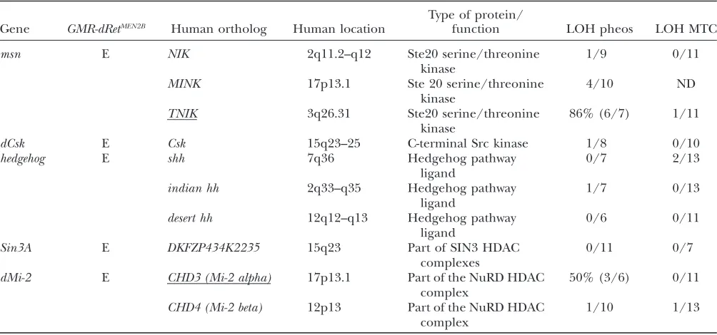
phenotypes and identified a large number of genes that participate in dRet signal transduction. Finally, with comparative genomics, we identified human orthologs for some of these genetic modifiers and examined their status in human tumor tissues. Two of these loci were somatically deleted at high rates within both sporadic
and MEN2-associated pheochromocytomas, suggesting that they may contribute to Ret-dependent oncogenesis.
MATERIALS AND METHODS
CloningdRetcDNAs, site-directed mutagenesis, and GMR-dRetflies:Full-lengthdRetcDNA clones were isolated from an adult head cDNA library using probes generated to known partialdRetcDNA sequences (Sugayaet al.1994). PCR-based site-directed mutagenesis was used to make dRetMEN2B (dRetM955T) anddRetMEN2A(dRetC695R). For the M955T mutation, codon 955 was changed to ACG from ATG. For the C695R mutation, codon 695 was changed to CGC from TGC. To create GMR-dRet transgenes, full-length dRet cDNAs were cloned into pGMR. Plasmids were injected intoy w;D2-3 Ki
flies, and multiple stable insertions were recovered. Isogenic stocks were generated for each independent insertion.
Immunohistochemistry and sectioning:In situhybridization was performed as described (Tautz and Pfeifle 1989). Digoxigenin-labeled DNA probes were made to thedRet59-end and were detected with alkaline phosphatase conjugated anti-bodies (Boehringer Mannheim, Indianapolis). For BrdU label-ing, live third instar larval imaginal discs were incubated with 8mg/ml bromodeoxyuridine (BrdU) for 1 hr, fixed in Carnoy’s solution, rehydrated, and treated with 2nHCl in 13PBS 0.3% Triton X-100. For other immunohistochemical stains, tissue was fixed in 13PBS 4% paraformaldehyde for 15–20 min. Stains were performed in 13PBS, 10% serum, 0.3% Triton X-100. 22C10, anti-BrdU, Sevenless, Bar, Boss, and antidiphospho-ERK (Promega, Madison, WI) antibodies were used at 1:100, 1:500, 1:2, 1:25, 1:100, and 1:200, respectively. Staining was detected with Cy3-conjugated secondary antibodies ( Jackson ImmunoResearch Labs, West Grove, PA). For adult sections, heads were fixed in 1% glutaraldehyde/2% osmium tetroxide/ PBS, dehydrated, and incubated 4 hr in 1:1 propylene oxide: Durcupan ACM resin, overnight in 100% resin, and finally at 65°to harden. Serial sections were stained with 0.5% methy-lene blue/0.1% toluidine blue. Photographs were taken on a Zeiss Axioplan.
Fly stocks and screens:Fly stocks were obtained from the Bloomington Stock Center unless otherwise noted.Src64BP1 was a gift of M. Simon.Stat92Ej6C8was a gift of S. Hou.RasD40b was a gift of N. Perrimon.phlandDsormutations were gifts of J. Skeath. A deficiency collection of 116 stocks was obtained from Bloomington. A total of 21 deficiency lines containing rough-eye markers, secondary mutations in knownGMR-dRet
modifiers, and/or undetectable balancers were excluded from analysis. A total of 1112P-element insertion lines were from Bloomington and 1412P-element lines were from the Szeged Stock Center. Crosses were performed at 22.5°. F1 progeny were sorted by visible markers to determine genotype. Eye phenotypes were scored on a dissecting microscope. Modifier penetrance was determined by calculating the percentage of enhanced or suppressed mutation-bearingGMR-dRetprogeny relative to the total number of mutation-bearing GMR-dRet
progeny. Crosses were repeated two to six times for lines showing modifier activity to determine if results were consis-tent.P-element lines that consistently showed enhancement or suppression with a penetrance.75% were considered to be
GMR-dRetMEN2B modifiers. For secondary screens, P-element flies were mated to tester flies and F1progeny were evaluated. Tester strains includedGMR-Gal4(gift of L. Zipursky), GMR-p21(gift of I. Hariharan),GMR-rpr(gift of H. Steller),GMR-hid
(gift of H. Steller),GMR-sina(gift of G. Rubin), anddEGFRElp (gift of N. Baker).
Inverse PCR and sequencing: Genomic DNA sequences flanking the insertion sites ofP-element modifiers were
iso-lated using an inverse PCR strategy (Spradlinget al.1999). Following an initial round of inverse PCR, a second nested PCR was done using products of the first PCR reaction as template. Final PCR products were sequenced using ABI Big Dye terminator cycle sequencing.
Outcrossing, complementation, excision, and additional alleles: P-element insertions were outcrossed to w; h kni e
flies and recombinant males were isolated and mated to
GMR-dRetfemales. F1progeny were scored for genetic inter-actions. P elements were excised from modifier stocks by crossing to y w; D2-3 Ki and isolating white-eyed progeny. Excision flies were mated toGMR-dRet flies and F1 progeny were scored.
Following sequence analysis, possible additional alleles of
P-element modifiers were tested. EP(2)2172, EP(3)3003,
DoaEP3602, anddTorEP2353were from Exelixis.dTorDPwas a gift of T. Neufeld.Sin3Aalleles were a gift of D. Pauli.Ras85Dc40b,scrb1, andscrib2were a gift of N. Perrimon.neur3,neur11,crb8F105, and
Pp1-87B1were from the Umea Stock Center.dMi-21anddMi-24 alleles were a gift of J. Muller. All others were provided by the Bloomington Stock Center. Complementation tests were for lethality only.
To characterize dMi-2, sev-Ras85Dv12 flies were mated to
dMi-2 mutant flies and F1 progeny were scored for dMi-2 -linked enhancement or suppression ofsev-Ras85Dv12rough-eye phenotypes.
Loss-of-heterozygosity and sequencing studies on human tumors: Human orthologs for GMR-dRet modifiers were identified by sequence comparisons, database searches, and literature. Loss-of-heterozygosity (LOH) analysis was per-formed using microsatellite repeat markers, essentially as previously described (Peiffer et al. 1995). For each locus, the human genomic sequence was searched for long (.15) CAnrepeats. Primers were designed to amplify the CA repeat (http://frodo.wi.mit.edu/cgi-bin/primer3/primer3_www.cgi) and reactions were optimized. Primers and PCR conditions are available upon request. Briefly, tumor and matched nor-mal blood DNA samples were prepared using conventional phenol-chloroform extractions. Representative portions of the tumor tissues used to prepare DNA were assessed histo-logically to ensure high neoplastic cellularity. The CA repeat markers were PCR amplified using a forward 32P-labeled primer. The amplified fragments were size separated on polyacrylamide urea gels and visualized with autoradiography. The products for each of the matched tumor-normal pairs were compared and those cases (tumors) for which there was substantial reduction in the relative intensity of one allelic fragment (60% or greater reduction) were classified as having LOH.
Sequencing of individual exons from patient and control samples for both TNIK and CHD3 were performed as de-scribed previously (Leyet al.2003). Primers chosen for exon 6 of TNIK, which contains a portion of the kinase domain, worked poorly repeatedly and this exon was not sequenced. CHD3 and TNIK sequences were assembled and compared using CONSED and POLYPHRED software (Nickersonet al. 1997; Gordonet al.1998). In cases with ambiguous results, sequence tracings for individual tumor and control samples were compared manually.
RESULTS
Characterization of the Drosophila ortholog dRet:
identified by others (Hahn and Bishop 2001). The
kinase domains of dRet and human Ret (hRet) share 52% identity (Figure 1A). Many hRet autophosphoryla-tion sites are conserved in dRet, including tyrosines required for Ret mitogenic and transforming activities (Figure 1A) (Iwashitaet al.1999). While the
extracel-lular domain of dRet showed only 22% identity with hRet, the dRet and hRet extracellular domains share a common structural organization, which includes a signal sequence, multiple glycosylation sites, a cadherin-like domain, and cysteine repeats (Figure 1B). dRet shows conservation of residues mutated in hRet in MEN2B: human M918 is equivalent to fly M955 and human A883 is equivalent to fly A920. dRet also shows conservation of a residue mutated in FMTC: human E768 is equivalent to fly E815. Within the cysteine repeat
region, dRet shows conservation of all cysteine residues reported as mutated in hRet in MEN2A and FMTC: human codons C609, C611, C618, C620, and C634 are equivalent to fly C628, C630, C636, C638, C691, and C695, respectively.
dRet is expressed in the developing central nervous system and peripheral nervous system in embryos (Sugayaet al.1994; Hahnand Bishop2001). In third
instar larval tissues, dRet expression was observed in a small number of cells in the brain and ventral ganglion (Figure 1, C and D) that morphologically appeared to be neuroendocrine cells (P. Taghert, personal
com-munication). dRet was also expressed in a restricted pattern in the leg, wing, and antennal imaginal discs (data not shown), but was expressed only weakly or not at all in the eye imaginal disc (Figure 1E). The Figure1.—DrosophiladRetis highly conserved compared to human Ret. (A) Detailed alignment of dRet (top sequence) and human Ret (bottom sequence) kinase domain and C terminus. Conserved kinase domain residues mutated in MEN2B patients are boxed in red; FMTC is boxed in blue. Conserved tyrosine residues are boxed in green; Y1015 in human Ret is the PLCg-binding site (Borrelloet al.1996), human Y900 and Y905 are autoregulatory tyrosines in the activation loop that are required for RetMEN2A activity, and Y864 and Y952 are required for RetMEN2Bactivity (Iwashitaet al.1996, 1999). Y1062 in human Ret, highlighted by a green asterisk, is not strictly conserved in dRet, but the C-terminal tail of dRet is tyrosine rich (orange asterisks) and some of these tyrosines are in motifs that may be Grb2-binding sites. (B) Schematic of dRet protein structure. The cysteine-repeat region con-tains 14 cysteine residues. Residues mutated to createdRetMEN2AanddRetMEN2Bare noted. (C–F) LarvaldRetexpression. (C and D)
dRetexpression within the larval brain and ventral nerve cord (vnc). Anterior is toward the right. (C) A low-magnification view. (D) A high-magnification view. Note the subset of strongly stained cells in the vnc (arrow) and the lighter-staining cells within the brain (arrow). (E and F)dRetexpression within the third instar eye-antennal disc. (E) A low-magnification view. The eye field proper is the larger disc to the left; note the diffuse staining that indicates either low-level gene expression or background. (F) A high-magnification view of thedRetexpressing developing ocelli (arrows), which are adjacent to the eye field.
developing ocelli, which are primitive light-sensing organs, expresseddRetas well (Figure 1F).
To date, no ligands or coreceptors for dRet have been described. Our analysis of the Drosophila genome did not uncover any obvious GDNF or GFRaorthologs. One gene,CG17204, shows weak similarity to GFRaproteins and contains a putative C-terminal GPI-anchor site, also suggestive of a GFRaortholog.
dRet overexpression within the developing Drosoph-ila retina:We used a gain-of-function approach to study oncogenic dRet. For this purpose, dRet was overex-pressed in the developing Drosophila retina, a neuro-epithelial tissue that shows remarkable evolutionary conservation of RTK signal transduction pathways. The adult retina is composed of750 unit eyes known as ommatidia; each ommatidium contains precisely 14 cells. In the late larval stages, eight photoreceptor neurons emerge within each ommatidium in a process that depends on the Drosophila epidermal growth factor receptor (dEGFR) and Sevenless RTKs and the Ras-ERK pathway (Simonet al.1991; Dicksonet al.1992;
Fortiniet al. 1992; Biggs et al.1994; Freeman 1996;
Karimet al.1996; Spenceret al.1998). Six glial-like cells
are then added to complete the 14-cell ommatidium., An interweaving lattice of support cells emerges in the pupa between ommatidia to precisely organize the
ommatidial array. This process requires proliferation, cell fate selection, cell morphogenesis, and pro-grammed cell death and is dependent on precisely regulated RTK signaling (e.g., Millerand Cagan1998;
Bakerand Yu2001). Disruption of these precisely
cho-reographed events typically leads to a rough-eye pheno-type that is readily scored with a dissecting microscope, making the fly retina an especially useful tool for iden-tifying abnormal gene activityin vivo.
To determine if wild-type dRet (dRetWT
) overexpres-sion had an effect in the fly, we fused thedRetWT
cDNA to the strong retinal-specific GMR promoter. We created six transgenic fly lines with a stably integrated GMR-dRetWT construct at various sites throughout the genome. Adults from five of these lines showed either no phenotype or a mildly rough-eye phenotype with a single copy of the transgene (Figure 2B). One GMR-dRetWT line showed a stronger single-copy phenotype (data not shown), presumably an effect of heightened gene expression due to insertion site. Two copies of GMR-dRetWT caused a severe phenotype in which the eye became an irregular mass that bulged from the side of the head (Figure 2C), likely due to spontaneous dRetWTactivation caused by high expression levels.
To model the effects of the MEN2B mutation, we created dRetMEN2B
by engineering a methionine-to-threonine Figure2.—Overexpression of activated dRet causes a dramatic rough eye.dRetWT,dRetMEN2B(M955T), anddRetMEN2A(C695R) overexpressed in the retina from theGMRpromoter. (B, D, and F) Phenotypes conferred by one copy of aGMR-Rettransgene,
point mutation at codon 955.dRetMEN2Bwas fused to the GMR promoter and 16GMR-dRetMEN2Bstable transgenic lines were created. All exhibited similar phenotypes that were more pronounced than that ofGMR-dRetWT. One copy of GMR-dRetMEN2B caused a dramatic rough eye with disorganized and fused ommatidia (Figure 2D). Two copies ofGMR-dRetMEN2Byielded a severe phenotype in which the eye became an irregular mass (Figure 2E). Interestingly, similar phenotypes have been observed with retinal specific overexpression of Ras85DV12, an
activated version of Ras85D, and Keren, a dEGFR ligand that activates the dEGFR pathway (Bishopand Corces
1988; Karimet al.1996; Halfaret al.2001; Reichand
Shilo2002).
Finally, to investigate MEN2A, we created transgenic flies that overexpress a MEN2A form ofdRet(dRetMEN2A). Site-directed mutagenesis was used to create a cysteine-to-arginine mutation at codon 695; this position is orthologous to cysteine 634 in human Ret, the most commonly mutated residue in MEN2A patients. Twelve stable lines of GMR-dRetMEN2A flies were created. All showed a phenotype nearly identical to that of GMR-dRetMEN2B
and clearly stronger than that ofGMR-dRetWT (Figure 2F), suggesting that both MEN2 forms ofdRet are hypermorphs. Two copies of GMR-dRetMEN2A
con-ferred a phenotype indistinguishable from the effects of two copies ofGMR-dRetMEN2B
(Figure 2G). Two copies of GMR-dRetWT
and of either GMR-dRetMEN2
isoform pro-duced similar phenotypes, suggesting that the MEN2 isoforms are not neomorphic, although we cannot rule out subtle effects.
Phenotypic analysis of dRet overexpression lines:
Given the similarities among GMR-dRetMEN2B, GMR-dRetMEN2A, andGMR-dRetWTadult phenotypes (and their genetic similarities; see below), we selected one trans-gene to examine in greater detail. Cell fate dif-ferentiation and GMR expression begin in the larval eye disc behind the anterior sweep of the morphoge-netic furrow. As the first neurons emerge behind the furrow, undifferentiated cells undergo a coordinated wave of mitosis referred to as the ‘‘second wave.’’ In GMR-dRetMEN2B
retinae, excess S-phase cells were ob-served within and ahead of the second wave, where the GMRpromoter is active (Figure 3, A and B). Cobalt sulfide preparations also indicated excess mitosis in GMR-dRetMEN2B larval retinas compared to wild-type controls (data not shown). However, GMR-dRetMEN2B retinas did not appear greatly enlarged relative to wild-type controls; this may be becauseGMR-dRetMEN2B larval retinas also showed increased apoptosis, as seen with acridine orange staining (data not shown).
Differentiation of the first neuronal cell type, R8, was unaffected byGMR-dRetMEN2B
(Figure 3, C and D); this was expected, as R8s begin differentiation prior to gene expression from the GMR promoter. Other types of photoreceptor neurons exhibited abnormal specifica-tion and patterning following the onset of dRetMEN2B
expression, as assessed by staining with antibodies to the photoreceptor-type-specific proteins Bar and Sev (Figure 3, E–H). Furthermore, the antibody 22C10, which highlights all photoreceptor neurons in the retina, demonstrated that cells between ommatidia were ectopically and inappropriately differentiating into neurons in response toGMR-dRetMEN2B(Figure 3, I and J). Ectopic neuronal differentiation is a defect pre-viously observed with aberrant Ras pathway activation in the fly eye (VanVactoret al.1991; Dicksonet al.1992;
Fortini et al.1992; Lai and Rubin1992). Consistent
with hyperactivation of the Ras pathway,GMR-dRetMEN2B larval retinae displayed excessive levels of active diphospho-ERK (Figure 3, K and L). Mammalian studies indicate that RetMEN2B aberrantly activates the
Ras pathway to trigger ERK phosphorylation and activation (Ohiwa et al. 1997; Califano et al. 2000;
Salvatoreet al.2001), and MEN2 mutations can lead
to excessive neuronal proliferation and differentiation (Wolfeet al.1973; Matias-Guiuet al.1995).
Later retinal development inGMR-dRetMEN2Bflies was severely affected, indicating that the retinal phenotype became progressively worse over time. For example, an antibody to the junctional marker Armadillo, which outlines the apical profiles of cells, showed that support cells (e.g., pigment cells) in GMR-dRetMEN2B
pupal eyes failed to attain a normal mature morphology (Figure 4, A–C).GMR-dRetMEN2A
pupal eye tissue showed indistin-guishable defects (data not shown). Similar pupal eye phenotypes have been reported for Ras85DV12
over-expression (Sawamoto et al. 1998). Histological
sec-tions of adult GMR-dRetMEN2B retinae showed variable numbers of photoreceptors, poorly spaced ommatidia, and large vacuolated spaces (Figure 4D). Later-stage GMR-dRetMEN2A and GMR-dRetMEN2B adult eyes typically exhibited degeneration (data not shown), indicative of a further progression of GMR-dRetMEN2
-mediated defects.
The Ras, Src, and JNK pathway members genetically interact with GMR-dRetMEN2
isoforms: Our phenotypic analysis ofGMR-dRetMEN2B
eyes suggested they have sev-eral cellular defects that have been associated with mam-malian RetMEN2, including Ras pathway activation and
excess proliferation. With these data in hand, we con-cluded that the GMR-dRetMEN2B and
GMR-dRetMEN2Aflies presented a novel and useful opportunity to examine RetMEN2function with the powerful genetic tools
avail-able in Drosophila. Importantly, a twofold increase in the dosage of anyGMR-Retconstruct increased the se-verity of the retinal phenotype, suggesting thatGMR-Ret phenotypes would be sensitive to altered dosage of endog-enous genes. To validate our Drosophila model of RetMEN2
signaling and to justify broader, unbiased screens, we examined genetic interactions betweenGMR-dRetMEN2B
, GMR-dRetMEN2A
, and/or GMR-dRetWT
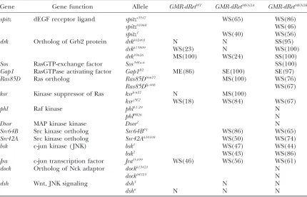
and mutations in orthologs of genes previously implicated in oncogenic mammalian Ret function (Table 1).
Ras pathway: Ligand-mediated or oncogenic activa-tion of Ret leads to binding of adaptor proteins, recruit-ment of the Grb2/SOS complex, activation of Ras, and induction of ERK activity (vanWeeringet al.1995; Ohiwa
et al.1997; Chiarielloet al.1998; Hayashiet al.2000;
Kurokawa et al.2001; Melillo et al. 2001). The Ras
pathway is remarkably well conserved between mam-mals and Drosophila. In a series of tests, we found that GMR-dRetWT,GMR-dRetMEN2B, andGMR-dRetMEN2Aall genet-ically interacted with Ras pathway components (Table 1). Mutations indrk (Grb2), Sos, Ras85D, and ksrall alle-viated (‘‘suppressed’’)GMR-dRet-mediated phenotypes. Conversely, GMR-dRetMEN2B
and GMR-dRetMEN2A
pheno-types were made more severe (‘‘enhanced’’) by muta-tions inGap1(RasGAP), a negative regulator of Ras.
GMR-dRetMEN2B
failed to interact with more downstream components of the Ras pathway, such asphl(Raf). Similar
results have been observed in modifier screens with the Sevenless RTK, illustrating that retinal phenotypes caused by activated RTKs are sensitive primarily to the gene dosage of upstream members of the Ras pathway. Mutations in the locus encoding the dEGFR ligand Spitz also suppressed GMR-dRet phenotypes, suggesting that reduction in parallel RTK signaling can inhibit the effects of dRetMEN2. Together, these data indicate thatdRet
over-expression led to activation of the Ras pathway, consistent with our immunohistochemical observations. Further-more, these data demonstrate that GMR-dRet flies can identify biochemical pathways linked to mammalian RetMEN2.
Src and Jun kinase pathways: GDNF-stimulated mam-malian Ret, RetMEN2A, and RetMEN2Bpromote Src kinase
ac-tivation in tissue culture (Melilloet al.1999; Encinas
et al. 2001). Src activity, in turn, is required for Figure 3.—GMR-dRetMEN2B di-rects excess proliferation, pattern-ing defects, excess neuronal differentiation, and ectopic ERK activation. Anterior is toward the right. (A and B) The larval eye field differentiates in a posterior-to-anterior wave, and the brackets denote the second wave of mito-ses; S-phase nuclei are visualized with BrdU (orange); note the ectopic S-phase cells in GMR-dRetMEN2B tissue. (C and D) Eye discs from GMR-dRetMEN2B flies (two copies) display a normal pat-tern of Boss staining (orange), in-dicating that early photoreceptor development (R8) is normal. Staining for Atonal (another marker for R8) inGMR-dRetMEN2B tissue is also normal (data not shown). These stains show that early development proceeds nor-mally before GMR-driven dRet
Ret-mediated cell survival, proliferation, and activation of ERK and AKT kinases, which themselves function in oncogenic Ret signaling (Chiariello et al. 1998;
Melilloet al.1999; Murakamiet al.1999b; Segouffin
-Cariou and Billaud 2000; Encinas et al. 2001).
Mutations in both Drosophila Src orthologs, Src42A andSrc64B(Takahashiet al.1996; Dodsonet al.1998),
suppressedGMR-dRetMEN2A
andGMR-dRetMEN2B
(Table 1), indicating that ectopic dRet requires Src activity for its function.
GDNF-stimulated Ret, RetMEN2A, and RetMEN2Bare also
known to promote JNK activation and c-jun phosphory-lation in numerous cell lines (Marshall et al. 1997;
Chiarielloet al.1998; Murakamiet al.1999a; Hayashi
et al.2000). This activity requires the Nck adaptor pro-tein (Murakamiet al.1999a). The Drosophila genome
contains a single JNK ortholog,basket(bsk), and a single c-jun ortholog, Jra. Both bsk and Jra mutations sup-pressed GMR-dRetMEN2A and GMR-dRetMEN2B (Table 1), indicating that dRet also requires JNK pathway activity. The dreadlocks(dock) locus encodes a Drosophila Nck ortholog (Garrityet al.1996);dockmutations failed to
modifyGMR-dRetMEN2B
(Table 1). JNK signaling can also be activated bydishevelled (dsh) (Boutroset al. 1998),
but mutations indshfailed to modifyGMR-dRet.
dRet modifier screens: Our data establish the GMR-dRet flies as a functional model for Ret signal trans-duction. Differences between the RetMEN2isoforms have
been proposed on the basis of previous mammalian work (Songyanget al.1995). To explore this issue, we
performed comparative genetic screens to identify factors common to all dRet isoforms and factors that distinguish one from another. We were especially in-terested in those genes that modified bothGMR-dRetMEN2 isoforms, as these would help identify (i) candidate factors that mediateRetMEN2
transformation, (ii) poten-tial ‘‘susceptibility loci’’ and/or tumor suppressors that promoteRetMEN2-dependent tumors when mutant, and, finally, (iii) attractive therapeutic targets for treatment of MEN2 tumors.
To this end, we used a genetic modifier screen to iden-tify second-site loci that enhanced or suppressed the GMR-dRet phenotypes. Modifiers represent genes that act as positive or negative regulators of dRet. We screened for dominant genetic modifiers. That is, we used hetero-zygotes: by altering only one genomic copy of each can-didate gene, we were able to test loci that are lethal when homozygous mutant. This approach allowed us to iden-tify truly novel functional dRetMEN2partners, providing
an importantin situcomplement to biochemical studies Figure 4.—Abnormal pattern-ing and differentiation of omma-tidia and interommaomma-tidial lattice cells in more mature GMR-dRetMEN2B retina. (A and B) Forty-two-hour pupal eye imaginal discs; apical profiles of cells are visual-ized with an antibody specific for the junctional protein Arma-dillo. (A) A wild-type pupal eye showing approximately a dozen ommatidia. Cells within and sur-rounding one ommatidium are labeled to indicate cone cells (c) and primary (1), secondary (2), and tertiary (3) pigment cells, and bristles (b) define an in-terommatidial lattice. (B) A single copy ofGMR-dRetMEN2Bresulted in a milder phenotype that included abnormal numbers of cone cells and a poorly patterned inter-ommatidial lattice. (C) GMR-dRetMEN2B (two copies) eyes show a marked lack of ommatidial orga-nization including an absence of clearly definable cells. The rounded cuboidal shape of these cells is typically seen in undiffer-entiated lattice cells early in nor-mal pupal retinal development. (D) Plastic sections from adult eye tissue. Wild type (inset) has a normal complement of seven photoreceptor neurons within each ommatidium; they are most easily seen by their rhabdomeres, which appear as solid blue circles in the section. Note that each rhabdomere array forms a stereotyped trapezoid that ‘‘points’’ upward (arrows in inset).GMR-dRetMEN2B(two copies) sections contain abnormally assembled and patterned ommatidia and large vacuolated spaces.
that have relied on testing candidate factors to identify RetMEN2effectors.
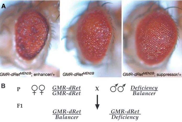
The screen design is diagrammed in Figure 5. Our first screen focused on genomic deficiencies, each of which typically removes dozens of genes.GMR-dRetMEN2B, GMR-dRetMEN2A, andGMR-dRetWTflies were crossed to flies heterozygous for deficiency mutations; each deficiency was maintained over a wild-type ‘‘balancer’’ chromo-some. We compared the following F1 progeny:
GMR-dRet; 1/1balancer
vs. GMR-dRet; Deficiency/1; GMR-dRet; Deficiency/1vs. GMR-dRet; and1/1when appropriate to control for balancer effects. Phenotypic differences between the two progeny types indicated that the mu-tations tested were genetic modifiers ofGMR-dRet.
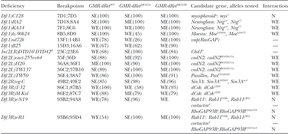
Comparative deficiency screen: We tested all three GMR-dRetisoforms using a set of 95 deficiencies that remove65% of the genome. Twenty-eight deficiencies that define at least 20 separate genomic regions genet-ically modified all threedRetisoforms (Tables 2 and 3). Candidate modifier genes for these regions were then identified through database searches and directly tested for genetic modification of the GMR-dRet isoforms (Tables 2 and 3). In some cases deficiencies overlapped, making identification of modifier regions more precise. We concentrated on candidate genes that regulate signal transduction, growth, proliferation, or differen-tiation within the eye. One caveat is that, for deficiencies
for which we could not identify matching modifier loci or overlapping modifier deficiencies, we cannot be confident that the modifier activity was caused by the deficiency or by another mutation elsewhere on the chromosome. Nevertheless, we identified 11 modifier loci with this approach, providing a crucial proof of principal for a broader, unbiased screen of single gene mutations.
Deficiency screens: suppressors of GMR-dRetWT
, GMR-dRetMEN2A
, and GMR-dRetMEN2B
: Nine genomic re-gions that contain GMR-dRet suppressors were identi-fied. Several of these regions contain known regulators of Ras signaling. For example,Df(3R)by10, a suppressor of both GMR-dRetMEN2 isoforms, deletes 85D8;85F1 (Table 2), a region that contains the Ras ortholog Ras85D. The suppressing deficiencies Df(2L)net-PMF (21A1;21B8) and Df(2L)al (21B8;21D1) contain the geneskismetandebi, respectively (Table 2). Bothkismet andebiare involved in chromatin remodeling and are positive regulators of dEGFR and Ras signaling in the retina (Therrienet al.2000; Tsudaet al.2002).
Muta-tions in either locus suppressedGMR-dRetphenotypes. Two interacting deficiencies are of special note. GMR-dRetWT
,GMR-dRetMEN2A
, andGMR-dRetMEN2B
were all sup-pressed byDf(2L)TW161, which removes a region that includes the endogenousdRetlocus (Table 2). Reduced endogenousdRet expression could suppressGMR-dRet
TABLE 1
GMR-dRetMEN2genetically interacts the Ras, Src, and JNK pathways
Gene Gene function Allele GMR-dRetWT GMR-dRetMEN2A GMR-dRetMEN2B
spitz dEGF receptor ligand spitzs3547 WS(65) WS(86)
spitz01068 WS(46)
spitz1 WS(40) WS(56)
drk Ortholog of Grb2 protein drkk02401 N N SS(95)
drkk13809 WS(23) N WS(100)
drk10626 MS(100) WS(24) SS(100)
Sos RasGTP-exchange factor Sos34Ea-6 SS(100)
Gap1 RasGTPase activating factor Gap1B2 ME(86) SE(100) SE(97)
Ras85D Ras ortholog Ras85D06677 MS(100) WS(76)
Ras85DDc40b WS(67)
ksr Kinase suppressor of Ras ksrS-627 N MS(100)
ksrj5E2 WS(18) WS(84) WS(67)
phl Raf kinase phl11.29 N
phlPB26 N
Dsor MAP kinase kinase Dsor1 N
Src64B Src kinase ortholog Src64BP1 WS(86) WS(65)
Src42A Src kinase ortholog Src42Ak10108 WS(50) WS(74)
bsk c-jun kinase ( JNK) bsk1 WS(47) WS(44)
bsk2 WS(43) WS(86)
Jra c-jun transcription factor JraIA109 WS(46) WS(56) WS(61)
dock Ortholog of Nck adaptor dockk13421 N
dock04723 N
dsh Wnt, JNK signaling dsh3 N N
dsh6 N N N
phenotypes by reducing overalldRet levels and would indicate that dRet is active in the retina. However, no dRetmutations are currently available to test this possi-bility. Also,Df(3R)DG2andDf(3R)Cha7define a region (91B2;91F5) that includes glass, the transcriptional
activator that drives gene expression from the GMR promoter. As expected, loss-of-functionglassmutations were confirmed as GMR-dRetWT
, GMR-dRetMEN2A , and GMR-dRetMEN2B
suppressors, presumably due to reduc-tion inGMRactivity.
Figure5.—Enhancer-suppressor screen for modifiers of GMR-dRetMEN2. (A) Examples of
GMR-dRetMEN2B modifier phenotypes, which were scored using a dissect-ing microscope. Enhanced flies had a worsened phenotype and suppressed flies had a milder phe-notype. (B) Diagram of parental (P) genetic crosses and F1progeny genotypes. GMR-dRetMEN2B,
GMR-dRetMEN2A, and GMR-dRetWT flies were crossed to flies heterozygous for loss-of-function deficiency mu-tations; each mutation was main-tained over a wild-type balancer chromosome (balancer chromo-somes contain breakpoints that prevent meiotic recombination and carry visible markers, but are otherwise genetically wild type and are denoted as such here). The phenotypes of different classes of progeny were compared and the penetrance was calculated for any observed genetic interactions.
TABLE 2
Deficiencies suppressGMR-dRetWT,GMR-dRetMEN2A, andGMR-dRetMEN2B
Deficiency Breakpoints GMR-dRetWT GMR-dRetMEN2A GMR-dRetMEN2B
Candidate gene,
alleles tested Interaction
Df(2L)net-PMF 21A1;21B8 WS(100) WS(86) WS(83) kismet:kisk10237, kisk13416 WS
Df(2L)al 21B8;21D1 SS(100) MS(100) SS(100) ebi:ebik16213 WS
Df(2L)TW161 38A6;40B1 WS(75) SS(96) SS(100) diaphanousa:diak07135 N
dRet —
Df(2R)X58-12 58D1;59A MS(80) SS(100) SS(100) plexus:pxk08316 WS
Df(2R)59AD 59A1;59D4 WS(78) SS(100) SS(100) —
Df(3L)Pc-2q 78C5;79A1 MS(100) WS(100) WS(100) SAKb:SAKc06612 N
Df(3R)ME15 81F3;82F7 MS(90) SS(97) SS(100) Gelsolinc —
Df(3R)Antp17 84A5;84D14 WS(67) SS(100) MS(100) —
Df(3R)Hu 84A6;84B6, 84D4;84F2 WS(76) MS(100) MS(82) —
Df(3R)by10 85D8;85F1 nt WS(97) WS(77) Ras85D:Ras85D06677,
Ras85DDc40b
WS-MS
Df(3R)DG2 89E1;91B2 SS(100) SS(100) SS(100) glass:gl2 SS
Df(3R)Cha7 90F1;91F5 WS (89) WS(100) WS(100) glass:gl2 SS
Each deficiency is listed by name. The cytological breakpoints for each are derived from the FlyBase and Bloomington Stock Center online databases. Genetic interactions are indicate according to strength of phenotype: W, weak; M, moderate; S, strong; and type of interaction: S, suppressor; N, no interaction; nt, not tested. Therefore, SS is a strong suppressor. Parentheses indicate the penetrance of the interaction calculated as a percentage. See text for details on candidate genes. —, indicates that there were no mutants in the candidate gene available for testing.
adiaencodes a formin homology protein involved in cytoskeleton remodeling and Rho signaling (Prokopenkoet al.1999). bEncodes a serine/threonine kinase, which is a putative effector of Src signaling (Yamashitaet al.2001).
cGelsolinencodes an actin-binding protein linked to Src signaling (Chellaiahet al.2000).
Deficiency screens: enhancers ofGMR-dRetWT, GMR-dRetMEN2A, andGMR-dRetMEN2B:Sixteen deficiencies that
define 11 genomic regions were isolated as enhancers of GMR-dRet(Table 3). Again, some of these deficiencies remove genes that are known negative regulators of RTKs and the Ras pathway (Table 3). The enhancing deficienciesDf(1)RA2andDf(1)KA14together define a region, 7F1;8A4, that includesNeuroglian(Nrg). In the developing eye,Nrgnegatively regulates dEGFR activity (Islam et al. 2003). Reducing Nrg gene dosage
en-hanced GMR-dRet activity, indicating that it also can negatively regulate dRet activity (Table 3). Df(2R)vg-C (49B2;49E2) behaved as a strong enhancer of GMR-dRetWT,
GMR-dRetMEN2A, and
GMR-dRetMEN2B (Table 3). Sin3A, which also enhanced all three forms ofdRet, maps to this interval and encodes a chromatin-remodeling factor that is a known regulator of the Ras pathway (Neufeldet al.1998; Rebayet al.2000).
Enhancer regions that contained regulators of Src signaling were also recovered. For example, two over-lapping deficiencies,Df(3R)T-32andDf(3R)M-Kxl, define 86E2;87B5. Smaller overlapping deficiencies (Kusano
et al. 2001) were used to more finely map this region: Df(3R)T-61(86E3;87A9) andDf(3R)pros235(86E2;86F4)
enhancedGMR-dRetMEN2B, whereasDf(3R)pros640(86E2; 86E11) and Df(3R)T7 (86F1;86F4) failed to modify theGMR-dRetMEN2Bphenotype, identifying an enhancer region within 86E11;86F1. A strong candidate that maps to 86E17 isdCsk, which encodes the Drosophila C-terminal Src kinase ortholog (Readet al.2004). A
strong hypomorphic mutation in dCsk enhanced GMR-dRetMEN2B
andGMR-dRetWT
. Mutations that reduce dCsk function lead to hyperactivation of dSrc activity (Readet al. 2004), suggesting that dRet acts through
Src. Other modifier deficiencies also deleted regulators of Src signaling (Tables 2 and 3). Unfortunately, muta-tions in many of these genes were not available, but as more Drosophila genes are mutated, these candidates and others can be tested.
Disregulation of cell adhesion contributes to tumor-igenesis and enhancing deficiencies identified multiple regions that contain regulators of cell adhesion: Df(1)lz-90b24(8B5;8D9) uncovers a locus encoding the Moesin ortholog Moe (Specket al.2003);Df(2L)TW50
(36E04-F01;38A06-07) defines a region that includes Drosoph-ila Paxillin, a component of focal adhesions; and a series of enhancing deficiencies [Df(2L)TW50, Df(2L)H20, andDf(2L)TW137] define two genomic regions, 36C2;
TABLE 3
Deficiencies enhanceGMR-dRetWT,GMR-dRetMEN2A, andGMR-dRetMEN2B
Deficiency Breakpoints GMR-dRetWT GMR-dRetMEN2A GMR-dRetMEN2B Candidate gene, alleles tested Interaction
Df(1)C128 7D1;7D5 SE(100) SE(100) SE(100) myospheroida:mys1 N
Df(1)RA2 7D18;8A4 SE(100) ME(100) ME(100) Neuroglian:Nrg14, Nrg17 WE
Df(1)KA14 7F1;8C6 WE(100) WE(100) ME(100) Neuroglian:Nrg14, Nrg17 WE
Df(1)lz-90b24 8B5;8D9 SE(100) WE(45) SE(100) Moesin: MoeG0404, MoeG0415 WE
Df(1)sd72b 13F1;14B1 WE(78) WE(26) ME(100) vap(RasGAP) —
Df(1)B25 15D3;16A6 WE(67) WE(62) WE(90) —
In(2LR)DTD16LDTD42R 23C;23E6 WE(68) SE(100) ME(84) Chd1b —
Df(2L)cact-255rv64 35F;36D SE(88) ME(92) SE(100) cadN2:cadN2BG02611a WE
Df(2L)H20 36A8;36F1 ME(100) SE(90) ME(100) cadN2:cadN2BG02611a WE
Df(2L)TW137 36C2;37B10 SE(89) SE(100) ME(100) cadN2:cadN2BG02611a WE
Df(2L)TW50 36E4;38A7 WE(86) SE(100) ME(91) Paxillin, PaxEY02020 WE
Df(2R)vg-C 49B2;49E2 SE(85) SE(98) SE(96) Sin3A:Sin3A08269, Sin3Aex4 WE
Df(3R)T-32 86C1;87B5 WE(100) WE (58) WE(93) dCsk:dCskj1D8 WE
Df(3R)M-Kxl 86E2;87C7 WE(88) ME(79) WE(79) dCsk:dCskj1D8 WE
Df(3R)e-N19 93B2;94A8 WE(78) SE(96) WE Rab11c:Rab1193Bi, Rab11j2D1 N
cortactind —
RhoGAP93Be:RhoGAP93BEY06358 N
Df(3R)e-R1 93B6;93D4 WE(54) SE(100) ME(100) Rab11c:Rab1193Bi, Rab11j2D1 N
cortactind —
RhoGAP93Be:RhoGAP93BEY07163 N
Each deficiency is listed by name. The cytological breakpoints for each are derived from the FlyBase online database and the Bloomington Stock Center. Genetic interactions are indicated according to strength of phenotype: M, moderate; S, strong; W, weak; and type of interaction: S, suppressor; E, enhancer; N, no interaction. Therefore, SS is a strong suppressor. Parentheses indicate penetrance of the interaction calculated as a percentage. —, indicates that there were no mutants in the candidate gene available for testing.
a
mysencodes a PS integrinb-subunit, which are proteins that regulate focal adhesions and Src signaling (Brown1994). b
Chd1encodes a chromatin-remodeling protein very similar to dMi-2 (Stokeset al.1996). c
Rab11encodes a small GTPase that regulates endosomal trafficking of transmembrane receptors (Culliset al.2002). d
cortactinencodes a known substrate for Src kinases (Wuet al.1991; Okamuraand Resh1995). e
36D and 36E4;36F1, one of which containscadN2, an N-cadherin ortholog, which mediates adherins junc-tions and regulates JNK signaling (Charrasse et al.
2002). Mutations in Moe, Paxillin, and cadN2 all en-hanced GMR-dRet isoforms (Table 3). Finally, not all deficiencies had clear candidate modifiers, highlighting the difficulties in using chromosomal deficiencies to identify truly novel modifiers.
Modifiers that may distinguish GMR-dRetMEN2A from GMR-dRetWT
and GMR-dRetMEN2B
: RetMEN2B has been
proposed to signal through pathways not targeted by
RetMEN2A or normal Ret signaling. However, no
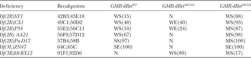
de-ficiency modifiers clearly distinguishedGMR-dRetMEN2B activity from that ofGMR-dRetMEN2A andGMR-dRetWTin our screen. Surprisingly, seven deficiencies distin-guished dRetMEN2A from dRetMEN2B anddRetWT (Table 4). Most of these showed weak interactions with onedRet isoform but not another; these data may simply reflect an inability to unambiguously score weak interactions. However, two deficiencies enhanceddRetMEN2A
but sup-presseddRetanddRetMEN2B
(Table 4). The C695R MEN2A mutation likely causes constitutive dimerization of the dRet extracellular domain, and perhaps these two deficiencies remove loci that regulate this process to affect dRetMEN2Ain a unique manner. However, we did
not finely map these regions or identify any correspond-ing loci for these deficiencies.
P-element screen: As a companion to the candidate gene and deficiency screens,P-element lines were simi-larly screened forGMR-dRetmodifiers. As part of a large-scale gene disruption project, the Berkeley Drosophila Genome Project (BDGP) has collected thousands of lethal mutations associated with stable insertions of modified P-element transposons (Deak et al. 1997;
Spradlinget al.1999). These collections, available from
the Drosophila stock centers, contained.2000 individ-ual disrupted genes at the time of our screen. The purpose of screening P-element lines was to identify unexpected genes that dominantly modifiedGMR-dRet. Such genes may represent novel regulators of
Ret-dependent oncogenesis. This screen allowed us to expand from the candidate approach inherent in the deficiency screen to take an unbiased functional genomics approach.
GMR-dRetMEN2B
flies were crossed to flies heterozygous for 2524 separateP-element mutations. For this screen we utilized flies from linesGMR-dRetMEN2B
-3Band GMR-dRetMEN2B
-3C, which carried single transgenes that confer intermediate phenotypes; these lines were especially sensitive to mutations in known RTK effectors. The screen design was similar to that of our comparative deficiency screen (Figure 5). Again, each cross gave two classes of F1progeny that were compared to each other.
Enhancers and suppressors were rescreened twice to confirm genetic interactions and to determine pene-trance. A total of 90 initialP-element modifiers were re-covered. These initial modifiers were then put through a series of genetic tests to remove nonspecific modifiers and to confirm specific modifier loci.
Secondary screens to eliminate nonspecific GMR-dRetMEN2B modifiers: We employed several secondary tests to elim-inate modifiers that alteredGMR-dRetMEN2B
phenotypes for reasons other than their effects on dRetMEN2B. Eleven
enhancers exhibited dominant rough-eye phenotypes even in the absence ofGMR-dRetMEN2B
constructs; these were discarded. Modifiers were screened with additional GMR-dRetMEN2Blines to determine if genetic interactions were line specific. Two modifiers interacted with only oneGMR-dRetMEN2Bline: these were discarded since they may alterGMR-dRetMEN2Bphenotypes due to site-specific insertion effects. To eliminate modifiers that acted on theGMRpromoter, all confirmed modifiers were tested with multiple unrelatedGMR-containing transgenes that also cause a dominant rough-eye phenotype (supplemen-tal Table S1 at http://www.genetics.org/supplemen(supplemen-tal/); five modifiers were eliminated by these tests. Together, these tests left 72 modifiers for further study.
Secondary screens for genetic verification of GMR-dRet modifiers:ManyP-element lines harbor additional muta-tions not associated with the P insertion (Spradling
et al.1999). Therefore, we attempted to verify that the
TABLE 4
Deficiencies that differentially interact withGMR-dRetMEN2A
Deficiency Breakpoints GMR-dRetWT GMR-dRetMEN2A GMR-dRetMEN2B
Df(2R)ST1 42B3;43E18 WS(15) N WS(88)
Df(2R)CX1 49C1;50D2 WS(48) WE(40) WS(93)
Df(2R)P34 55E2;56C11 WS(10) WE(24) MS(87)
Df(2R) AA21 56F9;57D12 WS(67) N MS(98)
Df(2R)Pu-D17 57B4;58B SS(97) N MS(100)
Df(3L)ZN47 64C;65C SE(100) N SE(100)
Df(3R)Dl-BX12 91F1;92D6 N WS(89) WS(17)
Each deficiency is listed by name. Breakpoints are derived from the FlyBase online database and the Bloo-mington Stock Center. Genetic interactions are indicated according to strength of the phenotype: W, weak; M, moderate; S, strong; and type of interaction: S, suppressor; E, enhancer; N, no interaction. For example, WS is a weak suppressor. Parentheses indicate the penetrance of the interaction calculated as a percentage.
genes disrupted by P-element insertions were indeed GMR-dRet modifiers. Some P-element lines were out-crossed to remove second-site mutations by recombina-tion and retested with GMR-dRetMEN2B (supplemental Table S2 at http://www.genetics.org/supplemental/); three lines lost their modifier activity upon out-crossing and were discarded. As another approach, we mobilized the P-element insertion in several lines using P trans-posase: ‘‘clean’’ excision of the P element out of the locus should revert modifier activity if it is due to P -element disruption (supplemental Table S2). For three lines, excision did not revert modifier activity, even in cases where excision reverted the insertion-associated lethality. Second-site mutations were the likely GMR-dRetmodifiers in these three lines. Sometimes excision did not revert the lethality of the affected chromosome, suggesting that either the excision process failed to restore the disrupted gene or the chromosome carried second-site lethal mutations; data from such lines were inconclusive. These tests left 66 lines.
Secondary screens with GMR-dRetMEN2Aand GMR-dRetWT: The purpose of these secondary screens was to distin-guish between modifiers common to alldRet isoforms and dRetMEN2B
-specific modifiers. Factors common to GMR-dRetMEN2B
and GMR-dRetMEN2A
were of particular interest since such factors may represent genes that are important forRetMEN2
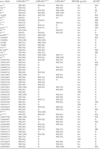
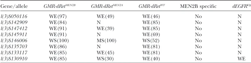
-mediated tumorigenesis. Our phenotypic data and deficiency screen suggested that all three isoforms of dRet were largely equivalent. We further tested this idea by screening GMR-dRetMEN2B modifiers for their ability to modify GMR-dRetWTand GMR-dRetMEN2A flies (Table 5). AllGMR-dRetMEN2B modi-fiers tested genetically interact withGMR-dRetMEN2Aand/ orGMR-dRetWT(Table 5), again suggesting that all three dRet isoforms rely on the same pathways. Interestingly, GMR-dRetMEN2A insertions that had strong phenotypes frequently showed weak or no interactions with weak enhancers of GMR-dRetMEN2B
, although insertions of GMR-dRetMEN2A
that gave rise to milder phenotypes did interact with most of these weak enhancers. This sug-gests that dRetMEN2Ais less sensitive than dRetMEN2B to
alterations in gene dosage of downstream targets, an observation that may explain the failure of some deficiencies to interact withdRetMEN2A(see above).
Secondary screens with dEGFRElp
: To identify dRetMEN2B modifiers that are also general regulators of RTKs, we rescreened them using dEGFRElp, which encodes an activated allele of the ortholog dEGFR.dEGFRElp/1flies have a dominant rough-eye phenotype caused by ectopic dEGFR activity (Lesokhinet al.1999). AllGMR-dRetMEN2B
modifiers were crossed todEGFRElpto determine their ability to modify an independent RTK (Table 5). As ex-pected, members of theRaspathway modifieddEGFRElp
. Surprisingly, many GMR-dRetMEN2B
modifiers failed to genetically interact with dEGFRElp
, suggesting that dRetMEN2Bmay act through a subset of genes not utilized
by dEGFR.
Molecular identification, verification, and function of GMR-dRet modifier loci: The major advantage to screening P-element lines is that the transposon pro-vides a molecular tag for identifying the disrupted locus. To help associateP-element modifier lines with corre-sponding disrupted genes, we sequenced genomic DNA flanking the transposon insertion sites. Concomitantly, other groups, including BDGP, also generated sequen-ces for many lines. Genomic regions flankingP-element insertions were mapped to the full Drosophila genome sequence. The surrounding sequences were searched for known genes, matching ESTs, and predicted ORFs using the FlyBase Genome and Gadfly browsers and annotated GenBank files. To date, flanking genomic sequence has been obtained and analyzed for 34 P -element modifiers (supplemental Table S3 at http:// www.genetics.org/supplemental/). A total of 33 yielded sequence that indicated that the P inserted in or near a gene. Other groups have characterized an additional six P-element modifiers and identified the disrupted genes (Table 4) (Salzberget al.1997; Spradlinget al.1999).
Thus, we were able to associate 40P-element modifier lines with disrupted genes, which were candidate mod-ifier loci.
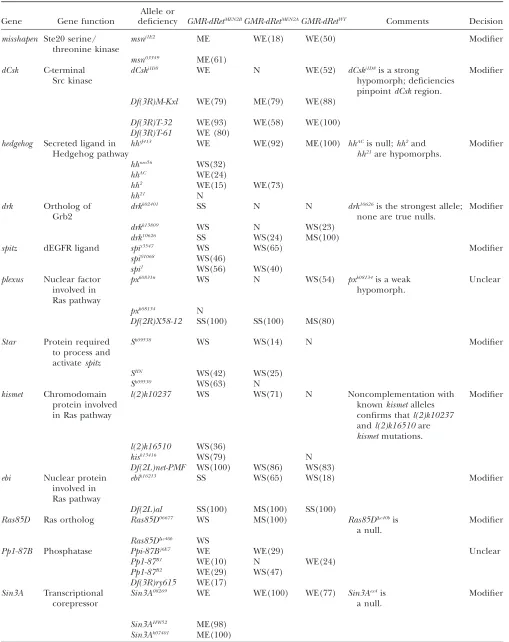
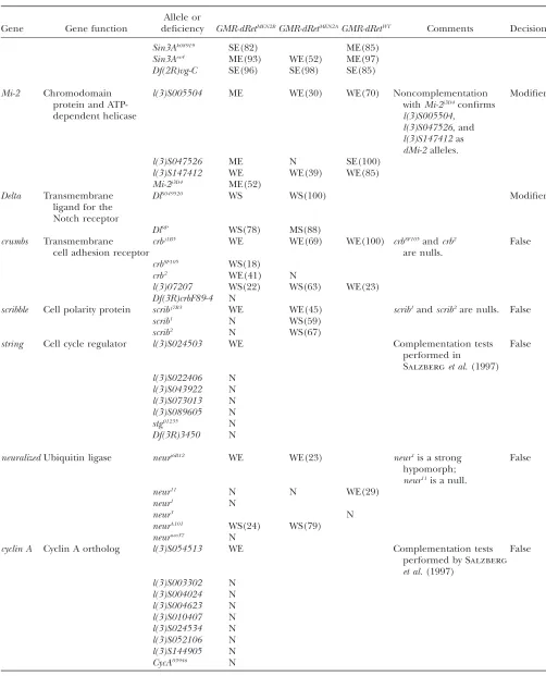
On the basis of previous studies, sequence data, and our own complementation tests, we identified addi-tional alleles for many modifiers and tested their ability to modify our GMR-dRet constructs (Table 6). If the ability of aP-element line to genetically modify GMR-dRetMEN2Bis due to disruption of a gene associated with the insertion, then other mutant alleles or matching deficiencies for that gene should show similar modifier activity. We concentrated on testing null alleles when-ever possible. Additional alleles of fiveP-element lines failed to modify GMR-dRet phenotypes: these lines probably contain second-site mutations that modified GMR-dRetMEN2B
(Table 6). In other cases, no additional alleles were available or problems with the strength of available alleles made such tests inconclusive (Table 6; data not shown). Importantly, we verified 12 genes from 16 P-element lines as GMR-dRet modifiers because multiple alleles or matching deficiencies for these genes proved to have similar modifier activity (Table 6). These verified loci were misshapen, Ras85D, drk, kismet, ebi, Sin3A, dMi-2, spitz, Star, Delta, hedgehog, and dCsk. We further investigated the functions of some of these loci. Ras pathway and related loci:As expected, we identified members of the Ras signal transduction pathway as modifiers ofGMR-dRet(Table 6). These include muta-tions in canonical pathway members such asdrk, a Grb2 ortholog, and Ras85D in addition to other regulators such asspitz,Star,kismet, andebi. Mutations that affect signaling through the dEGFR pathway also interacted with dRet. These include mutations inspitz, a ligand for dEGFR, andStar, a protein required for proper process-ing of Spitz protein (Leeet al.2001; Urbanet al.2002).
TABLE 5
Genetic interactions betweenGMR-dRetMEN2BP-element modifiers andGMR-dRetWT,GMR-dRetMEN2A, anddEGFRElp
Gene/allele GMR-dRetMEN2B GMR-dRetMEN2A GMR-dRetWT MEN2B specific dEGFRElp
msnj1E2 ME(88) WE(18) WE(50) No WE
dCskj1D8 WE(76) N WE(52) No N
hhrJ413 WE(84) WE(92) ME(100) No SE
crbj1B5 WE(93) WE(69) WE(100) No N
l(3)j2D5 ME(95) WE(76) WE(82) No WS
drkk02401 SS(95) N N? Noa WS
spis3547 WS(86) WS(65) No MEb
pxk08316 WS(73) N WS(54) No WE
Sk09538 WS(67) WS(14) N Noa MEb
kismetk10237 WS(80) WS(71) N No ND
drkk13809 WS(100) N WS(23) No N
ebik16213 SS(85) WS(65) WS(18) No N
Ras85D06677 WS(76) MS(100) No ND
l(3)06803 WS(95) MS(91) No WE
l(3)06906 WS(100) MS(100) No N
l(3)j5B6 WE(97) ME(100) No N
l(3)j4B9 WE(95) WE(68) No N
neurj6B12 WE(94) WE(47) No N
Pp1-87Bj6E7 WE(85) WE(29) No WE
scribj7B3 WE(98) WE(45) No ME
Sin3A08269 WE(86) WE(100) WE(77) No N
drk10626 SS(100) WS(24) MS(100) No N
l(3)S005504 ME(91) WE(30) WE(70) No N
l(3)S001405 WE(95) N WE(58) No WE
l(3)S003704 WE(94) N ND WE
l(3)S000718 WE(96) N WE(47) No N
l(3)S009515 WE(93) N WE(62) No N
l(3)S000710 WE(82) WE(26) No N
l(3)S012805 WE(100) N WE(97) No N
l(3)S016805 WE(88) WE(12) WE(96) No N
l(3)S023708 ME(94) WE(15) N No N
l(3)S023549 WE(82) WE(50) No N
l(3)S024833 WE(96) WE(33) No N
l(3)S023901 WE(100) WS(39) WE(79) No N
l(3)S022231 WE(91) N WE(80) No N
l(3)S024503 WE(92) N ND N
l(3)S026238 WE(100) N WE(65) No N
l(3)S026421 WE(94) N WE(90) No N
l(3)S024329 WE(88) WE(71) WE(52) No N
l(3)S049706 ME(85) N WE(82) No ND
l(3)S054513 WE(79) N ND N
l(3)S046604 WE(96) WE(52) ME(95) No N
l(3)S049902 WE(93) N WE(35) No WE
DlS049520 WS(85) WS(100) No N
bonS048706 WE(93) WE(50) WE(81) No WS
l(3)S047526 ME(100) N SE(100) No WE
l(3)S066607 WE(96) WE(22) WE(96) No N
l(3)S057101 WE(95) N WE(59) No WE
l(3)S068808 WE(93) WE(32) WE(84) No N
l(3)S083407 WE(90) WE(24) No N
l(3)S063512 WE(81) WE(15) WE(75) No ME
l(3)S056113 WE(88) N WE(50) No N
l(3)S090101 WE(100) WE(38) WE(81) No N
l(3)S090114 WE(79) N WE(76) No N
l(3)S141715 WE(84) N ND N
l(3)S136603 WE(97) N WE(90) No N
l(3)S092708 WE(94) N WE(83) No N
l(3)S003003 WE(82) N WE(100) No WE
(continued)
dominant rough-eye phenotypes, the Star mutation isolated in this screen had no phenotype on its own and, in fact, suppressed GMR-dRet rough-eye pheno-types. Mutations inkismet, which encodes an ortholog of chromatin-remodeling factors and is linked to dEGFR activity (Therrienet al. 2000), and ebi, a
chromatin-remodeling protein and nuclear regulator of bothRas andNotchsignaling (Donget al.1999; Tsudaet al.2002),
also modifiedGMR-dRetphenotypes. In addition,plexus, which encodes a nuclear factor, has also been implicated in RTK signaling (Matakatsuet al.1999). The ability of
dEGFR-specific regulators to modifyGMR-dRetsuggests either that these factors may also regulate the dRet recep-tor on some level or that genetic reduction of signal-ing through a parallel RTK may alleviate the effects of dRet.
misshapen: Mutations in misshapen (msn), which en-codes a Ste20 serine/threonine kinase, enhanced GMR-dRet (Table 6), suggesting that msn acts as a negative regulator of dRet. msn also enhanced dEGFRElp
. Msn activity is required for cytoskeletal remodeling and cell shape change during development in processes such as axon pathfinding and dorsal closure (Treismanet al.
1997; Suet al.1998; Ruanet al.1999) and can also act as a
negative regulator of Ras pathway signaling (Huangand
Rubin 2000). Msn functions in at least two separate
signaling pathways: downstream ofdock, the Drosophila ortholog of the Nck adaptor protein, and upstream ofbsk, the Drosophila ortholog of JNK (Suet al.1998; Paricio
et al.1999; Ruanet al.1999, 2002). Mutations indockdid
not modify GMR-dRetMEN2. Mutations in bsk did modify GMR-dRetMEN2B
, althoughbskmutations acted as suppres-sors (Table 1). Thus,bskandmsnmodifiedGMR-dRetin opposite directions, suggesting either thatmsnandbskare not in a simple linear pathway or that, in response to dRet, msnacts in a pathway that is separate fromdockorbsk.
Hedgehog and Delta-Notch signaling pathways: Muta-tions inDelta, which encodes a membrane-bound ligand of the Notch receptor, were found to be suppressors of GMR-dRet (Table 6). This interaction suggests a func-tional connection between Notch signaling and dRet activity. Connections between Notch and Ras pathway signaling have previously been noted: for example, mutations inDeltamodify Ras signaling (Maixneret al.
1998),Deltaexpression is regulated byRasactivity in the eye (Tsudaet al.2002), andNotchandRassignaling act
together in a number of developmental models (e.g., Baker and Rubin 1992; Miller and Cagan 1998;
Carmenaet al.2002). Mutations inhedgehog(hh), which
encodes a secreted activator of the Hh/Smo/Ptc signal transduction pathway, were also found to modify GMR-dRet signaling. Hh is expressed in the larval eye epithelium by maturing ommatidia and directs proper progression of the morphogenetic furrow (Heberlein
et al. 1995); its modification of GMR-dRetmay reflect disruption of furrow progression by aberrant dRet -expressing ommatidia. Alternatively, Hh is also known to modify Ras85D function (Karimet al.1996), and it
may modifydRetfunction through this capacity. dMi-2, Sin3A, and histone deacetylase complexes:We also identified components of the SIN3 and NuRD histone deacetylase complexes (reviewed in Ahringer2000), a
category of genes that has not been previously linked to Ret. For example, multiple alleles of Sin3A acted as GMR-dRetenhancers (Table 6). Sin3A, a part of the SIN3 complex, is a transcriptional corepressor (Neufeldet al.
1998; Pennettaand Pauli1998).Sin3Amutations also
dominantly enhance eye phenotypes caused by over-expression of the Yan transcriptional repressor (Rebay
et al. 2000) and dominantly suppress Sina overexpres-sion eye phenotypes (Neufeld et al. 1998); perhaps
significantly, both nuclear factors are downstream
TABLE 5 (Continued)
Gene/allele GMR-dRetMEN2B GMR-dRetMEN2A GMR-dRetWT MEN2B specific dEGFRElp
l(3)S050116 WE(97) WE(49) WE(46) No N
l(3)S142909 WE(84) N WE(85) No N
l(3)S147412 WE(91) WE(39) WE(85) No N
l(3)S145911 WE(91) N WE(69) No N
l(3)S146006 WS(100) MS(100) WS(52) No N
l(3)S135703 WE(86) N WE(81) No N
l(3)S133117 WE(85) WE(45) WE(81) No N
l(3)S130910 WE(85) WS(30) WE(40) No WE
EachGMR-dRetMEN2Bmodifier was crossed toGMR-dRetWTand/orGMR-dRetMEN2Aconstructs to determine the specificity ofdRetMEN2genetic interactions. Not allP-element lines could be tested since some lines died and were no longer available from the stock centers: these are indicated as ND (not determined).
a
Other alleles ofStaranddrkinteract withGMR-RetMEN2A(Table 6). This is probably due to the hypomorphic nature of the alleles isolated in the screen.
b
TABLE 6
Genetic interactions betweenGMR-dRetisoforms and additional alleles of candidateP-element modifiers
Gene Gene function
Allele or
deficiency GMR-dRetMEN2BGMR-dRetMEN2AGMR-dRetWT Comments Decision
misshapen Ste20 serine/ threonine kinase
msnj1E2 ME WE(18) WE(50) Modifier
msn03349 ME(61)
dCsk C-terminal Src kinase
dCskj1D8 WE N WE(52) dCskj1D8is a strong
hypomorph; deficiencies pinpointdCskregion.
Modifier
Df(3R)M-Kxl WE(79) ME(79) WE(88)
Df(3R)T-32 WE(93) WE(58) WE(100)
Df(3R)T-61 WE (80)
hedgehog Secreted ligand in Hedgehog pathway
hhrJ413 WE WE(92) ME(100) hhACis null;hh2and
hh21are hypomorphs.
Modifier
hhneo56 WS(32)
hhAC WE(24)
hh2 WE(15) WE(73)
hh21 N
drk Ortholog of Grb2
drkk02401 SS N N drk10626is the strongest allele; none are true nulls.
Modifier
drkk13809 WS N WS(23)
drk10626 SS WS(24) MS(100)
spitz dEGFR ligand spis3547 WS WS(65) Modifier
spi01068 WS(46)
spi1 WS(56) WS(40)
plexus Nuclear factor involved in Ras pathway
pxk08316 WS N WS(54) pxk08134is a weak
hypomorph.
Unclear
pxk08134 N
Df(2R)X58-12 SS(100) SS(100) MS(80)
Star Protein required to process and activatespitz
Sk09538 WS WS(14) N Modifier
SIIN WS(42) WS(25)
Sk09530 WS(63) N
kismet Chromodomain
protein involved in Ras pathway
l(2)k10237 WS WS(71) N Noncomplementation with
knownkismetalleles confirms thatl(2)k10237
andl(2)k16510are
kismetmutations.
Modifier
l(2)k16510 WS(36)
kisk13416 WS(79) N
Df(2L)net-PMF WS(100) WS(86) WS(83)
ebi Nuclear protein involved in Ras pathway
ebik16213 SS WS(65) WS(18) Modifier
Df(2L)al SS(100) MS(100) SS(100)
Ras85D Ras ortholog Ras85D06677 WS MS(100) Ras85DDc40bis
a null.
Modifier
Ras85DDc40b WS
Pp1-87B Phosphatase Ppi-87Bj6E7 WE WE(29) Unclear
Pp1-87B1 WE(10) N WE(24)
Pp1-87B2 WE(29) WS(47)
Df(3R)ry615 WE(17)
Sin3A Transcriptional corepressor
Sin3A08269 WE WE(100) WE(77) Sin3Aex4is
a null.
Modifier
Sin3AHW52 ME(98)
Sin3Ak07401 ME(100)
(continued)