Locus of
Drosophila simulans
Xian Zhang and Thomas H. Eickbush
1Department of Biology, University of Rochester, Rochester, New York 14627 Manuscript received November 19, 2004
Accepted for publication February 8, 2005
ABSTRACT
The rRNA gene (rDNA) loci of all arthropod lineages contain non-LTR retrotransposable elements that have evolved to specifically insert into the 28S rRNA genes. Extensivein vitroexperiments have been conducted to investigate the mechanism of R2 retrotransposition but little is known of the insertion frequency or cellular factors that might regulate R2 activity. In this article, isofemale lines obtained from a population ofDrosophila simulanswere surveyed for recent R2 insertions. Within most lines, all individuals showed the same collection of R2 insertions, providing no evidence for recent R2 activity. However, in a few of the isofemale lines, virtually all individuals differed in their R2 insertion profiles. The descendants of individual pairs of flies from these “active lines” rapidly accumulated new insertions. The frequent insertion of new R2 elements was associated with the elimination of old R2 elements from the rDNA locus. The existence of lines in which R2 retrotransposes frequently and lines in which the elements appear dormant suggests that cellular mechanisms that can regulate the activity of R2 exist. Retrotransposition activity was correlated with the number of full-length R2 elements but not with the size of the rDNA locus or the number of uninserted units.
T
RANSPOSABLE elements compose a significant other animal phyla (KojimaandFujiwara2004). When units within the rDNA array are inserted by R1 and/or proportion of eukaryotic genomes and have playedan important role in their evolution. To understand the R2, they can no longer synthesize functional 28S rRNA (Long and Dawid 1979; Jamrich and Miller 1984; dynamics of transposable element insertions, attempts
have been made, most often in the genus Drosophila, EickbushandEickbush2003). Despite this deleterious effect on the host, R1 and R2 elements appear to have to monitor the insertion of transposable elements over
been maintained in arthropods by vertical transmission time. Most studies have detected no activity or activity
since the origin of the phylum (Burkeet al.1998, 1999; on the order of 10⫺5–10⫺3new insertions per copy per
Gentileet al.2001). The long-term maintenance of R1 generation (NuzhdinandMackay1995; Dominguez
and R2 in arthropods is achieved without close regula-andAlbornoz1996;Masideet al.2001). Higher rates
tion of the fraction of the rDNA units they occupy. The of new insertion have been identified in dysgenic crosses
levels of R1 or R2 insertion can vary from only a small (Kidwell et al. 1977; Bucheton 1990; Petrov et al.
percentage to⬎70% of the total rDNA units in different 1995) or in isolated laboratory stocks (Prud’hommeet
Drosophila populations (Jakubczaket al.1992;Malik al.1995;Dessetet al.2003;HouleandNuzhdin2004),
and Eickbush 1998; Perez-Gonzalez and Eickbush which suggests that transposable element activity is
nor-2001). mally restrained. It is not known what percentage of the
Understanding how R1 and R2 are maintained re-transposition events in a population occurs by elements
quires information on how often these elements are occasionally escaping this regulation and what
percent-inserted into and eliminated from the rDNA locus. Se-age occurs by the periodic breakdown of regulation in
quence analysis from many Drosophila species revealed certain individuals, permitting high rates of
transposi-low levels of R1 and R2 sequence variation, strong selec-tion.
tion on their open reading frames, and few examples R1 and R2 elements are non-LTR retrotransposable
of elements containing mutations that disrupted their elements that insert specifically into the 28S genes of
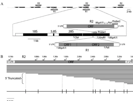
reading frames (EickbushandEickbush1995; Eick-the ribosomal RNA (rRNA) gene locus (Figure 1A).
bushet al.1995;Latheet al.1995;LatheandEickbush R1 and R2 elements exist in all lineages of arthropods
1997; Gentile et al.2001). Population surveys in
Dro-(Jakubczaket al.1991;Burkeet al.1998) as well as in
sophila melanogasterandD. simulanshave suggested that recombination eliminates individual copies of R1 and R2 from the rDNA locus more often than it amplifies 1Corresponding author:Department of Biology, Hutchinson Hall,
Uni-them (Jakubczaket al.1992;Perez-Gonzalezand
Eick-versity of Rochester, Rochester, NY 14627-0211.
E-mail: [email protected] bush2001). The recombination bias against these
using ImageQuant version 1.2. Within each lane, the weakest ments suggests they must propagate by
retrotransposi-bands were defined as representing single-copy elements; copy tion to remain within the rDNA locus. In the only studies
numbers of the remaining bands were estimated by dividing to directly follow the retrotransposition of new R1 and the signal of each band by the average signal of the single-R2 elements and the elimination of old copies, hun- copy bands.
Southern blots and the size of the rDNA locus:Southern dreds of new R1 insertions but few R2 insertions were
blots were conducted with genomic DNA extracted from 30 observed in the Harwich mutation accumulation lines of
adults of each Paradise line. To determine the proportions D. melanogaster(Perez-Gonzalez andEickbush2002;
of different types of rDNA units, genomic DNA was digested Perez-Gonzalez et al. 2003). The identification of withClaI,HindIII, andPstI, electrophoresed on a 1% agarose stocks in which new R2 integrations could be monitored gel, transferred to a nitrocellulose membrane and probed with a 280-bp segment from the 28S gene (as described in in vivowould be of significant value to understand the
Jakubczak et al. 1992) immediately downstream of the R1
dynamics of R2 retrotransposition and to complement
insertion site (see Figure 1A for the position of the probe and our in vitro studies of the R2 integration mechanism
the locations of restriction sites). Uninserted rDNA units, R1-(reviewed inEickbush2002). inserted units (including units that have both R1 and R2 In this article we have returned to a population ofD. insertions), and R2-inserted units gave rise to 1.9-, 1.1-, and 0.9-kb fragments, respectively. Using the PhosphorImager and simulans in which a previous survey had suggested a
ImageQuant to analyze the gel, the percentages of these three rapid turnover of R2 insertions (Perez-Gonzalezand
rDNA unit types were calculated for each line. To determine Eickbush 2001). Analysis of isofemale lines derived
the proportions of double-inserted rDNA units, the genomic from this population revealed lines in which the R2 DNA was digested withMspA1I and probed with a sequence elements appeared completely inactive and lines where from the 3⬘ end of the R2 element (as described in Perez
-GonzalezandEickbush2001). rDNA units inserted by R2
R2 frequently retrotransposed.
alone gave rise to a 2.3-kb fragment and units inserted with both a full-length R1 and either a full-length or a 5⬘-truncated R2 gave rise to a 1.2-kb fragment (see Figure 1A). The ratio MATERIALS AND METHODS
of double- to single-inserted units was again quantitated with a PhosphorImager. By combining data from these two Southern Fly stock, PCR amplification, and R2 5ⴕ-truncation profiles:
blots, the proportions of uninserted units, units inserted by The D. simulans isofemale lines were the gift of M. Turelli
R1 alone, units inserted by R2 alone, and units inserted by and have been previously described (Perez-Gonzalez and
both elements were calculated. The total number of rDNA
Eickbush 2001). Fifteen of the lines in the previous study
units in the locus of each line was estimated on the basis of were still in existence and were used in this report. The PCR
the number of R2 elements directly counted by virtue of their protocol was the same as described inPerez-Gonzalezand
different 5⬘ends (see above) divided by the fraction of the
Eickbush(2001) except that the Taq DNA polymerase and
rDNA locus containing an R2 insertion. PCR buffer were from Promega (Madison, WI). Genomic DNA
was extracted from single flies and prepared for PCR as de-scribed byGlooret al.(1993). A forward primer, 5⬘-TGCCCAGT
RESULTS GCTCTGAATGTC-3⬘, which anneals to the 28S gene 80 bp
upstream of the R2 insertion site, was used in combination with New R2 insertions within the rDNA locus of an organ-one of the following reverse primers: 5⬘-ATACCCACGCAGGT
ism can be characterized even though all elements insert TCCGC-3⬘, 5⬘-CGCTGGACGACAGCATACTGC-3⬘, 5⬘-GAAGAC
into the same site of the 28S gene because the mecha-GGTTCTGGCCAGTC-3⬘, 5⬘-TCGAATGCCTTGCTTACATC-3⬘,
5⬘-GATAGAAAATCCAACGTTCTGTC-3⬘, 5⬘-GTGCATGAAT nism of R2 insertion generates extensive sequence vari-GAATGCTATC-3⬘, 5⬘-CCCCTTGTAGTACGAGACTTC-3⬘, 5⬘- ants at their 5⬘junction with the 28S gene. Variation is GTCACCTGCGGCTTCGAATC-3⬘, and 5⬘-GTATGGAAATCT
generated because after initiating cDNA synthesis at ATCGAAAGATACT-3⬘, which bind, respectively, to R2
se-the 3⬘ end of the R2 RNA transcript, the R2 reverse quences 0.8, 1.0, 1.3, 1.6, 2.0, 2.4, 2.8, 3.2, and 3.6 kb from
transcriptase does not need to polymerize to the 5⬘end the 5⬘end of a canonical full-length R2 element (see Figure
1B). To construct an R2 5⬘-truncation profile, DNA extracted for a complete integration reaction to occur (Luanet from a single fly was PCR amplified with the above primer al.1993). Thus the R2 copies in any rDNA locus are a pairs and the PCR products were separated on 8.75% polyacryl- collection of “full-length” copies and copies that are 5⬘ amide gels. Lengths of the R2 5⬘truncations were calculated
truncated at various positions along the length of the on the basis of the sizes of the PCR products and the positions
element (see Figure 1B). The different 5⬘-truncated R2 of the reverse primers. To avoid scoring spurious bands that
are occasionally produced by PCR, every band was confirmed elements can be scored by PCR using one primer that by two adjacent primers before being scored as an authentic binds to the 28S gene upstream of the R2 insertion site
R2 5⬘-truncated copy. and a series of second primers that bind to different
Determination of the number of full-length R2 elements:
regions of R2 (Perez-GonzalezandEickbush2001). To score full-length R2 elements, the primer (5⬘-GACTTGAG
On the basis of an analysis of isofemale lines established TAAAGGAGAGACT-3⬘) that annealed to the R2 sequence 123
bp downstream of the 5⬘end of a full-length R2 was32P-end- from a population ofD. simulansin Paradise, California, it
labeled (Perez-GonzalezandEickbush 2002) and used in was found that a majority of the 5⬘-truncated R2 insertions combination with the primer complementary to the 28S gene within this population were of unique length (i.e., corre-80 bp upstream of the R2 insertion site. The products were
spond to a single copy per X chromosome) (Perez -separated on an 8% high-voltage denaturing polyacrylamide
Gonzalez and Eickbush 2001). In addition, the 5⬘ -gel, exposed in a PhosphorImager cassette, and quantitated
Figure1.—The rDNA locus and its R1 and R2 insertions. (A) The rDNA locus ofD. simulans, as in most eukaryotes, is composed of a tandem array of rDNA units each encoding 18S, 5.8S, and 28S rRNA. Each rDNA unit can be inserted by R1, R2, or both elements. The ORFs and untranscribed region of the R1 and R2 elements are shown. Also shown are the locations of restriction sites forClaI, HindIII,PstI,MspA1I and the positions of the probes used for the Southern blot experiments to determine the data in Table 1. (B) Sample summary diagram of the collection of 5⬘-truncated R2 elements present in the rDNA loci of the Paradise lines. The locations of R2 PCR primers used to determine the length of 5⬘-truncated and full-length R2 elements are shown above the diagram of the R2 element. The R2 copies in any organism are a collection of full-length copies and copies truncated at their 5⬘end. The length of each copy is represented by the closely spaced horizontal lines. The 5⬘-truncated elements are usually present only at a single copy per rDNA locus (each copy has a unique length). Multiple full-length R2 elements of similar length are also present. Differences of these full-length elements can be detected on high-resolution gels (see Figure 4). A summary (truncation profile) of the 5⬘-truncated R2 copies in an rDNA locus is shown in the diagram at the bottom. The horizontal line represents the length of a full-length R2 and vertical lines represent the different 5⬘-truncated R2 copies.
shared among the different isofemale lines. Thus the Variation in the R2 5ⴕ-truncation profiles of the Para-dise lines:It is straightforward to score the R2 profile R2 truncation patterns in these lines indicated that most
R2 insertions were sufficiently recent that they had nei- of individual rDNA loci inD. simulansbecause the rDNA units exist exclusively on the X chromosome;i.e., there ther spread by recombination to other chromosomes
in the population nor been amplified by recombination are no units on the Y chromosome, unlike most other Drosophila species (LoheandRoberts1990;Mecheva to higher numbers on the same chromosome. Such
rapid turnover of R2 elements can explain the⬍0.5% andSemionov1992). Therefore the R2 elements scored in individualD. simulansmales from each Paradise line nucleotide sequence divergence found in the full-length
and 5⬘-truncated R2 elements of all Drosophila species represented the R2 insertions on individual X chromo-somes in that line. As examples of the variation found studied to date (EickbushandEickbush1995;Lathe
andEickbush1997). in the different Paradise lines, the profiles of the 5⬘ -truncated R2 elements from three lines are shown in If R2 insertions and eliminations continued while
these isofemale lines were maintained as inbred stocks Figure 2. In line 1 (Figure 2A), the same 21 PCR frag-ments were found in all 18 males surveyed. The 5⬘ends within the laboratory, then changes in the R2 profile
over time would be expected. Therefore,ⵑ75 genera- of the truncated R2 copies that gave rise to these frag-ments are indicated by the vertical lines on the hori-tions after the initial characterization of the lines, the
Figure2.—R2 5⬘-truncation profiles of the X chromosome rDNA loci from Paradise lines 1, 31, and 58. rDNA units are only on the X chromosome ofD. simulans; thus the R2 profiles of individual males represent the profiles of individual X chromosomes in that population. As diagrammed in Figure 1B, the horizontal line represents the length of the complete R2 element, with vertical lines representing the different 5⬘-truncated copies. (A) Line 1. All 18 males had the same profile containing 21 5⬘ -truncated elements. (B) Line 31. Four different R2 truncation profiles were found in males from this line. The number to the right of each R2 truncation profile is the number of males in which the particular profile was found. The 5⬘-truncated elements marked with dots at the bottom represent R2 elements found in all males. (Bottom) A composite truncation profile, representing all R2 5⬘truncations seen in this line, is shown. Polymorphic truncations (truncations that are not present in all males sampled) are marked with the number of males in which the particular truncation was found. (C) Line 58. Ten different R2 truncation profiles were identified. Nine truncation copies were present in all males while 27 truncated copies were polymorphic.
element (see Figure 1B). All PCR products correspond- differences in R2 elements between any two X chromo-somes within this line ranged from 1 to 16. As shown ing to 5⬘-truncated copies were of similar intensity,
sug-gesting that they each correspond to a single R2 copy. by the composite R2 truncation profile, only 9 of 36 total R2 length variants were present in all males. This Thus there was no evidence of recent R2 insertions,
deletions, or amplifications in line 1. level of variation suggested many recent insertions and/ or deletions in line 58.
Unlike line 1, variation within the R2 insertion pattern
was found in D. simulans line 31 (represented by the A similar analysis was done for the remaining 12 Para-dise lines, and the composite R2 truncation profiles for four horizontal lines in Figure 2B). While 14 males
sampled had the same profile consisting of 23 5⬘-trun- all 15 lines are shown in Figure 3. The 15 lines could be divided into three groups on the basis of the level cated copies, 2 males were missing 7 R2 copies found
in all other males, and 1 male had 1 R2 copy missing. of R2 5⬘-truncation variation. The first group comprised 6 lines (top), in which all males within a line had identi-Finally, 1 male had 3 R2 copies that were not found on
any other chromosome. Thus new R2 insertions and/ cal R2 truncation profiles on their X chromosomes. The 11 (line 76) to 25 (line 96) 5⬘-truncated copies matched or eliminations had occurred in this line since the last X
chromosome fixation event. A composite R2 truncation the profiles scored 75 generations earlier. Therefore, there was no evidence in this group for insertions or profile is shown at the bottom of Figure 2B with the
number of males in which each unfixed R2 copy was deletions over a 3-year period.
The five paradise lines shown in the middle of Figure found indicated above the vertical line.
An even higher level of variation in R2 profiles was 3 composed a group in which two to four different X chromosome profiles were found among the males detected in line 58 (Figure 2C). Ten different X
chro-mosomes were found with no chromosome type present sampled from each line. These lines had from 14 to 28 5⬘-truncated R2 copies present on all X chromosomes in more than three of the males sampled. The number
Figure3.—R2 truncation profiles of all Paradise lines. Numbers at left are isofemale line numbers. Numbers at right are the number of X chromosome R2 truncation profile types found in that line. In the first six Paradise lines shown, all males sampled from each line had identical 5⬘-truncation profiles. For the remaining lines the profiles presented are composite R2 truncation profiles. Polymorphic R2 truncations are marked with the number of males in which the particular truncation was found. The number of males sampled in each line was 18 except for the sampling of 11 males in line 65, 13 males in line 61, and 16 males in line 58.
31 and 61) could be explained by one chromosome in percentage of full-length elements in each line). Fortu-nately, even individual full-length R2 insertions can be each line undergoing a deletion, which eliminated 7
R2 copies. Because the analysis in the R2 profile in the identified because the mechanism used by R2 to initiate second-strand DNA synthesis is imprecise, frequently original study (Perez-Gonzalez andEickbush 2001)
had been conducted with DNA derived from a pool of generating small deletions, insertions, and duplications at the 5⬘junction of the element (Georgeet al.1996). flies in each line, deletions as well as any insertions
present in only a few flies would have gone undetected. To determine if the many differences in the 5⬘-truncated R2 elements scored among males from Paradise line 58 Therefore while suggestive, this group of lines did not
provide definitive evidence for recent insertion or dele- were also true of full-length elements, a PCR primer that anneals to R2 sequences 123 bp from the 5⬘ end tion events.
Finally, the four lines in the third group (Figure 3, of full-length elements was end-labeled and used in com-bination with the primer that anneals to the 28S gene bottom) had 10 or more different X chromosomes in
each line. In line 89, all 18 males had a different R2 5⬘- 80 bp upstream of the R2 insertion site. The labeled PCR products were then electrophoresed on a high-truncation profile. In all four lines less than half of the
R2 copies were present in all males sampled, and the resolution denaturing gel to resolve small differences in product lengths. Figure 4 shows the results of individual R2 truncation profiles had dramatically changed
com-pared to the previous study (Perez-GonzalezandEick- males sampled from line 58. The full-length R2 profile of the males differed in the number of bands as well as bush 2001). These stocks had not become
contami-nated because the R1 truncation profiles showed little in the intensity of some bands. The canonical full-length R2 element as determined in the original study (Perez -variation from the profiles seen 75 generations earlier
(data not shown). The high level of variation seen within Gonzalez and Eickbush 2001) is the intense band identified with an asterisk. Many other R2 copies were these lines suggested the recent and rapid turnover of
R2 elements. up to 80 bp shorter or longer than this canonical length.
Nine different X chromosome types could be identified
Variation within the full-length R2 elements of line
58: Forty to 80% of the R2 elements in the Paradise solely on the basis of the combination of R2 copy lengths present. The intensity of some bands also differed be-isofemale lines do not have large 5⬘truncations and are
de-A, 16 new insertions (5 full length and 11 5⬘truncated) generated nine X chromosome profiles that differed from those in the original pair. Only two males tested in generation 5 contained an X chromosome identical to one of the parental X chromosome profiles. Three X chromosomes of the descendents, in addition to gaining new insertions, had lost four R2 elements present in the original pair.
In the B lineage, 16 new insertions (7 full length and 9 5⬘truncated) generated 14 different X chromosome types. This lineage had three X chromosomes with 4–7 deletions that involved in total 12 R2 copies. Interest-ingly 2 of the deleted R2 copies had also been deleted in the A lineage. None of the males tested in generation 5 contained an X chromosome identical to one of the parental types.
Finally in a third lineage (data not shown), new inser-tions were not detected but five R2 copies were deleted from one male. These deleted copies were among the same R2 copies deleted from lineages A and B, indicat-ing that certain R2 copies (or locations within the rDNA locus) were prone to elimination. The failure to find new insertions in this lineage suggested that all X chro-mosomes in line 58 did not contain equally active R2 elements.
Characterization of the rDNA loci of the Paradise lines: We next asked whether the R2 activity detected in some of the D. simulans lines could be correlated
Figure4.—5⬘ variation in the full-length R2 elements of
with the number of R2 elements, the fraction of the D. simulansline 58. DNA from 17 males of line 58 was
individu-rDNA units inserted with R2, or the size of the individu-rDNA ally PCR amplified with an end-labeled primer that annealed
to the 28S rRNA gene 80 bp upstream of the R2 insertion site locus (i.e., the total number of rDNA units). The num-and the 0.1-kb R2 primer shown in Figure 1B. The labeled ber of R2 insertions present within individuals of a line PCR products were run on a high-voltage, denaturing 8% can be accurately determined because of the extensive polyacrylamide gel and the signal was quantitated on a
Phos-sequence variation present at the R2 5⬘ends. We have phorImager screen (seematerials and methods). The
aster-previously shown that the many 5⬘-truncated bands isk indicates the length of canonical full-length R2 elements
as described inPerez-GonzalezandEickbush(2001). Base seen by PCR amplification of individual D. simulans positions relative to this canonical full-length R2 are shown males orD. melanogastermales are of equal intensity, and
at right. thus correspond to one copy per chromosome (Perez
-Gonzalez and Eickbush 2001, 2002; Eickbush and Eickbush2003;Perez-Gonzalezet al.2003). Therefore crease in the number of elements of these specific
the number of 5⬘-truncated copies can be determined lengths. By quantitating the amount of signal in each
by counting the number of distinct PCR bands detected PCR band on a PhosphorImager, virtually all X
chromo-in our 5⬘-truncation profiles (Figure 4). Polymorphic somes sampled from line 58 could be resolved as
differ-truncations (i.e., those not present in all X chromo-ent. Clearly the extensive variation in R2 profiles in line
somes sampled from each line) were counted as the 58 involved both 5⬘-truncated and full-length elements.
number of chromosomes in which the truncations were
R2 insertions and deletions rapidly accumulated in
observed divided by the number of X chromosomes
line 58:To determine how quickly variation in the R2
sampled. Because we have not shown that all 5⬘ -trun-profile could accumulate in these D. simulans lines, a
cated elements are single copy, these counts may repre-subline of line 58 was started with one pair of flies to
sent an underestimation of the number of 5⬘-truncated reduce the initial level of variation. Single pairs of flies
R2 elements in each line. The number of full-length R2 from this subline were mated and their progeny were
insertions was determined by using the end-labeled PCR maintained as separate lineages. R2 5⬘-truncation
pro-primer and separating the products on high-resolution files as well as full-length R2 profiles of individual male
denaturing gels (see Figure 4). Many of the full-length descendents after five generations were compared
(Fig-copies were present at one copy per chromosome. The ure 5). Because the R2 truncation profiles of the original
number of elements corresponding to the more intense pair were determined (top of each panel), new
Figure5.—R2 insertions and deletions rapidly accumulated in line 58. Single pairs of flies from line 58 were mated and their progeny were main-tained as separate lineages for five generations. Data from two lineages are presented. Shown at the top of A and B are the R2 profiles of the two X chromosomes from the original female and the single X chromosome from the original male used to establish the lineage. The profiles of the two X chromosomes of the female were determined by analysis of the R2 profiles of a collection of her sons. Below these progenitor profiles are the profiles of males collected at the fifth generation. The dotted line to the left of each R2 5⬘-truncation profile represents an expanded scale of differ-ences in the full-length R2 elements. Because there are many length R2 elements, only full-length elements not present in parental X chro-mosomes are shown. New R2 insertions are in bold and marked with a star. Deletions of the parental R2 copies are marked by solid ovals. Numbers on the right are the number of genera-tion 5 males in which this profile type was found.
in each band by the signal present in the single-copy inserted rDNA units were calculated (Table 1). The fraction of the rDNA locus inserted with R2 elements bands (seematerials and methods).
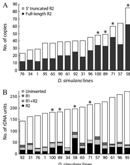
Shown in ascending order in Figure 6A are the total averaged 22% and varied from a low of 14% in line 95 to a high of 44% in line 58. The fraction of the locus number of 5⬘-truncated R2 elements (open area of the
bar) and full-length R2 elements (solid area of the bar) inserted with R1 elements averaged 10% and also showed significant variation among the lines. The frac-present in each of theD. simulans lines. The number
of R2 elements varied from a low of 23 in line 76 to a high tion of the rDNA units inserted with R1 showed no relationship to the fraction inserted with R2. The frac-of 85 in line 58 (3.7-fold range). Most frac-of the differences
between lines were due to the variation in the number of tion of the R1 and R2 double-inserted rDNA units varied from 3 to 14%, which, as we have noted previously the full-length R2 elements (range 12–62).
To determine the fraction of the rDNA locus inserted (Perez-GonzalezandEickbush2001), is significantly higher than what would be expected from the random with R1 and R2 elements, genomic DNA from 30 adults
of each line was extracted and two Southern blots were insertion of R1 and R2 elements into the rDNA units. Finally, to determine the size of the rDNA locus (the conducted. In the first Southern blot, restriction
en-zymes were used that cleaved the rDNA units into three total number of rDNA units) in each line, the total number of R2 elements (5⬘truncated and full length, fragments: uninserted, R1 inserted (including R1 and
R2 double inserts), and R2 inserted. The genomic blot Figure 6A) was divided by the fraction of the units that contained R2 insertions (Table 1). The size of the locus was probed with a fragment of the 28S gene downstream
of the R1 insertion site. In the second Southern blot, a averagedⵑ200 units varying from an estimated 140 in line 92 to 270 units in line 95 (shown in Figure 6B). restriction enzyme was used that cleaved the R2-inserted
units into two fragment sizes depending upon whether The calculated numbers of uninserted, R1 only inserted, R2 only inserted, and R1⫹ R2 double-inserted rDNA they were or were not also inserted with an R1 element.
This blot was probed with sequences from the 3⬘ end units are represented by the open, shaded, solid, and stippled areas of the bars, respectively. Lines with high of the R2 element (see Figure 1A andmaterials and
methodsfor a more detailed description of these blots). R2 activity are indicated with an asterisk. As will be described in thediscussion, R2 activity correlated with By determining the relative intensity of the bands from
these two Southern blots, the percentages of uninserted, the number of full-length R2 elements but not with the size of the rDNA locus.
double-TABLE 1
Percentages of uninserted, R1-inserted, R2-inserted, and double-inserted rDNA units in the Paradise lines
Fly Uninserted
line (%) R1 (%)a R2 (%)a R1⫹R2 (%)
1 83 11 16 10
31 71 17 25 14
34 86 8 14 8
57 66 15 29 10
58 51 14 44 9
61 83 5 16 3
65 77 11 18 6
71 72 7 28 7
76 85 9 14 7
89 72 9 27 8
90 79 11 17 6
92 73 15 20 8
95 85 5 14 4
96 80 8 17 6
100 73 12 27 11
aIncludes both single- and double-inserted (R1⫹R2) units.
Figure6.—R2 copy number and size of the rDNA locus in position events were found randomly distributed over
each Paradise line. (A) Average R2 copy number in each 19 sublines, suggesting a uniform low rate of new inser-Paradise line. The open portion of each bar represents the
tions. number of distinct 5⬘-truncated R2 elements scored in the
The isofemale lines of D. simulans characterized in PCR analysis (Figure 3), and the solid portion of each bar
represents the number of full-length elements as determined this report offered a different indication of the rate and by a PCR analysis similar to that in Figure 4. Polymorphic R2 pattern of R2 retrotransposition. Most lines were similar insertions were scored by the number of males containing the to the previously reported
D. melanogasterlines, and pro-element divided by the number of total males sampled. (B)
vided little evidence of active retrotransposition. The Average size of the rDNA locus in each Paradise line. The
insertion profiles of R2 elements in these lines were estimated number of uninserted, R1-inserted, R2-inserted, and
double-inserted (R1⫹R2) rDNA units are represented, re- similar or identical to the profiles scored 75 generations spectively, by the open, shaded, solid, and stippled portions earlier. On the other hand, virtually every rDNA locus of each bar. These values were determined by genomic DNA sampled from a few isofemale lines had differences in blots and using the calculated total number of R2 insertions
their R2 insertion profiles, suggesting retrotransposi-in each lretrotransposi-ine divided by the fraction of the total rDNA units
tion. Direct evidence for frequent retrotransposition was that were inserted by R2 to determine the total number of
units (Table 1) (seematerials and methodsfor details). In demonstrated by showing that the descendants of indi-A and B, lines with high R2 activity, as indicated by the level vidual pairs of flies from the “active” lines rapidly accu-of variation in the R2 praccu-ofiles (see Figure 3), are indicated mulated new R2 insertions.
with an asterisk.
The characterization of naturally occurring active and inactive lines of D. simulansprovides new insights into the dynamics of R2 element insertions and deletions. DISCUSSION
The data suggest that there are stable genetic conditions All laboratory stocks and geographical lines ofD. mela- under which new retrotransposition events can occur
nogasterandD. simulanscharacterized to date have con- frequently and stable genetic conditions under which tained significant numbers of R2 elements (Jakubczak events are rare or absent. Because some of the lines
et al.1992;EickbushandEickbush1995;Eickbushet contained intermediate levels of R2 variation, it will be
retrotrans-activity is turned off. The mechanisms that suppress the continued activity of these elements are believed to be cellular systems designed to detect the expression of any sequence present in multiple copies (Chaboissier et al. 1998; Roche and Rio 1998; Jensen et al. 1999; Boivinet al.2003). The R2 activity we have detected in theD. simulansisofemale lines clearly differs from that of hybrid dysgenesis in that both males and females from all lines contain many copies of R2 elements, and R2 activity is maintained for many generations. We do not know how long the R2 elements have been active in these lines but the rapid accumulation of new inser-tions in the descendants of single pairs has been ob-served over a 1-year period (ⵑ25 generations) in several independent trials with two of the active lines (58 and 89) (Figure 5; data not presented).
R2 activity is more similar to the activity associated with the LTR retrotransposons copia, gypsy, Zam, and
Idefix. The activity of these elements is associated with one or more alleles present at specific “control” loci in the genome (Prud’hommeet al. 1995; Nuzhdinet al.
1998;Dessetet al.2003), although a more general con-trol mechanism may be involved (Sarot et al. 2004).
Element activity continues for generations and does so Figure7.—Correlation between R2 activity and the number irrespective of the number of mobile element copies of full-length R2 elements or the fraction of the rDNA locus inserted with R2. The level of R2 activity was scored as the present in the organism.
level of variation within each line (i.e., the number of X chro-One likely locus that might control the activity of
mosome variant profiles from Figure 3). The Paradise lines R2 elements is the rDNA locus itself. In Figure 7A the
are divided into three groups on the basis of the number of number of full-length and, thus, potentially active R2 chromosome types found in sampled males from that line and elements has been plottedvs.the amount of R2 variation plottedvs. (A) the number of full-length R2 elements found in the Paradise lines and (B) the fraction (as percentage) of that was present in each subline (scored as the number
the rDNA units in each line inserted with R2. of X chromosome R2 profiles identified in 18 males).
The six lines with no R2 activity had on average 17 full-length R2 elements, while the four lines with high
intermediates, which overwhelms cellular control mech-activity (10 and more X chromosome R2 profiles)
aver-anisms. However, such a correlation between R2 copy aged 44 full-length elements or over 2.5 times that of
number and R2 activity was not exact. Line 96 contained the no activity lines. The five lines with 2–4 different
26 full-length R2 copies and no detectable R2 activity, R2 profiles averaged higher numbers of full-length
ele-while line 89 had 32 full-length elements and the highest ments but overlapped extensively with the no activity
level of R2 activity (as measured by the variation within lines. In contrast, there was no correlation between R2
the line), even higher than line 58, which had 65 full-activity and the size of the rDNA locus. The four active
length elements. lines had on average ⵑ195 rDNA units while the six
An alternative model to explain R2 activity is that the inactive lines averagedⵑ215 units (see Figure 6B).
How-rate of retrotransposition is unrelated to element copy ever, because the active lines had more R2 elements
number or the fraction of the locus inserted with R2; there was a positive correlation between R2 activity and
rather the larger numbers of R2 copies present in active the percentage of the locus that was inserted with R2
lines is the result (not the cause) of R2 activity. In this (averageⵑ15% for the inactive lines andⵑ31% for the
model, R2 activity could be controlled by a genetic locus active lines, Figure 7B).
other than the rDNA locus itself. We should be able to Transposable element insertion rates have previously
resolve between these two models by conducting crosses been correlated with the number of elements (Nuzhdin
between the active and inactive Paradise lines and
genet-et al.1996;Pasyukovaet al. 1998). Thus one possible
ically mapping control over R2 activity. model to explain the correlation of R2 activity with
Another important finding of this report is that the element number is that the larger number of R2
ele-insertion of many new R2 elements resulted in the elimi-ments or the greater fraction of inserted rDNA units is
Non-Burke, W. D., H. S. Malik, W. C. Lathe, III andT. H. Eickbush,
LTR retrotransposable elements have never been shown
1998 Are retrotransposons long-term hitchhikers? Nature392: to excise, thus these eliminations presumably involved 141–142.
Burke, W. D., H. S. Malik, J. P. JonesandT. H. Eickbush, 1999
recombination events that deleted the R2-inserted rDNA
The domain structure and retrotransposition mechanism of R2
units. These recombination events appeared to involve
elements are conserved throughout arthropods. Mol. Biol. Evol.
more than single rDNA units because four to seven R2 16:502–511.
Chaboissier, M.-C., A. BuchetonandD. J. Finnegan, 1998 Copy
copies were eliminated per event (Figure 5). The deletions
number control of a transposable element, the I factor, a
LINE-frequently involved the same regions of the rDNA locus
like element in Drosophila. Proc. Natl. Acad. Sci. USA95:11781–
because the same subset of R2 elements was often elimi- 11785.
Desset, S., C. Meignin, B. DastugueandC. Vaury, 2003 COM, a
nated (Figure 5). Hot spots for the elimination of R1
heterochromatic locus governing the control of independent
and R2 elements within the rDNA locus have also been
endogenous retroviruses fromDrosophila melanogaster.Genetics
seen in our studies of theD. melanogasterHarwich lines 164:501–509.
Dominguez, A., andJ. Albornoz, 1996 Rates of movement of
trans-(Perez-Gonzalez et al. 2003). In addition, deletions
posable elements inDrosophila melanogaster.Mol. Gen. Genet.251: within the rDNA locus in the presence of active R2
130–138.
insertion machinery are associated with ourin vivo R2 Eickbush, D. G., andT. H. Eickbush, 1995 Vertical transmission
of the retrotransposable elements R1 and R2 during the evolution
integration system (EickbushandEickbush2003). In
of the Drosophila melanogaster species subgroup. Genetics 139: this system the injection of purified R2 protein and
671–684.
RNA into Drosophila embryos results in the insertion Eickbush, D. G., andT. H. Eickbush, 2003 Transcription of
endog-enous and exogendog-enous R2 elements in the rRNA gene locus of
of marked R2 elements in the rDNA locus. These
inser-Drosophila melanogaster.Mol. Cell. Biol.23:3825–3836.
tions are frequently accompanied by large deletions of
Eickbush, D. G., W. C. Lathe, III,M. P. FrancinoandT. H. Eick-rDNA. Finally, retrotransposition of another non-LTR bush, 1995 R1 and R2 retrotransposable elements of
Drosoph-ila evolve at rates simDrosoph-ilar to those of nuclear genes. Genetics139: transposable element, L1, has been shown to cause large
685–695.
deletions (⬎70 kb) of host genomic DNA (Gilbertet
Eickbush, T. H., 2002 R2 and related site-specific non-long terminal
al.2002). repeat retrotransposons, pp. 813–835 inMobile DNA II, edited by
N. L.Craig, R.Craigie, M.Gellartand A. M.Lambowitz.
The elimination of R2-inserted rDNA units in the
American Society for Microbiology, Washington, DC.
active lines can explain why the rDNA loci in these lines
Gentile, K. L., W. D. BurkeandT. H. Eickbush, 2001 Multiple
are not extensively filled with R2 insertions. However, lineages of R1 retrotransposable elements can coexist in the
rDNA loci of Drosophila. Mol. Biol. Evol.18:235–245.
because the total number of rDNA units in the active
George, J., W. D. BurkeandT. H. Eickbush, 1996 Analysis of the
lines is similar to that of the inactive lines (Figure 6B),
5⬘junctions of R2 insertions with the 28S gene: implications for
the active lines must also be undergoing selection for non-LTR retrotransposition. Genetics142:853–863.
a compensating expansion of uninserted rDNA units. Gilbert, N., S. Lutz-PriggeandJ. V. Moran, 2002 Genomic dele-tion created upon LINE-1 retrotransposidele-tion. Cell110:315–325.
Finally there was no evidence for R1 activity in the four
Gloor, G. B., C. R. Preston, D. M. Johnson-Schlitz, N. A. Nassif, Paradise lines with R2 activity. This finding confirms R. W. Philliset al., 1993 Type I repressors ofP-element mobil-previous suggestions that R1 and R2 elements are sepa- ity. Genetics135:81–95.
Houle, D., andS. V. Nuzhdin, 2004 Mutation accumulation and
rately activated. For example, Jakubczak et al. (1992)
the effect ofcopiainsertions inDrosophila melanogaster.Genet. Res.
found that the levels of R1 and R2 elements were unre- 83:7–18.
lated in various geographical lines ofD. melanogaster. In Jakubczak, J., W. D. BurkeandT. H. Eickbush, 1991 Retrotrans-posable elements R1 and R2 interrupt the rDNA genes of most
the Harwich mutation accumulation lines, those lines
insects. Proc. Natl. Acad. Sci. USA88:3295–3299.
with the highest numbers of new R1 insertions did not Jakubczak, J., M. Zenni, R. WoodruffandT. H. Eickbush, 1992 have higher numbers of new R2 insertions (Perez-Gon- Turnover of R1 (type I) and R2 (type II) retrotransposable ele-ments in the ribosomal DNA ofDrosophila melanogaster.Genetics
zalez et al. 2003). Separate control over R1 and R2
131:129–142.
activity would minimize the direct competition between Jamrich, M., andO. L. Miller, Jr., 1984 The rare transcripts of these elements, thus helping to explain their coexis- interrupted rRNA genes inDrosophila melanogasterare processed
or degraded during synthesis. EMBO J.3:1541–1545.
tence in the rDNA locus since the origin of arthropods
Jensen, S., M. P. GassamaandT. Heidmann, 1999 Taming of
trans-(Burkeet al.1998, 1999;Gentileet al.2001). posable elements by homology dependent gene silencing. Nat.
Genet.21:209–212. We thank Bill Burke, Danna Eickbush, and Karin Tetzlaff for
discus-Kidwell, M. G., J. F. KidwellandJ. A. Sved, 1977 Hybrid dysgenesis sions and comments on the manuscript. We especially thank Naina
inDrosophila melanogaster: a syndrome of aberrant traits including Phadnis whose initial resurvey of the Paradise lines renewed our
inter-mutation, sterility, and male recombination. Genetics86:813– est in these lines. This research was supported by National Science
833. Foundation grant MCB-9974606 and National Institutes of Health
Kojima, K. K., andH. Fujiwara, 2004 Cross-genome screening of novel grant GM-42790. sequence-specific non-LTR retrotransposons: various multicopy RNA genes and microsatellites are selected as targets. Mol. Biol. Evol.21:207–217.
Lathe, W. C., III, andT. H. Eickbush, 1997 A single lineage of
LITERATURE CITED
R2 retrotransposable element is an active, evolutionarily stable component of the Drosophila rDNA locus. Mol. Biol. Evol.14: Boivin, A., C. Gally, S. Netter, D. AnxolabehereandS.
Rons-1232–1241.
seray, 2003 Telomeric associated sequences of Drosophila
re-Lathe, W. C., III,W. D. Burke, D. G. EickbushandT. H. Eickbush, cruit polycomb-group proteinsin vivoand can induce
pairing-1995 Evolutionary stability of the R1 retrotransposable element sensitive repression. Genetics164:195–208.
in the genus Drosophila. Mol. Biol. Evol.12:1094–1105.
Bucheton, A., 1990 I transposable elements and I-R hybrid
Luan, D. D., M. H. Korman, J. L. JakubczakandT. H. Eickbush,
and R2 elements in the rDNA locus ofDrosophila simulans. Genet-1993 Reverse transcription of R2Bm is primed by a nick at the
ics158:1557–1567. chromosomal target site—a mechanism for non-LTR
retrotrans-Perez-Gonzalez, C. E., andT. H. Eickbush, 2002 Rates of R1 and position. Cell72:595–605.
R2 retrotransposition and elimination from the rDNA locus of
Malik, H. S., andT. H. Eickbush, 1998 Retrotransposable elements
Drosophila melanogaster.Genetics162:799–811. R1 and R2 in the rDNA units ofDrosophila mercatorum: abnormal
Perez-Gonzalez, C. E., W. D. BurkeandT. H. Eickbush, 2003 R1 abdomen revisited. Genetics151:653–655.
and R2 retrotransposition and deletion in the rDNA loci on the
Maside, X., C. Bartolome, S. AssimacopoulosandB. Charlesworth,
X and Y chromosomes ofDrosophila melanogaster.Genetics165:
2001 Rates of movement and distribution of transposable
ele-675–685. ments inDrosophila melanogaster:in situhybridization vs. Southern
Petrov, D. A., J. L. Schutzman, D. L. HartlandE. R. Lozovskaya, blotting data. Genet. Res.78:121–136.
1995 Diverse transposable elements are mobilized in hybrid
dys-Mecheva, I. S., andE. P. Semionov, 1992 Localization of ribosomal
genesis inDrosophila virilis.Proc. Natl. Acad. Sci. USA92:8050–8054. DNA insertion elements in polytene chromosomes ofDrosophila
Prud’homme, N., M. Gans, M. Masson, C. TerzianandA. Bucheton,
simulans, Drosophila mauritiana and their interspecific hybrids.
1995 Flamenco, a gene controlling thegypsyretrovirus of
Drosoph-Genetica85:223–229.
ila melanogaster.Genetics139:697–711.
Nuzhdin, S. V., andT. F. Mackay, 1995 The genomic rate of
trans-Roche, S. E., and D. C. Rio, 1998 Trans-silencing by P elements posable element movement inDrosophila melanogaster.Mol. Biol. inserted in subtelomeric heterochromatin involves the Drosoph-Evol.12:180–181. ila polycomb group gene,
enhancer of zeste.Genetics149:1839–
Nuzhdin, S. V., E. G. PasyukovaandT. F. Mackay, 1996 Positive 1855.
correlation betweencopia transposition rate and copy number Sarot, E., G. Payen-Groschene, A. Buchetonand A. Pelisson, inDrosophila melanogaster.Proc. R. Soc. Lond. B Biol. Sci.263: 2004 Evidence for apiwi-dependent RNA silencing of thegypsy
823–831. endogenous retrovirus by the Drosophila melanogaster flamenco
Nuzhdin, S. V., E. G. Pasyukova, E. A. MorozovaandA. J. Flavell, gene. Genetics166:1313–1321. 1998 Quantitative genetic analysis ofcopiaretrotransposon