| INVESTIGATION
Context-Dependent Sensitivity to Mutations
Disrupting the Structural Integrity of Individual EGF
Repeats in the Mouse Notch Ligand DLL1
Karin Schuster-Gossler,* Ralf Cordes,*,1Julia Müller,*,2Insa Geffers,*,3Patricia Delany-Heiken,* Manuel Taft,†Matthias Preller,†,‡and Achim Gossler*,4
*Institut für Molekularbiologie and†Institut für Biophysikalische Chemie, Medizinische Hochschule Hannover, 30625 Germany, and‡Centre for Structural Systems Biology, Deutsches Elektronen-Synchrotron, 22607 Hamburg, Germany
ABSTRACTThe highly conserved Notch-signaling pathway mediates cell-to-cell communication and is pivotal for multiple developmental processes and tissue homeostasis in adult organisms. Notch receptors and their ligands are transmembrane proteins with multiple epidermal-growth-factor-like (EGF) repeats in their extracellular domains.In vitrothe EGF repeats of mammalian ligands that are essential for Notch activation have been defined. However,in vivothe significance of the structural integrity of each EGF repeat in the ligand ectodomain for ligand function is still unclear. Here, we analyzed the mouse Notch ligand DLL1. We expressed DLL1 proteins with mutations disrupting disulfide bridges in each individual EGF repeat from single-copy transgenes in the HPRT locus of embryonic stem cells. In Notch transactivation assays all mutations impinged on DLL1 function and affected both NOTCH1 and NOTCH2 receptors similarly. An allelic series in mice that carried the same point mutations in endogenous Dll1, generated using a mini-gene strategy, showed that early developmental processes depending on DLL1-mediated NOTCH activation were differently sensitive to mutation of individual EGF repeats in DLL1. Notably, some mutations affected only somite patterning and resulted in vertebral column defects resembling spondylocostal dysostosis. In conclusion, the structural integrity of each individual EGF repeat in the extracellular domain of DLL1 is necessary for full DLL1 activity, and certain mutations in Dll1 might contribute to spondylocostal dysostosis in humans.
KEYWORDSNotch signaling; Notch-ligand interaction; Notch activation; mouse DLL1; targeted mutagenesis/allelic series
C
OMMUNICATION between adjacent cells mediated by the evolutionary conserved Notch-signaling pathway regu-lates multiple developmental processes in different tissues and species (reviewed in Andersson et al.2011). Notch re-ceptors and their ligands encode type 1 transmembrane pro-teins with multiple EGF-like repeats in their extracellular domains. The ligands contain an additional conserved extra-cellular cysteine-rich so-called DSL domain and a disulfi de-bond stabilized module at the N terminus called MNNL(reviewed in Chillakuri et al. 2012). Mammalian genomes encode four Notch receptors, two Delta (DLL1 and DLL4), two Serrate-type ligands [called Jagged (JAG) 1 and 2] that activate Notch, and the untypical ligand DLL3 that can in-teract with, but does not activate, Notch (Ladiet al.2005; Gefferset al.2007; Chapmanet al.2011). Mutations in Notch pathway components underlie human diseases such as Alagille syndrome (ALGS) (mutations in JAG1 and NOTCH2) and CADASIL syndrome (mutations in NOTCH3) or spondy-locostal dysostosis (mutations in DLL3, HES7, LFNG, and MESP2) (reviewed in Pentonet al.2012).
EGF repeats 11 and 12 constitute the major ligand binding site of Notch receptors, although additional repeats are essential for full Notch activation and function in vitroand in vivo(Shimizuet al.1999; Hambletonet al.2004; Xuet al. 2005; Cordleet al.2008b; Andraweset al.2013; Lucaet al. 2015). The MNNL and DSL domains and EGF2 ofDrosophila Delta were shown to be required for Notch activationin vivo (Parks et al. 2006). In binding assays, the DSL domain of Copyright © 2016 by the Genetics Society of America
doi: 10.1534/genetics.115.184515
Manuscript received November 9, 2015; accepted for publication January 16, 2016; published Early Online January 20, 2016.
Supporting information is available online at www.genetics.org/lookup/suppl/ doi:10.1534/genetics.115.184515/-/DC1.
1Present address: Ascenion GmbH, Helstorfer Str. 7, 30625 Hannover, Germany. 2Present address: Gasteiner Str. 31, 10717 Berlin, Germany.
3Present address: Max-Planck-Institut für Biophysikalische Chemie, Am Fassberg 11, 37077 Göttingen, Germany.
4Corresponding author: Institut für Molekularbiologie, Medizinische Hochschule Hannover, Carl-Neuberg-Str. 1, 30625 Hannover, Germany.
mouse Jag1 was essential for binding to mouse Notch2, and the presence of EGF1 and 2 enhanced this interaction (Shimizuet al.1999). Likewise, a fragment of human Jag1 encompassing the DSL domain and first three EGF repeats was shown to bind to fragments of human Notch1 encom-passing EGF repeats 10–13 (Cordleet al.2008a), and dele-tion analyses of human DLL1 and DLL4 showed that the regions containing the MNNL to EGF3 were necessary and sufficient for full activation of Notch1 (Andraweset al.2013). Recently, the structure of a complex of EGF repeats 11–13 of Notch1 with the N-terminal portion of DLL4 up to and in-cluding EGF2 was published, showing that EGF repeats 11 and 12 of Notch interact with the DSL and MNNL domains of the ligand, respectively, placing EGF1 and -2 outside the es-sential Notch interaction surface (Lucaet al.2015).
While thefirst three EGF repeats of Notch ligands appear to be important for activation of Notch, the significance of other EGF repeats in the extracellular domains of ligands is less clear. Parks et al.(2006) identified cysteine missense mutations in EGF re-peats 4 and 9 inDrosophilaDelta that affect Notch signaling in some contexts, but were associated with aberrant subcellular localization and trafficking. Similarly, missense mutations in EGF repeats distant from the DSL domain in JAG1 of ALGS patients caused intracellular retention of the mutant protein (Morrissette et al.2001; Bauer et al.2010), preventing firm conclusions on how the region C-terminal to the interaction domain contributes to ligand function. Here, we focus on mouse DLL1, which has eight EGF-like repeats in its extracellular do-main (Bettenhausenet al.1995). To address the significance of all EGF repeats for DLL1 function, we disrupted the same two disulfide bridges individually in each EGF repeat. We intro-duced constructs expressing these protein variants as single-copy transgenes into the HPRT locus of embryonic stem (ES) cells and analyzed DLL1-mediated Notch activation in cell-based transactivation assaysin vitro.In vivo we generated an allelic series introducing the same mutations into the endog-enousDll1gene and analyzed somitogenesis, myogenesis, neurogenesis, and establishment of left–right asymmetry, devel-opmental processes known to requireDll1function. Our analy-ses show that disrupting disulfide bridges in any EGF repeat impairs ligand activity and reveals context-dependent different sensitivity of developmental processes to reduced DLL1-mediated Notch signaling, anterior–posterior patterning of so-mites being most sensitive. Mutations inDll1that specifically affected somitogenesis showed vertebral column malformations resembling spondylocostal dysostosis of varying severity, a hu-man condition known to be caused by abnormal Notch signaling during somitogenesis (reviewed in Pentonet al.2012), but not yet associated with mutations inDll1.
Materials and Methods
Ethics statement
Animal experiments were performed according to the German rules and regulations (German Animal Welfare Act Tierschutzgesetz) and approved by the ethics committee of
Lower Saxony for care and use of laboratory animals (Lower Saxony State Office for Consumer Protection and Food Safety). Mice were housed in the central animal facility of Hannover Medical School Zentrales Tierlaboratorium (ZTL) and were maintained as approved by the responsible Veterinary Officer of the City of Hannover. Animal welfare was supervised and approved by the Institutional Animal Welfare Officer (Tierschutzbeauftragter).
Site-directed mutagenesis of EGF repeats
A 1.1-kbNotI/NdeI fragment of the Dll1 complementary DNA (cDNA) coding for the DSL domain and all eight EGF repeats was subcloned into pGem5zf. The fourth andfifth cysteine co-dons (TGT or TGC) of each individual EGF repeat were changed to glycine codons (GGT and GGC, respectively) using the Quick Change mutagenesis kit (Stratagene) according to the manu-facturer’s instructions and the following primer pairs: EGF1 (CAAACCAGGGGAGGGCAAGGGCAGAGTTGGCTGG) and EGF1_R (CCAGCCAACTCTGCCCTTGCCCTCCCCTGGTTTG); EGF2 (CAGCAACCCTGGCAGGGTAACGGCCAGGAAGGC) and EGF2_R (GCCTTCCTGGCCGTTACCCTGCCAGGGTTGCTG); EGF3 (GGGGAGCTACACAGGTTCCGGCCGACCTGGG) and EGF3_R (CCCAGGTCGGCCGGAACCTGTGTAGCTCCCC); EGF4 (GGACAGCTTCTCTGGCACCGGCCCTCCCGGC) and EGF4_R (GCCGGGAGGGCCGGTGCCAGAGAAGCTGTCC); EGF5 (CGGAGGCTACACCGGCCATGGCCCCTTGGGC) and EGF5_R (GCCCAAGGGGCCATGGCCGGTGTAGCCTCCG); EGF6 (GCAACTCTTACCTGGGCCGGGGCCAGGCTGGC) and EGF6_R (GCCAGCCTGGCCCCGGCCCAGGTAAGAGTTGC); EGF7 (GAACGACTTCTCCGGTACCGGCCCACCTGGC) and EGF7_R (GCCAGGTGGGCCGGTACCGGAGAAGTCGTTC); and EGF8 (GCCAGCGCTACATGGGTGAGGGCGCCCAGGGCTATG) and EGF8_R (CATAGCCCTGGGCGCCCTCACCCATGTAGCGCTGGC).
All cDNA fragments were verified by sequencing.
Expression vectors
To generate expression constructs for DLL1 variants, theNotI/ NdeI fragment of the wild-type cDNA in pTRACER was replaced with a mutated NotI/NdeI fragment generated by site-directed mutagenesis. For protein purification, the extra-cellular domains (ECDs) of DLL1 WT and EGF4m were fused to the Fc fragment of human IgG1 as theEcoRI/HindIII frag-ment in pCMV5. The plasmids were introduced in pTracerCMV asEcoRI/XbaI fragments. From a Notch1 cDNA containing the complete ORF the bases encoding the C-terminal 56 amino acids (PEST domain) were deleted and a C-terminal Flag tag was added using a fragment synthesized in vitro (MWG/ Operon) and conventional cloning. The modified Notch1 cDNA (NOTCH1DC-Flag) was cloned into pcDNA3, resulting in pcDNA3-NOTCH1DC-Flag.
Constructs for introduction of Dll1 cDNAs into the HPRT locus
2012) using these sites. The stop cassette was excised by Cre-mediated recombination in SW106 bacteria, bringing the cDNA expression under the control of the CAG promoter.
Construct for introduction of a Notch reporter into the HPRT locus
Four copies of a synthetic DNA fragment containing paired RBP-binding sites (TGAAAGTTACTGTGGGAAAGAAAGTTTGG GAAGTTTCACACGAGCCGTTCGCGTGCAGTCCCAGATATA TATAGAGGCCGCCAGGGCCTGCGGATCACACAGGATCTG GAGCTGGTG) were cloned into pGa981-6 (Minoguchiet al. 1997), in front of theb-globin minimal promotor followed by thefirefly luciferase gene and the SV40 polyadenylation signal generating RBP4xluc. The RBP4xluc cassette was cloned into a modified version of the HPRT-targeting vector pMP8 (referred to as pMP8-RBP4xluc) (Bronsonet al.1996; Altenet al.2012).
Constructs for introduction of Notch1 or Notch2 and a Notch reporter into the HPRT locus
Notch1DC-Flag or Notch2-Flag cDNA linked to SV40pA and driven by the CAG promoter were introduced into pMP8-RBP4xluc. The chicken b-globin insulator (kind gift of Bernhard Herrmann) was cloned between the Notch cDNAs and RBP4xluc, resulting in H-Notch1DC-luc and H-Notch2-luc. Equal orientation of the insulator (39-59) was ascertained by PCR using primer pairs CGGATCTGATCAGCACGTGTT GAC and TCCTTTGCAACCCAGGCGTTC and CCACTGCAG CACCGCTCTTTG and GTTTAAACGAATTCGCCCTTATGTCG.
Constructs for introducing EGF mutations into the Dll1 locus
The strategy to modify endogenous Dll1 uses a Dll1 mini-gene build from a 2.2-kbEcoRI/NdeI cDNA fragment (encompass-ing exons 1 through part of exon 9) that was fused with a 1.6-kb genomicNdeI/EcoRI fragment that includes the remain-ing part of exon 9, intron 10, exon 10, intron 11, and exon 11 including the endogenous poly(A) signals. The 59homology region is a genomic 3.7-kb SalI (site derived from a phage vector)/EcoRI fragment. Three prime to the mini-gene the neo geneflanked by loxP sites (a 2-kbEcoRI/SalI fragment from pPNT lox2) was added in inverse orientation, followed by the 39homology region (a genomic 2.9-kbSalI/EcoRI frag-ment, in whichNotI andNsiI sites were destroyed). On both sides a 1.1-kb Diphtheria toxin A cassette taken from pKO SelectDT was included. To generate the targeting vectors for the individual EGF repeat mutations, theNotI/NdeI fragment of the wild-type mini-gene was replaced with the fragments generated by site-directed mutagenesis. All vector constructs were generated by standard cloning techniques and were ver-ified by sequencing.
ES cell culture
Embryonic stem cells were cultured in DMEM, supplemented with penicillin/streptomycin, glutamax, sodium pyruvat, es-sential amino acids, (Invitrogen), 15% fetal calf serum (Bio-chrom),b-mercaptoethanol, and leukemia inhibitory factor.
ES cells carrying Dll1 transgenes in the Hprt locus
Dll1 Hprt vectors were linearized withFseI and electroporated into HPRT-deficient E14tg2a ES cells (Hooperet al. 1987) that were re-derived from hybrid 129Sv/CD1 E14tg2a mice in our laboratory. Cells were selected with HAT in a concen-tration of 1:300 (Gibco). E14tg2a ES cells carry a deletion at the Hprt locus, allowing for efficient selection of single-copy transgene insertions into this locus using a targeting vector that restores HAT resistance (Hooper et al. 1987; Bronson et al. 1996; Redeker et al. 2013). Correct integration of HAT-resistant clones was verified by long-range PCR using the primers HPRT typ 59F3 GAT GGA CAA GGC CCT AAC TAG GTG AAC TG and HPRT typ 59F2 GGG AAC CTG TTA GAA AAA AAG AAA CTA TGA AGA AC and CAG rev GGC TAT GAA CTA ATG ACC CCG. Expression of DLL1 wild-type and mutant proteins was confirmed by Western blot analysis us-ing anti-Flag-POD (SigmaA8592) diluted 1:4000. These cells are referred to as H-Dll1flag, H-Dll1EGF1mutflag, and H-Dll1EGF2mutflag, etc.
ES cells carrying RBP4xluc in the Hprt locus
pMP8-RBP4xluc was linearized withSacII, electroporated in-to E14Tg2a/CD1 ES cells, and selected as described before. HAT-resistant clones were verified for correct integration us-ing the followus-ing primers: TGA GTG GGG GGG TTG ATA ATC TTG G and GTT TAA ACG AAT TCG CCC TTA TGT CG. These ES cells are referred to as H-RBPluc.
ES cells carrying Notch receptors and RBP4xluc in the Hprt locus
H-Notch1DC-RBPluc and H-Notch2-RBPluc constructs were linearized with SalI and electroporated into E14Tg2a/CD1 ES cells and selected as described before. Correctly targeted ES cells were identified by PCR using the primers described for ligand integrations. Expression of Notch1 and Notch2 was confirmed by Western blot analysis using anti-Flag-POD (SigmaA8592) diluted 1:4000. ES cells carrying these con-structs are referred to as H-Notch1DCFlag-RBPluc and H-Notch2Flag-RBPluc.
H-RBPluc ES cells carrying randomly inserted Notch1DC
pcDNA3-N1DC-Flag was linearized with PvuI and electropo-rated into H-RBPluc ES cells. Clones carrying stable integrations were selected using 150mg/ml G418. Notch1-expressing clones were identified by Western blot analysis using anti-Flag-POD (SigmaA8592) diluted 1:4000. ES cells are referred to as
“Notch1DC-Flag/H-RBPluc.”
ES cells carrying targeted mutations in Dll1
was confirmed by Southern blot analyses. Probes were the fol-lowing: a 59320-bpAvaII/BamHI fragment derived from a PCR-amplified genomic fragment obtained with primers melta 121 GCGGAAAATGGACAGAAGGG and melta 122 AATGGGTGGA-TAGGGCAGACTC; 39a 500-bp genomic PCR fragment
ampli-fied with primers melta 124 CCTGTGAGACTTTCTACGTTGCTC and melta 125 CACAACCATGTCACCTTCTAGATTC cloned into pGemT Easy. These probes detect a 10-kb wild-type fragment. After homologous recombination the 59probe detected an 8-kb fragment and the 39 probe a 6.5-kb fragment. Independently targeted ES cell clones obtained with each construct were used for chimera production.
Analysis of protein expression
Cell lysates were analyzed by Western blotting using anti-Flag antibodies. Expression levels were analyzed using ImageJ (Schneideret al. 2012) and compared usingb-tubulin (de-tected by anti-tubulin antibodies; Sigma T7816 1:250000) as a reference. Chinese hamster ovary (CHO) cells transfected with expression vectors for DLL1 variants were analyzed by immunofluorescence using anti-Flag (M2, Sigma, 1:5000) antibodies and Alexa 488-coupled secondary antibodies (Invitrogen). Expression of ECD-Fc fusion proteins in stably transfected CHO cells was verified by Western blotting using an antibody against Fc (Dianova 209-005-088 1:1000) and as secondary antibody anti-mouse HRPOD (GE Healthcare NA031V 1:7000). For analysis under nonreducing conditions cell lysates were separated by PAGE in sample buffer without b-mercaptoethanol and probes were not boiled.
Purification of Fc fusion proteins
Six 150-mm dishes of CHO cells stably expressing DLL1-Fc fusion proteins were grown to confluency in medium contain-ing fetal calf serum (FCS). Cells were extensively washed with PBS and DMEM/F12 and were grown for another 5 days in FCS free medium (ZAP, Invitria). Supernatants were briefly centrifuged to remove cell debris, concentrated with Pierce concentrators 20 ml/20 K (#89887; 40003g, 30 min), and incubated overnight with Sepharose G beads (GE Healthcare #17-0618-01) in the presence of protease inhibitors (Com-plete EDTA-free Roche #04693132001). Beads were washed several times with 50 mM sodium phosphate buffer, pH 7, containing 500 mM NaCl, 0.5% Triton, and protease inhibi-tor, followed by washes with 50 mM sodium phosphate buffer, pH 7, without Triton and Proteaseinhibitor. Protein was eluted with 0.1 M glycin/HCl, pH 2.0, and buffered with 0.5 M sodium phosphate, pH 8. Quality of the purified pro-teins was assessed by silver staining of 7.5% SDS-PAGE gels following standard procedures.
Circular dichroism
Circular dichroism(CD) spectra of Fc-fusion constructs of the entire extracellular DLL1 domain were measured at 25°in a buffer containing sodium phosphate (pH 8) and 0.1 M glycine. Melting temperatures of the constructs were determined by monitoring the temperature-dependent changes of ellipticity
at 218 nm using a temperature-controlledp*-180 spectrometer equipped with a circular dichroism unit (Applied Photophysics, Leatherhead, UK), and a temperature gradient of 1°min-1.
Surface biotinylation
Surface biotinylation and analysis of ES cells expressing DLL1 variants from the Hprt locus were performed essentially as described (Brauneet al.2014). Proteins were detected using anti-Flag antibodies (M2, Sigma, diluted 1:4000) and quan-tified using ImageJ (Schneideret al.2012). Surface presen-tation was calculated as percentage of precipitated protein to the calculated total in the input. The surface presentation of DLL1 wild type was set to 1, and the surface presentations of DLL1 EGF mutants were normalized to the surface presenta-tion of DLL1 wild-type protein.
Notch transactivation assay
To analyze Notch activation by DLL1EGF mutant proteins, Notch1DC-Flag/H-RBPluc or H-Notch2Flag-RBPluc ES cells were cocultered for 48 hr with ES cells expressing no, wild-type, or EGF mutant DLL1 proteins at a ratio of 1:12.5 in gelati-nized 30-mm dishes (total cell number 1 3106). Cells were harvested in 13Cell Culture Lysis Reagent (Promega), and luciferase activity was measured using Luciferase Assay Reagent II (LARII Promega) in a TurnerBioSystems luminometer with Glomax software. Every lysate was measured three times. The activation potential of H-DLL1wtFlag ES cells compared to ES cells without DLL1 was set to 1, and the activation levels of the H-DLL1EGF mutants were normalized to the activation obtained by coculturing H-Dll1wt with Notch1deltaPest Flag/H-RBPluc ES cells or HPRT-Notch2Flag-insulator 39-59RBPluc ES cells. Proliferation of ES cells expressing DLL1 variants over the duration of the coculture was determined by seeding 13106cells and counting cells after 48 hr (three in-dependent experiments, each experiment counted three times).
Generation of mice carrying targeted mutations in Dll1
Chimeric mice were generated as described (Alten et al. 2012).
Removal of the neo cassette
PCR fragment including theNotI/NdeI EGF cassette using the primers EGF-clone_FOR GCAACAGAAAACCCAGAAAGACTC and EGF-neo_REV GCCAGTCAGTTCCCAGTAAGAAGTC.
Mouse husbandry
Initially, all transgenic mouse lines were kept on the 129SV/ ImJ genetic background (Dll1Dll1Ki,Dll1EGF5m,Dll1EGF6m, and
Dll1EGF7mmice as homozygotes). Due to increasingly
deteri-orating breeding performance precluding the efficient collec-tion of sufficient numbers of embryos, all lines were outcrossed to CD1 after the initial gross characterization and kept on a mixed 129Sv/ImJ/CD1 genetic background for further analyses.
Genotyping of mice and embryos
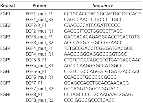
PCR typing was performed using genomic DNA isolated from tail biopsies or embryonic yolk sacs, respectively, using the allele-specific primer pairs listed in Table 1.
Whole-mount in situ hybridization
Whole-mount in situ hybridizations were done by standard procedures. Mutant and wild-type embryos were analyzed in parallel with a given probe under identical conditions. cDNAs for the generation of probes were originally obtained from M. Gessler (HeyL), M. Goulding (NeuroD,Neurogenin), A. Kispert (Uncx4.1,Tbx18,Pitx2,Tbx5), J. Rossant (Nodal), and T. Braun (Myogenin,MyoD). Probes were labeled with anti-digoxigen AP (Roche), and embryos were stained using BM purple. Skeletons of mouse fetuses were stained following standard procedures (Cordeset al.2004). Pictures were taken with a MD628 micro-scope with a DFC 420 camera utilizing the Firecam software v3.4.1 (all Leica). All photos were processed equally using Adobe Photoshop.
Immunohistochemistry
Specimens werefixed in Bouin’sfixative for 24 hr, embedded in paraffin, and sectioned at 8mM. Antibodies used were My32 (SigmaM4276) and NeuN (Millipore MAB377) at a dilution of 1:400. For NeuN, the MOM Kit from Vector Laboratories was used. Mouse biotinylated secondary antibody and the ABC Kit from Vector Laboratories were applied to sections incubated with My32. Detection of the signal was achieved with the DAB Kit (Vector Laboratories). Pictures were taken using a Leica DM5000B microscope and processed using Adobe Photoshop.
Statistical analyses
Statistical analyses were performed using Prism software (GraphPad). ImageJ quantifications and luciferase measure-ments were analyzed by one-way ANOVA, and compared using Bonferroni’s Multiple Comparison Test with a signifi -cance level of 0.05. Expression levels of NOTCH1 and NOTCH2 were analyzed using the Student’st-test.
Data and reagent availability
Cell lines, constructs, and strains are available upon request to the corresponding author.
Results
Generation and characterization of cell lines expressing DLL1 variants
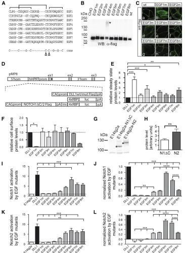
Since an EGF-like domain is thought to be an independently folding module (Downinget al.1996), we reasoned that, by disrupting disulphide bridges in individual EGF repeats, the intrinsic structure of neighboring repeats should be only mildly affected, if at all (Suk et al.2004; Mor-Cohenet al. 2012), and thus allows one to study the relevance of the structural integrity of individual EGF repeats in the context of full-length DLL1. We decided to exchange cysteine resi-dues 4 and 5 of each individual EGF repeat by glycine, an amino acid that displays intrinsically highflexibility, to dis-rupt two disulfide bridges of each repeat (Figure 1A). To test how these mutations affect DLL1 function in cultured cells, we generated expression vectors for Flag-tagged DLL1 pro-teins carrying the Cys-Gly mutations in individual EGF re-peats (from hereon referred to as “EGF#m”). Transiently transfected CHO cells expressed all mutant proteins, and all but EGF1m were predominantly detected at the cell mem-brane (Figure 1, B and C). For better comparability, we expressed the Flag-tagged DLL1 proteins from single-copy insertions into the Hprt locus of E14tg2a ES cells and used these cells for further analyses (Figure 1D andMaterials and Methods). To obtain thefirst hints of whether the EGF mu-tants differ with respect to global structural disruptions or the formation of aberrant disulfide bridges, we compared their migration patterns during electrophoreses under nonreduc-ing conditions. All mutant proteins except EGF1m showed a migration pattern similar to wild-type DLL1 (Supporting In-formation, Figure S1A), suggesting that these mutant pro-teins do not form mixed disulfides with other proteins during folding and transport to the cell surface. In addition, CD spectra of Fc-fusion proteins of the ECD of wild type or EGF4m (chosen as an example) were indistinguishable from each other (data not shown). Measuring the corresponding melting curves of these proteins revealed no significant Table 1 Allele-specific primers used for genotyping
Repeat Primer Sequence
EGF1 EGF1_mut_F1 CCTGCACCTACGGCAGTGCTGTCACG
EGF1_mut_R3 CAGCCAACTCTGCCCTTGCC
EGF2 EGF2-3_F1 CAACCCCATCCGATTCCCC
EGF2_mut_R1 CAGCCTTCCTGGCCGTTACC
EGF3 EGF3_mut_F1 GACCACACAGAGGCACCTCACTGTG
EGF3_mut_R2 ACCCAGGTCGGCCGGAACC
EGF4 EGF4_mut_F1 TCTGCCGACCTCGGGATGACGCC
EGF4_mut_R1 AAGCCGGGAGGGCCGGTGCC
EGF5 EGF5-6_F1 CTGTCTGCCAGGGTGTGATGACCAAC
EGF5_mut_R1 AGCCCAAGGGGCCATGGCC
EGF6 EGF5-6_F1 CTGTCTGCCAGGGTGTGATGACCAAC
EGF6_mut_R1 CCAGCCTGGCCCCGGCC
EGF7 EGF7_F1 GGAGCCACCTGCACCAACACG
EGF7_mut_R2 GCCAGGTGGGCCGGTACC
EGF8 EGF8_F1 CCTAGCCCCTGCAAGAACGGAGC
differences in the melting temperatures, suggesting that no massive changes in the overall protein stability occur due to the introduction of the mutations (Figure S1, B and C). Quan-tification showed higher steady-state levels of EGF3m, EGF5m, EGF8m, and particularly EGF1m, compared to DLL1 wild type (P.0.05; Figure 1E andTable S1). Surface biotinylation and quantification showed that all mutant
pro-teins except EGF1m were present on the cell surface at sim-ilar relative levels (Figure 1F and Table S2). EGF8m consistently showed lower relative cell-surface levels that were, however, statistically not significant (P . 0.05 in one-way ANOVA).
We also generated ES cells that carry the RBP4xluc Notch reporter gene (Serth et al. 2015) in the Hprt locus of Figure 1 Analyses of EGF repeat mu-tant DLL1 proteins in cultured cells
in vitro. (A) Alignment of amino acid sequences of DLL1 EGF repeats. The characteristic disulfide bridges are indi-cated by brackets above the sequence; arrowheads in the consensus sequence point to the mutated cysteine residues. (B) Western blot analysis of cell lysates of CHO cells transfected with expression vectors for DLL1 proteins as indicated at the top. CHO cells expressed all mutant proteins at the expected molecular weight. In the case of EGF4m, an addi-tional slower-migrating protein species was observed (asterisk). This high-molecular-weight species was not shifted to a lower molecular weight by treatment with reducing agents (DTT and iodoacetamide) and thus is unlikely to be caused by aberrant disulfide bridges. When the extracellular domain of EGF4m was expressed as a soluble Fc fusion, only a protein of the expected size was observed (data not shown), suggesting that the intracellular domain or localization at the cell membrane leads to an as-yet-unknown modifi ca-tion of some EGF4m protein. For quan-tification, both protein species were taken into account. (C) Detection of DLL1 proteins in CHO cells by
immuno-fluorescence. All DLL1 variants except EGF1m are at the cell surface. (D) Sche-matic representation of constructs used to generate single-copy transgene inser-tions in the Hprt locus. (E) Quantifi ca-tion of DLL1 proteins in E14tg2a ES cells expressing DLL1 variants from the Hprt locus (mean values and SEM;n= 4;
E14tg2a ES (referred to as H-RBPluc) cells. In addition, either NOTCH1 or NOTCH2 expression cassettes were in-troduced into these cells upstream of the reporter (Figure 1C) separated by the chicken b-globin insulator sequence (Vidigalet al.2010). Since in our hands NOTCH1 is hard to detect by Western blot analysis, we deleted the C-terminal 56 amino acids composing the PEST domain (referred to as
“NOTCH1DC”). For unknown reasons, only NOTCH2 expressed in the Hprt locus of ES cells (referred to as “ H-Notch2Flag-RBPluc cells”) could be activated by DLL1 in coculture assays. Therefore, we introduced a Notch1DC expression construct ran-domly into E14tg2a H-RBPluc cells (referred to as Notch1D C-Flag H-RBPluc cells). From NOTCH1DC-expressing clones, we selected the one that most closely matched NOTCH2 expression levels and (based on the S1-cleavage products) reached56% of NOTCH2 levels (Figure 1, G and H, andTable S3).
Analysis of Notch activation by DLL1 variants
Notch1DCFlag H-RBPluc and H-Notch2Flag-RBPluc ES cells were cocultured with ES cells expressing wild-type or mutant DLL1. DLL1wt activated NOTCH1DC on average 11-fold and NOTCH29-fold (Figure 1, I and K, andTable S4). As expected, EGF1m did not activate NOTCH1DC or NOTCH2 since it does not reach the cell surface and served as addi-tional negative control. Likewise, EGF2m and EGF3m did not activate either receptor (Figure 1, I and K). All other DLL1 mutants activated NOTCH1DC and NOTCH2 although with different efficiencies ranging from20 to 80% (Figure 1, I and K, un-normalized data). To compare the activity of EGF mutant proteins to DLL1wt, we also normalized their activity (setting DLL1 activity to 1; Figure 1, J and L) and corrected for expression levels [for EGF3m, EGF5m, and EGF8m, which showed significant (P . 0.05) differences in their steady-state levels compared to wild type; no correction was made for EGF1m because it was not present on the cell surface]. Both receptors showed a highly similar profile of activation by the EGF mutants. EGF4m, EGF5m, and EGF8m showed
15–20% activity of DLL1wt, EGF6m activated NOTCH1DC and NOTCH2 to80% of wild-type levels, and EGF7 reached between 50 and 60% of activity (Figure 1, J and L, andTable S5). To exclude that different proliferation rates and thus different numbers of ES cells expressing DLL1 variants affect the results of the transactivation assays, we counted the num-ber of ES cells after 48 hr of culture and did notfind signif-icant differences (Figure S1E). Thus, based on thein vitro Notch activation assays, disruption of the structural integrity of EGF repeats 2–8 impinges on DLL1-mediated Notch acti-vation, and activation of both NOTCH1 and NOTCH2 is sim-ilarly affected. The significance of EGF1 for DLL1 ligand function cannot be assessed due to the intracellular retention of the mutated protein.
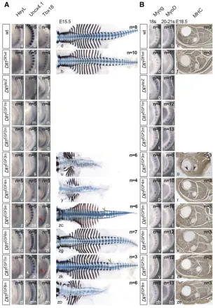
Generation and analysis of an allelic series of Dll1 mutations in mice
To assess the contribution of EGF2-8 to full DLL1 function under physiological conditions, we generated an allelic series
of these Dll1mutations in mice based on the knock-in of a Delta1 mini-gene (Figure 2A andFigure S1F). All heterozy-gous EGF repeat mutants were normal, indicating that one wild-type allele ofDll1is sufficient for normal development and that the mutant proteins do not exert obvious dominant-negative effects on wild-type DLL1. Initially, homozygous mutants were analyzed on the isogenic 129Sv/ImJ genetic background. The Dll1Dll1wt allele carrying the Dll1
wild-type mini-gene was indistinguishable from wild-wild-type mice (Figure 2B, a–f and g–l), indicating that the mini-gene com-pensates the disrupted endogenous gene. As previously de-scribed, mice lacking DLL1 (Dll1lacZ) were hemorrhagic from
embryonic day 10.5 (E10.5) on and died around E12.5 (Fig-ure 2B, m–o). Essentially the same phenotype was observed with Dll1EGF1m(Figure 2B, p–r) and Dll1EGF2m(Figure 2B,
s–u) embryos.Dll1EGF3membryos displayed a milder
pheno-type: embryos developed to E18.5, had only mild hemor-rhages and a severely reduced body axis (Figure 2B, v–z), and were motionless. Dll1EGF4m (Figure 2B, za–ze) and
Dll1EGF8m(Figure 2B, zx–zzb) embryos displayed a shortened
body axis and did not survive after birth. Dll1EGF5mand
Dll1EGF7mmutants were viable and fertile with shortened
kinky tails, Dll1EGF5m being more strongly affected than
Dll1EGF7m(Figure 2B, zf–zk and zr–zw).Dll1EGF6membryos
were indistinguishable from wild-type andDll1Dll1wt, viable,
and fertile (Figure 2B, zl–zq). Thus, the structural integrity of all but EGF repeat 6 appears to be important for DLL1 as a sufficiently active Notch ligandin vivo. For further compar-ative analyses of early developmental processes known to re-quire DLL1, all alleles were outcrossed for three generations and analyzed on a mixed genetic background because the poor breeding performance of isogenic 129Sv/ImJ mice precluded the efficient collection of embryos. Mutants showed a virtually identical range of phenotypes on the mixed and the isogenic background (Figure S1H).
Somite patterning
DLL1 signaling is instrumental for establishment of anterior– posterior somite polarity and subsequent axial skeleton de-velopment (Hrabe de Angeliset al.1997; Cordeset al.2004). To compare the impact of EGF repeat mutations on somito-genesis, we analyzed expression of the Notch target HeyL (Leimeister et al. 2000), andUncx4.1 and Tbx18, markers for posterior and anterior somite compartments, respectively, in E9.5 embryos (Neidhardtet al.1997; Krauset al.2001). The expression patterns of these genes as well as the axial skeletons of homozygous Dll1Dll1wt (Figure 3A, e–h) and
Dll1EGF6m(Figure 3A, zd–zg) embryos were indistinguishable
from wild type (Figure 3A, a–d).Dll1EGF1m, Dll1EGF2m, and Dll1EGF3membryos (Figure 3A, l–t) resembled embryos
lack-ing DLL1 (Figure 3A, i–k), and Dll1EGF3m E15.5 skeletons
showed massive defects (Figure 3A, u). Dll1EGF4m and
Dll1EGF8mmutants displayed somewhat milder A–P polarity
defects than Dll1EGF3m, Dll1EGF4m being more severely
preparations revealed misshapen vertebrae and ribs similar toDll1EGF3mmutants (Figure 3A, y and zo).Dll1EGF5m(Figure
3A, zc) andDll1EGF7m(Figure 3A, zk) mutants displayed mild
skeletal defects, which might underlie the slightly reduced breeding performance of these mutant lines (Figure S1G). AlthoughHeyLexpression was clearly reduced (Figure 3A, z and zh),Uncx4.1andTbx18showed only minor irregular-ities (arrowheads in Figure 3A, za, zb, and zi, zj).
Myogenesis
Loss of DLL1 leads to premature differentiation of myoblasts and severely reduced skeletal muscles (Schuster-Gossleret al. 2007). To compare the mutations in this context, we analyzed expression of Myog and MyoD, two regulators of myogenesis (Arnold and Braun 2000), in age-matched somite-stage (ss) 18 and 20–21 embryos and stained skeletal muscles in cross sections of hind limbs at similar proximo-distal
positions for myosin heavy chain (MHC). In wild-type 18 ss embryos, Myog was expressed in the anterior 7–8 somites, and at ss 20–21 faintMyoDexpression was detected (Figure 3B, a and b). Dll1Dll1wt, Dll1EGF5m, Dll1EGF6m, andDll1EGF7m
embryos expressed Myog and MyoD virtually identical to wild-type embryos (Figure 3B, d, e, s, t, v, w, y, z) and had apparently normal skeletal muscles at E18.5 (compare Figure 3B, c, and Figure 3B, f, u, x, za). InDll1knockout embryos, MyogandMyoDexpression was upregulated (Figure 3B, g and h), which was similarly observed inDll1EGF1m,Dll1EGF2m, and
Dll1EGF3membryos (Figure 3B, i–n). Hind limbs of motionless
Dll1EGF3mmutants lacked skeletal muscles with the exception
of a few MHC-positive remnants (arrowheads in Figure 3B, o) resembling the phenotype of embryos that are heteroallelic for theDll1null and a hypomorphic allele (Schuster-Gossleret al. 2007).MyogandMyoDexpression inDll1EGF4mandDll1EGF8m
embryos appeared slightly upregulated (Figure 3B, p, q and zb, Figure 2 Generation of an allelic series of EGF mutations in DLL1 and external phenotypes. (A) Structure ofDll1before and after homologous recombination and of the targeting vector. Black boxes indicate coding and white boxes noncoding regions ofDll1. The cDNA portion is outlined in red. (B) Morphology of wild-type 129Sv/ImJ embryos and mice and of isogenic embryos and mice homozygous for the Dll1
zc); however, in cross sections through the hind limbs skeletal muscles were indistinguishable from wild type (Figure 3B, r, ze).
Neurogenesis
DLL1-mediated Notch activation represses neuronal differen-tiation (de la Pompaet al.1997; Rochaet al.2009). To com-pare the EGF-repeat mutations in this context, we analyzed expression ofNeurog1andNeuroD, a neuronal determination and differentiation gene, respectively, in 23 ss embryos (Ma et al.1996). To analyze whether the EGF mutations lead to obvious overall alterations of the architecture of the central nervous system at later stages, we stained cross sections of the cervical spinal cord of E18.5 embryos for NeuN, a neuron-specific nuclear protein (Mullenet al.1992) that labels all neurons. Like wild type, Dll1Dll1wt and Dll1EGF6m embryos
showed normal expression of Neurog1 and NeuroDin the brain and dorsal neural tube and in cranial and spinal ganglia (Figure 4A, a, b, d, e, v, w) and an indistinguishable
cytoarchi-tecture of the cervical spinal cord (Figure 4A, c, f, x). In Dll1EGF1m, Dll1EGF2m, and Dll1EGF3membryos,Neurog1 and
NeuroDexpression was upregulated in the spinal cord, and premature expression domains were detected in the nasal placodes (white and red arrowheads, respectively, in Figure 4A, i–n).Neurog1was additionally upregulated in the mid-and forebrain (yellow mid-and black arrowheads, respectively, in Figure 4A, i, k, m), resemblingDll1knockout embryos (Fig-ure 4A, g and h). Unexpectedly, despite the clearly enhanced neuronal differentiation in early embryos, the spinal cords of homozygous Dll1EGF3m embryos appeared enlarged rather
than reduced in size, and cellularity was increased (Figure 4A, o). This contrasts with the reduction of neural tube size at E12.5 that was observed in embryos with cell type-specific deletion of Dll1 in neural tube progenitors (Rocha et al. 2009). The development of this phenotype is unclear at pre-sent and will require a detailed analysis in the future. A total of 23 ss Dll1EGF4mand Dll1EGF8m embryos displayed weak Figure 3 Somite patterning and skeletal muscle develop-ment in mutants homozygous for individualDll1EGF al-leles. (A) Whole-mount in situ hybridizations (WISH) of E9.5 and skeletal preparations of E15.5 embryos. Alleles are indicated at the left, probes at the top. White and red arrowheads point to irregularities ofUncx4.1andTbx18
premature expression of Neurog and NeuroD in the nasal placodes (red arrow heads in Figure 4A, p, q, zb, zc). How-ever, their spinal cords appeared indistinguishable from wild type (Figure 4A, r, zd).
Left–right asymmetry
DLL1-mediated Notch activation is essential for establishment of left–right asymmetry (Krebs et al. 2003). We analyzed expression ofnodalin E8.0,Pitx2in E8.5 embryos, and heart looping at E10.5 in embryos hybridized to aTbx5probe that shows strong expression in the left ventricle (Bruneauet al. 1999). In wild-type embryos,nodalis first expressed
asym-metrically around the node followed by expression in the left lateral plate mesoderm (LPM) (Figure 4B, a) and (Loweet al. 1996). Subsequently,Pitx2expression is activated in the LPM (Figure 4B b), and (Yoshiokaet al.1998), and at E10.5 right-ward looping of the heart indicates correct left–right deter-mination (Figure 4B, c). Normal expression of nodal and Pitx2 and consecutive normal heart looping was observed in all Dll1Dll1ki, Dll1EGF4m, Dll1EGF5m, Dll1EGF6m, Dll1EGF7m,
looping was abnormal (Figure 4B, g, h, i). About 25% of Dll1EGF1m,Dll1EGF2m, andDll1EGF3membryos obtained from
matings between heterozygous mutants showed no nodal expression even after prolonged staining, absent or random-izedPitx2expression in the LPM, and randomization of heart looping (Figure 4B, j–r;Table S6;Table S7).
Discussion
To obtain insights into the significance of each EGF repeat in the ectodomain of the Notch ligand DLL1 for its function, we disrupted two disulfide bridges in each repeat individually by substituting cysteine with glycine residues. These mu-tations should disrupt and destabilize the domain structure of the respective repeat as has been observed for cysteine substitutions in EGF repeats of other proteins (Suket al. 2004; Mor-Cohenet al.2012) and should increase the in-trinsicflexibility of the mutated repeats. Analyses of single cysteine substitutions (disruption of a single disulfide bridge) infibrillin showed only localized structural effects but did not exclude the possibility of different effects if other disulfide bridges were disrupted (Suket al.2004). In addition, although we disrupted the same disulfide bridges in each EGF repeat, the severity of the disruption
of the individual domain structure might vary between in-dividual mutated EGF repeats due to their different amino acid compositions, which could contribute to different ductions in DLL1 function. Nonetheless, our mutations re-veal the consequences of disrupting the structural integrity of individual EGF repeats in the context of the full ligand protein.
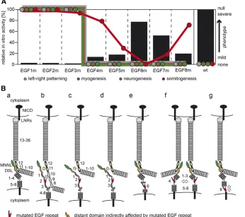
To analyze how our EGF mutations affect DLL1 ligand function, we first tested the ability of mutant proteins to activate the Notch1 and Notch2 receptors in cell-based trans-activation assays and observed very similar activity patterns ranging from complete absence of ligand activity to levels up to 80% of the wild-type level (Figure 5A). This suggests that DLL1 interacts with both Notch receptors very similarly, at least under the conditions of our transactivation assays. Gen-erating the same individual EGF mutations in endogenous DLL1, we established an allelic series in mice in vivo and observed similar effects of our EGF repeat mutations on DLL1 ligand activity: mutations in EGF repeats close to the MNNL-DSL binding interface resulted in null or strong hypo-morphic mutations, mutations more distant affected ligand activity successively less up to EGF6, and mutations close to the membrane interfered with DLL1 function again more strongly (summarized in Figure 5A).
Figure 5 Schematic summary of results and potential effects of EGF repeat mutations on DLL1. (A) Schematic representation of DLL1 ac-tivity in transactivation assays in vitro (black bars; scale on lefty-axis) and severity of mutant phenotypes (colored circles; arbitrary scale on righty-axis). The position of colored circles in-dicates the severity of the mutant phenotype with respect to left–right patterning, myogen-esis, neurogenmyogen-esis, and somitogenesis. Above a certain threshold corresponding to ,20% DLL1 activity measured in in vitro, left–right patterning, myogenesis, and neurogenesis pro-ceed apparently normal. In contrast, somito-genesis shows a graded response to reduced DL1 activity. (B) Model depicting potential ef-fects of EGF repeat mutations on DLL1 func-tion. (a) Interaction between MNNL (green) and DSL (orange) domains of DLL1 (dark gray) with EGF 11 and 12 (graded yellow/black ovals) of Notch (light gray) based on Luca et al.
As the consequences of our mutations may extend beyond the mutated EGF repeat, their impact on DLL1 activity may be explained by interference local and/or distant DLL1–Notch interactions. Mutations in EGF2 and EGF3 completely abol-ished activation of Notch1 and Notch2 in vitro. In mice, EGF2m behaved as a null allele indistinguishable from Dll1lacZ orDll1EGF1mwhereas EGF3m behaved like a severe
hypomorph (Figure 5A). Thesefindings are consistent with results of previousin vitrostudies using purified ligand pro-tein fragments showing that the presence of EGF2 and EGF3 in these fragments is essential for effective binding to, and activation of, Notch (Shimizu et al. 1999; Andrawes et al. 2013). Since the MNNL and the DSL domain of DLL4 (and most likely also of DLL1) are in direct contact with the ligand binding interface of the receptor (EGF 11 and 12) (Lucaet al. 2015), EGF2 and EGF3 could contribute to the direct inter-action of ligand and receptor by binding to Notch adjacent to EGF 11 (Figure 5B, b), which is consistent with the observa-tion that addiobserva-tional EGF repeats outside the major ligand binding site of Notch receptors are essential for full Notch activation and function (Shimizu et al. 1999; Hambleton et al. 2004; Xuet al.2005; Cordleet al. 2008b; Andrawes et al.2013). Such an interaction could not be observed by Lucaet al. (2015) since EGF repeats of Notch1 potentially involved in this interaction were not included in their study. Additionally, the linear arrangement of the DSL, EGF1, and EGF2 domains, which was detected in the crystal structure of Notch1 EGF 11–13 with the DLL4 N-terminal domains (Luca et al.2015), or in the uncomplexed structures of DLL1 and JAG1 (Cordle et al.2008a; Kershaw et al.2015), might be destabilized by EGF2m and EGF3m, resulting in ineffective interaction of the MNNL/DSL domains with EGF 11 and 12 of Notch (Figure 5B, c). Furthermore, it is possible that muta-tions in EGF repeats close to the MNNL and DSL domains impinge on the structure of these domains and thereby affect receptor ligand interaction (Figure 5B, d). Likewise, the mu-tation of EGF4 (or EGF5) might spread beyond the repeat boundary and thereby perturb the likely interaction of EGF3 (and EGF2) with Notch. If the effects of our EGF mu-tations spread to adjacent domains, their impact on the DLL1– Notch binding interface and DLL1 activity can be expected to decrease with increasing distance from the binding interface. This possibility might be reflected by the apparent correlation between decreasing phenotypic strength and distance of our mutations from the binding interface up to EGF 6.
In our transactivation assays, NOTCH1 and NOTCH2 were activated by EGF6m at80% of wild-type levels, andin vivo this allele was indistinguishable from wild type in all ana-lyzed processes (Figure 5A). Eighty percent of DLL1 activity should be sufficient for normal development, since mice that are heterozygous for the Dll1 null allele, and presumably have 50% of DLL1 activity, are normal. EGF6 is found to be located next to a bent potentially present in DLL1 (Kershaw et al. 2015), which might explain why DLL1 tolerates the disruption of two disulfide bridges in EGF6 and the presumed increasedflexibility fairly well (Figure 5B, e). Alternatively,
the integrity of EGF6 is not important for DLL1 function and the mutation does not propagate into EGF repeats important for ligand receptor interaction, or the mutation affects the intradomain stability of EGF6 only mildly, possibilities that we cannot distinguish at present.
Disruption of EGF repeats close to the cell membrane are less likely to directly affect the interaction of ligand with Notch. Recycling of DLL1 molecules is thought to lead to their clustering in microdomains at the cell surface, which in certain contexts is required for Notch activation (Musseet al.2012). It is conceivable that mutations in EGF repeats impinge on and weaken homophilic DLL1 interactions and thus affect ligand clustering and density on the cell surface leading to reduced DLL1 activity (Figure 5B, f). Upon ligand binding, the extra-cellular domain of Notch is endocytosed into the signal-sending cell, generating a pulling force that is necessary for a conformational change in the Notch extracellular domain and subsequent S2 cleavage (Meloty-Kapella et al. 2012; Musseet al. 2012; Gordonet al.2015). An increased
flexibility of EGF repeats might compromise DLL1’s ability to exert the necessary pulling force on the Notch extracellular domain and thus weaken Notch activation (Figure 5B, g). In addition, interactions with as-yet-unknown proteins on the cell surface might be affected.
With respect to somitogenesis, myogenesis, neurogenesis, and left–right determination—early developmental processes known to require DLL1 activity—EGF3m is virtually indistin-guishable from the Dll1lacZnull allele. However,DLL1EGF3m
mutants did not show the severe hemorrhages observed in theDll1lacZ,Dll1EGF1m, andDll1EGF2mnull alleles and survived
until E18.5, suggesting some residual DLL1 activity that we did not detect in ourin vitrotransactivation assays. Alterna-tively, the rescue of the early lethality might reflect a DLL1 function independent from Notch. No such activity is known for mammalian DLL1, but such a function has been suggested for Drosophila Delta (Mok et al. 2005). In the Dll1EGF4m,
Dll1EGF5m,Dll1EGF7m, andDll1EGF8membryos, DLL1-dependent
myogenesis, neurogenesis, and left–right determination pro-ceeded apparently normally (Figure 5A), although we cannot exclude minor abnormalities, suggesting that these processes require fairly low DLL1 activity. In contrast, all these mu-tants displayed anterior–posterior polarity defects in somites and, consequently, skeletal malformations of varying degrees (Figure 5A).
in vitro, which is similar to EGF4m (Figure 1, J and L, and Figure 5A), but much higherin vivo, allowing for survival of homozygous mutants (Figure 5A).
Only somitogenesis appears to be affected by alterations of the structural integrity of EGF repeats distant from the ligand– receptor interface and responds in a graded manner (Figure 5A) consistent with our previous observation that somitogen-esis is fairly sensitive to reduced Notch activity (Schuster-Gossleret al.2009). Reduction of proteinO-fucosyltransferase 1 (POFUT1), which links fucose to specific Ser or Thr resi-dues in EGF repeats and is essential for Notch signaling (Shi and Stanley 2003), also affects only somite patterning (Schuster-Gossleret al.2009).O-linked fucose can be further modified by Fringe glycosyltransferases, which modulate NOTCH activation by ligands (reviewed in Stanley 2007). Lunatic fringe (LFNG) is expressed in the presomitic meso-derm, required for normal somitogenesis (Zhang and Gridley 1998), and appears to act there as a negative regulator of Notch signaling (Morimotoet al.2005). LFNG modification of NOTCH might affect the interaction of EGF-mutant DLL1 with NOTCH and might contribute to the specific impact of the EGF repeat mutations on somitogenesis, which requires future analyses. However, the tissue-specific differences in phenotypic outcomes clearly indicate context-dependent ef-fects of disrupted EGF repeats in DLL1. DLL1 is also a sub-strate for POFUT1 (Paninet al.2002; Mülleret al.2014). The mutations that we generated do not affect O-fucosylation sites, although the structure of mutated EGF repeats might affectO-fucosylation (Wanget al.1996). However, in vitro POFUT1 was dispensable for DLL1 function (Müller et al. 2014), and in Drosophila O-linked fucose was dispensable in signal-sending cells (Okajima and Irvine 2002), suggesting that altered sugar modification of DLL1 does not contribute to its reduced activity.
Mutations in the Notch pathway components DLL3, LFNG, HES7, and MESP2 underlie the severe vertebral abnormalities in the autosomal recessive human condition spondylocostal dysostosis (SCD; reviewed in Pentonet al.2012). However, 70% of the patients do not have mutations in these genes (Chapmanet al.2011). Our alleles define somite patterning as particularly sensitive to disruption of the intrinsic second-ary structure of EGF repeats of DLL1, raising the possibility that such mutations in DLL1 in humans can underlie cases of SCD. Collectively, ourin vitroandin vivoanalyses discrimi-nate context-dependent requirements of Delta–Notch inter-action and show that disruption of the intrinsic domain structure of each EGF repeat impinges on DLL1-mediated Notch activation, and hence the structural integrity of each of these EGF repeats is required for full ligand activity.
Acknowledgments
We thank Alain Israel, Shigeru Chiba, Raphael Kopan, and Andreas Kispert for providing plasmids; Thomas Klein, Dietmar Manstein, and Matthias Gaestel for critical com-ments and discussion; and Anatoli Heiser for technical
assistance. This work was supported by grant no. GO 449/ 10-1 from the German Research Council (to A.G.) and by funding from the Cluster of Excellence “From Regenerative Biology to Reconstructive Therapy”(to A.G.).
Literature Cited
Alten, L., K. Schuster-Gossler, M. P. Eichenlaub, B. Wittbrodt, J. Wittbrodt et al., 2012 A novel mammal-specific three partite enhancer element regulates node and notochord-specific Noto expression. PLoS ONE 7: e47785.
Andersson, E. R., R. Sandberg, and U. Lendahl, 2011 Notch sig-naling: simplicity in design, versatility in function. Development 138: 3593–3612.
Andrawes, M. B., X. Xu, H. Liu, S. B. Ficarro, J. A. Martoet al., 2013 Intrinsic selectivity of Notch 1 for like 4 over Delta-like 1. J. Biol. Chem. 288: 25477–25489.
Arnold, H. H., and T. Braun, 2000 Genetics of muscle determina-tion and development. Curr. Top. Dev. Biol. 48: 129–164. Bauer, R. C., A. O. Laney, R. Smith, J. Gerfen, J. J. D. Morrissette
et al., 2010 Jagged1 (JAG1) mutations in patients with tetral-ogy of Fallot or pulmonic stenosis. Hum. Mutat. 31: 594–601. Bettenhausen, B., M. Hrabe de Angelis, D. Simon, J. L. Guénet, and
A. Gossler, 1995 Transient and restricted expression during mouse embryogenesis of Dll1, a murine gene closely related to Drosophila Delta. Development 121: 2407–2418.
Braune, E.-B., K. Schuster-Gossler, M. Lyszkiewicz, K. Serth, K. Preusseet al., 2014 S/T Phosphorylation of DLL1 is required for full ligand activity in vitro but dispensable for DLL1 func-tion in vivo during embryonic patterning and marginal zone B cell development. Mol. Cell. Biol. 34: 1221–1233.
Bronson, S. K., E. G. Plaehn, K. D. Kluckman, J. R. Hagaman, N. Maedaet al., 1996 Single-copy transgenic mice with chosen-site integration. Proc. Natl. Acad. Sci. USA 93: 9067–9072. Bruneau, B. G., M. Logan, N. Davis, T. Levi, C. J. Tabin et al.,
1999 Chamber-specific cardiac expression of Tbx5 and heart defects in Holt-Oram syndrome. Dev. Biol. 211: 100–108. Chapman, G., D. B. Sparrow, E. Kremmer, and S. L. Dunwoodie,
2011 Notch inhibition by the ligand DELTA-LIKE 3 defines the mechanism of abnormal vertebral segmentation in spondylocostal dysostosis. Hum. Mol. Genet. 20: 905–916.
Chillakuri, C. R., D. Sheppard, S. M. Lea, and P. A. Handford, 2012 Notch receptor-ligand binding and activation: insights from molecular studies. Semin. Cell Dev. Biol. 23: 421–428. Cordes, R., K. Schuster-Gossler, K. Serth, and A. Gossler,
2004 Specification of vertebral identity is coupled to Notch sig-nalling and the segmentation clock. Development 131: 1221–1233. Cordle, J., S. Johnson, J. Z. Y. Tay, P. Roversi, M. B. Wilkinet al., 2008a A conserved face of the Jagged/Serrate DSL domain is involved in Notch trans-activation and cis-inhibition. Nat. Struct. Mol. Biol. 15: 849–857.
Cordle, J., C. Redfieldz, M. Stacey, P. A. van der Merwe, A. C. Willis et al., 2008b Localization of the delta-like-1-binding site in human Notch-1 and its modulation by calcium affinity. J. Biol. Chem. 283: 11785–11793.
de la Pompa, J. L., A. Wakeham, K. M. Correia, E. Samper, S. Brown et al., 1997 Conservation of the Notch signalling pathway in mammalian neurogenesis. Development 124: 1139–1148. de Vries, W. N., L. T. Binns, K. S. Fancher, J. Dean, R. Mooreet al.,
2000 Expression of Cre recombinase in mouse oocytes: a means to study maternal effect genes. Genesis 26: 110–112. Downing, A. K., V. Knott, J. M. Werner, C. M. Cardy, I. D. Campbell
Marfan syndrome and other genetic disorders. Cell 85: 597– 605.
Geffers, I., K. Serth, G. Chapman, R. Jaekel, K. Schuster-Gossler et al., 2007 Divergent functions and distinct localization of the Notch ligands DLL1 and DLL3 in vivo. J. Cell Biol. 178: 465–476.
Gordon, W. R., B. Zimmerman, L. He, L. J. Miles, J. Huanget al., 2015 Mechanical allostery: evidence for a force require-ment in the proteolytic activation of Notch. Dev. Cell 33: 729–736.
Hambleton, S., N. V. Valeyev, A. Muranyi, V. Knott, J. M. Werner et al., 2004 Structural and functional properties of the human Notch-1 ligand binding region. Structure 12: 2173–2183. Hooper, M., K. Hardy, A. Handyside, S. Hunter, and M. Monk,
1987 HPRT-deficient (Lesch-Nyhan) mouse embryos derived from germline colonization by cultured cells. Nature 326: 292–295.
Hrabe de Angelis, M., J. McIntyre, and A. Gossler, 1997 Maintenance of somite borders in mice requires the Delta homologue DII1. Na-ture 386: 717–721.
Kershaw, N. J., N. L. Church, M. D. W. Griffin, C. S. Luo, T. E. Adams et al., 2015 Notch ligand delta-like1: X-ray crystal structure and binding affinity. Biochem. J. 468: 159–166. Kraus, F., B. Haenig, and A. Kispert, 2001 Cloning and expression
analysis of the mouse T-box gene Tbx18. MOD 100: 83–86. Krebs, L. T., N. Iwai, S. Nonaka, I. C. Welsh, Y. Lan et al.,
2003 Notch signaling regulates left-right asymmetry determi-nation by inducing Nodal expression. Genes Dev. 17: 1207– 1212.
Ladi, E., J. T. Nichols, W. Ge, A. Miyamoto, C. Yaoet al., 2005 The divergent DSL ligand Dll3 does not activate Notch signaling but cell autonomously attenuates signaling induced by other DSL ligands. J. Cell Biol. 170: 983–992.
Leimeister, C., N. Schumacher, C. Steidl, and M. Gessler, 2000 Analysis of HeyL expression in wild-type and Notch pathway mutant mouse embryos. MOD 98: 175–178.
Lowe, L. A., D. M. Supp, K. Sampath, T. Yokoyama, C. V. Wright et al., 1996 Conserved left-right asymmetry of nodal expres-sion and alterations in murine situs inversus. Nature 381: 158– 161.
Luca, V. C., K. M. Jude, N. W. Pierce, M. V. Nachury, S. Fischer et al., 2015 Structural basis for Notch1 engagement of Delta-like 4. Science 347: 847–853.
Ma, Q., C. Kintner, and D. J. Anderson, 1996 Identification of neurogenin, a vertebrate neuronal determination gene. Cell 87: 43–52.
Meloty-Kapella, L., B. Shergill, J. Kuon, E. Botvinick, and G. Weinmaster, 2012 Notch ligand endocytosis generates me-chanical pulling force dependent on dynamin, epsins, and actin. Dev. Cell 22: 1299–1312.
Minoguchi, S., Y. Taniguchi, H. Kato, T. Okazaki, L. J. Stroblet al., 1997 RBP-L, a transcription factor related to RBP-Jkappa. Mol. Cell. Biol. 17: 2679–2687.
Mok, L.-P., T. Qin, B. Bardot, M. LeComte, A. Homayouniet al., 2005 Delta activity independent of its activity as a ligand of Notch. BMC Dev. Biol. 5: 6.
Mor-Cohen, R., N. Rosenberg, Y. Einav, E. Zelzion, M. Landauet al., 2012 Unique disulfide bonds in epidermal growth factor (EGF) domains of b3 affect structure and function ofaIIbb3 and avb3 integrins in different manner. J. Biol. Chem. 287: 8879–8891.
Morimoto, M., Y. Takahashi, M. Endo, and Y. Saga, 2005 The Mesp2 transcription factor establishes segmental borders by suppressing Notch activity. Nature 435: 354–359.
Morrissette, J. D., R. P. Colliton, and N. B. Spinner, 2001 Defective intracellular transport and processing of JAG1 missense muta-tions in Alagille syndrome. Hum. Mol. Genet. 10: 405–413.
Mullen, R. J., C. R. Buck, and A. M. Smith, 1992 NeuN, a neuro-nal specific nuclear protein in vertebrates. Development 116: 201–211.
Müller, J., N. A. Rana, K. Serth, S. Kakuda, R. S. Haltiwanger et al., 2014 O-fucosylation of the Notch ligand mDLL1 by POFUT1 is dispensable for ligand function. PLoS One 9: e88571.
Musse, A. A., L. Meloty-Kapella, and G. Weinmaster, 2012 Notch ligand endocytosis: mechanistic basis of signaling activity. Semin. Cell Dev. Biol. 23: 429–436.
Neidhardt, L. M., A. Kispert, and B. G. Herrmann, 1997 A mouse gene of the paired-related homeobox class expressed in the cau-dal somite compartment and in the developing vertebral col-umn, kidney and nervous system. Dev. Genes Evol. 207: 330– 339.
Okajima, T., and K. D. Irvine, 2002 Regulation of notch signaling byO-linked fucose. Cell 111: 893–904.
Panin, V. M., L. Shao, L. Lei, D. J. Moloney, K. D. Irvine et al., 2002 Notch ligands are substrates for protein O-fucosyltransferase-1 and Fringe. J. Biol. Chem. 277: 29945–29952.
Parks, A. L., J. R. Stout, S. B. Shepard, K. M. Klueg, A. A. Santos Dos et al., 2006 Structure-function analysis of Delta trafficking, re-ceptor binding and signaling inDrosophila. Genetics 174: 1947– 1961.
Penton, A. L., L. D. Leonard, and N. B. Spinner, 2012 Notch sig-naling in human development and disease. Semin. Cell Dev. Biol. 23: 450–457.
Redeker, C., K. Schuster-Gossler, E. Kremmer, and A. Gossler, 2013 Normal development in mice over-expressing the intra-cellular domain of DLL1 argues against reverse signaling by DLL1 in vivo. PLoS ONE 8: e79050.
Rocha, S. F., S. S. Lopes, A. Gossler, and D. Henrique, 2009 Dll1 and Dll4 function sequentially in the retina and pV2 domain of the spinal cord to regulate neurogenesis and create cell diver-sity. Dev. Biol. 328: 54–65.
Schneider, C. A., W. S. Rasband, and K. W. Eliceiri, 2012 NIH Image to ImageJ: 25 years of image analysis. Nat. Methods 9: 671–675.
Schuster-Gossler, K., R. Cordes, and A. Gossler, 2007 Premature myogenic differentiation and depletion of progenitor cells cause severe muscle hypotrophy in Delta1 mutants. Proc. Natl. Acad. Sci. USA 104: 537–542.
Schuster-Gossler, K., B. Harris, K. R. Johnson, J. Serth, and A. Gossler, 2009 Notch signalling in the paraxial mesoderm is most sensitive to reduced Pofut1 levels during early mouse de-velopment. BMC Dev. Biol. 9: 6.
Serth, K., K. Schuster-Gossler, E. Kremmer, B. Hansen, B. Marohn-Köhn et al., 2015 O-fucosylation of DLL3 is re-quired for its function during somitogenesis. PLoS ONE 10: e0123776.
Shi, S., and P. Stanley, 2003 Protein O-fucosyltransferase 1 is an essential component of Notch signaling pathways. Proc. Natl. Acad. Sci. USA 100: 5234–5239.
Shimizu, K., S. Chiba, K. Kumano, N. Hosoya, T. Takahashiet al., 1999 Mouse jagged1 physically interacts with notch2 and other notch receptors. Assessment by quantitative methods. J. Biol. Chem. 274: 32961–32969.
Singh, R., W. M. Hoogaars, P. Barnett, T. Grieskamp, M. S. Rana et al., 2012 Tbx2 and Tbx3 induce atrioventricular myocardial development and endocardial cushion formation. Cell. Mol. Life Sci. 69: 1377–1389.
Stanley, P., 2007 Regulation of Notch signaling by glycosylation. Curr. Opin. Struct. Biol. 17: 530–535.
factor-like domain 30 of humanfibrillin-1. J. Biol. Chem. 279: 51258–51265.
Vidigal, J. A., M. Morkel, L. Wittler, A. Brouwer-Lehmitz, P. Grote et al., 2010 An inducible RNA interference system for the func-tional dissection of mouse embryogenesis. Nucleic Acids Res. 38: e122.
Wang, Y., G. F. Lee, R. F. Kelley, and M. W. Spellman, 1996 Identification of a GDP-L-fucose:polypeptide fucosyl-transferase and enzymatic addition of O-linked fucose to EGF domains. Glycobiology 6: 837–842.
Xu, A., L. Lei, and K. D. Irvine, 2005 Regions of Drosophila Notch that contribute to ligand binding and the modulatory influence of Fringe. J. Biol. Chem. 280: 30158–30165.
Yoshioka, H., C. Meno, K. Koshiba, M. Sugihara, H. Itoh et al., 1998 Pitx2, a bicoid-type homeobox gene, is involved in a lefty-signaling pathway in determination of left-right asymme-try. Cell 94: 299–305.
Zhang, N., and T. Gridley, 1998 Defects in somite formation in lunatic fringe-deficient mice. Nature 394: 374–377.