Phage Assisted Continuous Evolution (PACE):
a How-to Guide for Directed Evolution
Serban C. Popa,1 Ichiro Inamoto,1 Benjamin W. Thuronyi,2 and Jumi A. Shin1,*
1 Department of Chemistry, University of Toronto, 3359 Mississauga Road, Mississauga, ON, L5L
1C6, Canada.
2 Department of Chemistry, Williams College, 47 Lab Campus Drive, Williamstown, MA, 01267,
USA.
* To whom correspondence should be addressed: [email protected], (905) 828-5355
Keywords: continuous evolution, protein design, protein engineering, phage, bacterial one-hybrid, plaque assay, mutational analysis, DNA sequencing.
ABSTRACT
Directed evolution methods are becoming increasingly popular, as they are extremely powerful toward developing new biomolecules with altered/novel activities, e.g., proteins with new catalytic functions or substrate specificities, and nucleic acids that recognize an intended target. Especially useful are systems that have incorporated continuous evolution, where the protein to be evolved undergoes continuous mutagenesis to evolve a desired trait with little to no input from the researcher once the system is started. However, continuous evolution methods can be challenging to implement in the lab and daunting for researchers to invest time and resources. Our intent is to provide basic information and helpful suggestions that we have gained from our experience with bacterial phage-assisted continuous evolution (PACE). Specifically, we review factors to consider before adopting PACE for a given evolution scheme, different types of selection circuits that can be utilized with particular focus on the PACE-B1H selection system, what optimization of a PACE selection circuit may look like using directed evolution of ME47 as a case study, and additional techniques that may be incorporated into a PACE experiment. With this information, researchers will be better equipped to determine if PACE is a valid strategy to use to evolve their proteins and how to set up a valid selection circuit.
INTRODUCTION
Although directed evolution can be applied toward generating novel nucleic acids and
nucleic-acid-encoded biomolecules, researchers have primarily focused on the continuous evolution of
proteins. Proteins are the vehicle through which biological processes take place that allow for life
to occur as we know it. For decades, scientists have taken advantage of proteins to advance science:
examples include restriction enzymes, DNA polymerases, antibodies, fluorescent proteins, and
CRISPR-associated proteins. A wealth of technologies has been developed to engineer and modify
these proteins toward a specific need. One example of protein design success is the variety of
available fluorescent proteins with different spectral properties, made by modifying the original
green fluorescent protein from Aequoria victoria (reviewed in refs. [1, 2]), as well as fluorescent
proteins from other marine organisms; this demonstrates the utility and function that protein
engineers have achieved by manipulating the protein scaffold.
Protein engineering & principles of directed evolution
Methods for protein engineering can be classified into two major categories: rational design and
directed evolution. Rational design refers to the use of literature, modeling, and knowledge of the
protein scaffold to generate novel proteins with desired traits[3, 4]. For example, we can alter the
specificity of an enzyme by mutating amino acids in the enzyme's active site. Although the rational
approach is powerful and rapid, a limitation is the availability of useful information: e.g., we must
know an enzyme's structure and function—in particular, the structure of its active site—in order
to mutate amino acids that can impact ligand-binding and catalysis. Rationally designed libraries
tend to be small as they are laborious to create, which limits their utility. In addition, because
designed modifications will give the intended outcome.
Directed evolution harnesses genetic systems toward improving the protein by subjecting it
to multiple rounds of mutagenesis and selection[5, 6]. By selecting proteins with improved
attributes while introducing random mutations into their sequences, directed evolution can
circumvent the limitations of rational design. During directed evolution, mutations are typically
incorporated into the protein's DNA-coding sequence (CDS) through methods such as error-prone
PCR and chemical mutagenesis, thereby generating a DNA library for the protein of interest. The
resulting library can then be screened; library members are sorted to find those rare individuals
with the desired change. Alternatively, a selection can be carried out where only library members
with a level of beneficial activity above a specified threshold are observed. Screens and selections
exist on a continuum, where the trait that is meant to be evolved determines the appropriate strategy
to be adopted[7]. The CDSs for the desired proteins are then isolated and inserted into the next
cycle of mutagenesis and selection. This process can be repeated indefinitely, and it is similar to
natural evolution: the mutant proteins represent individuals of a population competing with each
other to pass their genes into the next generation, and the selective assay represents the selective
pressure that defines the "fitness" required for the organism to survive. After multiple iterations of
mutagenesis and selection, advantageous mutations can accumulate in the protein CDS, thereby
allowing the protein to acquire the desired property, as defined by the selective assay.
In contrast to rational design, directed evolution can be performed with less knowledge of
the protein. A solid understanding of the protein's activity and mode of action is required, as these
are vital toward building an appropriate selective assay for the protein. However, structural
information is typically unnecessary, as directed evolution allows you to work with larger libraries
that directed evolution can identify mutations in unexpected regions of the protein that would have
been impossible to foresee by using high-resolution structural information. However, the use of
directed evolution brings new challenges to the table, one of which is the design of a selective
assay that appropriately guides the protein's evolution in the desired direction. Other challenges
include the generation of large, unbiased DNA libraries, avoidance of false positive and negative
signals, and the significant effort involved in manually performing multiple generations of
mutagenesis and selection[8, 9].
PACE remedies some of these challenges by automating the maintenance of the mutagenesis
and selection cycles. PACE accelerates the evolutionary process by exploiting the rapid,
replicative lifecycle of the M13 phage and the ease of recovery of the phage genome from the host
cell. Although PACE has been successfully applied to protein evolution, this system can be used
toward evolution of any genetically encoded molecule, including nucleic-acid aptamers.
Directed Evolution & the M13 bacteriophage
Phage-assisted continuous evolution (PACE) is atrue evolution system, in which evolving genes
are subjected to continuous, seamless cycles of mutagenesis and selection[10-12]. Liu and
coworkers developed PACE to provide an environment like natural evolution, where random
mutations in DNA are produced in every generation at a rate that is much higher than what occurs
naturally, and expressed proteins are selected for their fitness in situ; this contrasts with
conventional methods of directed evolution, in which mutagenesis and selection are performed in
discrete steps that require mutations to be introduced by scientists at every iteration of
evolution[13].
continuous infection of Escherichia coli host cells by a modified version of M13 bacteriophage.
The mature M13 bacteriophage particle features a rod-shaped protein shell carrying a circular
single-stranded phage DNA (Fig. 1). The protein shell contains five different phage coat proteins.
The majority of the coat is built from >2000 copies of phage protein pVIII, while smaller numbers
of proteins pIII, pVI, pVII, and pIX are found at the ends of the rod-shaped shell. All coat proteins
are essential for the maturation of M13 phage[14].
Figure 1. Schematic of M13 bacteriophage. The five phage coat proteins (pIII, pVI, pVII, pVIII, pIX) and single-stranded circular DNA are labeled[14].
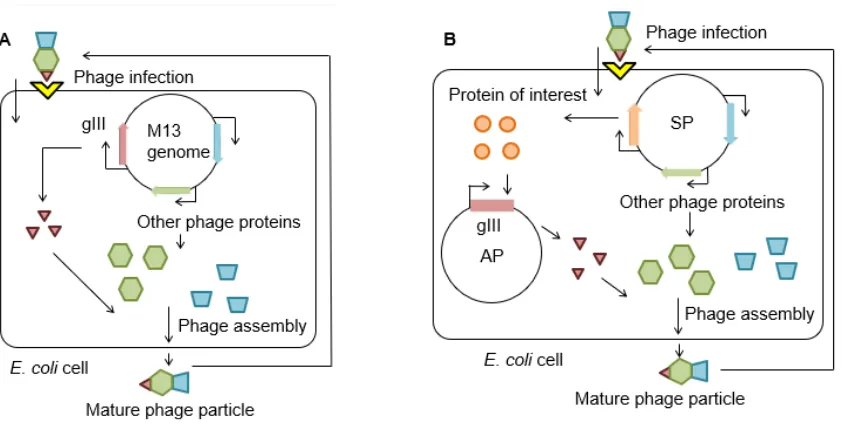
During infection, the M13 particle attaches to F+E. coli via the F pilus using the N2 domain
of the gIII protein, which "reels" in the phage and allows the N1 domain of pIII to interact with
(Fig. 2A)[14, 15]. Once inside the cytoplasm of host E. coli, phage DNA uses the host's DNA
replication machinery to produce a circular double-stranded version of itself and starts to express
various phage proteins required for production of mature M13 particles. 30 minutes after phage
infection, dozens of double-stranded, transcriptionally active phage molecules are present within
the host cell[14]. Phage are made via rolling-circle replication from the initial viral single-stranded
genome—note this is an important factor when considering gene dosage effects for PACE (more
on this later)[16]. The phage coat proteins assemble on the surface membrane of the host E. coli,
and a copy of the circular single-stranded phage DNA is packaged inside. Once assembly is
complete, the M13 particle detaches from the bacterial membrane and exits to the environment to
start another cycle of infection. Unlike other bacteriophage, M13 is not lytic: the host E. coli is
kept alive, while mature M13 particles release from the cell membrane. The host growth rate is
significantly diminished, however, as a result of producing phage particles, but host cells can
continue to grow, divide, and further produce M13 phage indefinitely.
mediated by interaction of the gIII coat protein with the TolQRA complex (yellow)[14, 15]. Upon entry into the host, the single-stranded viral genome is converted to a double-stranded genome by host DNA polymerases and expression of the M13 genome can begin by utilizing host machinery[16].
Phage protein pIII, which is encoded by phage gene gIII, is essential for phage maturation
and infectivity[17]. The infectivity of M13 phage scales with increasing levels of pIII over a range
of two orders of magnitude[18]. PACE utilizes a mutant M13 bacteriophage whose gIII gene is
replaced by that for the protein of interest (the mutant phage is called Selection Phage, SP, Fig.
3)[12]. Thus, the SP expresses the protein instead of pIII in host E. coli; the SP cannot produce
mature phage particles by itself. To complement the SP, the gIII gene is supplied on a separate
plasmid in the host E. coli (Accessory Plasmid, AP) as part of a selection system that activates pIII
production (the "gIII selection system") in response to the activity of the protein of interest. SP can
only propagate by expressing the protein from phage DNA, followed by expression of gIII that is
mediated by the protein’s activity (Fig. 2B). Thus, successful SP propagation is linked to the
activity of the protein of interest. SP carrying a mutant protein with enhanced activity will have a
fitness advantage over other SP particles, because the enhanced protein activity allows for
increased pIII production, thereby increasing offspring production. Over time, SP harboring the
coding sequences expressing improved proteins will outcompete others in the population. If
phage-dependent activity produces sufficient pIII product, then there will not be a fitness advantage
gained from producing additional pIII, which makes fine-tuning the stringency of the selection
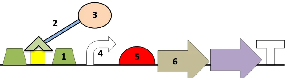
Figure 3. Schematics of the M13 genome, SP, and AP displaying their critical genetic elements. The gIII and protein of interest are shown in brown and orange, respectively. Other phage genes are represented in green and blue. Antibiotic resistance genes are shown in yellow. Swapping the native gIII protein from the M13 genome with the CDS of the protein (and, optionally, KanR) renders the SP dependent on the AP in order to be capable of producing pIII to
assemble mature M13 bacteriophage[10-13].
Compared to the directed evolution system described previously, the PACE gIII selection
system is the equivalent of the "selective assay" that establishes the direction of evolution, and the
SP is the vector that expresses the protein of interest. The competition between SP particles
carrying mutations in their proteins and selection for their maximum fitness as defined by the gIII
selection system is the basis of evolution in PACE.
Continuous evolution in PACE
A defining feature of PACE is the continuous evolution of the SP that can be achieved by the
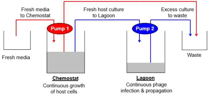
set-up shown in Figure 4. The simplest PACE system consists of two flasks: the Chemostat and
Lagoon are connected by tubes driven by two separate pumps. The Chemostat contains a culture
of host E. coli, incubated at 37 ºC. The first pump continuously delivers fresh media into the
Chemostat, while transporting excess media and cells out of the Chemostat to the waste container
(Fig. 4, red lines). Thus, the bacterial culture in the Chemostat is maintained under healthy growth
conditions, while its volume remains constant. The Lagoon is filled with a culture of host E. coli
infected with SP; the second pump continuously delivers Chemostat culture into the Lagoon, while
the same pump transports excess media, cells, and phage out of the Lagoon to waste (Fig. 4, blue
lines). Therefore, the Lagoon is constantly inoculated with fresh, uninfected host E. coli for SP
particles to infect, while the already-infected cells in the Lagoon exit to waste. Again, the volume
the Lagoon for hundreds of generations without interruption.
Figure 4. Schematic diagram of the PACE pump system. Host cells transformed with the AP construct are maintained in the Chemostat and continuously pumped into the Lagoon at the optimal flow rate to permit M13 propagation and minimize host replication. By utilizing pumps to regulate the flow of media into/out of the Chemostat and Lagoon, PACE can proceed uninterrupted[10, 13].
The flow rate of the Chemostat culture into the Lagoon plays a key role in PACE, mainly
by preventing false positive mutations (mutations that decouple gIII expression from the activity
of interest to be evolved) from accumulating in the E. coli genome[10-12]. When established
properly, the Lagoon culture only allows SP to propagate while preventing host E. coli cells from
dividing, thereby preventing E. coli offspring from lingering in the Lagoon. Typically, the flow
rate into the Lagoon is set at more than double the Lagoon volume per hour; e.g., the flowrate of
a 30 mL Lagoon may be 80 mL/hr[10]. At this rate, the culture in the Lagoon is refreshed in less
than 30 minutes, allowing E. coli cells to linger in the Lagoon for less than a half hour. Since the
fastest doubling time for E. coli is approximately 20 minutes, an average uninfected cell in the
Lagoon has barely enough time to divide; however, host cells infected with M13 phage will
experience a significant increase in their doubling times[14]. This limits the ability of the host E.
in their genome that might remove the selective pressure established by the gIII selection system
by allowing for pIII expression without the desired protein activity.
The challenge for the phage is to infect a host, replicate, and detach from the host within a
predetermined time frame to avoid being washed out of the Lagoon. Increasing the flow rate
through the Lagoon is one way to increase the stringency of the PACE system, but it is
recommended to switch to a more stringent selection circuit rather than to rely on flow rates into
the Lagoon to alter the stringency of the selection[19]. As a result of all these factors, PACE allows
only mutations in the phage DNA (SP genome) to accumulate, since phage DNA is the only genetic
material that can pass to future generations in the Lagoon. This limits the amount of “cheating”
that can occur within the Lagoon as a result of activity-independent expression of pIII; however,
cheater phage can still arise that have recombined gIII or some other component of the selection
circuit into its genome that will express gIII outside of the host circuit. Minimizing the number of
recombination hotspots in the phage genome can decrease the likelihood of this occurring, and
having phage that robustly activate the selection circuit through the intended mechanism may help
to avoid enriching the recombinant phage within the Lagoon.
During PACE, the mutagenesis rate during DNA replication is greatly increased using the
MP (Mutagenesis Plasmid) that increases the rate of protein evolution[12]. This is accomplished
through the arabinose-dependent expression of a dominant-negative variant of the E. coli DNA
Pol III proofreading domain on the MP, alongside other genes that facilitate mutagenesis[12, 20].
Currently, the most effective variant of the MP is MP6 (available from Addgene), which increases
the mutagenesis rate to >300,000-fold over the basal level of mutation in E. coli[20]. This high
mutagenesis rate allows for the rapid accumulation of beneficial mutations in the protein after
very first generation, with enrichment of fitness-improving mutations within 1-3 days of
continuous propagation. PACE experiments usually run for several days and multi-experiment
evolutionary trajectories have exceeded 20 days. PACE duration depends on the nature of the
desired changes and the evolutionary trajectories the protein takes to achieve the desired
function[21].
Workflow for building the PACE system
This protocol aims to be a guide for establishing a basic, economical PACE system on a standard
lab bench. Our protocol is written for researchers with strong foundations in molecular biology
and microbiology. A solid understanding of evolutionary concepts, such as selective pressure and
fitness is a useful asset while running and troubleshooting a PACE evolution.
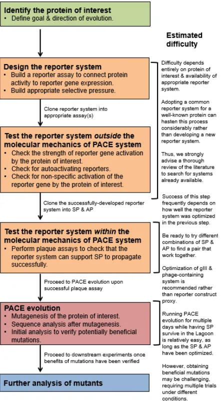
The workflow for developing a PACE system is shown in Figure 5. We describe this
development in two separate steps: 1) developing the gIII selection system, and 2) installing the
Chemostat-Lagoon pump system for PACE evolution. We roughly estimate that it takes 1-2 years
for a lab to complete this workflow from scratch, although the timeline will vary depending on the
lab’s experience with PACE, the complexities of the protein of interest, and the existence of
selection assays that are valid for the protein of interest. Adapting an existing selection scheme to
Figure 5. Typical workflow for the development of a PACE selection circuit. Before embarking on adopting PACE to evolve a protein, considerations must be made as to what is the goal of the evolution, the availability of a well-characterized selection circuit, the nature of the selective pressure, and whether it is worth it the effort invested to carry out refinement of various systems before obtaining a successful PACE circuit. Designing and constructing the selection could take anywhere from 0.5-1.5 years to achieve depending on how much troubleshooting needs to take place. Evolving the protein of interest in PACE once the system is set up could take up to a year to carry out the actual experiment, interpret the results, and examine potentially beneficial mutations.
The two major steps of PACE development will be described in separate sections. In the
first section, the development of a PACE selection circuit will be described. It is impractical to
different for each protein of interest. Instead, we will use the development of the PACE-B1H gIII
selection circuit as an example to demonstrate the key concepts during this step. In the second
section, we will discuss the development of the Chemostat-Lagoon pump system in detail,
followed by a brief section describing the expected results from PACE evolution. This protocol
will focus on the basic PACE pump system. Protocols for more complex PACE will not be
provided, although they will be discussed at the end of this paper.
The Liu lab has also developed an intermediate technique between plate-based selections
and PACE called phage assisted non-continuous evolution (PANCE) that can be developed and
utilized in tandem with PACE[19, 21]. PANCE can use the same selection circuit that PACE
utilizes, but instead of media continuously flowing from a Chemostat into the Lagoon, we
subculture the contents of the Lagoon into fresh media[22, 23]. This makes PANCE slower than
PACE at developing mutations, but PANCE has the advantage of not requiring a specialized
set-up. PANCE is useful for preliminary evolution of a protein of interest to obtain variants with high
enough activity to support PACE, which requires a certain minimum propagation efficiency to
sustain phage titers. PANCE also demonstrates high utility as it can easily be parallelized, and it
can be used to assess the feasibility of developing a selection circuit before committing significant
time and resources to adopting full-blown PACE[23].
Protocol #1: Developing a PACE selection system
Establishing a proper PACE selection circuit is key to successful PACE. Here, we discuss
guidelines for the construction of a PACE selection circuit by using the development of our
PACE-B1H as an example. The PACE selection circuit comprises a gIII cassette (located on the AP) that
protein activity leads to pIII production, which allows for production of next-generation phage
particles (Fig. 2B). When developing the selection circuit, three major aspects must be considered:
1) tuning the amplitude of gIII activation to yield sufficient pIII expression required for phage
propagation; 2) confirming that the gIII cassette is not auto-activated by endogenous molecules,
i.e., that the system has low background; 3) verifying that the gIII cassette is only activated through
specific interaction with the protein of interest, i.e., that the system has high specificity. Typically,
development of the PACE selection circuit begins by identifying both the gIII cassette and protein
of interest that together produce a strong signal. Then, this promising pair is investigated to verify
specificity of interaction. The goal is to produce a gIII cassette-protein pair that produces a strong
signal (high phage propagation characterized by plaque forming units, PFU) or high
signal-to-noise ratio (strong signal relative to background from an indirect reporter like luciferase).
Initial development of the selection circuit may be done independently from the phage-E.
coli infrastructure. The phage-E. coli interaction is an already complicated system on its own,
making this system unsuitable as an environment for the construction of a unique selection system.
Instead, the selection circuit should be developed using a reporter gene in lieu of gIII that is more
straightforward: these reporter genes include LacZ, luciferase, and HIS3. Once the reporter system
has been developed "outside" of PACE, its components will be cloned into the AP and SP to test
whether those components can properly control gIII expression and phage propagation.
Alternatively, you could develop an AP construct that has a reporter cassette where the
stop codon of gIII overlaps with the start codon of the reporter; e.g., the Liu lab utilizes LuxA/B
as the reporter, and the gIII stop codon overlaps with luxAB by 1 base (TAATG)[19, 21, 24].
and translation such that luciferase activity can be used as an indirect means to monitor changes
in gIII expression.
Goal of SP-ME47 PACE
In our PACE-B1H system, our protein of interest is ME47, a rationally designed basic
helix-loop-helix (bHLH) protein comprising 68 amino acids that specifically targets the E-box motif
(5’-CACGTG)[25]. Our goal was to use the molecular mechanisms of the B1H system to make ME47
control expression of an box-regulated gIII gene to generate ME47 mutants that bind to the
E-box with higher affinity and specificity. A mutation in the ME47 sequence that improves its E-E-box
binding capability should increase gIII expression and give the SP carrying the mutated ME47 a
selective advantage over other SPs in the population. Over the course of PACE, SPs carrying
beneficial mutations would then outcompete other SPs and dominate the Lagoon population. These
mutations can then be isolated for further analysis.
Developing the B1H for ME47: initial screening
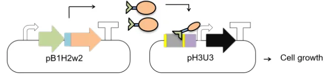
The bacterial-one-hybrid (B1H) system was developed by the Wolfe lab to assay the affinity and
specificity of a given protein-DNA interaction in E. coli[26-28]. At the molecular level, the B1H
functions similarly to related systems in yeast (Fig. 6A)[29].[29] Briefly, in the B1H, the protein of
interest is expressed from the pB1H2w2 vector as a fusion protein to the RNA polymerase omega
subunit: this fusion protein, therefore, behaves as a transcriptional activation domain (AD)[26-28,
30]. The system uses HIS3 as the reporter gene, which is located on a second vector,
pH3U3[26-28, 31]. HIS3 is a yeast gene that encodes an essential enzyme in the histidine biosynthesis
gene (hisB is the bacterial analog of the yeast HIS3 gene and will be used to refer to the HIS3
reporter gene) was deleted, to grow in histidine-deficient media[26-28]. To test how strongly the
AD construct interacts with the cognate DNA to mediate hisB expression, the doubly transformed
US0 cells are grown in histidine-deficient media supplemented with various concentrations of
3-Amino-1,2,4-triazole (3-AT), an inhibitor of the hisB gene[32]. Thus, in the B1H, the strength of
the protein-DNA interaction is represented by the amount of bacterial growth when challenged to
grow in histidine-deficient media.
Figure 6. Schematic diagram illustrating A) B1H system and B) PACE-B1H system. The omega subunit of RNAP fused to ME47 (green and orange, respectively) acts as an activator domain (AD). ME47 binds to the E-box (blue) present on the pH3U3/AP vector and positions the omega subunit of RNAP so that it can recognize the -10/-35 elements of the weak lac promoter (purple). This leads to expression of the downstream gene (brown) that is needed for E. coli growth and phage propagation[26-28].
To construct the B1H reporter system in E. coli, we cloned the sequence expressing the
ME47 bHLH into the pB1H2w2 vector; the expressed protein contains ME47 fused to the
N-terminus of the RNA polymerase omega subunit separated by a 12 amino-acid linker (Fig. S1).
Similarly, we cloned a variant of the E-box sequence, the AT-E-box (5’ ACCACGTGGT, core
E-box underlined, flanking AT base pair in bold), upstream of the hisB gene in the pH3U3 vector,
promotor. We chose these parameters as a starting point and used materials already available at
the time of cloning. Subsequently, we doubly transformed E. coli US0 with pB1Hw2w/ME47 and
pH3U3/-13 AT-E-box-containing plasmids. Unfortunately, these doubly transformed host cells
did not grow in histidine-deficient media, indicating that this B1H pair (ME47 vs. -13 AT-E-box)
was incapable of reporter gene activation (Fig. S2).
In our search for a protein-DNA pair capable of producing a B1H signal, we adjusted two
features in the hisB reporter gene. First, we used an alternate E-box variant as the target sequence
for ME47. The new sequence, the GC-E-box (GCCACGTGGC), has flanking GC nucleotides in
place of the AT nucleotides in the AT-E-box. Nucleotides in this position were shown previously
to influence the protein-DNA interaction for this class of DNA binding proteins[33], and therefore,
we surmised that this alteration might achieve a successful B1H pair. Second, it is known that the
relative position of the target DNA sequence (in our case, the E-box) within the rest of the hisB
promoter influences the strength of reporter gene activation. To account for this, we generated
variants of hisB reporters containing different numbers of nucleotides between the edges of the
E-box and the -35 promoter. Thus, we generated a library of E-E-box reporters that contained either
the AT- or GC-E-box, 7 to 25 bp away from the -35 weak promoter (Fig. S2).
We tested ME47 against the various GC- and AT-E-box reporter constructs (Fig. S2). As
before, we saw no growth when ME47 was paired against the -13 AT-E-box. In contrast, other
reporters, such as -7 GC-E-box and -11 AT-E-box, gave varying degrees of bacterial growth.
Therefore, these reporters do activate the hisB gene and were candidates for the PACE-B1H
reporter.
Following their identification, we screened the candidate reporters for specificity. First, we
checked for autoactivation that can result from endogenous cellular molecules activating the
reporter. The candidate reporters were singly transformed into E. coli US0, and the B1H assay was
repeated. Cells transformed with non-autoactivating reporter constructs will not grow, because
these cells lack ME47 to activate the reporter. In contrast, cells transformed with an autoactivating
reporter will still grow. The results are presented in Figure S3A. As expected, many cells were
incapable of growing: e.g., cells harboring the -7 GC-E-box or -11 AT-E-box. However, other
cells—such as those carrying the -9 GC-E-box—showed strong growth in the absence of ME47.
Reporters that allow cells to grow without ME47 were eliminated from the candidate list, as they
were most likely autoactivating.
Developing the B1H for ME47: testing for specificity
We tested the remaining candidates for specificity of the protein-DNA interaction to ensure that
reporter gene activation was controlled by the specific molecular interaction between ME47 and
E-box in the reporter gene. To do so, we synthesized a "nonspecific" version of each candidate
reporter gene by replacing the central 8 bp sequence with a nonspecific sequence known not to
interact with ME47 (NS-DNA, 5’ TTCCAAGG[25]).We doubly transformed E. coli US0 cells
with ME47 and the NS-DNA reporter constructs and then performed the B1H. Figure S3B clearly
shows that all candidates exhibit minimal cell growth, even at the highest serial dilution. Compared
to the cell growth shown by their E-box-containing counterparts, growths resulting from
nonspecific reporters were at least six orders of magnitude lower: we note that although the B1H
is not a quantitatively linear assay, this is a strong signal-to-noise ratio, confirming the specificity
of interaction between ME47 and E-box reporters in the B1H.
As the final step in developing the PACE selection circuit, we cloned ME47—still fused to the
RNA-polymerase omega subunit—into the SP to give SP-ME47. Also, we cloned the four
candidate E-box constructs (-7 and -11 GC-E-boxes, -9 and -11 AT-E-boxes) into the AP,
upstream of the gIII gene. We obtained mature phage particles of SP-ME47 after transforming the
SP-ME47 genome into E. coli 1059 and purifying the supernatant from its overnight culture. The
SP genome can be constructed and manipulated like a regular plasmid and does not require any
special handling[34]. We then transformed the four AP/E-box vectors separately into E. coli
1030[35]. There are two strains of host cells used in PACE: S1059 and S1030. Both strains have
the F pilus to allow for M13 infection via gIII. The S1059 strain has a constitutive AP that produces
gIII regardless of activity from the desired protein from the SP and is a permissive host. The S1030
cell lacks the constitutive AP of S1059 and needs to be transformed with an AP, whose design is
based on the initial screening process outlined earlier. Recently, S2060 cells were developed as an
updated version of the S1030 host. S2060 contains a lacZ gene under the control of a phage shock
promoter that produces blue plaques when X-gal/Bluo-Gal are used in the plaque assay. This
facilitates the identification of plaques and is recommended for use in the plaque assay. S1059
cells can be replaced with “S2208” cells that are S2060 cells transformed with pJC175e (the
constitutive AP, available from Addgene)[19].
In theory, SP-ME47 should be able to activate the gIII gene on the AP/E-boxes via the
omega-ME47 construct (Fig. 6B). To test this, we performed a plaque assay using the purified
SP-ME47 phage particles against E. coli S1030 transformed with the AP/E-boxes. Activity-dependent
plaquing is a stringent test for propagation, but it is not quantitative. However, overnight
propagation tests will give a quantitative measure of how fit the phage are on the developed
phage, active phage (contain the protein of interest cloned into the SP), and empty phage with gIII
deleted.For this discrete assay, cultures containing the appropriate AP are grown to mid-log phase
in Davis rich medium and infected with a sub-saturating amount of SP-ME47 for 10 min at 37 °C
(typically 106 PFU/mL is adequate). It is important that a sub-saturating amount of SP be used, as
not all the host cells will be initially infected, which allows the phage to propagate and enrich
overnight. The culture is then spun down and the supernatant is saved for titering on a plaque assay
on S1030 E. coli cells carrying the desired AP. To estimate the instantaneous propagation rate of
the phage, cells post-infection can be reinoculated into fresh Davis rich medium and grown to
OD600 ~0.8 rather than overnight, then pelleted, and the supernatant can be saved for titering as
described before. The expected results should show >105-fold propagation for wild-type phage and ≥103-fold difference between active and inactive/empty phage, ideally as large a difference as
possible. Active phage should enrich at least 10-fold before considering PACE, and 100-fold is
safer; otherwise, the continuous flow of PACE will lead to phage washout before
fitness-improving mutations have a chance to accumulate and be enriched.
The results are shown in Figure S4. For the -7 GC E-box AP, we observed numerous
plaques after the plaque assay, whereas the -11 AT/GC E-boxes did not produce any plaques. This
indicates that the -7 GC E-box B1H selection circuit—now harboring the SP and AP—could
activate the gIII gene, thereby allowing phage propagation. The successful plaque assay
established that our selection circuit was fit for use in the Chemostat-Lagoon pump system.
However, if the starting phage propagates too robustly on the selection circuit, there will not be a
selective pressure on the protein to improve. We would advise not doing PACE with a circuit that
gives >103-fold overnight propagation on a given circuit, though it may possibly still give useful
Protocol #2: PACE & related techniques
Handling of sterile materials
• Autoclaving should be done under standard sterilization conditions. Adjust the sterilization time as appropriate to account for large volumes of media.
• Use 0.22 µm filters for filter sterilization.
• Note that these filters are used to remove bacterial cells (and larger particles) from the solution. Phage particles will pass through these filters.
• Use standard aseptic techniques to handle sterile materials. We regularly use a Bunsen burner or laminar flow cabinet for this purpose.
Recipes for general media & stock solutions 2x YT Top Agar
3.1 g 2x YT media 0.75 g Agar 100 ml ddH2O
Autoclave to sterilize
Davis rich medium (DRM) base solution
35 g Anhydrous potassium phosphate, dibasic 10 g Potassium phosphate, monobasic
5 g Ammonium sulfate 5 L ddH2O
Autoclave to sterilize
Alternatively, pre-mixed DRM is available from US Biological, 001 and CS050H-003.
DRM supplement solution 90 g D-Glucose
10 g Sodium citrate, tribasic
0.52 g Magnesium sulfate (heptahydrate) 10 g Casein hydrolysate
0.5 g L-leucine 500 mL ddH2O
Filter sterilized
Note: An updated recipe for DRM is currently available for use, and we invite others to explore it[17]. However, we have routinely using this older recipe for its simplicity and consistency. For large quantities of DRM supplement, it is feasible to dissolve chloramphenicol/other antibiotics directly in the supplement before filter sterilizing.
Note: Introducing 1.5 M arabinose solution at 0.5 mL/hr to a Lagoon receiving bacterial culture at 100 mL/hr will result in a final arabinose concentration of approximately 7 mM in the Lagoon.
LB
10 g Tryptone 10 g NaCl 5 g Yeast extract 1 L ddH2O
Autoclave to sterilize
Ampicillin (1000x stock)
50 mg/mL solution in H2O, filter sterilized
Kanamycin (1000x stock)
30 mg/mL solution in H2O, filter sterilized
Chloramphenicol (1000x stock)
25 mg/mL solution in anhydrous EtOH
Description of bacteria, phage and plasmids
Bacteria
Escherichia coli DH5α (F-)
• Used for general molecular biology purposes, such as cloning.
• Does not contain the F plasmid (F-); will not be infected by M13 phage and derivatives.
• Can be replaced with other generic lab strains of E. coli.
E. coli S1030 (F+, no AP, no MP) • Host strain for PACE.
• Carries the F plasmid, allowing infection by M13 phage and derivatives.
• Lacks AP; by itself, does not support propagation of SP.
• Lacks MP; by itself, does not introduce increased mutagenesis rate.
• AmpS, KanS, CmS, TetR.
E. coli S2060 (F+, no AP, no MP) • Host strain for PACE.
• Carries the F plasmid, allowing infection by M13 phage and derivatives.
• Lacks AP; by itself, does not support propagation of SP.
• Lacks MP; by itself, does not introduce increased mutagenesis rate.
• AmpS, KanS, CmS, TetR.
• E. coli S2060 possess a LacZ gene under the control of a phage shock promoter that will make visualizing plaques easier when grown on Xgal-containing media[19].
E. coli S1059 (F+, + APconstitutive, no MP)
• Carries the F plasmid, allowing infection by M13 phage and derivatives.
• Carries AP with constitutively active gIII gene, thereby allowing SP propagation regardless of the SP genotype.
• Lacks MP; by itself, does not introduce increased mutagenesis rate.
• AmpR, KanS, CmS, TetR.
E. coli S2208 (F+, + AP
constitutive, no MP)
• Host strain used for constitutive propagation of SP.
• Carries the F plasmid, allowing infection by M13 phage and derivatives.
• Carries AP with constitutively active gIII gene, thereby allowing SP propagation regardless of the SP genotype.
• Lacks MP; by itself, does not introduce increased mutagenesis rate.
• AmpR, KanS, CmS, TetR.
• E. coli S2208 possess a LacZ gene under the control of a phage shock promoter that will make visualizing plaques easier when grown on Xgal-containing media[19].
• Can be made by transforming S2060 cells with pJC175e.
Phage
Selection Phage (SP)
• Mutated derivative of the M13 bacteriophage.
• Will infect any F+E. coli.
• Lacks one gene essential for bacteriophage propagation (gIII) that has been replaced by the CDS of the protein of interest.
• gIII needs to be provided by the host E. coli strain, typically on the AP as a gIII reporter gene activatable by the protein of interest.
• Phage propagation depends on the protein (expressed from SP genome) successfully activating the gIII reporter gene found on AP.
• Carries a KanR gene upstream of the cloning site for the gene of interest. SP without KanR
can be used to increase speed of phage propagation by decreasing the size of the phage genome.
• Techniques to assemble the SP genome via Golden Gate cloning are outlined in the Supplementary Information of Thuronyi et al., 2019 and the genome fragments used for this purpose are available from Addgene[34].
Plasmid vectors
Accessory plasmid (AP)
• Carries gIII reporter gene.
• AmpR (typically).
• Low-copy SC101 origin (typically).
• Plasmids with other characteristics, or multiple plasmids, may be needed to implement a given selection circuit.
Mutagenesis plasmid (MP)[20]
• Carries the genes necessary for increasing the mutagenesis rate during PACE.
• Cm.
• VERY IMPORTANT: Cell strains hosting MP should always be handled carefully and grown in a media containing 25 mM glucose to suppress the mutagenic activity of MP. Avoid serially propagating these strains as much as possible, as this may cause accumulation of mutations in the host genome.
Handling bacteria
Bacterial cultures
Overnight cultures of E. coli strains are prepared by inoculating a single bacterial colony in LB containing appropriate antibiotics and incubating for 16 hr, 37 °C,with shaking at 200 RPM. A loopful of frozen bacterial glycerol stock can also be used to start an overnight culture. Similarly, an overnight culture can be started from a plaque of SP-infected E. coli; a sterile pipette tip can be used to stab the plaque to transfer material into the culture tube.
Transformation
All E. coli strains in this protocol can be transformed using common transformation methods, such as chemical transformation and electroporation.
Note: Attempts to doubly transform E. coli S1030 with AP and MP using chemical transformation may suffer from very low transformation efficiency. We advise to first transform E. coli 1030 with AP alone and to make competent cells from the AP-transformed E. coli S1030. MP can be easily transformed into chemically competent E. coli S1030 + AP.
Storing bacterial glycerol stocks
1. Mix an overnight culture of the bacterial strain 1:1 (v:v) with sterile, 50% glycerol. The final concentration of glycerol will be 25%. Mix well.
2. Store this mixture at -80 °C. The glycerol stock must always be kept frozen to retain maximum viability.
E. coli can be stored this way for years without completely losing viability, provided the stock is kept frozen. Bacteria infected with M13 phage (or SP) can be stored in the same way without losing their capacity to produce and release phage particles.
Handling phage
Note on disinfection
2% bleach solution should be used to disinfect equipment and surfaces that come into contact with SP. 70% EtOH is not recommended for this purpose.
"Plasmid-like" properties of SP genome
The SP genome is a circular, single-stranded DNA inside the bacteriophage protein shell. When SP infects E. coli, the SP injects its genome into the bacterial cytoplasm, and the genome is synthesized into a circular double-stranded DNA. This circular-double stranded form of the SP genome can be handled similarly to a regular plasmid. (From here, the acronym "SP genome" will indicate the circular, double-stranded form of the phage genome.)
Thus, it is possible to
common miniprep protocols and commercially available kits.
• Modify the SP genome by conventional methods, such as restriction cloning, USER cloning, Golden Gate cloning, and site-directed mutagenesis.
• Transform the SP genome back into E. coli using common transformation methods. o When the SP genome is introduced this way into an E. coli host suitable for phage
propagation, the SP genome will function normally, producing the phage proteins and protein of interest, eventually leading to production and secretion of mature phage particles to the environment.
o Transformation efficiency of this procedure is usually very low. While this is not a problem when transforming with a uniform clone of the SP genome, it may cause difficulties when developing libraries of SP in vitro.
Note on isolating plasmids from E. coli 1030 or E. coli 1059
E. coli strains S1030 and S1059 infected with the SP genome carry multiple plasmids, such as the AP, SP genome, MP, and F plasmid, which will not be differentiated during plasmid purification. Therefore, the extracted plasmid solution will contain a mix of these plasmids, and further processing (such as transforming E. coli DH5α with this solution followed by replica plating) is necessary to obtain these plasmids in pure form. Alternatively, the SP can be isolated using commercially available kits that are specifically made for this purpose.
Handling cultures of E. coli host infected with SP
Since M13 (and its derivative SP) do not kill the cells in order to propagate, SP-infected E. coli can be used to start an overnight culture to produce more SP-infected cells. The supernatant of this culture will contain mature phage particles released from the cells. Typical phage titers from overnight growth of activity-independent propagation host cells are 108-1012 depending on the
phage propagation efficiency.
Crude isolation of phage particles from an infected E. coli host culture
1. Start an overnight culture of the appropriate, SP-infected E. coli strain in LB + Amp. 2. Centrifuge the overnight culture at 3000 x g, 5 min, to isolate the E. coli cells.
3. Pass the culture supernatant through a 0.2 µm syringe filter to completely remove any E. coli cells remaining in the supernatant.
4. The supernatant is ready to be used as a source of purified phage.
Storage of purified phage particles
1. Mix the solution of purified phage with sterile glycerol to a final glycerol concentration of 50%. Typically, we add 1.7 mL purified phage solution to 3.3 mL sterile 75% glycerol to obtain ~50 % final glycerol concentration.
2. Store the phage solution in 50% glycerol at -20 °C. Ensure that the solution does not freeze.
Plaque assay Materials:
• Overnight culture of the host E. coli strain.
• Purified phage solution.
• 4 mL aliquots of molten 2x YT top agar.
o On the day of the plaque assay, melt the top agar using a microwave oven and aliquot 4 mL into sterile culture tubes. Keep the aliquots at 50 °C until use.
• LB plates.
o Pre-warmed before use. Cold plates make it difficult to spread the top agar.
Procedure:
1. Make a 1:50 dilution of the overnight E. coli culture in 5 mL fresh LB broth + Amp. 2. Grow the diluted culture at 37 °C, 200 RPM, until OD600 0.6-0.8. This typically takes 2-3
hr incubation.
3. Perform a serial dilution of the phage stock solution in ddH2O until 10-6 dilution. We
typically perform the dilution in 200 µL total volume per tube.
4. Separately transfer 30 µL serially diluted phage solution into fresh 1.5 mL tubes.
• Note: These aliquots may be kept at room temperature for 30-60 min without adverse effects. Samples may be kept on ice for longer waiting periods.
5. Once OD600 of the E. coli culture reaches 0.6-0.8, add 270 µL culture to each of the tubes
containing 30 µL diluted phage solutions. Incubate for 10 min to allow phage to infect host
E. coli cells.
• Note: Incubation times can be varied from 5-20 min without adversely affecting the result, although extra precautions should be taken if the absolute quantity of plaques is important.
• Note: Alternatively, cultures of host cells grown to OD600 0.6-1.0 can be stored at
4 °C for up to two weeks. Stored cells can be used in the plaque assay straight from the refrigerator after vortexing to resuspend the cells.
6. After incubation, transfer all 300 µL phage + E. coli mixture into a tube of 4 mL molten top agar. Mix well and immediately pour everything onto a LB plate. Allow the top agar to spread evenly across the plate. Agitation/tilting of the plate while the top agar is solidifying may cause clumping.
7. The top agar will solidify within minutes. Incubate the plate at 37 °C, overnight.
Instead of using a standard 100 mm petri dish, plaque assays can be poured using quarter well petri dishes and culture plates of various sizes to save space and reagents. Reagent volumes should be adjusted accordingly.
Isolating a single clone of phage particle from a solution of phage
1. Perform a plaque assay of the phage stock using E. coli 1059 as the host.
2. You should get plaques forming on the plates the next day. It is reasonable to assume that an individual plaque represents a uniform clone of phage particles.
3. Using a sterile pipette tip, transfer a single plaque from the plaque assay to 5 mL fresh LB + Amp. Grow the culture for 16 hr, 37 °C, 200 RPM.
using directly for downstream steps is not recommended, since only low amounts of phage DNA may be present. We recommend outgrowth of plaques, which refers to allowing plaques to grow overnight to increase the initial amount of phage genome.
MP Check
Due to its strong mutagenic activity, MP stored as a glycerol stock in E. coli can still mutagenize DNA, including itself, even when the storage media is appropriately supplemented with 25 mM glucose. Therefore, we advise checking the mutagenic activity of MP each time before a PACE experiment to ensure that the plasmid is functioning properly. This procedure is called the “MP Check.”
1. Prepare a pair of culture tubes containing E. coli S1030 that will be used for the PACE experiment in 5 mL LB with the appropriate amounts of Amp and Cm added.
2. Supplement the first tube with 25 mM glucose. This should suppress MP activity. 3. Supplement the second tube with 1 mM Arabinose. This should induce MP activity. 4. Incubate both culture tubes overnight and observe for bacterial growth the following day.
Note: The MP check can also be carried out by plating serially diluted cells onto 2xYT plates containing Amp+Cm that have been supplemented either with glucose or arabinose, as has been described[20].
After the overnight incubation, the culture tube supplemented with 25 mM glucose should have saturated bacterial growth, as expected. In contrast, there will be no growth in the tube supplemented with arabinose if the MP is functioning properly, since the mutagenic activity from a fully induced MP will quickly destroy the genetic integrity of the host cell, hampering bacterial growth. No growth in the MP Check confirms that the E. coli strain is ready for PACE. In contrast, observing growth in the arabinose-supplemented tube may mean that the MP has lost its functionality, or that the arabinose concentration was too low.
The MP Check should be done before setting up PACE evolution to avoid terminating the experiment if the check fails. Alternatively, the MP Check can be performed on the exact same E. coli cells that were used to prime the Chemostat flask; this can be done easily by setting up the MP Check as you also set up the Chemostat. Either way, performing this simple test on a routine basis will help maintain the integrity of a much longer experiment. MP checks can be conducted periodically on Chemostat cells to verify their integrity over the course of a long PACE.
PACE Checklist
A shopping list of the various parts listed in the selection below can be found in Figure S15, along with the corresponding part numbers to facilitate building your own PACE setup.
Pumps, shakers, & incubator
• A pair of peristaltic pumps, one each for the Chemostat and the Lagoon (Fig. S5A).
• A syringe pump for pumping arabinose (Fig. S5B).
• An air pump to aerate the Chemostat (optional).
• A tabletop incubator with an access hole on one of its sides.
Assemble the pumps as per the manufacturer's instructions. In many cases, the speed dials of the peristaltic pumps must be calibrated to obtain the desired flow rate (done by assembling the pump with their proper tubing and testing the flow with water). We advise you to become accustomed to assembling, disassembling, and operating the pumps before installing them in the incubator. Then, assemble these components in the incubator (Fig. S5C).
Tubes (Assemble tubes according to Fig. S6. Pictures of these tubes are shown in Figure S7.):
• Media to Chemostat.
• Chemostat to Lagoon.
• Chemostat to Waste.
• Lagoon to Waste.
• Air to Chemostat (if using).
• Air to Lagoon (if using).
• Air pump to Air filter (if using).
o Note: it is possible to run PACE anaerobically with vents included in the set-up to relieve pressure buildup.
• Arabinose filter to Lagoon.
• Arabinose pump to Arabinose filter.
Chemostat & Lagoon flasks (Fig. S7):
• two 125 mL flasks with stirring rod inside.
• two silicone-tube caps.
Media Bottles:
• two 5 L bottles of DRM (Fig. S8A). You may use larger bottles if your autoclave has the capacity.
• two tubed-bottle caps (Fig. S8B).
Note: All tubes, flasks, caps, and media must be autoclaved before use. The open ends of these components should be covered with aluminum foil for autoclaving (Fig. S9).
Bacteria and phage:
• Colonies of E. coli S1030 freshly transformed with the appropriate AP and MP, with MP activity confirmed by MP Check. To prevent accumulation of mutations in the host genome, cells transformed with MP should not be stored for more than 2 days at 4 °C after the initial overnight incubation at 37 °C.
• Purified solution of the desired SP. A glycerol stock of the SP will work as well.
Disposables:
• 0.22 µm syringe filters.
• 60 mL disposable syringes.
• 1" sterile needles.
• 1000x Ampicillin.
• 1000x Chloramphenicol.
Note: Here, we use disposable needles and a silicone cap as an easy solution to connect the tubes to the Chemostat and Lagoon. Alternately, re-usable, blunt-end needles can be used with a compatible tube cap.
PACE: Pump parameters & timeline
Chemostat & Lagoon parameters
Chemostat
• culture volume = 80 mL.
• flow speed of incoming media = 80 mL/hr.
• turnover rate = 1 culture volume/hr.
• flow rate may need adjustment for different host cell growth rates
Lagoon
• culture volume = 30 mL.
• flow speed of incoming media = 60 mL /hr.
• turnover rate = 2 culture volumes/hr.
• generation time = 2 infection cycles/hr.
Note: These parameters are for a 125 mL flask. Make adjustments when using containers of other sizes.
PACE timeline
• Prep-Day1: Setting up the Chemostat.
• Prep-Day2: Setting up the Lagoon and inducing mutagenesis with arabinose.
• Day1: First day after the start of arabinose induction. Sample the Lagoon to detect and analyze surviving SP.
• Day2, onwards: Sample the Lagoon every day for analysis. Experiment may end after Day3 but can be extended for a longer time.
Setting up PACE
Refer to Figures S10 and S11 for a detailed schematic diagram of the system.
Note: Aseptic techniques while setting up PACE.
Typically, brief exposure of sterile tubings and needles to air does not cause contamination in the PACE system. Consciously minimizing the handling time of sterile components, as well as using sprays of 2% bleach, prevents contamination.
Note: Items to check regularly during a PACE run
• All equipment functioning properly (pumps, tubes, stirring rods, air supply, etc.).
• Chemostat and Lagoon cultures look healthy. Both cultures should have a dense and well-established bacterial growth, but not be overgrown. Chemostat ODs should typically be between 0.1 to 0.8.
• Appropriate amount of media in the DRM bottle.
• Waste containers emptied before overflowing.
Prep-Day1: Installing the Chemostat
1. Let incubator warm to 37 °C.
2. Assemble the DRM bottle (Fig. S8C and S10).
a. Add 2.5 mL 1000x Amp, 2.5 mL 1000x Cm, and 125 mL DRM supplement to the autoclaved 5 L DRM base solution. Mix well.
b. Place a tubed-bottle cap on the 5 L DRM bottle and add a 0.2 µm syringe filter to its "filter" tube. The other end of the tube should be kept sealed with aluminum foil until it is connected to the Chemostat.
3. Assemble the Chemostat (Fig. S11B).
a. Using a sterile loop, transfer a single colony of E. coli S1030 + AP + MP into a sterile 125 mL flask. Alternatively, an overnight culture of the bacteria can be used to prime the flask, as shown in Figure S11b. Immediately seal the flask with the silicone cap.
i. Optional: Set up an MP Check as you assemble the Chemostat.
b. Connect PACE tubings to the Chemostat using disposable needles (Fig. S11a). After attaching the needle to the appropriate end of each tube, insert the needle into the 125 mL tube through the silicone cap.
i. The Chemostat end of (1) Media to Chemostat tube connects to a 1" needle. ii. The Chemostat end of (3) Chemostat to Waste tube connects to a 5" needle. iii. The Chemostat end of (5) Air filter to Chemostat tube connects to a 5"
needle.
iv. The Air filter end of (5) Air filter to Chemostat tube connects to a 0.2 µm syringe filter.
c. Adjust the height of the 5" needles. The needle for the Chemostat to waste tube should be set at the 80 mL mark of the Chemostat. The needle for the air to Chemostat tube should be set below the needle for the Chemostat to Waste tube. 4. Place the 5 L DRM bottle and a waste container beside the 37 °C incubator beside the
access hole (Fig. S11C).
5. Place the Chemostat on top of the Chemostat Stirrer inside the 37 °C incubator (Fig. S11C). 6. Connect the Chemostat to Air Pump (Fig. S11C).
a. Connect the (8) Air Pump to filter tube to the (5) Air filter to Chemostat tube via the 0.2 µm syringe filter attached to the (5) Air filter to Chemostat tube.
7. Turn on the Chemostat Pump. Turn on the Chemostat Stirrer.
8. Connect the (1) Media to Chemostat tube to the Chemostat Pump (Fig. S11B). Make sure that the tube is flowing toward the Chemostat. Pass the Media end of this tube through the access hole and connect this end to the Chemostat tube of the 5 L DRM bottle.
10.Turn the dial of the Chemostat Pump to start introducing DRM to the Chemostat. Once the flow has been established, allow DRM to fill the Chemostat to the 80 mL mark.
11.Check that the air pump and stirrer are working properly for the Chemostat.
12.Let the bacterial culture grow overnight in the Chemostat with the Chemostat Pump turned on. This will keep the culture healthier overnight, as glucose will not be depleted and the MP will not be induced.
Prep-Day2: Setting up the Lagoon and inoculating it with SP
1. Check that the bacterial culture is growing in the Chemostat. The culture should be at OD600
0.1-0.8 to maintain the culture in log phase that will facilitate phage infection and induction of the error-prone DNA polymerase from the MP.
2. Assemble the Lagoon (Fig. 7).
a. Transfer 1 mL purified phage solution into a 125 mL flask. Immediately seal the flask with the silicone cap.
b. Connect PACE tubings to the Lagoon using disposable needles (Fig. 7A). After attaching the needle to the appropriate end of each tube, insert the needle into the 125 mL tube through the silicone cap.
i. The Lagoon end of the Chemostat to Lagoon tube connects to a 1" needle. ii. The Chemostat end of the Chemostat to Lagoon tube connects to a 5"
needle. Keep the needle placed in its package as this needle will be inserted into the Chemostat while installing the Lagoon in the incubator.
iii. The Lagoon end of (4) Lagoon to Waste tube connects to a 5" needle. iv. The Lagoon end of (6) Air to Lagoon tube connects to a 1" needle.
v. The Air filter end of (6) Air to Lagoon tube connects to a 0.2 µm syringe filter.
c. Adjust the height of the 5" needles. The needle for the (4) Lagoon to Waste tube should be set at the 30 mL mark of the Lagoon.
3. Place a second Waste Container beside the 37 °C incubator, on the side of the access hole. 4. Place the Lagoon on top of the Lagoon Stirrer inside the 37 °C incubator (Fig. 7B). 5. Turn on the Lagoon Pump and the Lagoon Stirrer.
6. Install the (2) Chemostat to Lagoon tube in the Lagoon Pump. Make sure that the tube is flowing towards the Lagoon. Insert the 5" needle on the Chemostat end of this tube into the Chemostat. Position this needle below the needle for the Chemostat to Waste tube. 7. Install the (4) Lagoon to Waste tube in the Lagoon Pump. Ensure that the tube is flowing
towards the Waste Container. Pass the Waste end of this tube through the access hole and place the tube over the second Waste Container. Seal the Waste Container with aluminum foil.
8. Turn the dial of Lagoon Pump to start introducing the culture in the Chemostat to the Lagoon. Once the flow has been established, allow the Lagoon to be filled with 30 mL bacterial culture and check that the (4) Lagoon to Waste tube is working properly.
9. Prepare the arabinose solution.
10.Assemble and install the arabinose syringe and pump.
b. Attach the Arabinose Syringe end of the (9) Arabinose Syringe to Filter tube to a new 60 mL syringe. Load the syringe with the arabinose solution by dipping the filter end of this tube into the arabinose solution and pulling the plunger of the 60 mL syringe. The arabinose solution does not need to be kept sterile at this point. c. Attach the Filter end of the (9) Arabinose Syringe to Filter tube to the 0.2 µm
syringe filter that is already attached to the (7) Arabinose Filter to Lagoon tube. d. Install the 60 mL plunger in the Arabinose Pump.
e. Start the arabinose pump to begin adding arabinose solution into the Lagoon. It may take time for arabinose to start dripping into the Lagoon.
11.Take note of the approximate time when you started arabinose induction. 12.Leave the system running overnight.
13.Start an overnight culture of E. coli S1059.
Note: It is not necessary to have the 0.2 µm syringe filter in between the syringe and the Lagoon, if the arabinose solution is filter sterilized first and then loaded in the arabinose pump aseptically. Refer to Figure S12 for how the PACE system should look like once fully assembled.
Figure 7. Setting up the Lagoon. A) Lagoon flask after assembly. The numbers correspond to Figure S6. B) Lagoon flask installed in the incubator.
Day1-Day3 Post Induction: Sampling SP surviving in the mutagenesis-induced Lagoon
2. Alternatively, samples can be collected directly from the Lagoon using a syringe. This may be helpful if biofilm formation in the waste line is a concern.
3. Use the sampled Lagoon culture to prepare a purified phage solution.
4. Perform a plaque assay on the purified phage solution using E. coli S1059 as the host. 5. Keep the PACE running without any changes.
6. Every day after mutagenesis has been induced, take a sample of the Lagoon culture (as per Step 1 of Post induction) and:
a. Store a portion of the Lagoon culture as a glycerol stock.
b. Purify phage from the Lagoon culture to perform a plaque assay. c. Store a portion of the purified phage solution in 50% glycerol.
7. PACE can be extended beyond 3 days if desired. The culture within the Chemostat can be maintained for ~4 days before needing to be replaced to avoid having the cells adapt to Chemostat conditions, at which point they may begin to mutate.
Dismantling the PACE set-up
After the PACE experiments, all tubes and flasks must be disinfected with 2% bleach, washed with mild detergent, rinsed with water and dried. To begin the washing process, the pumps can be used to run bleach and water through the tubes and flasks before dismantling; this lowers the chance of phage contamination while dismantling the system. Disinfect the surfaces in the incubator with bleach, as appropriate. Washing the PACE components with MilliQ water will prevent corrosion and will allow for the re-using of the components several times before having to replace them due to wear and tear.
Expected results from PACE
Here, we present the seven-day evolution of SP-ME47 as an example. Details regarding SP-ME47
can be found under the Protocols section.
Experimental parameters of SP-ME47 PACE
• Protein of interest: ME47, a designed bHLH E-box-binding protein, expressed from SP (SP-ME47).
• Reporter system: PACE-B1H with E-box-controlled gIII reporter gene, located in the AP (AP/-7gcEbox).
• Selective pressure: ME47 must bind to its target E-box site in order for the SP genome to survive. Improved E-box binding by ME47 mutants leads to improved survival and propagation of their SPs.
• Expected direction of evolution: ME47 will evolve to bind the E-box sequence with 1)
higher affinity, and/or 2) higher specificity.
• Induction of mutagenesis: Done using MP6, continually induced at ~7 mM arabinose.
• Duration of evolution: Seven days after starting the arabinose induction of MP6.
Evolution of SP-ME47 & analysis of sequencing data
The plaque assay analyzing the Lagoon supernatant one day after arabinose induction ("Day 1")
produced plaques at an efficiency of 2.3 x 10⁶ PFU/mL. The efficiency increased at Day 2 (7.0 x
107 PFU/mL) and Day 3 (6.8 x 10⁸ PFU/mL) before stabilizing. Presence of plaques at Day 7
indicated successful phage survival over the duration of the experiment. Five plaques were selected
for sequencing analysis for each of the seven days. Multiple sequence alignment (MSA) of the
resulting sequences is shown in Figure 8.
Figure 8. Multiple sequence alignment of ME47 CDS after 7 days of PACE. ME47 sequence corresponding to its basic region is shown. The sequence of original ME47 before mutagenesis is shown on top of the alignment, followed by representative sequences from Days 1-7 after arabinose induction. Mutations highlighted in yellow were present in less than half of the sampled population. Mutations shown in green were present in 50% of the population, while mutations in blue were present in >75% of the population. The R12C mutation was of interest, as it became the predominant variant of ME47 in the Lagoon by Day 6, whereas the N22D mutation did not persist in the Lagoon; we presumed that this was a chance mutation with no impact on phage propagation and did not explore it further.
When analyzing Figure 8, we must distinguish beneficial mutations from spontaneous ones
that occur by chance. These beneficial mutations allow their SP to outcompete other SPs in the
Lagoon, and therefore SP genomes carrying these mutations should increasingly dominate the
Lagoon population over the seven days. In Figure 8, two mutations fit the criteria. Both mutations
occur at the same location (residue 12) of the ME47 CDS, where the wild-type residue is R at this
![Figure 1. Schematic of M13 bacteriophage. The five phage coat proteins (pIII, pVI, pVII, pVIII, pIX) and single-stranded circular DNA are labeled[14]](https://thumb-us.123doks.com/thumbv2/123dok_us/7978076.1323054/5.612.265.354.242.563/figure-schematic-bacteriophage-proteins-single-stranded-circular-labeled.webp)


![Figure 12. Stepping-stone strategy. Adapted from Leconte et al[11]. Promoter sequences used to change the sequence from which T7 RNAP enables transcription, with sites that were changed from the initial T7 promoter shown in red](https://thumb-us.123doks.com/thumbv2/123dok_us/7978076.1323054/42.612.189.425.126.286/stepping-strategy-adapted-promoter-sequences-sequence-transcription-promoter.webp)