1556-6811/06/$08.00⫹0 doi:10.1128/CVI.00034-06
Copyright © 2006, American Society for Microbiology. All Rights Reserved.
Analysis of Serum Antibodies in Patients Suspected
of Having Inflammatory Bowel Disease
Troy D. Jaskowski,
1* Christine M. Litwin,
1,2and Harry R. Hill
1,2,3Associated Regional and University Pathologists Institute for Clinical and Experimental Pathology1and Departments of Pathology2
and Pediatrics and Medicine,3University of Utah School of Medicine, Salt Lake City, Utah 84108
Received 27 January 2006/Returned for modification 2 March 2006/Accepted 23 March 2006
Inflammatory bowel disease (IBD) is the general term used for a heterogeneous group of intestinal disorders, including Crohn’s disease (CD) and ulcerative colitis (UC). Serological markers such as anti-Saccharomyces
cerevisiaeantibodies (ASCA) and atypical perinuclear antineutrophilic cytoplasmic antibody (atypical pANCA)
have proven useful in the diagnosis and differentiation of CD and UC. Immunoglobulin A (IgA) antibody directed against the outer membrane protein C (OmpC) ofEscherichia coliis said by one group to have clinical utility in diagnosing IBD, specifically in ASCA-negative CD patients. Our objective in this study was to compare the results obtained from two separate laboratories offering similar IBD tests using sera from suspected IBD patients. One hundred ninety-seven sera received for IBD testing were included in the study. The agreement between the two laboratories was 93.4% for ASCA IgA, 90.9% for ASCA IgG, and 87.8% for atypical pANCA IgG. There were 25 sera with ASCA-negative/OmpC-positive results reported by one labora-tory. Thirteen of these 25 (52.0%) ASCA-negative/OmpC-positive sera were also atypical pANCA positive (9 as determined by both laboratories, 3 by one, and 1 by the other). Atypical pANCA antibody is found primarily in IBD patients with UC and colon-limited CD (Crohn’s colitis). We conclude that the ASCA and atypical pANCA assays showed good agreement between the two laboratories, but the data for ASCA-negative/OmpC-positive sera suggest that many (52.0%) of these patients were more likely to have had UC or Crohn’s colitis based on the presence of an atypical pANCA.
Crohn’s disease (CD) and ulcerative colitis (UC) are the two major forms of inflammatory bowel disease (IBD). Both CD and UC are chronic, affecting children and adults (men and women almost equally), and are most common in northern Europe and North America. The onset of CD and UC is usually between the ages of 15 and 30, with a second, smaller peak of incidence between the ages of 50 and 70 (4, 23). Approximately 20% of individuals with CD have a biological relative with some form of IBD, and several reports have noted an increase in the prevalence of CD and UC in various geo-graphic regions (2, 17, 24, 36).
Although there are many theories about the etiology of CD and UC, none have been proven. Many of the symptoms of CD and UC are similar, and diagnosis is often difficult, time-con-suming, and invasive. Since CD and UC are treated differently, correct diagnosis and differentiation are medically important. Two serological markers have been found to have clinical utility in diagnosing IBD and aiding in the differentiation of CD from UC: anti-Saccharomyces cerevisiaeantibody (ASCA) (immunoglobulin A [IgA] and/or IgG), using enzyme immu-noassay (EIA) techniques, and antineutrophil cytoplasmic an-tibody (ANCA) (IgG) that demonstrates atypical perinuclear staining (pANCA), using indirect fluorescent-antibody assay (IFA) techniques. ASCA is directed against mannose se-quences in the cell wall ofSaccharomyces cerevisiae (26, 35) and is significantly more prevalent in patients with CD than in those with UC and healthy controls (15, 18, 19, 25, 31, 32, 35).
ASCA IgA is found in 35 to 50% of patients with CD but in
⬍1% of patients with UC. ASCA IgG is found in 50 to 80% of CD patients but only 20% of UC patients. Multiple studies have reported the simultaneous presence of ASCA IgA and IgG to be highly specific for CD (3, 31, 33). ASCA antibodies are currently identified using commercial or in-house-devel-oped EIAs. Independent studies have shown differences in sensitivity and specificity between some of the ASCA EIAs that are currently available (13, 16, 20, 41).
The atypical pANCA (ethanol-positive/formalin-negative) IFA pattern seen in patients with UC and autoimmune hepa-titis is directed against an antigen(s) on the inner side of the nuclear membrane of the neutrophil (37–39). One group has suggested that histone H1 is the target antigen associated with atypical pANCA (5), but histone H1 is not specific to neutro-phils and histone H1 as a target is not supported by other researchers (37–39).
Atypical pANCA is found in 70% of patients with UC but in only 20% of patients with CD (8–10). Atypical pANCA has also been reported to be present in a subgroup of CD patients with colon-limited disease (12, 40). The “typical” pANCA pat-tern observed in vasculitis patients, using ethanol-fixed neutro-phils, will convert to a cytoplasmic ANCA (cANCA) pattern on formalin-fixed neutrophils. These antibodies are usually directed against myeloperoxidase (MPO). In contrast, the “atypical” pANCA pattern found in IBD patients will not convert to a cANCA pattern on formalin-fixed neutrophils but rather exhibits an essentially negative pattern. ANCA systems that replace formalin-fixed neutrophils with an enzyme (DNase I) digest step are rarely used outside the group (the Cedars Sinai IBD group of Stephen R. Targan) that developed this method.
* Corresponding author. Mailing address: ARUP Institute for Clin-ical and Experimental Pathology, 500 Chipeta Way, Salt Lake City, UT 84108. Phone: (801) 583-2787, ext. 2817. Fax: (801) 584-5109. E-mail: jaskowtd@aruplab.com.
655
on August 17, 2020 by guest
http://cvi.asm.org/
Detection of IgA antibody against the outer membrane porin C (OmpC) ofEscherichia colihas been promoted as a way to identify patients with Crohn’s disease that are seroneg-ative for ASCA (14). Thus far, independent studies have re-ported poor sensitivity for OmpC IgA in IBD, and false posi-tives are common (6, 43).
Our objectives in this study were to compare the results obtained from two different reference laboratories that offer similar IBD tests using sera from suspected IBD patients.
MATERIALS AND METHODS
Clinical samples.One hundred ninety-seven consecutive sera from patients suspected of having IBD, sent to our laboratory for IBD testing, were included in the study. All patient sera were processed according to the University of Utah Institutional Review Board approved protocol no. 13433 and meet the Health Information Portability and Accountability Act patient confidentiality guidelines. Physicians of patients having ASCA-negative/OmpC-positive results as deter-mined by Prometheus Laboratories were contacted by phone in an attempt to obtain the diagnosis.
IBD immunoassays by ARUP Laboratories.ASCA IgA and IgG were detected using EIAs purchased from INOVA Diagnostics (San Diego, CA). These assays were performed at Associated Regional and University Pathologists (ARUP) Laboratories (Salt Lake City, UT), an esoteric reference laboratory owned by the University of Utah, according to the manufacturer’s product insert. These assays have been approved by the Food and Drug Administration (FDA) for in vitro diagnostic use with patients suspected of having Crohn’s disease.
Atypical pANCA IgG was detected using ANCA IFA substrate slides pur-chased from INOVA Diagnostics. This assay utilizes the standard IFA method (ethanol- and formalin-fixed neutrophils) for detecting ANCA and is the most accepted and widely used method in detecting atypical pANCA (1, 11, 21, 22, 28–30, 34, 37–39). This assay has been approved by the FDA for in vitro diag-nostic use with patients suspected having systemic vasculitis.
Ethanol- and formalin-fixed neutrophils are run in parallel for each patient, and all steps are performed at room temperature. Patient serum is diluted 1:20 in phosphate-buffered saline and allowed to incubate with fixed neutrophils for 30 min in a moist chamber. Slides are then rinsed, submerged in phosphate-buffered saline for 5 min, and then incubated with fluorescent conjugate for 30 min in a moist chamber. Slides are then washed again as described above, and coverslips are applied with mounting medium. Neutrophils were observed at a magnification of⫻400 using a Nikon Eclipse E200 fluorescence microscope. Sera demonstrating a perinuclear (pANCA) pattern with 1⫹or greater fluorescence on ethanol-fixed neutrophils but negative on formalin-fixed neutrophils were considered positive for atypical pANCA, which is characteristic of patients hav-ing ulcerative colitis.
Additional neutrophil and nuclear immunoassays by ARUP Laboratories.
EIAs detecting IgG antibody against MPO and proteinase 3 (PR3) were pur-chased from The Binding Site, Inc. (San Diego, CA). EIAs detecting IgG anti-body against ribonucleoprotein (RNP), histone, and chromatin were purchased from INOVA Diagnostics, as was an IFA for detecting IgG antibody against double-stranded DNA (dsDNA). These assays were performed at ARUP Lab-oratories according to the manufacturer’s product insert, and all have been approved by the FDA for in vitro diagnostic use for various autoimmune dis-eases. These assays, which have been validated for clinical use, were utilized to assess atypical pANCA discrepant sera for the presence of additional autoanti-bodies against other known neutrophil/nuclear antigens.
IBD immunoassays by Prometheus Laboratories.Samples were sent to Pro-metheus Laboratories (San Diego, CA) for IBD testing, which is offered under various test panel names (IBD First Step, IBD Diagnostic System, IBD
Confir-matory System) and includes the following assays: ANCA IgG, ASCA IgA and IgG, and OmpC IgA (all by EIA) and pANCA IgG (by IFA) (excluded from IBD First Step). These assays were, apparently, developed in-house at Prometheus Laboratories and have not, to our knowledge, been approved by the FDA. The ANCA IFA method used at Prometheus replaces formalin-fixed neutrophils with an enzyme (DNase I) digest step. Although Prometheus does not use the term “atypical,” pANCA-positive sera that were “DNase sensitive” were considered positive for atypical pANCA. We were unable to obtain any further information on the protocols for these assays.
Anti-OMP IgA testing by EIA.Sera reported to be ASCA negative/OmpC positive by Prometheus were sent to INOVA Diagnostics (San Diego, CA) for anti-outer membrane protein (OMP) IgA testing by EIA. This prototype EIA uses a proprietary blend of OMPs from two strains of colonic bacteria isolated from patients with Crohn’s disease. Results were compared to those obtained from OmpC IgA testing performed at Prometheus.
RESULTS
The agreement between the two laboratories for the ASCA IgA was 93.4%, 90.9% for ASCA IgG and 87.8% for atypical pANCA IgG (Tables 1 to 3). The ASCA IgA assay performed at Prometheus gave positive results for 11 additional sera com-pared to the ASCA IgA performed at ARUP (Table 1). Nine of these 11 sera (81.8%; no. 57, 65, 68, 103, 158, 189, 195, 210, and 212) gave negative results for ASCA IgG, as determined by both laboratories (Table 4). There were two sera that were ASCA IgA negative as determined by Prometheus and that gave positive results as determined by ARUP (Table 2). One (no. 133) of these two sera was also positive for ASCA IgG, as determined by ARUP (Table 4). The Prometheus ASCA IgG gave negative results for 18 of the 32 positive sera (56.3%) detected by the ASCA IgG assay performed at ARUP (Table 2). In addition, eight (no. 14, 30, 113, 136, 138, 172, 193, and 194) of these 18 sera (44.4%) were ASCA IgA positive, as determined by both laboratories (Table 4).
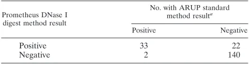
The DNase digest method detected atypical pANCA (DNase sensitive) in 22 sera in addition to the 33 that were in positive agreement with the standard IFA method (Table 3). The standard IFA method detected atypical pANCA (ethanol positive/formalin negative) in two sera that were negative by the DNase digest method (Table 3). We assessed these 24 discrepant atypical pANCA sera for IgG antibodies against
TABLE 1. Comparison of results from two different laboratories for ASCA IgA by EIA in 197 patient sera
Prometheus ASCA IgA result
No. with ARUP ASCA IgA resulta
Positive Negative
Positive 29 11
Negative 2 155
aAgreement, 93.4%.
TABLE 2. Comparison of results from two different laboratories for ASCA IgG by EIA in 197 patient sera
Prometheus ASCA IgG result
No. with ARUP ASCA IgG resulta
Positive Negative
Positive 14 0
Negative 18 165
aAgreement, 90.9%.
TABLE 3. Comparison of results from two different laboratories for atypical pANCA IgG by IFA in 197 patient sera
Prometheus DNase I digest method result
No. with ARUP standard method resulta
Positive Negative
Positive 33 22
Negative 2 140
aAgreement, 87.8%.
on August 17, 2020 by guest
http://cvi.asm.org/
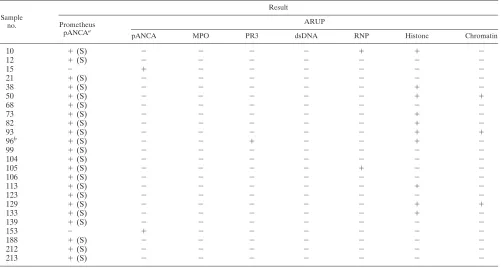
other known specific neutrophil/nuclear antigens (MPO, PR3, dsDNA, RNP, histone, and chromatin). Eleven of the 22 atyp-ical pANCA discrepant sera by the DNase digest method (50.0%) gave positive antibody results for one or more of the six antigens tested (Table 5). The two atypical pANCA dis-crepant sera by the standard IFA method gave negative anti-body results for all six antigens (Table 5). Cytoplasmic (cANCA) staining was not observed with any of these 24 dis-crepant sera.
Thirty-eight of 197 sera tested by Prometheus (19.3%) were positive for OmpC IgA. Twenty-five of these 38 sera (65.8%) were ASCA negative (IgA and IgG) as determined by Pro-metheus, while only 22 of 38 (57.9%) were ASCA negative as determined by ARUP. Three (no. 49, 133, and 135) of the 25 ASCA-negative/OmpC-positive sera detected by Prometheus (12.0%) were ASCA positive (one IgA, one IgG, and one IgA and IgG) by ARUP (Table 6). Twelve of the 25 (48.0%) ASCA-negative/OmpC-positive sera, as determined by Prome-theus, were also positive for atypical pANCA (DNase sensi-tive), 9 of which (69.2%) were in agreement with the standard IFA performed at ARUP (Table 6), suggesting that these pa-tients were more likely to have UC or Crohn’s colitis. Four of six (66.7%) patients in this group for whom clinical and biopsy information was available actually had UC (Table 7).
Of the 197 sera tested for OmpC IgA by Prometheus, only 9 (4.6%) had a positive OmpC result in the absence of other antibodies in IBD, and 8 of these had very low antibody con-tent (16.8 to 22.8 EIA units [EU]/ml) (Table 6). The majority (19 of 25; 76.0%) of sera with ASCA-negative/OmpC-positive results as determined by Prometheus gave very low values
TABLE 4. ASCA results for 29 discrepant sera from two different laboratories
Sample no.
Resulta
ASCA IgA ASCA IgG
ARUP Prometheus ARUP Prometheus
14 ⫹ ⫹ ⫹ ⫺
26 ⫺ ⫺ ⫹ ⫺
30 ⫹ ⫹ ⫹ ⫺
47 ⫺ ⫺ ⫹ ⫺
49 ⫺ ⫺ ⫹ ⫺
57 ⫺ ⫹ ⫺ ⫺
65 ⫺ ⫹ ⫺ ⫺
68 ⫺ ⫹ ⫺ ⫺
70 ⫺ ⫺ ⫹ ⫺
99 ⫺ ⫺ ⫹ ⫺
103 ⫺ ⫹ ⫺ ⫺
113 ⫹ ⫹ ⫹ ⫺
118 ⫺ ⫹ ⫹ ⫹
133 ⫹ ⫺ ⫹ ⫺
135 ⫹ ⫺ ⫺ ⫺
136 ⫹ ⫹ ⫹ ⫺
138 ⫹ ⫹ ⫹ ⫺
148 ⫺ ⫺ ⫹ ⫺
158 ⫺ ⫹ ⫺ ⫺
162 ⫺ ⫹ ⫹ ⫺
168 ⫺ ⫺ ⫹ ⫺
172 ⫹ ⫹ ⫹ ⫺
189 ⫺ ⫹ ⫺ ⫺
193 ⫹ ⫹ ⫹ ⫺
194 ⫹ ⫹ ⫹ ⫺
195 ⫺ ⫹ ⫺ ⫺
200 ⫺ ⫺ ⫹ ⫺
210 ⫺ ⫹ ⫺ ⫺
212 ⫺ ⫹ ⫺ ⫺
aSamples 133 and 162 carry double discrepancies.
TABLE 5. Assessment of 24 atypical pANCA discrepant sera for additional IgG autoantibodies against other known neutrophil/nuclear antigens
Sample no.
Result
Prometheus pANCAa
ARUP
pANCA MPO PR3 dsDNA RNP Histone Chromatin
10 ⫹(S) ⫺ ⫺ ⫺ ⫺ ⫹ ⫹ ⫺
12 ⫹(S) ⫺ ⫺ ⫺ ⫺ ⫺ ⫺ ⫺
15 ⫺ ⫹ ⫺ ⫺ ⫺ ⫺ ⫺ ⫺
21 ⫹(S) ⫺ ⫺ ⫺ ⫺ ⫺ ⫺ ⫺
38 ⫹(S) ⫺ ⫺ ⫺ ⫺ ⫺ ⫹ ⫺
50 ⫹(S) ⫺ ⫺ ⫺ ⫺ ⫺ ⫹ ⫹
68 ⫹(S) ⫺ ⫺ ⫺ ⫺ ⫺ ⫺ ⫺
73 ⫹(S) ⫺ ⫺ ⫺ ⫺ ⫺ ⫹ ⫺
82 ⫹(S) ⫺ ⫺ ⫺ ⫺ ⫺ ⫹ ⫺
93 ⫹(S) ⫺ ⫺ ⫺ ⫺ ⫺ ⫹ ⫹
96b ⫹(S) ⫺ ⫺ ⫹ ⫺ ⫺ ⫹ ⫺
99 ⫹(S) ⫺ ⫺ ⫺ ⫺ ⫺ ⫺ ⫺
104 ⫹(S) ⫺ ⫺ ⫺ ⫺ ⫺ ⫺ ⫺
105 ⫹(S) ⫺ ⫺ ⫺ ⫺ ⫹ ⫺ ⫺
106 ⫹(S) ⫺ ⫺ ⫺ ⫺ ⫺ ⫺ ⫺
113 ⫹(S) ⫺ ⫺ ⫺ ⫺ ⫺ ⫹ ⫺
123 ⫹(S) ⫺ ⫺ ⫺ ⫺ ⫺ ⫺ ⫺
129 ⫹(S) ⫺ ⫺ ⫺ ⫺ ⫺ ⫹ ⫹
133 ⫹(S) ⫺ ⫺ ⫺ ⫺ ⫺ ⫹ ⫺
139 ⫹(S) ⫺ ⫺ ⫺ ⫺ ⫺ ⫺ ⫺
153 ⫺ ⫹ ⫺ ⫺ ⫺ ⫺ ⫺ ⫺
188 ⫹(S) ⫺ ⫺ ⫺ ⫺ ⫺ ⫺ ⫺
212 ⫹(S) ⫺ ⫺ ⫺ ⫺ ⫺ ⫺ ⫺
213 ⫹(S) ⫺ ⫺ ⫺ ⫺ ⫺ ⫺ ⫺
a(S), DNase sensitive.
bCytoplasmic pattern (cANCA) absent, as determined by both laboratories.
on August 17, 2020 by guest
http://cvi.asm.org/
(⬍30.0 EU/ml) for OmpC IgA (Table 6). The OMP IgA (pro-totype EIA) performed at INOVA Diagnostics gave 9 positive, 5 equivocal, and 10 negative results for 24 of these 25 sera (Table 6) (the quantity was not sufficient for sample 205).
DISCUSSION
The ASCA and pANCA assays showed good agreement between the two reference laboratories (Tables 1 to 3). The Prometheus ASCA IgG assay, however, detected only 14 of the 32 positives (43.8%) detected by ARUP (Table 2). These find-ings are in agreement with a study from Belgium by Vermeire et al. (41), which showed the INOVA ASCA IgG assay to be 23% more sensitive than the ASCA IgG assay performed at Prometheus. Discord between these ASCA EIAs is most likely due to differences in their cutoff values, as was demonstrated in the study by Vermeire et al. using clinically defined sera (41). Of the three assays compared between the two laboratories, the atypical pANCA IgG IFA had the most discrepancies. The DNase I digest method detected atypical pANCA in 22 addi-tional sera, while the standard IFA method detected it in only 2 additional sera. Further assessment of these atypical pANCA discrepant sera for autoantibodies against other known neu-trophil/nuclear antigens (MPO, PR3, dsDNA, RNP, histone, and chromatin) showed 50.0% of the DNase IFA discrepant sera to contain other autoantibodies against one or more of the specific antigens tested (Table 5). In contrast, the two discrep-ant sera by the standard IFA method gave negative results for
all six additional markers (Table 5) and are more likely to be true atypical pANCA. The presence of antinuclear autoanti-bodies in patients with CD (18%) or UC (43%) has previously been reported (7). In addition, Reumaux et al. (27) detected IgG autoantibodies against several histone peptides primarily in patients with CD.
Since Prometheus utilizes a nonspecific ANCA (neutrophil) EIA for the screening of atypical pANCA, the presence of other autoantibodies as mentioned above could result in a positive ANCA EIA test that may affect the subsequent sub-jective neutrophil IFA pattern interpretation. Sera with strong homogeneous antinuclear reactivity could be misinterpreted as pANCA positive. If these specimens were then subjected to DNase digestion, sera with reactivity to dsDNA, histone, or chromatin (as shown to be the case for some of the atypical pANCA discrepant sera) would be interpreted as DNase sen-sitive. These results would be interpreted as supporting a di-agnosis of ulcerative colitis when they are actually more likely to be false positives due to the presence of antibodies to other specific neutrophil/nuclear antigens. A serum that was positive for dsDNA antibody was a control (it was DNase sensitive) used in the study first describing the DNase IFA methodology for detecting atypical pANCA (42).
The reports for IBD testing from Prometheus indicate that the purpose of their ANCA (neutrophil) EIA is to validate positive pANCA IFA sera, meaning that both tests must be positive before the result is reported to be “IFA perinuclear pattern detected.” For sera that are ANCA EIA positive/ pANCA IFA negative, “elevated levels” of ANCA IgG are reported. Sixteen of 140 pANCA IFA-negative sera were ANCA EIA positive, giving the Prometheus ANCA EIA a false-positive rate of 11.4%. These patients must then continue with the Prometheus “IBD Diagnostic System” or “IBD Con-firmatory System” to determine if the “IBD First Step”-posi-tive ANCA EIA result is actually due to atypical pANCA. Six of 57 (10.5%) Prometheus pANCA IFA-positive sera (all six DNase sensitive) were reported to be “IFA perinuclear pattern not detected” since they were ANCA EIA negative. Thus, the IBD reports from Prometheus indicate that false positives oc-cur often (10.5% in this study) with their DNase digest IFA method for detecting pANCA.
Currently, the OmpC IgA assay is performed only at Pro-metheus, whose reports indicate that this assay detects an additional 21% of CD patients who are seronegative for ASCA (14). It should be noted that only 56% of their CD patients were found to be ASCA seropositive (14). IBD test reports
TABLE 6. Results for 25 sera that were ASCA negative/OmpC positive as determined by Prometheus Laboratories
Sample no.
Result
Prometheus OmpC IgA (EU/ml)a
INOVA OMP IgAb
ARUP ASCA IgA
ARUP ASCA IgG
ARUP pANCA
IgG
Prometheus pANCA
IgGc
6 20.0 EQV ⫺ ⫺ ⫹ ⫹(S)
16 19.7 ⫺ ⫺ ⫺ ⫺ ⫺
24 18.0 EQV ⫺ ⫺ ⫺ ⫺
49 37.7 ⫹ ⫺ ⫹ ⫺ ⫺
55 20.4 EQV ⫺ ⫺ ⫺ ⫹(R)
69 27.9 ⫹ ⫺ ⫺ ⫹ ⫹(S)
79 26.3 ⫹ ⫺ ⫺ ⫹ ⫹(S)
80 21.3 ⫺ ⫺ ⫺ ⫺ ⫺
82 20.2 ⫺ ⫺ ⫺ ⫹ ⫹(S)
89 16.8 ⫺ ⫺ ⫺ ⫺ ⫺
93 40.2 ⫹ ⫺ ⫺ ⫺ ⫹(S)
102 22.8 ⫺ ⫺ ⫺ ⫺ ⫺
107 25.9 ⫺ ⫺ ⫺ ⫹ ⫹(S)
108 41.4 ⫹ ⫺ ⫺ ⫹ ⫹(S)
111 18.1 ⫺ ⫺ ⫺ ⫺ ⫺
116 16.9 EQV ⫺ ⫺ ⫺ ⫹(R)
133 ⬎111.0 ⫹ ⫹ ⫹ ⫺ ⫹(S)
135 20.6 ⫺ ⫹ ⫺ ⫹ ⫹(S)
137 23.2 EQV ⫺ ⫺ ⫹ ⫹(S)
150 19.3 ⫺ ⫺ ⫺ ⫺ ⫺
152 20.1 ⫺ ⫺ ⫺ ⫹ ⫹(S)
153 18.6 ⫹ ⫺ ⫺ ⫹ ⫺
166 54.0 ⫹ ⫺ ⫺ ⫺ ⫺
188 60.6 ⫹ ⫺ ⫺ ⫺ ⫹(S)
205 17.0 QNS ⫺ ⫺ ⫺ ⫺
a
16.5 EU/ml or greater is considered positive. b
EQV, equivocal; QNS, quantity not sufficient. c
(S), DNase sensitive; (R), DNase resistant.
TABLE 7. Diagnosis based on clinical and biopsy data for ASCA-negative/OmpC-positive patients for whom
information was availablea
Sample
no. Age (yr) Gender Diagnosis
55 75 Female Ulcerative colitis
80 30 Female Ulcerative colitis
93 38 Male Ulcerative colitis
137 54 Male Crohn’s disease
152 45 Male Ulcerative colitis
166 61 Female Crohn’s disease
aSee Table 6 for IBD testing results.
on August 17, 2020 by guest
http://cvi.asm.org/
from Prometheus show a positive predictive value of 85% for CD if OmpC IgA is the only positive result in their panel. If the results show OmpC positive/pANCA positive (DNase sensi-tive), the report states “results suggestive of ulcerative colitis.” Studies performed at Prometheus indicate that this pattern of results (ASCA negative/OmpC positive/pANCA positive) oc-curred two times more frequently in patients with UC than in those with Crohn’s disease. In view of this information and the data generated from this study, OmpC IgA appears to be more prevalent in UC, since many of the patients having ASCA-negative/OmpC-positive results were also atypical pANCA positive (Table 6). Of the limited number of patients who were ASCA negative/OmpC positive and from whom we were able to obtain clinical data (Table 7), 66.7% actually had ulcerative colitis, suggesting the limited diagnostic value of OmpC IgA in Crohn’s disease.
REFERENCES
1.Bansi, D. S., R. W. Chapman, and K. A. Fleming.1996. Prevalence and diagnostic role of antineutrophil cytoplasmic antibodies in inflammatory bowel disease. Eur. J. Gastroenterol. Hepatol.8:881–885.
2.Bernstein, C. N., J. F. Blanchard, P. Rawsthorne, and A. Wajda.1999. Epidemiology of Crohn’s disease in a central Canadian province: a popula-tion-based study. Am. J. Epidemiol.149:916–924.
3.Canani, R. B., M. T. Romano, L. Greco, G. Terrin, C. Sferlazzas, A. Barabino, M. Fontana, P. Roggero, G. Guariso, G. De Angelis, S. Fecarotta, G. Polito, and S. Cucchiara.2004. Effects of disease activity on anti- Saccha-romyces cerevisiaeantibodies: implications for diagnosis and follow-up of children with Crohn’s disease. Inflamm. Bowel Dis.10:234–239.
4.Coche, J. C., and J. F. Colombel.1998. Heterogeneity of inflammatory bowel disease: clinical subgroups of patients. Res. Clin. Forums20:135–145. 5.Eggena, M., O. Cohavy, M. H. Parseghian, B. A. Hamkalo, D. Clemens, S. R.
Targan, L. K. Gordon, and J. Braun.2000. Identification of histone H1 as a cognate antigen of the ulcerative colitis-associated marker antibody pANCA. J. Autoimmun.14:83–97.
6.Elitsur, Y., Z. Lawrence, and N. Tolaymat.2005. The diagnostic accuracy of serologic markers in children with IBD: the West Virginia experience. J. Clin. Gastroentrol.39:670–673.
7.Folwaczny, C., N. Noehl, S. P. Endres, W. Heldwein, K. Loeschke, and H. Fricke. 1997. Antinuclear autoantibodies in patients with inflammatory bowel disease. High prevalence in first-degree relatives. Dig. Dis. Sci.42:
1593–1597.
8.Freeman, H., B. Roeck, D. Devine, and C. Carter.1997. Prospective evalu-ation of neutrophil autoantibodies in 500 consecutive patients with inflam-matory bowel disease. Can. J. Gastroenterol.11:203–207.
9.Freeman, H. J.1997. Atypical perinuclear antineutrophil cytoplasmic anti-bodies in patients with Crohn’s disease. Can. J. Gastroenterol.11:689–693. 10.Frenzer, A., W. Fierz, E. Rundler, B. Hammer, and J. Binek.1998. Atypical, cytoplasmic and perinuclear anti-neutrophil cytoplasmic antibodies in pa-tients with inflammatory bowel disease. J. Gastroenterol. Hepatol.13:863– 864.
11.Hardarson, S., D. R. Labrecque, F. A. Mitros, G. A. Neil, and J. A. Goeken.
1993. Antineutrophil cytoplasmic antibody in inflammatory bowel disease and hepatobiliary diseases. High prevalence in ulcerative colitis, primary sclerosing cholangitis, and autoimmune hepatitis. Am. J. Clin. Pathol.99:
277–281.
12.Klebl, F. H., F. Bataille, C. R. Bertea, H. Herfarth, F. Hofstadter, J. Scholmerich, and G. Rogler.2003. Association of perinuclear antineutrophil cytoplasmic antibodies and anti-Saccharomyces cerevisiae antibodies with Vienna classification subtypes of Crohn’s disease. Inflamm. Bowel Dis.
9:302–307.
13.Klebl, F. H., F. Bataille, F. Hofstadter, H. Herfarth, J. Scholmerich, and G. Rogler.2004. Optimising the diagnostic value of anti-Saccharomyces cerevi-siae-antibodies (ASCA) in Crohn’s disease. Int. J. Colorectal Dis.19:319– 324.
14.Landers, C. J., O. Cohavy, R. Misra, H. Yang, Y. C. Lin, J. Braun, and S. R. Targan.2002. Selected loss of tolerance evidence by Crohn’s disease-asso-ciated immune response to auto- and microbial antigens. Gastroenterology
123:689–699.
15.Lindberg, E., K. E. Magnusson, C. Tysk, and G. Jarnerot.1992. Antibody (IgG, IgA, and IgM) to baker’s yeast (Saccharomyces cerevisiae), yeast man-nan, gliadin, ovalbumin and betalactoglobulin in monozygotic twins with inflammatory bowel disease. Gut33:909–913.
16.Linskens, R. K., R. C. Mallant-Hent, Z. M. Groothuismink, L. E. Bakker-Jonges, J. P. van de Merwe, H. Hooijkaas, B. M. von Blomberg, and S. G. Meuwissen.2002. Evaluation of serological markers to differentiate between
ulcerative colitis and Crohn’s disease: pANCA, ASCA and agglutinating antibodies to anaerobic coccoid rods. Eur. J. Gastroenterol. Hepatol.14:
1013–1018.
17.Loftus, E. V., Jr., M. D. Silverstein, W. J. Sandborn, W. J. Tremaine, W. S. Harmsen, and A. R. Zinsmeister.1998. Crohn’s disease in Olmsted County, Minnesota, 1940–1993: incidence, prevalence, and survival. Gastroenterol-ogy114:1161–1168.
18.Main, J., H. McKenzie, G. R. Yeaman, M. A. Kerr, D. Robson, C. R. Pennington, and D. Parratt.1988. Antibody toSaccharomyces cerevisiae
(baker’s yeast) in Crohn’s disease. BMJ297:1105–1106.
19.McKenzie, H., J. Main, C. R. Pennington, and D. Parratt.1990. Antibody to selected strains ofSaccharomyces cerevisiae(baker’s and brewer’s yeast) and
Candida ablicansin Crohn’s disease. Gut31:536–538.
20.Moore, M. M., D. Fabricatorian, and W. S. Selby.2002. Assessment and relevance of enzyme-linked immunosorbent assay for antibodies to Saccha-romyces cerevisiaein Australian patients with inflammatory bowel disease. J. Intern. Med.32:349–352.
21.Mulder, A. H., G. Horst, E. B. Haagsma, J. H. Kleibeuker, and C. G. Kallenberg.1993. Anti-neutrophil cytoplasmic antibodies (ANCA) in auto-immune liver disease. Adv. Exp. Med. Biol.336:545–549.
22.Mulder, A. H., J. Broekroelofs, G. Horst, P. C. Limburg, G. F. Nelis, and C. G. Kallenberg.1994. Anti-neutrophil cytoplasmic antibodies (ANCA) in inflammatory bowel disease: characterization and clinical correlates. Clin. Exp. Immunol.95:490–497.
23.National Institute of Diabetes and Digestive and Kidney Diseases.1998. Crohn’s disease. NIH publication no. 98–3410. National Institutes of Health, Bethesda, Md.
24.Niv, Y., G. Abuksis, and G. M. Fraser.1999. Epidemiology of Crohn’s disease in Israel: a survey of Israeli kibbutz settlements. Am. J. Gastro-enterol.94:2961 2965.
25.Panaccione, R., and W. J. Sanborn.1999. Is antibody testing for inflamma-tory bowel disease clinically useful? Gastroenterology116:1001–1008. 26.Quinton, J. F., B. Sendid, D. Reumaux, P. Duthilleul, A. Cortot, B.
Grandbastien, G. Charrier, S. R. Targan, J. F. Colombel, and D. Poulain.
1998. Anti Saccharomyces cerevisiae mannan antibodies combined with anti-neutrophil cytoplasmic autoantibodies in inflammatory bowel disease: prev-alence and diagnostic role. Gut42:788–791.
27.Reumaux, D., C. Meziere, J. F. Colombel, P. Duthilleul, and S. Mueller.
1995. Distinct production of autoantibodies to nuclear components in ulcer-ative colitis and in Crohn’s disease. Clin. Immunol. Immunopathol.77:349– 357.
28.Roozendaal, C., and C. G. Kallenberg.1999. Are anti-neutrophil cytoplasmic antibodies (ANCA) clinically useful in inflammatory bowel disease (IBD)? Clin. Exp. Immunol.116:206–213.
29.Roozendaal, C., K. Pogany, G. Horst, T. G. Jagt, J. H. Kleibeuker, G. F. Nelis, P. C. Limburg, and C. G. Kallenberg.1999. Does analysis of the antigenic specificities of anti-neutrophil cytoplasmic antibodies contribute to their clinical significance in the inflammatory bowel diseases? Scand. J. Gastroenterol.34:1123–1131.
30.Roussomoustakaki, M., J. Satsangi, K. Welsh, E. Louis, G. Fanning, S. Targan, C. Landers, and D. P. Jewell.1997. Genetic markers may predict disease behavior in patients with ulcerative colitis. Gastroenterology112:
1845–1853.
31.Ruemmele, F. M., S. R. Targan, G. Levy, M. Dubinsky, J. Braun, and E. G. Seidman.1998. Diagnostic accuracy of serological assays in pediatric inflam-matory bowel disease. Gastroenterology115:822–829.
32.Rutgeerts, P., and S. Vermeire.1998. Clinical value of the detection of antibodies in the serum for diagnosis and treatment of inflammatory bowel disease. Gastroenterology115:1006–1022.
33.Saibeni, S., C. Folli, R. de Franchis, G. Borsi, and M. Vecchi.2003. Diag-nostic role and clinical correlates of anti-Saccharomyces cerevisiaeantibodies (ASCA) and anti-neutrophil cytoplasmic antibodies (p-ANCA) in Italian patients with inflammatory bowel disease. Dig. Liver Dis.35:862–868. 34.Seibold, F., P. Weber, R. Klein, P. A. Berg, and K. H. Wiedmann.1992.
Clinical significance of antibodies against neutrophils in patients with inflam-matory bowel disease and primary sclerosing cholangitis. Gut33:657–662. 35.Sendid, B., J. F. Colombel, P. M. Jacquinot, C. Faille, J. Fruit, A. Cortot, D.
Lucidarme, D. Camus, and D. Poulain.1996. Specific antibody response to oligomannosidic epitopes in Crohn’s disease. Clin. Diagn. Lab. Immunol.
3:219–226.
36.Shivananda, S., J. Lennard-Jones, R. Logan, N. Fear, A. Price, L. Carpenter, and M. van Blankenstein.1996. Incidence of inflammatory bowel disease across Europe: is there a difference between north and south? Results of the European collaborative study on inflammatory bowel disease (EC-IBD). Gut
39:690–697.
37.Terjung, B., H. J. Worman, V. Herzog, T. Sauerbruch, and U. Spengler.
2001. Differentiation of antineutrophil nuclear antibodies in inflammatory bowel and autoimmune liver disease from antineutrophil cytoplasmic anti-bodies (p-ANCA) using immunofluorescence microscopy. Clin. Exp. Immu-nol.126:37–46.
38.Terjung, B., V. Herzog, H. J. Worman, I. Gestmann, C. Bauer, T. Sauerbruch, and U. Spengler.1998. Antineutrophil cytoplasmic antibodies with
on August 17, 2020 by guest
http://cvi.asm.org/
clear fluorescence in chronic inflammatory bowel diseases and hepatobiliary disorders colocalize with nuclear lamina proteins. Hepatology28:332–340. 39.Terjung, B., U. Spengler, T. Sauerbruch, and H. J. Worman.2000. “Atypical
p-ANCA” in IBD and hepatobiliary disorders react with a 50-kilodalton nuclear envelope protein of neutrophils and myeloid cell lines. Gastroenter-ology119:310–322.
40.Vasiliauskas, E. A., S. E. Plevy, C. J. Landers, S. W. Binder, D. M. Ferguson, H. Yang, J. I. Rotter, A. Vidrich, and S. R. Targan. 1996. Perinuclear antineutrophil cytoplasmic antibodies in patients with Crohn’s disease define a clinical subgroup.Gastroenterol.6:1810–1819.
41.Vermeire, S., S. Joossens, M. Peeters, F. Monsuur, G. Marien, X. Bossuyt, P.
Groenen, R. Vlietinck, and P. Rutgeerts.2001. Comparitive study of ASCA (anti-Saccharomyces cerevisiaeantibody) assays in inflammatory bowel dis-ease. Gastroenterology120:827–833.
42.Vidrich, A., K. Lee, E. James, L. Cobb, and S. Targan.1995. Segregation of pANCA antigenic recognition by DNase treatment of neutrophils: ulcerative colitis, type 1 autoimmune hepatitis, and primary sclerosing cholangitis. J. Clin. Immunol.15:293–299.
43.Zholudev, A., D. Zurakowski, W. Young, A. Leichtner, and A. Bousvaros.
2004. Serologic testing with ANCA, ASCA, and anti-OmpC in children and young adults with Crohn’s disease and ulcerative colitis: diagnostic value and correlation with disease phenotype. Am. J. Gastroenterol.99:2235–2241.