Western University Western University
Scholarship@Western
Scholarship@Western
Electronic Thesis and Dissertation Repository
9-7-2017 12:00 AM
Bio-modulation of Primary Human Tenon’s Capsule Fibroblasts
Bio-modulation of Primary Human Tenon’s Capsule Fibroblasts
Using a Novel Application of Coated Magnesium
Using a Novel Application of Coated Magnesium
Xiangji LI
The University of Western Ontario
Supervisor Dr.Hutnik
The University of Western Ontario Graduate Program in Surgery
A thesis submitted in partial fulfillment of the requirements for the degree in Master of Science © Xiangji LI 2017
Follow this and additional works at: https://ir.lib.uwo.ca/etd
Recommended Citation Recommended Citation
LI, Xiangji, "Bio-modulation of Primary Human Tenon’s Capsule Fibroblasts Using a Novel Application of Coated Magnesium" (2017). Electronic Thesis and Dissertation Repository. 4917.
https://ir.lib.uwo.ca/etd/4917
This Dissertation/Thesis is brought to you for free and open access by Scholarship@Western. It has been accepted for inclusion in Electronic Thesis and Dissertation Repository by an authorized administrator of
Abstract
Background: Glaucoma is the leading cause of irreversible blindness. The last decade has seen the emergence of minimally invasive glaucoma surgical procedures,
with the most recent utilizing implantable devices. The first generation of
micro-incisional devices has been made of titanium. Since then other materials have
also been introduced. In each case, the presence of a permanent foreign body has
been associated with undesirable wound healing responses characterized by fibroblast
proliferation and surgical failure. Magnesium alloys have attracted much attention as
biodegradable implantable materials due to their excellent biodegradable and
biocompatibility properties. The purpose of this research was to evaluate the in vitro
biocompatibility and anti-proliferative properties of differently coated magnesium
alloys in a primary culture of human Tenon’s capsule fibroblasts (HTCFs). The goal
of the research was to establish a proof of principle for further exploration of this
novel material as a potential adjunct to glaucoma surgery.
Materials and Methods: Pure magnesium was cut into discs measuring 14.5 mm in diameter and 1 mm thick. These were coated with Hydroxyapatite (HA), Dicalcium
phosphate dehydrate (DCPD) and Dicalcium phosphate dehydrate + Stearic acid
(DCPD+SA), respectively. Coated magnesium alloys were immersed in simulated
aqueous humor to examine the corrosive properties, the released ions, and the
disks in 24-well culture and incubated at 37 degrees for 2-7 days.. Glass disks were
used as control. The MTT and LDH assays were used to determine cellular metabolic
activity and cytotoxicity during the logarithmic phase of HTCFs, respectively. The
BrdU assay was used to evaluate cellular proliferation. Western blot was used to
assess the expression of alpha-smooth muscle actin (α-SMA).
Results: A total of 453 coated magnesium alloy disks were evaluated. Corrosion was observed in 67 disks (14.8%). The corrosion rate of HA (12.6%) was lower than other
two coatings (DCPD 15.9%, DCPD+SA 15.9%, respectively), but the difference was
not statistically significant different (p=0.851). The immersion test showed that the
HA coating had better ability to prevent the coating dissolution and corrosion. The
cellular metabolic activity of different coated magnesium alloys gradually declined
during the logarithmic phase of HTCFs, and each type of coated magnesium alloy
demonstrated significant decreased metabolic activity of HTCFs when compared to
the control (p<0.001). The cytotoxicity of different coated magnesium alloys
slightly increased during the logarithmic phase. The group of DCPD+SA showed
higher cytotoxicity than the other coatings, but it was not statistically significant
different when compared to the control (p=0.976). Significant inhibition of
proliferation was observed with the DCPD+SA coating (p=0.47). The expression of
α-SMA was decreased in the cells when seeded on all of the coated magnesium alloy
Chemical coatings are able to affect the corrosive properties of magnesium. HA was
the most resistant to corrosion. No significant difference was found in metabolic
activity or necrosis at different times during the logarithmic phase of HTCFs.
DCPD+SA demonstrated a stronger ability to reduce metabolic activity while its
cytotoxic profile was the same as the titanium and glass controls. In comparison to
titanium, coated magnesium alloys attenuated HTCFs proliferation. Coated
magnesium alloys reduced the expression of α-SMA. The expression of α-SMA was
significantly decreased in cells exposed to the HA coated magnesium
Conclusions: Different chemical coatings on magnesium were able to affect the corrosive properties which, in turn, influenced the morphology and function of human
Tenon’s capsule fibroblasts. These results support the further study of coated
magnesium for its potential modulatory role in Tenon’s capsule wound healing.
Keywords:
Glaucoma, glaucoma drainage device, fibrosis, tenon’s capsule fibroblasts,
biodegradable, magnesium, magnesium alloys, coating
Acknowledgments
My deepest gratitude goes to my supervisor Dr. Cindy Hutnik, for providing me with
the opportunity to work on this wonderful project. Thank you for your continued
insight, guidance, and patience as I embarked on the road to becoming a better
researcher and intellectual.
I would like to acknowledge my advisory committee, Dr. Amin Rizkalla, Dr. Nicholas
Power, and Dr. Weiping Min, for their knowledge, encouragement, and humor. I
always left our committee meetings with renewed fervor for my work and new
complexities to ponder.
Thank you to Hong Liu, for her kind help and support within the Ophthalmology
research lab.
Finally, I would like to thank my family for their continued support and avid
Table of Contents
Abstract... i
Acknowledgments ... iv
Table of Contents ………..……….……..…… v
List of Tables ... ix
List of Figures ... x
List of Abbreviations ... xi
Review ... xiv
Chapter 1 ... 1
1.1 Glaucoma... 1
1.1.1 Epidemiology of glaucoma... 1
1.1.2 Aqueous humor and outflow ... 2
1.1.3 Pathogenesis... 3
1.2. Glaucoma treatment options ... 4
1.2.1 Filtration surgery... 4
1.2.2 Glaucoma drainage devices... 5
1.3 Fibrosis... 6
1.3.1 Anti-fibrotic agents... 6
1.3.2 Permanent foreign body under tenos’s capsule stimulates fibrosis……. 7
1.4 Magnesium... 8
1.4.2 Magnesium alloys ... 9
1.4.3 Coating techniques improve corrosive property……… 10
1.5 Hypothesis and Objectives……… 11
1.5.1 Hypothesis………..… 11
1.5.2 Objectives………... 11
Chapter 2……….. 13
2.1 Materials and methods……….……….. 13
2.1.1 Primary HTCFs culture………... 13
2.1.2 Assessment of morphology……….. 13
2.1.3 Fabrication of differently coated magnesium alloys……….…... 14
2.1.4 Magnesium sample preparation……….……….. 16
2.1.5 Statistical analysis……….………... 17
2.2 Results……….………..…….. 17
2.2.1 Cell morphological observation and identification of HTCFs by immunofluorescence staining. ……….……… 17
2.2.2 Corrosion rate of different coated magnesium alloys………..… 20
2.2.3 The corrosion susceptibility of different coatings from day 2 to day 7…... 21
2.3 Discussion……….. 22
Chapter 3……….. 24
3.1 Background……….... 24
3.2 Materials and methods………...……….... 25
3.3 Results……….……….. 26
3.3.1 Microstructure of different coatings……….……….. 26
3.3.2 pH variations of BSS……….………...….. 28
3.3.3 Sample weight changes in BSS……….……….. 29
3.3.4 Changes of ion concentrations during immersion test. ……….. 30
3.4 Discussion………... 32
3.4.1 Effect of coating on corrosion process………... 32
3.4.2 Ion release of different samples during immersion test……… 34
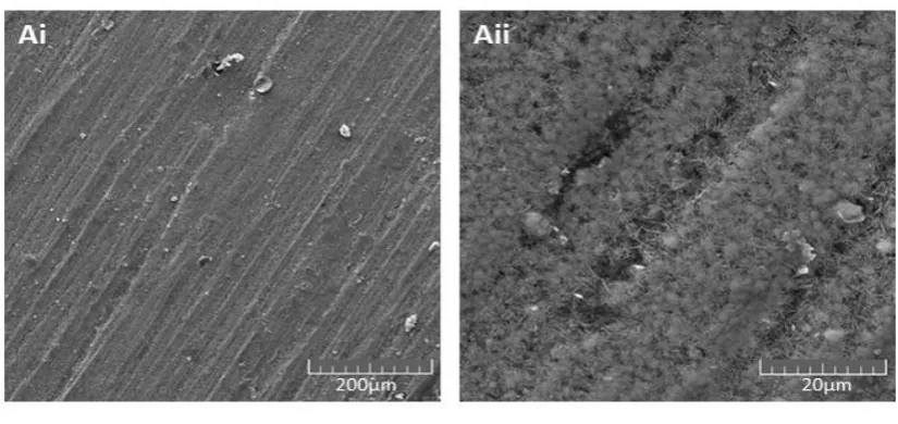
3.4.3 SEM morphologies of different coatings after immersion test………. 35
Chapter 4……… 36
4.1 Background ………….……… 36
4.2 Materials and methods……….…… 37
4.2.1 MTT assay……….… 37
4.2.2 Lactate dehydrogenase cytotoxicity(LDH) assay………..… 37
4.2.3 Statistical analysis………..…… 38
4.3 Results………..………. 38
4.3.1 Time and cellular metabolic activity curves………..…….39
4.3.2 Time and cytotoxicity curves………..…... 40
4.4 Discussion ……….…... 41
4.4.1 The influence of time………..…... 41
4.4.2 Biocompatibility of coated magnesium alloys………..…... 41
5.1 Background………..…... 44
5.2 Materials and methods………..…... 45
5.2.1 BrdU assay………..…... 45
5.2.2 Protein extraction and Western blotting analysis... 46
5.3 Results………..…... 47
5.3.1 Cell proliferation study………..…... 47
5.3.2 Western blotting analysis………..…... 48
5.4 Discussion………..…... 49
5.4.1 Coated magnesium alloys attenuate HTCFs proliferation... 49
5.4.2 HA coating inhibits α-SMA expression………50
Chapter 6……… 52
6.1 Summary of results………..……… 52
6.2 Limitations of the study………...……… 55
6.3 Future directions……….. 57
6.4 Concluding remarks………. 58
6.5 Conclusions of full text……….... 59
References………. 61
List of Tables
Table 2.2.2 Corrosion rate of differently coated magnesium alloys (events)... 21
Table 2.2.3 The corrosion susceptibility of different coatings at different days
(events/disks)………. 22
List of Figures
Figure 1. Magnesium deficiency contributes to the pathogenesis of cataract ... xxiv
Figure 2. Magnesium deficiency contributes to the pathogenesis of glaucomatous neropathy………... xxvii
Figure 1.1.2 The trabecular meshwork outflow pathways……... 3
Figure 1.3.1 Clinical presentation of filtering blebs……… 6
Figure 1.3.2 Conjunctival scarring following filtering glaucoma surgery... 8
Figure 2.1.3SEM images of different coatings... 16
Figure 2.1.4 Different sample groups for experiments..………..17
Figure 2.2.1Immunostaining and characterization of primary human Tenon’s capsule cultures... 20
Figure 3.3.1Scanning electron microscopic (SEM) images of coated magnesium samples after immersion at with different magnifications... 27
Figure 3.3.2 Variation of BSS pH of coated and uncoated samples with time... 29
Figure 3.3.3Weight changes of coated and uncoated samples with time…………... 30
Figure 3.3.4 Changes of ions concentration during immersion test………... 31
Figure 4.3.1Cellular metabolic activity curves of different samples. ………….…... 39
Figure 4.3.2 Time and cytotoxicity curves of different samples………. 40
Figure 5.3.1 Relative proliferation rate of HTCFs. ………...………. 48
List of Abbreviations
POAG Primary open angle glaucoma
PACG Primary angle closure glaucoma
IOP Intraocular pressure
MMC Mitomycin C
5-FU 5-Fluorouracil
ECM Extracellular matrix
HA Hydroxyapatite
DCPD Dicalcium phosphate dehydrate
DCPD+SA Dicalcium phosphate dehydrate +Stearic acid
HTCFs Human tenon’s capsule fibroblast
SEM Scanning electron microscope
DMSO Dimethyl sulfoxide
FBS Fetal bovine serum
MTT 3-(4,5-dimethyl-2-thiazolyl)-2, 5-diphenyl-2-H-tetrazolium bromide
LDH Lactate dehydrogenase
BrdU 5-Bromo-2-deoxyUridine
α-SMA α-Smooth muscle actin
GAPDH Anti-glyceraldehyde-3phosphate dehydrogenase
DMEM Dulbecco’s Modified Eagle Medium
mRNA Messenger RNA
ng/mL Nanograms per milliliter
P/S Penicillin/Streptomycin
BSS Balanced salt solution
ANOVA Analysis of variance
BSA Bovine serum albumin
RGCs Retinal ganglion cells
Al Aluminum
Mg Magnesium
Ca Calcium
K Potassium
Na Sodium
AH Aqueous humor
TM Trabecular meshwork
TRAB Trabeculectomy
PBS Phosphate buffered saline
CaP Calcium phosphate
mmHg Millimeter of mercury
CO2 Carbon dioxide
Review
A feasibility study of using biodegradable magnesium
alloy in glaucoma drainage device
Abstract
Technological advances in glaucoma have challenged the traditional treatment
paradigm. Historically, incisional surgery has been used in cases of advanced
disease and/or uncontrolled intraocular pressures resistant to medical or laser
interventions. Despite these trends, surgical manipulation of the tissues and
unpredictability of wound healing continue to result in surgical failure. Magnesium is
an essential element for the human body and plays a critically important role for
maintaining the functional and structural integrity of several tissues, including the eye.
Due to several of its advantageous properties such as
non-toxicity, biodegradability, and high biological compatibility, magnesium alloy has
attracted great attention as a novel biomaterial. Biodegradable cardiovascular stents
made of magnesium alloy have already been introduced into clinical practice. The
purpose of this review is to determine if bio-absorbable magnesium alloys can be
utilized as a promising candidate for the development of a new generation of
glaucoma surgical assistive devices.
devices, Magnesium alloy, Coating, Anti-scarring.
Introduction
Magnesium is an essential trace element for human life, and also is one of the most
important regulatory cations involved in several biological processes. Magnesium is
the second most common cation in the intracellular fluid. Mg2+ plays a crucial role in
regulating vascular functions and energy metabolism as well as maintaining water and
electrolyte balance1,2. The level of Mg2+in the serum ranges from 0.8 to 1.2mmol/L,
with homeostasis being maintained by the kidney and intestine. Hypomagnesemia
(serum Mg2+<0.8mmol/L) has been suggested to be associated with several disorders,
such as vascular spasm and arrhythmia. Magnesium deficiency may also lead to
cardiovascular, respiratory, and digestive illnesses, as well as abortion, fetal
abnormalities and other obstetric diseases3. A large number of studies have shown that
dietary intake of magnesium can prevent osteoporosis and femoral neck fracture 4-7,
and help to manage diabetes and coronary artery disease8,9. Serum Mg2+ levels
exceeding 1.2mmol/L may cause muscular paralysis, respiratory distress and
hypotension10; however hypermagnesemia is rare due to the efficient excretion of the
element in the urine11.
Glaucoma is the second most common cause of blindness and the leading cause of
irreversible worldwide blindness12. Surgery is an important treatment option for
glaucoma Incisional surgery, with and without glaucoma drainage devices, is a major
reach a pressure that halts progression. Long-term complications are often related to
variability in the wound healing process. Devices used adjunctively in surgical
glaucoma management represent a foreign body under the conjunctiva, causing
fibroblast proliferation. This may be due to the persistent effect of inflammatory
mediators and cytokines at the level of the conjunctival-Tenon-episcleral interface
resulting in fibrosis and obstruction of the fistula13,14. There continues to be
significant interest in adjunctive approaches to minimize scarring in the
subconjunctival space, reducing inflammation, and in slowing or halting the excessive
fibrotic healing process.
Bleb scarring is a major contributor to increased intraocular pressure (IOP) and
surgical failure. In order to improve the success rate of glaucoma surgery, various
intraoperative anti-metabolites have been used to inhibit fibroblast proliferation, such
as 5-fluorouracil (5-FU) and mitomycinC (MMC). However, these antimitotic agents
interfere indiscriminately with cellular proliferation, and can contribute to a number
of postoperative adverse events, including bleb leaks, hypotony, choroidal detachment,
endophthalmitis, and keratitis15,16. There continues to be a significant need for a safe,
non-toxic, and effective approach to reduce postoperative scarring and adhesions after
glaucoma surgery. The relatively recent introduction of bleb-forming micro-invasive
glaucoma surgery has heightened this interest.
properties such as high strength, light weight, and high biological compatibility.
Additionally, magnesium alloy has been shown to be able to reduce irritation and
inflammatory reactions in the body17-20. We were thus motivated to determine the
feasibility of developing a magnesium-based bio-absorbable device for modulation of
wound healing associated with glaucoma surgery. Moreover, the possibility that the
slow release of Mg2+ by the device could have a local neuroprotective role is an
intriguing concept worthy of future investigation.
Application of magnesium alloy in modern medicine
The application of magnesium alloy as a bio-absorbable material in clinical practice
can be traced back to the early 1900s. The first use of magnesium was reported by
Lambotte et al. in 1907, who utilized a plate of pure magnesium with gold-plated steel
nails to treat a fracture involving the bones of the lower leg. The initial attempt
failed as the pure magnesium plate corroded too rapidly in vivo, disintegrating only
eight days after surgery and producing a large amount of gas under the skin21,22. In
1938, McBride et al. developed magnesium alloy fixtures which successfully treated
20 cases of fracture without any significant adverse effects, and the fixtures were
completely absorbed three months after surgery 23. In 1944, Troitskii et al. applied
cadmium magnesium alloy as an internal fixation device to secure the bones of 34
consecutive fracture patients. They observed that the fixture could maintain the
mechanical integrity up to two months, and it was completely absorbed after 10-12
A hard callous was found around the fracture site with no increase in serum levels of
magnesium and no obvious inflammatory reactions to the implantin all 34 patients
who were studied23. Znamenski et al. reported similar results in 1945, where
magnesium alloy containing 10% aluminum (Al) was used to treat two patients with
gunshot wounds. The magnesium implant and nails were completely absorbed after
4-6 weeks23. These early reports demonstrated that magnesium alloy was a non-toxic
biomaterial, and it had the ability to promote bone healing. However, due to its
characteristics of hydrogen emission and low corrosion resistance in the electrolytic
and aqueous environments of the physiological system, biomaterial research on
magnesium alloy was suspended. Thereafter, stainless steel materials were widely
applied in bone internal fixation devices. Until recent years, with the use of advanced
techniques, more complicated alloy compositions have been introduced.
Advancements in corrosion protection technologies have effectively reduced the
production of hydrogen gas. It is for these reasons that interest in the medical use of
magnesium alloys has once again increased.23.
Numerous in vitro and in vivo studies have focused on the use of magnesium alloys in
internal fixation devices for fracture repairs. The corrosion resistance of different
types of coated magnesium alloys has been studied in cytological and animal
experiments to determine the reduction in hydrogen evolution and tissue compatibility
24,25. In vitro experiments confirmed that there were minimal cytotoxicity and cell
magnesium alloys25. In vivo experiments found that Mg2+ could enhance bone
formation during the degradation process of the implant, and no inflammation was
observed26,27. F.Witte et al. successfully implanted magnesium alloy stents into the
femur of rabbits, and the magnesium implants substantially degraded after three
months. Their study also showed that the degradation process of the magnesium alloy
stents could promote trabecular bone formation and resorption, without any
significant harm to their neighboring tissues. They concluded that even fast-degrading
magnesium alloy stents could show favourable biocompatibility thus establishing a
more convincing potential role, in musculoskeletal surgery28,29. In addition, F.Witte et
al. also carried out an in vitro experiment to investigate the properties of a metallic
matrix composite made of magnesium alloy AZ91 as a matrix with hydroxyapatite
(HA) particles as reinforcements. The results revealed that the HA particles could
stabilize the corrosion rate of the magnesium alloy, and this biodegradable metallic
matrix composite HA was a cyto-compatible biomaterial with adjustable mechanical
and corrosive properties30. Okazaki M et al. developed a novel material containing
magnesium, calcium, and phosphate for use as oral implants. They found that
magnesium increased the metabolic rate of osteoblasts31. Zreiqat et al. found that the
protein levels were significantly higher in human bone-derived cells cultured on
[Mg]-Al2O3 (alumina doped with magnesium ions) compared with those grown on
Al2O3 alone32. Hunt Jet al. reported that the magnesium-coated Ti-6Al-4v implant
could activate bone cell signal transduction and hence improve protein synthesis and
magnesium have been used to develop novel cardiovascular stents, so as to maintain
the endothelial function of coronary arteries and to reduce the risk of coronary
ischemia and occlusion34-37. Based on a large number of clinical trials38,39, magnesium
coronary stents have been introduced into clinical practice40.
The important roles of Mg2+ in the eye
Mg2+ is important for maintaining the structural and functional integrity of several
vital ocular tissues such as the cornea; lens and retina. The concentration of
magnesium in aqueous humor is 2.97 ± 0.75 mg/ 100 ml. The magnesium levels in the
lens are far higher than those in the anterior chamber and vitreous body. The
concentration in the lens periphery is four times greater than the axial regions41. Mg2+
plays an extremely important role in maintaining retinal function. The reason for this
is because magnesium acts as a co-factor involved in the catalytic function of more
than 350 enzymes in the body and regulates neuro-excitability. Membrane associated
ATPase functions that are crucial in regulating the intracellular ionic environment, are
also magnesium-dependent. As a result, a reduced level of Mg2+may affect the
functions of Na+/K+ ATPase and calcium dependent ATPase, causing an increase in
the intracellular concentrations of calcium and sodium as well as a decrease in the
potassium42 concentration. Such ionic imbalances induced by magnesium deficiency
may contribute to the pathogenesis of many eye disorders, such as cataract, corneal,
Maintenance of corneal structure and function
Mg2+ is one of the most important cations in the cornea as it is involved in the
metabolism and maintenance of corneal transparency45. As early as 1920, Kirkpatrick
et al. reported the use of magnesium sulfate in the treatment of keratitis, conjunctivitis,
and corneal ulceration46. In 1985, Bachman and Wilson performed an animal
experiment and found that the epithelial surface of the excised rabbit cornea was
maintained best with a buffered solution containing Mg2+, K+, and Ca2+ 47. Hogan et al.
reported that Mg2+ loss in the corneal stroma was associated with corneal edema48. In
2001, Gong et al. investigated the effect of magnesium deficiency on the cornea in
rats that were fed a low magnesium diet for 3 weeks. Their findings revealed that
magnesium deficiency affected the structural and functional integrity of the cornea49.
Keratoconus is defined as a progressive eye disease which causes thinning and
fragmentation of membranes, degenerated cells and collagen fibers, swelling of the
mitochondria, and biochemical abnormalities in protein synthesis and expansion of
central area of the cornea. Thalasselis et al. reported that hypomagnesemia was
commonly seen in the serum samples of keratoconus patients, and magnesium
deficiency could pathologically affect the integrity of the cornea50. Prior studies also
showed that magnesium deficiency was associated with a reduced number of
microvilli and their irregular arrangement. Vesicular degeneration and swelling of the
mitochondria were observed in the cytoplasm of epithelial cells, resulting in abnormal
and fragmentation of the Bowman’s layer, which may be an early change leading to
keratoconus53,54.
Supporting lens metabolism and preventing cataract
Cataract is the most common cause of blindness worldwide. It is characterized by
progressive lenticular opacities. It is known that cataractous lenses have an abnormal
intracellular ionic environment with lower concentrations of potassium and
magnesium and higher concentrations of sodium and calcium relative to the cytosol of
most cells. The lens membrane has increased permeability in the presence of a
cataract55. Studies have explored he relationship between magnesium deficiency and
cataract. These studies have shown that the alterations in lenticular redox status and
ionic imbalances form the basis of the relationship between magnesium deficiency
and cataract. It is believed that Mg2+ plays an important role not only in maintaining a
low lenticular Ca2+ and Na2+ concentration but also in preserving the lens redox status,
which has been shown to reduce lenticular oxidative stress56,57. Nagaiet al. reported
that the incidence of cataract was significantly lower in Shumiya rats fed 200mg/L
magnesium compared to controls. It was noted that the intracellular Ca2+ level of the
lenses was also significantly lower58. Based on these findings, they concluded that an
appropriate supplementation of magnesium could delay cataract genesis, probably by
inhibiting the increase in Ca2+ levels in the lens59. After a thorough literature review,
Agarwal et al. concluded that magnesium supplementation might be of therapeutic
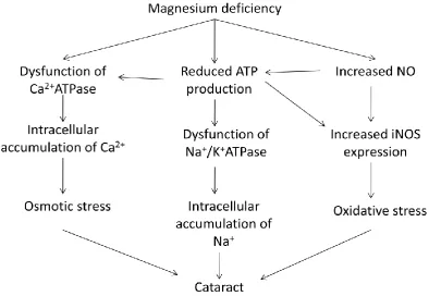
Oxidative stress is another important contributor to the pathogenesis of cataract. It has
been shown in many studies that nitric oxide (NO) could be a risk factor in cataract
formation61. NO production via inducible nitric oxide synthase (iNOS) causes an
oxidative stress response in the lens62. Mg2+ has been shown to prevent the increase of
NO in the lens which provides protection by inhibiting the nitrosylation of gap
junctional proteins and maintaining membrane permeability63. It is also known to
block iNOS expression and reduce the oxidative stress response. A cytological
experiment found that the expression of iNOS was 6 times higher in the lens epithelial
cells cultured in the magnesium-deficient medium compared to those cultured in the
magnesium-supplemented medium. In addition, Mg2+ was shown to regulate Ca2+
ATPase thus modulating the concentration of Ca2+ in the lens.
Both magnesium deficiency and excessive NO production have been shown to
decrease ATP levels58. The reduction in ATP levels affects the membrane-associated
Ca2+ ATPase and Na+/K+ ATPase, causing ionic imbalance in the lens. Interestingly,
the decreased ATP levels in turn may have inhibitory effects on the expression of
iNOS64. Therefore, it has been speculated that magnesium deficiency may cause an
Figure 1. Magnesium deficiency contributes to the pathogenesis of cataract development.
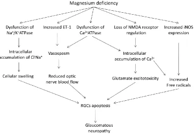
Glaucomatous neuroprotection
Elevated intraocular pressure (IOP) is not the sole risk factor for chronic
glaucomatous neuropathy. In fact, the search for putative non-IOP dependent factors
continues to be of significant interest. Vasomotor dysfunction has also been suggested
to contribute to optic neuropathy, by mediating abnormal hemodynamics and
oxidative stress65. In an epidemiological study, Bonomi et al. reported that reduced
diastolic perfusion pressure could be an important risk factor for primary open-angle
glaucoma66. Leske et al. reported an association between cardiovascular disease and
glaucoma which suggested a vascular role in glaucomatous progression67. It has been
in turn, prevents ischemia and provides protection to the optic nerve. A large number
of studies concerning ocular blood flow and oxidative stress response have confirmed
that Mg2+ may regulate the current strength and in activation process of Ca2+
channels68,69. Even small changes in extracellular Mg2+ levels may have significant
effects on vascular tone. Mg2+ has been shown to have a direct vasodilatory effect70-72
while magnesium deficiency has been shown to increase intracellular Ca2+levels
leading to vasoconstriction73,74. Moreover, as Mg2+ regulates Na+/ K+ ATPase,
magnesium deficiency is associated with reduced activity of Na+/ K+ ATPase and
increased intracellular Na+ and Cl- levels causing cellular swelling and apoptosis of
retinal ganglion cells (RGCs) 75.
Other studies have demonstrated an association between plasma endothelin-1 (ET-1)
levels and normal tension glaucoma (NTG). Patients with NTG in the initial stage of
visual field loss demonstrated higher plasma ET-1 levels than those with moderate
visual field damage76. An increase in extracellular Mg2+ levels inhibits ET-1, which
may induce constriction and vasospasm of the ciliary arteries, with reduced blood
supply to the optic nerve. Furthermore, ET-1 may affect the functions of axons and
astrocytes, and accelerate the apoptosis of RGCs77. It has been suggested that the
favorable effects of magnesium on the visual field may be attributed to ET-1inhibition,
suppressing vasoconstriction, improving the ocular blood flow, and preventing
The ion channel of the N-methyl-D-aspartate (NMDA) receptor is calcium dependent
and subject to voltage-dependent regulation by Mg2+. Mg2+ can regulate the
glutamate-gated ion channel which, in turn, has been shown to prevent excitotoxicity
and cell apoptosis78-81. In the presence of a magnesium deficiency, the toxic effects of
glutamate on RGCs are mediated by the over stimulation of NMDA receptors, leading
to glutamate excitotoxicity and loss of RGCs in glaucoma patients. Lambuk et al.
reported that intravitreal Mg2+ could prevent retinal and optic nerve damage induced
by NMDA82. Moreover, intracellular accumulation of Ca2+associated with magnesium
deficiency has been associated with the production of free radicals83,84.
Magnesium deficiency directly increases the expression of iNOS. Lower levels of
Mg2+ have been associated with vasospasm and retinal ischemia which also enhance
the expression of iNOS85, and produce large amounts of free radicals86. Numerous
studies have shown that Mg2+ plays a neuroprotective role in inhibiting the elevated
iNOS activity of neurons in retinal ischemia87,88.
In a study by Gaspar et al., 10 glaucoma patients including 6 with primary open-angle
glaucoma (POAG) and 4 with NTG were administered magnesium 121.5mg, twice a
day for one month. Results showed that magnesium significantly increased the ocular
blood flow and improved the peripheral circulation, exerting a beneficial effect on the
visual field in glaucoma patients with vasospasm89. Aydin et al. also reported that in15
improvement in the visual field was observed, but the ocular blood flow remained
unchanged. They speculated that mechanisms other than increased ocular blood flow
may be responsible for the improvement in the visual field when given oral
magnesium therapy90. Based on these findings, it was suggested that Mg2+may play an
important role in optic neuroprotection in patients with glaucoma (Figure 2).
Figure 2. Magnesium deficiency contributes to the pathogenesis of glaucomatous neuropathy.
The history and prospects of using magnesium materials in glaucoma drainage surgery
Due to the unique properties of magnesium and its satisfactory bio-compatibility, the
in the middle of the 20th century. In 1940, Troncoso utilized pure magnesium
implants to increase the aqueous outflow in the treatment of glaucoma91. Five years
later, Boshoff used this technique to treat a patient with neovascular glaucoma who
refused enucleation, Twelve hours after operation, the patient’s whole anterior
chamber was filled with gas and the IOP was significantly increased, resulting in
severe pain. The pressure of the gas was relieved by venting with a thin hypodermic
needle. The IOP normalized on the sixth postoperative day, but huge bubbles were
still found in the anterior chamber and the conjunctiva, which disappeared on the
tenth day. Interestingly, before the operation the cornea was opaque at an IOP of 54
mmHg, but it became absolutely clear when the anterior chamber was full of gas,
despite the elevated IOP which varied from 49 to 55 mmHg. The postoperative IOP
was controlled at 1-4 months after surgery, but elevated to 45mmHg again at the fifth
month. Gonioscopy revealed complete circumferential iridocorneal synechiae. Coarse
pigment granules were observed in the dependent part of the angle. Eventually, the
eye was removed. Pathological examination revealed a closed filter channel and
massive fibroblast proliferation around the incision92.
These experiences indicated that pure magnesium had significant adverse effects due
to the excessive corrosion when in contact with body fluids due to hydrogen gas
emission. This limited its potential therapeutic benefits when used as a pure, uncoated
material. Since then, advancements in the area of coating technology have allowed
(containing aluminum, zinc, manganese, calcium, praseodymium, or neodymium)
allowing control over the corrosive properties. Coated magnesium alloys have shown
significant promise as effective biodegradable materials in vivo.
During the last 15 years, biodegradable metallic stents have been developed and
investigated as alternatives for the currently-used permanent cardiovascular stents93-95.
Traditional cardiovascular stents have significant drawbacks including irritation and
damage to the vascular endothelium, leading to stenosis and blockage 96-99. In order to
solve these problems, absorbable magnesium materials have shown success. In vitro
studies have demonstrated that magnesium cardiovascular stents can maintain the
integrity and function of endothelial cells, as well as reduce inflammation and
fibroblast proliferation. In these studies, stents could be fully absorbed within 2-4
months with few complications observed during a two years of follow-up period37,39.
Subsequently, a large number of clinical trials were conducted37,38. In 2007, a
prospective multi-center trial in patients with coronary heart disease was published in
the Lancet, which showed that biodegradable magnesium stents achieved an
immediate angiographic result similar to the result of traditional metal stents and was
safely degraded after 4 months 100.
The introduction of magnesium cardiovascular stents has led to interest in how their
use can be expanded into other areas of the body. In particular, their potential utility as
developments in the area of micro-invasive glaucoma procedures. The excellent
biocompatibility and bio-degradability have the potential to allow magnesium alloy
devices to effectively decrease postoperative irritation, reduce scar formation,
improve the success rate of glaucoma surgery, and minimize the long-term effects of
permanent implants which tend to produce local complications related to tissue
compression. The liberation of Mg2+ cations during the natural degradation process
has many potential beneficial effects on the cornea, lens, retina, choroid, and optic
nerve.
In conclusion, bio-absorbable coated magnesium alloys may be a very promising
1
Chapter 1
Introduction
1.1 Glaucoma
The glaucoma is a group of diseases that have in common a characteristic optic
neuropathy with associated visual field loss for which elevated intraocular pressure
(IOP) is one of the primary risk factors.
Most patients with early glaucoma are asymptomatic. The great majority of patients
lack pain, ocular inflammation, or halos. Significant peripheral vision can be lost
before the patient notices visual disability. If glaucoma is detected early and treated
medically or surgically, blindness can be prevented.
1.1.1 Epidemiology of glaucoma
The World Health Organization ranks glaucoma as the second most common cause of
blindness, and as the leading cause of irreversible blindness worldwide101,102. Since
2010 there are about 60.5 million patients who suffered from primary open angle
glaucoma (POAG) and primary angle-closure glaucoma (PACG) all over the world.
The amount will become 79.6 million considerably, and it has been estimated that
over 5.9 million people worldwide will be bilaterally blind with open-angle glaucoma
by 2020103. Two main types of glaucoma can be classified according to etiology:
primary angle closure glaucoma and primary open-angle glaucoma. The highest
2
angle-closure glaucoma occurs in the Inuit.
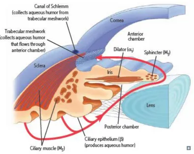
1.1.2 Aqueous Humor and Outflow
Elevated IOP is a well-known causative risk factor for both the development and
progression of glaucoma. Within the eye is a mechanism for the continuous
production and drainage of fluid. This fluid is called aqueous humor (AH). Intraocular
pressure is regulated by aqueous humor production in the ciliary body and drainage
from the eye occurs by way of the trabecular meshwork (TM) and uveoscleral
pathways.
Aqueous humour is secreted into the posterior chamber by the ciliary body,
specifically the non-pigmented epithelium of the ciliary body. It flows from the ciliary
processes into the posterior chamber, bounded posteriorly by the lens and the zonules
of Zinn, and anteriorly by the iris, to escape through the pupil into the anterior
chamber, and then to drain out of the eye via the trabecular meshwork. From here, it
drains into Schlemm's canal by one of two ways: directly, via aqueous vein to the
episcleral vein, or indirectly, through collector channels to the episcleral vein by
intrascleral plexus into the veins of the orbit andeventually general blood circulation.
Because of some resistance to the flow of aqueous through the trabeculum and
3
Figure 1.1.2 The trabecular meshwork outflow pathways.
1.1.3 Pathogenesis
Although the pathogenesis of glaucoma is not fully understood, the level of
intraocular pressure is related to retinal ganglion cell death. Intraocular pressure can
cause mechanical stress and strain on the posterior structures of the eye, notably the
lamina cribrosa and adjacent tissues. In addition, elevated intraocular pressure is not
the only risk factor for chronic glaucomatous neuropathy, In fact, the search for
putative non-IOP dependent factors continues to be of significant interest. Vasomotor
dysfunction has also been suggested to contribute to optic neuropathy, by mediating
4
1.2 Glaucoma treatment options
Slowing disease progression and preservation of quality of life are the main goals for
glaucoma treatment. Current management guidelines from the American Academy of
Ophthalmology Preferred Practice Patterns recommend lowering the intraocular
pressure toward a target level, which is a value or range of values at whichthe rate of
disease progression will be slowed sufficiently to avoid functional impairment from
the disease104. The target intraocular pressure should be achieved with the fewest
medications and/or surgeries, and with a minimum of adverse effects. When glaucoma
is no longer controlled by maximally tolerated medical therapy or laser
trabeculoplasty, incisional ab externo filtering surgery has been the traditional next
step in the therapeutic management.
1.2.1 Filtration surgery
Filtration surgery still plays a mainstream role in the treatment of glaucoma despite
the fact its development dates back to the 19th century. Trabeculectomy (TRAB) is
still the most popular surgical intervention in patients who affected by primary
glaucoma, and is the most commonly performed incisional surgical procedure to
lower intraocular pressure. The trabeculectomy was introduced almost 50 years ago
with very few modifications since that time. Trabeculectomy is generally
recommended for patients with glaucoma that continues to progress despite use of
medications and/or laser treatments. It consists of excision of a small portion of the
trabecular meshwork tissue to provide a drainage route for aqueous humor from
5
trabeculectomy can contribute to a number of adverse effects, such as: scaring,
bleeding, infection, malignant glaucoma. Therefore, a new class of glaucoma
procedures, termed microinvasive glaucoma surgery, has emerged, which aims to fill
the gap between conservative medical management and more invasive surgery.
1.2.2 Glaucoma drainage devices
Although the procedure itself has not changed much in the last 50 years, chemical
agents that modulate the fibroblastic response to the surgery have been more recently
introduced. These agents, known as 5-fluorouracil and mitomycin C have effects at
the cellular level in blunting the would healing response. In addition,
trabeculectomy is not the only choice of ab externo glaucoma surgery . These
procedures have been augmented, traditionally in more advanced cases, with macro-
glaucoma drainage devices and implantations. Glaucoma drainage devices are
designed to divert aqueous humor from the anterior chamber to an external reservoir,
where a fibrous capsule forms about 4-6 weeks after surgery and regulates flow.
Lowering IOP can be achieved by increasing aqueous humour drainage through this
artificial route. These devices have shown success in controlling intraocular pressure
(IOP) in eyes with previously failed trabeculectomy and in eyes with insufficient
conjunctiva because of scarring from prior surgical procedures or injuries. They also
have demonstrated success in complicated glaucomas, such as uveitic glaucoma,
neovascular glaucoma, and pediatric and developmental glaucomas, among others.
6
surgery. Currently, the glaucoma drainage devices are available in different materials.
These “macro” ab externo adjunctive devices provided a basis for the emergence
ofthe first generation of micro-incisional devices. The latter have been made of
various materials with one of the first being titanium.
1.3 Fibrosis
1.3.1 Anti-fibrotic agents
In general, filtering procedures such as trabeculectomy or placement of
subconjunctival drainage implants, aim at allowing aqueous humor to leave the
anterior chamber through a novel transscleral route towards the subconjunctival space.
Scarring is a major cause of increased intraocular pressures and surgical failure. In
order to prevent bleb scarring and obstruction of the fistula, the inhibition of cellular
proliferation and inflammation is both necessary and desirable after surgery for
glaucoma. Currently, various substances have been used for the modulation of wound
healing in filtering glaucoma surgery. In filtering glaucoma surgery, the most
frequently used anti-fibrotic agents are mitomycinC (MMC), and 5-fluorouracil
(5-FU). These anti-fibrotic agents improve the success rate for long-term intraocular
pressure (IOP) control and reduce the risk of bleb failure105,106. but their use is
associated with an increased complication rate including thin-walled blebs107,
hypotony108, wound leakage109 and infection110. Until now, no satisfactory methods
7
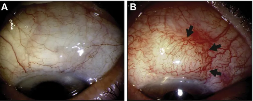
Figure 1.3.1 Clinical presentation of filtering blebs.
(A) Unscarred diffuse filtering bleb 12 months following trabeculectomy. (B) Scarring filtering bleb
with beginning encapsulation (arrows) and enhanced vascularity, 6 weeks following trabeculectomy. G.
Schlunck et al. / Experimental Eye Research 142 (2016) 76-82.
1.3.2 Permanent foreign body under tenos’s capsule stimulates fibrosis
Currently, permanent and inert metals like Titanium alloy, Polypropylene, and
Silicone are the most common materials used in adjunctive glaucoma surgical
devices. . However, it has been found that these materials can be associated with
long-term complications. One reason is that the presence of the permanent foreign
body induces inflammation stimulation under the tenon’s capsule. The initial process
of inflammatory is characterized by the increasing of monocytes and lymphocytes
with the early proliferation of connective tenon’s capsule tissue111. Thisis a main risk
factor cause fibroblast proliferation112-114 and fibrosis115,116.
Inflammatory responses of organisms against foreign body are a natural defense
8
permanent implantable devices117,118. The foreign body may be associated with
subconjunctival inflammation due to toxic and allergic responses. The inflammatory
phase is characterized by the activation of the innate immune system and the release
of inflammatory cytokines. Chronic inflammation is an established risk factor for
fibrosis due to the release of mediators such as interleukin-1(IL-1) and tumor
necrosis factor-alpha (TNF-α)which lead to increased tissue levels of TGF-β.
Chronic inflammatory reactions associated with the foreign body can accelerate
wound scarring, resulting in failure of the glaucoma surgery. It is for these reasons
that higher surgical success rates may occur if the inhibitory substances remain in
intimate contact with the tissue whose proliferation should be prevented, while
supporting the well-being of the tissues whose integrity is necessary to prevent
complications and promote healthy function. Ideally, the implantable material would
do its job in the acute phases of wound healing and then auto-degrade to limit the
potential for chronic responses to a foreign body.
Figure 1.3.2 Conjunctival scarring following filtering glaucoma surgery.
9
sutures, (2) blood, vessel-derived cells, cytokines and growth factors, (3) aqueous humor-derived
growth factors, (4) shear force stimulation by interstitial fluid flow, (5) signaling molecules released
from ECM storage sites, (6) myofibroblast transdifferentiation leading to matrix deposition and tissue
compaction. G. Schlunck et al. / Experimental Eye Research 142 (2016) 76-82
1.4 Magnesium
1.4.1 The important roles of Mg
2+Magnesium is an essential trace element for human life, and is one of the most
important regulatory cations involved in several biological processes. Magnesium is
the most common divalent cation in the intracellular fluid, and plays an important role
in regulating vascular functions and energy metabolism, as well as maintaining water
and electrolyte balance. Mg2+ is important for maintaining the structural and
functional integrity of several vital ocular tissues such as the cornea; lens and retina119.
Magnesium deficiency may contribute to the pathogenesis of many eye disorders,
including, cataract, conjunctival diseases, corneal diseases, choroidal diseases, and
retinal diseases. Magnesium is a nature’s calcium blocker120, increase blood flow to
tissues through endothelin-1 and endothelial nitric oxide pathways121. As well as,
magnesium has been shown neuroprotective role to glaucoma patients122,123 that
prevent oxidative stress and apoptosis124.
1.4.2 Magnesium alloys
10
biodegradable, non-toxic, high strength, light weight, and high biological
compatibility125. Due to the unique properties and satisfactory biocompatibility the
application of magnesium alloy as a bio-absorbable material in clinical practice can be
traced back to the early 1900s for treatment of bone fracture patients, which
demonstrated it was a non-toxic biomaterial21-23. Magnesium alloy also has been
considered as a potentially appropriate biomaterial for glaucoma drainage devices.
Various studies have found that magnesium alloys are biologically safe and have
demonstrated positive biological reactions in ocular tissue126. In comparison with
polymers, the degradation products of the alloys which includes magnesium ions are
needed in the human body for physiological functions, with consumption lying in the
range 250–500mg day. About 20g of Mg is always present in the average 70kg human
body127.
1.4.3 Coating techniques improve corrosive property
Magnesium as a pure metal has poor corrosion resistance, which has been a major
obstacle to their application as a human implantable device. If corrosion is rapid and
not homogenous, it limits its potential in vivo applications. Another issue is the
formation of hydrogen gas during corrosion: if evolution of the gas is too rapid it
cannot be absorbed and a balloon effect takes place. Fortunately, in recent years, the
most attention in the magnesium alloy field has been the study of coatings or surface
modification to slow the degradation rates of various Mg alloys. This has caused a
11
large number of possible coating technologies for Mg biomaterials, including
anodisation, metal–metal coatings, plasma spray, chemical vapour deposition, pulsed
laser deposition, ion beam assisted deposition, solution coatings, calcium phosphate
(CaP) deposition achieved by various means and the well known methods of
electrodeposition and conversion coating. The CaP coatings have garnered the
majority of attention due to their intrinsic biocompatibility128-130, due to the formation
is similar to the mineral phase of bone, including hydroxyapatite (HA), dicalcium
phosphate dehydrate (DCPD) and dicalcium phosphate dehydrate +Stearic acid
(DCPD+SA).
Nowadays, the advancement techniques have fabricated a new generation of
magnesium alloys, especially the development of coating technique which effectively
reduced the production of gas and rapid corrosion in the body. The coated magnesium
alloys have been used to develop novel cardiovascular stents resulting in a number of
clinic trials37-39. It is for these reasons that we were interested in exploring the
feasibility of coated magnesium devices as a potential surgical adjunct to glaucoma
surgery.
1.5 Hypothesis and Objectives
The preliminary data showed that coated magnesium alloy may reduce metabolic
activity and potentially inhibit human tenon’s capsule fibroblasts(HTCFs)cells
growth and proliferation. The results indicated that different coated magnesium alloys
12
associated with the capacity of blocking cell cycle.
1.5.1 Hypothesis
Bio-degradable coated magnesium alloys will inhibit cellular proliferation and reduce
myofibroblast activity in a primary culture of human Tenon’s capsule fibroblasts.
1.5.2
Objectives
The purpose of this study was to evaluate the biocompatibility and anti-proliferative
potential of different coated magnesium alloys on the activity of human Tenon’s
13
Chapter 2
Primary cell culture and sample preparations
2.1 Materials and methods
2.1.1 Primary HTCFs culture
The tenon’s capsule samples were obtained from 32 glaucoma patients without a past
history of ocular surgery. The study has been approved by the Research Ethics
Committee of St. Joseph’s Hospital, Western University, London, Canada. All the
patients provided informed consent, and the study was conducted in accordance with
the Declaration of Helsinki. The capsule samples were excised during the surgery, and
cultured in a 60 mm dish. The culture medium, consisting of Dulbecco’s modified
Eagle’s medium (DMEM, Gbico, USA) supplemented with 10% fetal bovine serum
(FBS, USA) and 1% penicilin/streptomycin (Gbico, USA), was changed every 2-3
days and the cells were allowed to reach 80% confluence. Subsequently, the cells
were disaggregated with 0.25% trypsin and 0.02% EDTA at 37oC for 5 min. And then,
the HTCFs cells were transferred to 25 cm2 flasks (BD Falcon, BD Biosciences,
Broendby, Denmark), which were placed at 37oC in 5% CO2 incubator for subculture.
The cells were seeded in a 24-well plate when they reached 80% confluence.
2.1.2 Assessment of morphology
The cells were analyzed 2 days after seeding. Cell counting was performed by
fluorescence microscopy (Axio Observer. Z1; Carl Zeiss, Germany) using Heochst
14
of incubation with the fluorescent dye, the cells were fixed with 4% buffered
formaldehyde in phosphate buffered saline (PBS) for 10 minutes, and then
permeabilized with 0.1% Triton-X 100 in PBS for 5 minutes. Thereafter, the samples
were rinsed with PBS, stained with a 1:100 dilution of monoclonal mouse
anti-vimentin and 1:500 monoclonal rabbit anti-keratin, and then left overnight at 9oC.
After a PBS rinse, the samples were incubated in a 1:625 dilution of anti-mouse Alexa
Fluor 488-conjugated goat anti-mouse IgG for 1 hour at room temperature. For double
labeling, the samples were also incubated in a 1:500 dilution of Alexa Fluor
568-conjugated donkey anti-rabbit IgG for 1 hour at room temperature. Finally, the
samples were rinsed twice with PBS and kept in PBS at 4oC until the observation
time.Image sampling, cellular morphology assessment and cell counting were
performed using custom-written routines for the AxioVision rel.4.7 software (Carl
Zeiss).
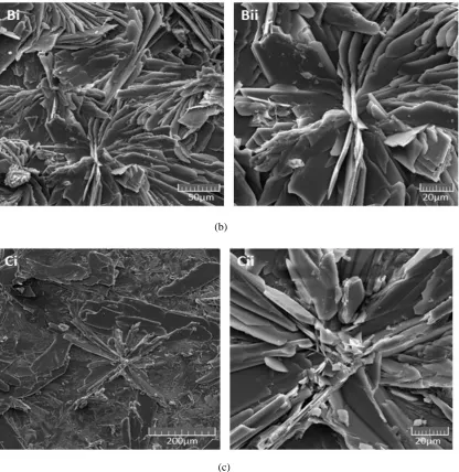
2.1.3 Fabrication of differently coated magnesium alloys
Magnesium alloys with a purity of 99.99% were obtained from the National
Engineering Research Centre for Magnesium Alloys, Chongqing University, China.
The pure magnesium was cut into disks with 14.5 mm in diameter and 1 mm in
thickness after pretreatment at 550oC for 24 hours of annealing. The surface of the
disks was polished using 320-800# abrasive paper. All of the disks were ultrasonically
cleaned in acetone, air dried and weighed. Thereafter, the magnesium disks were
15
(CaHPO4·2H2O, DCPD, Figure 2.1.3.B), and DCPD + stearic acid (DCPD + SA,
Figure 2.1.3.C), respectively. The HA coating was composed of 0.25 mol/L
ethylenediaminetetracetic acid calcium disodium salt hydrate (Ca-EDTA) and 0.25
mol/L potassium dihydrogenphosphate (KH2PO4), and the thickness of coating was
2um. The DCPD coating was composed of 3.1% sodium hydrogen phosphate
(Na2HPO4) and 5.3% calcium nitrate (Ca(NO3)2). The HA and DCPD coatings formed
with chemical bond of calcium phosphate and magnesium matrix. The thickness of
DCPD coating was 3um. The DCPD + SA coating was treated based on the
DCPD-coated magnesium disks with the addition of stearic acid.The combination of
SA depends on hydrogen bond and physisorption. The thickness of DCPD+SA
coating was 4um. Finally, all of the coated magnesium disks were ultrasonically
cleaned.
16
(b)
(c)
Figure 2.1.3 Scanning electron microscope (SEM) images of different magnesium coatings. (A) SEM morphology of HA; (B) SEM morphology of DCPD; (C) SEM morphology of DCPD +
SA.
2.1.4 Magnesium sample preparation
The coated magnesium samples were immersed in 70% ethanol for 10 minutes, and
rinsed twice with distilled water, and then dried under UV light. The samples were
introduced into a separate 24-well plate (BD Falcon, BD Biosciences, Broendby,
Denmark), with 5 x 10⁴ primary HTCF cells per well (Invitrogen Countess™). The
17
During the experiments, if a coated magnesium disk emitted hydrogen gas, the gas
would elevate the disk. We defined this phenomenon as corrosion, recorded it and did
not use such disks for our experiments.
Figue.2.1.4 Different sample groups for experiments.
2.1.5 Statistical analysis
The corrosion rate of differently coated magnesium alloys were compared using
chi-square test. The corrosion susceptibility of different coatings from day 2 to day 7
was compared using two-way analysis of variance (ANOVA). Statistical significance
was accepted at P < 0.05. All analyses were performed using SPSS 24.0 software.
2.2 Results
18
immunofluorescence staining.
The cells migrated out from the tissue after 7-10 days of adherent culture.
Microscopically, the cells exhibited a fusiform appearance with clear outline, and the
cellular plasma was abundant, bright, and uniform-sized. The cells were arranged in
fasciculus or swirling patterns. No differences were observed between the recovered
cells and those before cryopreservation in terms of morphology and growth
characteristics. The results revealed that the cells were positive for the expression of
vimentin, and the specific fluorescence could be observed within the cytoplasm
(Figure 2.2.1). The cells were negative for the expression of keratin. This supported
their identity as fibroblasts.
19
B
20
Figure 2.2.1 Immunostaining and characterization of primary human Tenon’s capsule
cultures. Cells were stained for nuclei (blue), vimentin (green), and keratin (red) under 40x
magnification. Image A shows a complete image while B, C, and D show isolated images for
nuclei, vimentin, and keratin stains respectively. Images displayed are representative of all images
of samples taken.
2.2.2 Corrosion of different coated magnesium alloys
A total of 453 coated magnesium alloy disks were used in our experiments: 399 for
21
for western blot analysis (Table 2.2.2). The phenomenon of corrosion was observed in
67 disks, accounting for 14.8%. The coating of DCPD and DCPD + SA had the same
corrosion proportion (both 15.9%), while the HA coating had the lowest corrosion
proportion (12.6%). The difference, however, was not statistically significant
(chi-square test, χ2 = 0.876, P = 0.645).
Table 2.2.2 Corrosion rate of different coated magnesium alloys (events).
DCPD DCPD+SA HA Total
Corrosion 24 24 19 67
No corrosion 127 127 132 386
Total 151 151 151 453
Proportion 15.9% 15.9% 12.6% 14.8%
The difference was not statistically significant (chi-square test, χ2 = 0.876, P = 0.645).
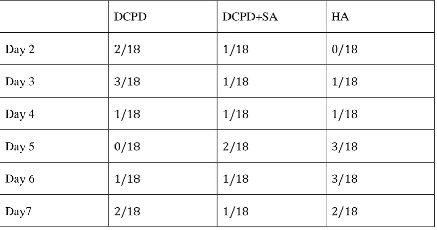
2.2.3 The corrosion susceptibility of different coatings from day 2 to day
7
A total of 324 samples were used for assessment of metabolic activity and
cytotoxicity from day 2 to day 7. As shown in Table 2.2.3, corrosion of the HA
coating was more frequently found during the last three days (8/10), while corrosion
of the DCPD coating was more commonly seen during the first three days (6/9). The
two-way ANOVA analysis revealed that the corrosion susceptibility of different
22
Table 2.2.3 The corrosion susceptibility of different coatings on different days (events/disks).
DCPD DCPD+SA HA
Day 2 2/18 1/18 0/18
Day 3 3/18 1/18 1/18
Day 4 1/18 1/18 1/18
Day 5 0/18 2/18 3/18
Day 6 1/18 1/18 3/18
Day7 2/18 1/18 2/18
The difference was not statistically significant (two-way ANOVA, PCoating=0.729, PDay=0.903).
2.3 Discussion
Coatings increased the corrosion resistance of magnesium alloys
The total corrosion rate was 14.8%, and the HA coating showed a relatively more
stable property compared with DCPD and DCPD + SA.
Mg alloys have unique properties, providing the mechanical benefits of a metal
combined with the degradable and biological advantages of biomaterials 131. However,
in spite of significant recent research, corrosion is a major challenge to successful
implementation of Mg-based materials in the body. In addition, the wear resistance of
Mg and Mg alloys is not very high. Therefore, a broad range of coating systems have
23
Recently, calcium phosphate-based coatings have attracted special interests for
biomedical application as bone substitutes and orthopaedic materials 134, including
DCPD, DCPD + SA, and HA. Particularly, the formation of a HA layer is similar to
the mineral phase of bone 135-137,and more smoothly than other two coatings. In our
experiments, the HA coating was more stable than DCPD or DCPD + SA.
Interestingly, in the MTT and LDH assays, where disks were immersed in the culture
medium for 7 days, most of the corrosion events of the HA-coated magnesium alloys
occurred during the last three days, accounting for 80%. Therefore, we conclude that
the HA coating has better corrosion resistance at the beginning of the cellular
24
Chapter 3
Corrosive properties of coated magnesium alloys in simulated
aqueous humor
3.1 Background
Due to their excellent bio-compatibility, magnesium alloys are being considered as
promising implant materials. However, the poor corrosion resistance becomes a major
obstacle to their widespread applications. Currently, the focus of bio-magnesium
studies is on coatings or surface modification to slow the degradation rates of Mg
alloys. If coated magnesium alloys are going to be used a biomaterial candidate for
glaucoma drainage device, not only gradual degradation is needed to control the
overall corrosion process, but also pH maintenance and control of ions released into
the anterior chamber are also required to maintain a non-toxic concentration of each
ions. These conditions very rapidly alter as a result of the corrosion process, with
rapid changes in pH levels and metal ion concentrations, as well as presence of
soluble corrosion products and hydrogen gas evolution. Thus, we need a complicated
solution that considers the corrosion resistance, metal ion concentrations, pH and
mechanical performance of coated magnesium alloy implants when immersed in the
aqueous humor. It is essential to fully understand the long term corrosion engineering
and potential biological interactions. Thus we invited our collaborators (National
Engineering Research Center for Magnesium Alloys, Chongqing University, China)
using balanced salt solution to test the variation of pH, sample’s weight, and ion