1556-6811/08/$08.00⫹0 doi:10.1128/CVI.00154-08
Copyright © 2008, American Society for Microbiology. All Rights Reserved.
High-Level Antigen Expression and Sustained Antigen Presentation in
Dendritic Cells Nucleofected with Wild-Type Viral mRNA
but Not DNA
䌤
Nada M. Melhem,
1Sherrianne M. Gleason,
1Xiang Dong Liu,
1and Simon M. Barratt-Boyes
1,2*
Center for Vaccine Research and Department of Infectious Diseases and Microbiology1and Department of Immunology,
University of Pittsburgh, Pittsburgh, Pennsylvania 152612
Received 30 April 2008/Returned for modification 3 July 2008/Accepted 21 July 2008
Dendritic cells (DC) are potent antigen-presenting cells that hold promise as cell-based therapeutic vaccines for infectious diseases and cancer. Ideally, DC would be engineered to express autologous viral or tumor antigens to ensure the presentation of relevant antigens to host T cells in vivo; however, expression of wild-type viral genes in primary cell lines can be problematic. Nucleofection is an effective means of delivering transgenes to primary cell lines, but its use in transfecting DNA or mRNA into DC has not been widely investigated. We show that nucleofection is a superior means of transfecting human and monkey monocyte-derived DC with DNA and mRNA compared to lipofection and conventional electroporation. However, the delivery of DNA and mRNA had significantly different outcomes in transfected DC. DC nucleofected with DNA encoding green fluorescent protein (GFP) had poor antigen expression and viability and were refractory to maturation with CD40 ligand. In contrast, >90% of DC expressed uniform and high levels of GFP from 3 h to 96 h postfection with mRNA while maintaining a normal maturation response to CD40 ligation. Monkey DC nucleo-fected with wild-type, non-codon-optimized mRNA encoding simian immunodeficiency virus Gag stimulated robust antigen-specific effector T-cell responses at 24 h and 48 h postnucleofection, reflecting sustained antigen presentation in transfected DC, whereas no detectable T-cell response was noted when DC were nucleofected with DNA encoding the same Gag sequence. These data indicate that mRNA nucleofection may be an optimal means of transfecting DC with autologous tumor or viral antigen for DC-based immunotherapy.
Monocyte-derived dendritic cells (DC) are currently being used as therapeutic vaccines for cancer and infectious diseases, and approaches to express tumor-associated or viral antigens in DC are being widely sought (3, 16, 32, 38, 39). Viral vectors based on adenovirus, poxviruses, and lentivirus induce high-level antigen expression in DC but may be associated with safety concerns and generally require DNA codon optimiza-tion to overcome poor gene expression (11, 12, 14, 23, 35, 49). An attractive alternative to vector-mediated delivery of anti-gens into DC is nonviral gene transfer based on DNA or mRNA. DNA transfection of DC has been used successfully in some studies but is limited by poor expression levels and tox-icity (2, 29, 41, 45, 49), although methods to enhance expres-sion based on DC maturation have been proposed (28). mRNA transfection of DC is being increasingly utilized for cancer immunotherapy and in vitro stimulation of virus-spe-cific T cells, but approaches to deliver mRNA into primary DC cultures have been limited (16, 18, 25, 39, 40, 43, 48, 50). Transfection of DC with mRNA offers the potential for high-level expression of wild-type viral transgenes that have poor expression in mammalian cells when traditional methods of gene delivery are used (8, 26). This is advantageous since expression of autologous viral or tumor genes in a patient-specific, DC-based vaccine would ensure stimulation of rele-vant T-cell responses in vivo (42, 48).
Recently, nucleofection has emerged as a superior method for the delivery of transgene to primary cell lines, including cytokine-induced killer cells, neurons, keratinocytes, macro-phages, and DC (9, 15, 21, 27, 29, 33, 37, 47). Nucleofection has been used primarily as a method of introducing DNA into cells as it is reported to deliver DNA directly into the nucleus, enhancing gene expression (17). However, recent studies sug-gest that nucleofection of DC with mRNA is an effective means of inducing high-level antigen expression (27, 33). While conventional methods of gene delivery have been com-pared using monocyte-derived DC (41, 51), a limited number of studies have employed the more-efficient process of nucleo-fection to evaluate DNA and mRNA delivery into these cells (27), and none to our knowledge have compared the capacity for mRNA- and DNA-nucleofected DC to stimulate antigen-specific T-cell responses.
In the present study, we did a comprehensive analysis of DNA and mRNA transfection of human and monkey mono-cyte-derived DC, comparing liposomal transfection methods and conventional electroporation with nucleofection. We con-firm that nucleofection is a superior method for the delivery of both DNA and mRNA into primary DC lines, although the outcome of nucleofection differed substantially depending on the source of genetic material. DC nucleofected with mRNA encoding the marker gene green fluorescent protein (GFP) had rapid and sustained antigen expression with limited toxic-ity and were responsive to maturation stimuli, whereas DC nucleofected with GFP DNA had limited antigen expression and poor viability and were refractory to maturation with CD40 ligand (CD40L). Notably, monkey DC nucleofected with
* Corresponding author. Mailing address: University of Pittsburgh, 3501 Fifth Avenue, Pittsburgh, PA 15261. Phone: (412) 383-7537. Fax: (412) 624-4440. E-mail: [email protected].
䌤Published ahead of print on 30 July 2008.
1337
on August 17, 2020 by guest
http://cvi.asm.org/
wild-type simian immunodeficiency virus (SIV) gag mRNA induced a robust virus-specific T-cell response in cells isolated from immune monkeys, whereas DC nucleofected with viral DNA were unable to stimulate detectable T-cell responses.
MATERIALS AND METHODS
Plasmid and mRNA generation. Generation of the pSP73/GFP/A64 and pSP73/SIVmac239Gag/A64 plasmids and in vitro transcription of mRNA were performed as described previously (33). pSP73/SIVmac239Gag/A64 encodes a wild-type, non-codon-optimized Gag sequence isolated from a rhesus macaque 2 weeks postinfection with the SIVmac239 SIV isolate (33). pSP73/SIVmac239Gag/ A64 was used as a template for the amplification of Gag by PCR using the following forward and reverse primers, respectively: pGagN1F, 5⬘GCGCTCGAGGCCAC CATGGGCGTGAG3⬘; and pGagN1R, 5⬘CGCGCGGCCGCTTACTTGCCCA ACTGCATGTAG 3⬘. pEGFP-N1 DNA was digested with XhoI and NotI, and the larger vector band was retrieved by gel purification. XhoI- and NotI-digestedgag
PCR product was inserted to the XhoI- and NotI-digested pEGFP-N1 DNA to generate pSIVmac239Gag-N1 encoding the wild-type sequence.
Cells. DC were cultured from the purified blood monocytes of SIV-naı¨ve rhesus macaques or healthy human volunteers as described previously (6). In some experiments, DC were matured for 24 h or 48 h with 3g/ml recombinant trimeric CD40L (Immunex, Seattle, WA) as described previously (6). Approval was obtained from the institutional review board prior to experiments involving human samples and from the institutional animal use and care committee for all experiments involving rhesus macaque samples. K562 cells were grown in Iscove’s modified Dulbecco’s medium (HyClone, Logan, UT) supplemented with 10% fetal bovine serum.
Transfection of K562 cells and DC.K562 cells were washed twice with Opti-MEM (Gibco Invitrogen Corporation, Frederick, MD) and transfected with TransFast transfection reagent (Promega, Madison, WI) or TransMessenger reagent (Qiagen, Valencia, CA) by adding 2 or 4g GFP mRNA or DNA in Opti-MEM with other reagents provided by the manufacturers to 1⫻106cells as described previously (33). For electroporation, 1⫻106
K562 cells were electroporated with 10g mRNA or DNA in a total volume of 250l in a 0.4-cm cuvette using the Gene Pulser II electroporation system (Bio-Rad, Hercules, CA) with voltage/capacitance settings of 260 V/150F or 300 V/150F, respec-tively (50). Immediately after electroporation, cells were incubated for 5 min on ice. For nucleofection, 10g of DNA or mRNA was added to 1⫻106K562 cells in Opti-MEM in a final volume of 100l and transfected in a 2-mm-wide electroporation cuvette (BTX, San Diego, CA) using the T-16 program of the amaxa nucleofector (Amaxa, Koln, Germany). Smaller amounts of mRNA and DNA were used for lipofection than for electroporation and nucleofection as toxicity was noted at higher amounts with the former methods (data not shown). K562 cells were cultured in Iscove’s modified Dulbecco’s medium for 24 h at 37°C following transfection. DC were transfected as immature cells (day 5 of culture) or as mature cells after a 24-h incubation with CD40L (day 6 of culture). Lipofection using TransFast or TransMessenger reagents was done as for K562 cells. For electroporation, 1⫻106to 2⫻106DC in Opti-MEM were mixed with 5g to 20g mRNA or DNA, depending on the experiment, and electroporated as for K562 cells using voltage/capacitance settings of 300 V/150F and 250 V/300F, respectively, as described previously (33, 41). Nucleofection of DC was done as described for K562 cells using 5g to 20g mRNA or DNA and the U-02 or T-01 Amaxa program. Details relating to the electrical parameters of electroporation using the Amaxa nucleofector are proprietary. Nucleofection was performed according to the manufacturer’s instructions, with some modifi-cations. In preliminary studies, we determined that nucleofection of cells in Opti-MEM and buffers provided by the manufacturer resulted in similar effi-ciencies of transfection (data not shown), and we therefore used Opti-MEM for all transfection experiments. Following transfection, DC were cultured in pre-warmed complete RPMI medium supplemented with granulocyte-macrophage colony-stimulating factor and interleukin-4 for 24 h or 48 h at 37°C, with and without CD40L, as described previously (33).
Confocal microscopy.DC nucleofected with pEGFP-N1 DNA orgfpmRNA 24 h previously were harvested using 20 mM EDTA and resuspended in phos-phate-buffered saline prior to being settled onto glass slides at 37°C for 1 h. Adhered cells were fixed with 2% paraformaldehyde for 15 min and washed in phosphate-buffered saline. Gelvatol was used to apply coverslips to slides. DC were imaged for GFP and differential interference contrast using an Olympus FluoView 500 laser scanning confocal microscope (Olympus, Center Valley, PA). Images were collected using MetaMorph software (Molecular Devices, Sunnyvale, CA).
Flow cytometric analysis.GFP expression in transfected K562 cells and DC and expression of HLA-DR, CD80, CD83, CD86, and CD40 on transfected DC were done as described previously (33).
ELISPOT assay.Gag-specific gamma interferon (IFN-␥) enzyme-linked im-munospot (ELISPOT) assays were done as described previously (33). Briefly, immature monkey DC were nucleofected with 10g pSIVmac239Gag-N1 DNA or withgagmRNA transcribed from pSP73/SIVmac239Gag/A64 and simulta-neously matured with CD40L (3g/ml) for 24 h or 48 h prior to incubation with autologous peripheral blood mononuclear cells (PBMC) at a 1:10 ratio. Control cells were nucleofected with pEGFP-N1 DNA orgfpmRNA transcribed from pSP73/GFP/A64 or mock transfected. IFN-␥spot-forming cells were developed and enumerated as described previously (4).
RESULTS
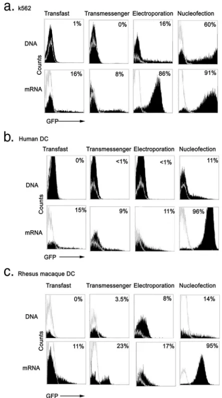
Nucleofection with mRNA is a superior method of introduc-ing transgene into primary human and monkey monocyte-derived DC. We performed a side-by-side comparison of TransFast and TransMessenger lipofection methods along with electroporation and nucleofection using a GFP reporter construct. Cells were transfected with either pEGFP-N1 DNA or in vitro-transcribedgfpmRNA that was generated from the plasmid pSP73/GFP/A64 (33) and monitored for GFP expres-sion at 24 h by flow cytometry. For lipofection methods, we used 4 g DNA or mRNA as toxicity was noted at higher amounts (not shown), whereas for electroporation and nucleo-fection we used 10g DNA or mRNA. In initial experiments, we used the chronic myelogenous leukemic cell line K562 that is readily transfected with plasmid DNA or mRNA (7, 50). Transfection of K562 cells with EGFP-N1 DNA using the two lipofection methods produced no detectable GFP fluores-cence, whereas transfection via electroporation resulted in 16% GFP-expressing cells at 24 h. In contrast, 60% of K562 cells expressed GFP following nucleofection with EGFP-N1 DNA using the T-16 program of the nucleofector device, which was shown in preliminary experiments to generate the highest efficiency of transfection with this cell line (Fig. 1a and data not shown). Transfection with mRNA was significantly more ef-fective than transfection with DNA by all methods, with 8% to 16% GFP expression following lipofection and 86% and 91% of K562 cells expressing GFP following electroporation and nucleofection, respectively (Fig. 1a).
We next tested the capacities of lipofection, electroporation, and nucleofection to transfect human and monkey immature monocyte-derived DC. In addition to measuring GFP expres-sion at 24 h posttransfection, we assessed the effect of trans-fection on DC viability by using the standard approach of trypan blue exclusion. Transfection of DC with DNA using lipofection or electroporation produced negligible GFP ex-pression and significant cell death, with viability ranging from 40% to 57% (Fig. 1b and c) (Table 1). Nucleofection with DNA using the U-02 program was more effective at generating GFP-expressing DC than the other methods but was associated with enhanced cell death, with only 24% of DC remaining viable at 24 h posttransfection (Fig. 1b and c) (Table 1), similar to other reports (29). Switching the nucleofector program to T-01 as favored by others (29) did not enhance transfection efficiency, and increasing the quantity of DNA to 20g was associated with a significant increase in toxicity (data not shown). Lipofection with mRNA resulted in variable transfec-tion of DC, with maximum expression noted in monkey DC following TransMessenger lipofection, although the
on August 17, 2020 by guest
http://cvi.asm.org/
tion of viable cells was only 52% to 55% (Fig. 1b and c) (Table 1). In contrast to K562 cells, electroporation of DC with mRNA was inefficient, with expression levels not exceeding 17% and viability averaging only 46% (Fig. 1b and c) (Table 1). However, nucleofection with mRNA using the U-02 program was a highly effective means of transfecting human and mon-key monocyte-derived DC, with the proportion of DC express-ing GFP reachexpress-ing 96% at 24 h posttransfection (Fig. 1b and c) (Table 1). Nucleofection with mRNA maintained the highest cell viability of all methods, although the proportion of viable cells at 24 h posttransfection was still only 68% (Table 1), consistent with results of other reports (27). Similar
transfec-tion efficiency and DC viability were noted when nucleofectransfec-tion was performed with a range of mRNA from 5g to 20g (data not shown).
Flow cytometric analysis of nucleofected cells indicated that GFP expression varied in intensity and uniformity depending on whether DNA or mRNA was used, with DNA producing a wide range of fluorescence and mRNA generating uniform expression in almost all cells (Fig. 1b and c). To evaluate this in more detail, we examined immature monkey DC by confocal microscopy 24 h after DNA and mRNA nucleofection. The majority of mRNA-transfected DC showed a similar intensity of GFP expression when examined individually by microscopy, whereas GFP expression in DNA-transfected DC was highly variable, with some cells having intense fluorescence and a majority having weak or undetectable fluorescence (Fig. 2). Expression of GFP in DNA- or mRNA-transfected DC was cytoplasmic in distribution, as expected (Fig. 2). Similar results were found using human monocyte-derived DC (data not
FIG. 1. Evaluation of DNA and mRNA transfection of K562 cells and human and monkey DC. K562 cells (a) or immature human (b) or rhesus macaque (c) monocyte-derived DC were transfected with pEGFP-N1 DNA (top row) (filled histograms) orgfpmRNA (bottom row) (filled histograms) or were mock transfected (empty histograms) using the methods indicated and analyzed by flow cytometry for GFP expression 24 h later. A total of 4g DNA or mRNA was used for lipofection with TransFast and TransMessenger reagents, whereas 10
g DNA or mRNA was used for electroporation and nucleofection. Numbers represent the percentage of cells expressing GFP based on mock transfection.
TABLE 1. Gene expression and cell viability following transfection of immature monkey DC
Method
DNAa
RNAb
GFP
expressionc Viabilityd
GFP
expression Viability
TransFaste 0 57 (3) 7 (3) 52 (4)
TransMessengere 2 (1) 49 (1) 14 (8) 55 (4)
Electroporationf 4 (7) 40 (2) 9 (7) 46 (3)
Nucleofectiong 10 (4) 24 (5) 91 (3) 68 (6)
aEGFP-N1 DNA.
bIn vitro-transcribedgfpmRNA.
cPercentage of cells expressing GFP at 24 h posttransfection. Mean (SEM) of
two to five experiments.
dPercentage of viable cells at 24 h posttransfection. Mean (SEM) of two to five
experiments.
eA total of 4g DNA or mRNA was used with both lipofection methods. fCells were electroporated with 10g DNA or mRNA using 250 V/300F or
300 V/150F settings, respectively.
gCells were nucleofected with 10g DNA or mRNA using the U-02 program.
FIG. 2. Expression of GFP in mRNA- and DNA-nucleofected DC. Immature monkey monocyte-derived DC were nucleofected withgfp
mRNA (a and c) or pEGFP-N1 DNA (b and d) and examined by confocal microscopy 24 h later. Shown are GFP expression (left) and differential interference contrast (right). (a and b) Original magnifica-tion,⫻200. (c and d) Original magnification,⫻1,500.
on August 17, 2020 by guest
http://cvi.asm.org/
shown). Taken together, these data indicate that nucleofection with mRNA is a superior method of introducing transgenes into DC, both with respect to antigen expression and to cell viability.
mRNA nucleofection of DC produces rapid and sustained expression of transgene. A potential limitation of mRNA transfection of DC for immunotherapy is that mRNA is labile in cells and may be degraded rapidly, resulting in limited du-ration of antigen expression. We therefore assessed the dura-bility of transgene expression in immature monkey DC by harvesting cells at various intervals after DNA or mRNA trans-fection and determining the proportion of cells expressing GFP by flow cytometry. For these and all other experiments, we focused on nucleofection as the preferred method of trans-fection. GFP expression following nucleofection of DC with pEGFP-N1 DNA was detectable at 3 h posttransfection and maintained relatively constant levels of expression from 6 h to 48 h posttransfection, after which the proportion of GFP-expressing cells markedly declined (Fig. 3), similar to other reports (29, 45). In contrast, the proportion of monkey DC expressing GFP following nucleofection with in vitro-tran-scribed gfp mRNA reached near-maximal levels by 3 h and stayed at this high level for 96 h, the duration of the experiment (Fig. 3). Similar rapid and sustained kinetics of transgene ex-pression were noted in other studies following mRNA delivery to human DC using either electroporation or nucleofection (27, 31, 33, 46, 50). These findings indicate that the delivery of mRNA rather than DNA generates a more-durable expression of transgene in monocyte-derived DC.
Relationship between DNA and mRNA nucleofection and DC maturation.Immature DC are specialized in antigen up-take, and as such, transfection of DC with either DNA or mRNA is traditionally done at the immature stage of differ-entiation. However, electroporation of mature human DC with mRNA is reported to be efficient (22, 31), and in the murine system, DNA transfection may be enhanced in mature DC (28). To determine the influence of DC maturation on trans-fection efficiency, we nucleofected human monocyte-derived DC with DNA and mRNA encodinggfpwith and without prior treatment of cells with CD40L for 24 h to induce maturation. Maturation was confirmed by phenotypic analysis using flow cytometry (data not shown). Prior DC maturation did not enhance GFP expression, as nucleofection of DC with DNA or mRNA generated similar levels of transgene expression
re-gardless of the maturation state at the time of gene delivery (Fig. 4a).
We next evaluated the responsiveness of DNA- and mRNA-nucleofected DC to maturation. Human immature DC were nucleofected with pEGFP-N1 DNA or gfp mRNA or were mock nucleofected and either left untreated or treated imme-diately with CD40L. Cells were harvested and their phenotype was analyzed 24 h postnucleofection by flow cytometry. To ensure that only the phenotype of transgene-expressing cells was analyzed, cells were gated based on GFP expression. Mock-nucleofected immature DC expressed relatively high levels of HLA-DR, CD86, and CD40 and lacked expression of CD80 and CD83, as expected (Fig. 4b and c) (6). Nucleofec-tion of immature DC with pEGFP-N1 DNA did not induce maturation and in fact resulted in a modest downregulation in CD40 expression compared to that of mock-nucleofected cells (Fig. 4b). Moreover, immature DC nucleofected with DNA were refractory to CD40L, as GFP-expressing DC had negli-gible increases in CD83, CD86, and CD80 expression following CD40 ligation compared to those of mock-nucleofected con-trols (Fig. 4b). In contrast, immature DC nucleofected withgfp mRNA had a minor shift in expression of CD83 and CD80 compared to that of mock-nucleofected DC and responded fully to subsequent CD40 ligation, with increases in CD83, CD86, and CD80 expression, similar to those of mock-nucleo-fected cells (Fig. 4c). These data indicate that mRNA-nucleo-fected DC are responsive, whereas DNA-nucleomRNA-nucleo-fected DC are refractory to CD40L-mediated maturation.
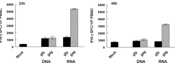
mRNA- but not DNA-nucleofected DC stimulate robust vi-rus-specific T-cell responses when expressing wild-type virus genes.A key issue in DC transfection for immunotherapy ap-plications is the capacity to stimulate T-cell responses to the specific transgene being introduced. This has been evaluated in the past using lipofection and electroporation approaches to deliver DNA or mRNA into monocyte-derived DC (41, 51); however, the ability to stimulate T-cell responses following DNA and mRNA transfection of DC via nucleofection, which we have shown is a superior method of transfection, has not been compared. To evaluate this, we cultured monocyte-de-rived DC from a rhesus macaque that had a robust CD8⫹ T-cell response to SIV Gag through vaccination (33). Imma-ture DC were nucleofected with pGag-N1 DNA encoding a SIVmac239gag sequence isolated from an SIV-infected ani-mal, or the correspondinggagmRNA (33), and immediately matured with CD40L for various intervals to induce matura-tion. To ensure applicability to immunotherapeutic vaccine strategies designed to stimulate T-cell responses to autologous viral sequences (48), we used mRNA and DNA encoding wild-type, non-codon-optimizedgagsequences. The transfected DC were then cultured with autologous PBMC at a 1:10 ratio in a short-term IFN-␥ELISPOT assay to determine their capacity to stimulate Gag-specific effector T cells (33). DC that had been nucleofected withgagmRNA 24 h earlier induced robust Gag-specific T-cell responses with a frequency of greater than 5,000 IFN-␥ spot-forming cells per million PBMC (Fig. 5). Importantly, DC that had been nucleofected withgag- express-ing mRNA 48 h previously induced reduced but still strong T-cell responses, supporting the notion that antigen expressed following the mRNA delivery of transgene is processed and presented in DC for extended periods. In contrast to DC
FIG. 3. High-level and durable GFP expression in monkey DC nucleofected withgfpmRNA but not with pEGFP-N1 DNA. Imma-ture monkey monocyte-derived DC were nucleofected with 10ggfp
mRNA or pEGFP-N1 DNA, and GFP expression was determined at various intervals posttransfection by flow cytometry.
on August 17, 2020 by guest
http://cvi.asm.org/
nucleofected with mRNA, DC nucleofected with DNA encod-ing wild-typegaggenerated no detectable responses above the nonspecific responses generated by DC nucleofected with p-EGFP-N1 DNA at either 24 h or 48 h postnucleofection (Fig. 5). These data indicate that DC nucleofected with wild-type viral mRNA have a durable presentation of antigen to T cells and stimulate robust virus-specific T-cell responses, whereas DC nucleofected with a comparable DNA construct are ineffective.
DISCUSSION
Non-viral-based approaches for delivering genes to DC for cancer immunotherapy have distinct advantages over recom-binant virus-based strategies, in particular because DNA or RNA can be amplified directly from a tumor and serve as a
source of polyvalent patient-specific antigens (2, 19, 42). This strategy is being employed in the design of therapeutic DC-based vaccines for human immunodeficiency virus (HIV)-in-fected individuals as well, as viral mRNA expressing patient-derived sequences or sequences from SIV-infected monkeys can readily be introduced into monocyte-derived DC for stim-ulation of autologous T cells (33, 48). Our studies confirm that nucleofection is superior to conventional electroporation and lipofection for introducing DNA and mRNA into human and monkey monocyte-derived DC, although nucleofection offered no advantage over electroporation when mRNA was intro-duced into the K562 cell line. However, mRNA was a signifi-cantly better source of genetic material than DNA for nucleo-fection of DC, producing higher-level, more-uniform, and
FIG. 4. Differential responsiveness of human DC nucleofected withgfpmRNA and pEGFP-N1 DNA to CD40L. (a) Human monocyte-derived DC were nucleofected with 10g pEGFP-N1 DNA or 10ggfpmRNA (solid line) or mock nucleofected (dotted line) with and without prior treatment of cells with 3g/ml CD40L as indicated for 24 h, and GFP expression was determined by flow cytometry. Numbers represent the percentage of cells expressing GFP based on mock transfection. (b and c) Immature human DC were nucleofected with DNA (b) (solid histograms) or mRNA (c) (solid histograms) or mock transfected (open histograms) and cultured with or without 3g/ml CD40L for 24 h as indicated. Cells were labeled with phycoerythrin-conjugated antibodies as indicated and analyzed by flow cytometry. Of the cells nucleofected with DNA or mRNA, only GFP-expressing DC are shown based on gating.
on August 17, 2020 by guest
http://cvi.asm.org/
modurable antigen expression with limited toxicity. The re-sults we observed following transfection of DC withgfpmRNA using the standard methods of electroporation and lipofection were consistent with those of previous reports (41). Moreover, our results are similar to those observed by others following electroporation of DC with DNA encoding GFP (27, 41). However, a number of studies reported electroporation to produce at least 50% GFP-positive DC following mRNA transfection (25, 27, 39, 40, 50). This discrepancy between our results and those of others using electroporation might be explained by the use of different electroporation devices lead-ing to differences in the delivery of electric current to cells.
In our studies, the efficiency of nucleofection was such that as little as 5g mRNA resulted in near-total transfection of DC, yet at least 50g mRNA can be safely delivered to DC without apparent toxicity (22). This provides the advantage of being able to introduce relatively small amounts of mRNA encoding multiple genes into DC simultaneously, such as dif-ferent patient-derived viral genes for immunotherapy of HIV infection. Alternatively, mRNA encoding individual virus or tumor antigens could be introduced together with genes de-signed to enhance DC function and Th1-stimulating capacity, such as OX40 ligand (10) or interleukin-12 p70 (24), or to prolong DC survival, such as the gene encoding the antiapop-totic protein Bcl-xl (20). In contrast, increasing the amount of DNA had a negative impact on DC viability (data not shown), limiting the number of DNA species that could potentially be expressed. The toxicity of DNA is most pronounced when delivered to DC via nucleofection as we and others have shown (29), presumably because greater quantities of DNA enter cells using this method.
Prior or concurrent maturation of human monocyte-derived DC with CD40L did not alter nucleofection efficiency with either DNA or mRNA in our studies, similar to results in previous reports (29, 34), although in other studies, electropo-ration of mRNA into mature DC led to a modest increase in transgene expression (22). Larregina et al. reported that simul-taneous transfection and CD40-mediated maturation of mu-rine bone marrow-derived DC significantly enhanced DNA transfection efficiency (28), suggesting that maturation may have a greater effect on gene expression in the murine system. In our studies, human DC nucleofected with DNA were
re-fractory to maturation with CD40L, as has previously been shown using lipopolysaccharide (29), which is presumably as-sociated with DNA toxicity. In contrast, DC nucleofected with mRNA were fully responsive to CD40 ligation, similar to re-sults of other reports (29, 33). These studies suggest that the mRNA-based delivery of genes will retain DC viability and responsiveness to maturation stimuli that are critical for T-cell stimulatory capacity, whereas the delivery of antigen via DNA could potentially be deleterious.
A critical factor in gene transfection of DC for immunother-apy is the capacity for the sustained presentation of antigen to tumor- or virus-specific T cells. We found that DC nucleo-fected with non-codon-optimized SIVgagmRNA up to 48 h earlier were capable of stimulating robust antigen-specific ef-fector T-cell responses in PBMC isolated from a vaccinated macaque, equivalent to levels previously shown using peptide-pulsed DC (33), although responses were somewhat dimin-ished from those induced 24 h following nucleofection. In contrast, DC nucleofected with DNA encoding non-codon-optimized Gag were unable to induce detectable Gag-specific T-cell responses even at 24 h posttransfection. Similarly, B lymphoblastoid cells transfected with mRNA encoding HIV nefprotein were capable of stimulating Nef-specific T cells for at least 72 h posttransfection (25), and mature DC transfected with mRNA encoding the MART-1 tumor-associated antigen remained effective targets for MART-1-specific cytotoxic T cells for at least 96 h postelectroporation (31). Other compar-ative studies have shown that human DC expressing mRNA-encoded influenza virus matrix protein by electroporation were far superior in their capacity to stimulate M1-specific cytotoxic T cells than DC expressing DNA-encoded antigen (41). The durable antigen presentation in mRNA-nucleofected DC is likely to be a function of sustained protein expression and presentation rather than the persistence of intracellular mRNA, as introduced mRNA is rapidly degraded in cells (13, 22).
It is notable that we used a wild-type sequence of SIVgagfor DC transfection rather than a codon-optimized sequence to reflect the conditions of DC-based immunotherapy using viral antigens derived from autologous virus sequences (33, 48). SIV and HIVgag mRNA are known to have multiple inhibitory/ instability elements that substantially limit viral protein
expres-FIG. 5. Monkey DC nucleofected with non-codon-optimized mRNA but not with DNA encoding SIVgagstimulate potent Gag-specific effector T-cell responses. Immature DC propagated from an SIV Gag-vaccinated monkey were mock nucleofected (Mock) or nucleofected with DNA or mRNA encoding GFP or SIVmac239 Gag as indicated and matured with CD40L for 24 h (left) or 48 h (right) prior to incubation with autologous PBMC in an IFN-␥ELISPOT assay. IFN-␥spot-forming cells (SFC) were measured 24 h later. Data shown are means⫾standard errors of the means (SEM) of triplicate determinations.
on August 17, 2020 by guest
http://cvi.asm.org/
sion (1), yet Gag protein was readily detected in DC 24 h post-mRNA nucleofection when monkey-derived viral se-quences were used (33). We do not have data on the long-term expression of Gag within nucleofected DC, and it is possible that expression does not persist as long as that of GFP, which is known to be highly stable (30). Nevertheless, the relative stability of antigen presentation should allow mRNA-trans-fected DC to traffic to lymph nodes and engage antigen-spe-cific T cells following intradermal or subcutaneous delivery to patients (5, 36, 44). It is likely that the use of codon-optimized as opposed to wild-type DNA sequences would allow for suf-ficient antigen expression in DC for detectable T-cell re-sponses to be elicited (28).
In summary, these data indicate that nucleofection of pri-mary DC cultures with wild-type mRNA is an effective and nonperturbing means of delivering antigen for DC-based im-munotherapy of cancer or infectious diseases, providing uni-form and sustained antigen expression and presentation for stimulation of antigen-specific T cells. In contrast, while nucleofection is a more-effective means of introducing DNA into DC than other methods, the resulting low viability, refrac-toriness to maturation, and poor T-cell-stimulating capacity of transfected DC make this approach unfavorable for autolo-gous DC-based vaccination.
ACKNOWLEDGMENTS
This work was supported by NIH grant no. AI52052 to S. M. Barratt-Boyes.
We thank Nicole Banichar for assistance with animal procedures and M. Murphey-Corb, A. Gambotto, P. Kalinski, and C. Rinaldo for helpful suggestions.
REFERENCES
1.Afonina, E., M. Neumann, and G. N. Pavlakis.1997. Preferential binding of poly(A)-binding protein 1 to an inhibitory RNA element in the human immunodeficiency virus type 1 gag mRNA. J. Biol. Chem.272:2307–2311. 2.Artusio, E., B. Hathaway, J. Stanson, and T. L. Whiteside.2006.
Transfec-tion of human monocyte-derived dendritic cells with native tumor DNA induces antigen-specific T-cell responses in vitro. Cancer Biol. Ther.5:1624– 1631.
3.Banchereau, J., and A. K. Palucka. 2005. Dendritic cells as therapeutic vaccines against cancer. Nat. Rev. Immunol.5:296–306.
4.Barratt-Boyes, S. M., A. C. Soloff, W. Gao, E. Nwanegbo, X. Liu, P. A. Rajakumar, K. N. Brown, P. D. Robbins, M. Murphey-Corb, R. D. Day, and A. Gambotto.2006. Broad cellular immunity with robust memory responses to simian immunodeficiency virus following serial vaccination with adenovi-rus 5- and 35-based vectors. J. Gen. Virol.87:139–149.
5.Barratt-Boyes, S. M., S. C. Watkins, and O. J. Finn.1997. In vivo migration of dendritic cells differentiated in vitro: a chimpanzee model. J. Immunol. 158:4543–4547.
6.Barratt-Boyes, S. M., M. I. Zimmer, L. A. Harshyne, E. M. Meyer, S. C. Watkins, S. Capuano III, M. Murphey-Corb, L. D. Falo, Jr., and A. D. Donnenberg.2000. Maturation and trafficking of monocyte-derived dendritic cells in monkeys: implications for dendritic cell-based vaccines. J. Immunol. 164:2487–2495.
7.Baum, C., P. Forster, S. Hegewisch-Becker, and K. Harbers.1994. An op-timized electroporation protocol applicable to a wide range of cell lines. BioTechniques17:1058–1062.
8.Berg, O. G., and C. G. Kurland.1997. Growth rate-optimised tRNA abun-dance and codon usage. J. Mol. Biol.270:544–550.
9.Curti, A., S. Pandolfi, M. Aluigi, A. Isidori, I. Alessandrini, C. Chiodoni, N. Testoni, M. P. Colombo, M. Baccarani, and R. M. Lemoli.2005. Interleu-kin-12 production by leukemia-derived dendritic cells counteracts the inhib-itory effect of leukemic microenvironment on T cells. Exp. Hematol.33: 1521–1530.
10.Dannull, J., S. Nair, Z. Su, D. Boczkowski, C. DeBeck, B. Yang, E. Gilboa, and J. Vieweg.2005. Enhancing the immunostimulatory function of dendritic cells by transfection with mRNA encoding OX40 ligand. Blood105:3206– 3213.
11.Dullaers, M., K. Breckpot, S. Van Meirvenne, A. Bonehill, S. Tuyaerts, A. Michiels, L. Straetman, C. Heirman, C. De Greef, P. Van Der Bruggen, and
K. Thielemans.2004. Side-by-side comparison of lentivirally transduced and mRNA-electroporated dendritic cells: implications for cancer immunother-apy protocols. Mol. Ther.10:768–779.
12.Engelmayer, J., M. Larsson, M. Subklewe, A. Chahroudi, W. I. Cox, R. M. Steinman, and N. Bhardwaj.1999. Vaccinia virus inhibits the maturation of human dendritic cells: a novel mechanism of immune evasion. J. Immunol. 163:6762–6768.
13.Eppler, E., H. Horig, H. L. Kaufman, P. Groscurth, and L. Filgueira.2002. Carcinoembryonic antigen (CEA) presentation and specific T cell-priming by human dendritic cells transfected with CEA-mRNA. Eur. J. Cancer38: 184–193.
14.Gao, W., A. Rzewski, H. Sun, P. D. Robbins, and A. Gambotto. 2004. UpGene: application of a web-based DNA codon optimization algorithm. Biotechnol. Prog.20:443–448.
15.Ga¨rtner, A., L. Collin, and G. Lalli.2006. Nucleofection of primary neurons. Methods Enzymol.406:374–388.
16.Gilboa, E.2007. DC-based cancer vaccines. J. Clin. Investig.117:1195–1203. 17.Gresch, O., F. B. Engel, D. Nesic, T. T. Tran, H. M. England, E. S. Hickman, I. Korner, L. Gan, S. Chen, S. Castro-Obregon, R. Hammermann, J. Wolf, H. Muller-Hartmann, M. Nix, G. Siebenkotten, G. Kraus, and K. Lun.2004. New non-viral method for gene transfer into primary cells. Methods33:151– 163.
18.Heiser, A., D. Coleman, J. Dannull, D. Yancey, M. A. Maurice, C. D. Lallas, P. Dahm, D. Niedzwiecki, E. Gilboa, and J. Vieweg.2002. Autologous den-dritic cells transfected with prostate-specific antigen RNA stimulate CTL responses against metastatic prostate tumors. J. Clin. Investig.109:409–417. 19.Heiser, A., M. A. Maurice, D. R. Yancey, N. Z. Wu, P. Dahm, S. K. Pruitt, D. Boczkowski, S. K. Nair, M. S. Ballo, E. Gilboa, and J. Vieweg.2001. Induc-tion of polyclonal prostate cancer-specific CTL using dendritic cells trans-fected with amplified tumor RNA. J. Immunol.166:2953–2960.
20.Hou, W. S., and L. Van Parijs.2004. A Bcl-2-dependent molecular timer regulates the lifespan and immunogenicity of dendritic cells. Nat. Immunol. 5:583–589.
21.Jacobsen, F., J. Mertens-Rill, J. Beller, T. Hirsch, A. Daigeler, S. Langer, M. Lehnhardt, H. U. Steinau, and L. Steinstraesser.2006. Nucleofection: a new method for cutaneous gene transfer? J. Biomed. Biotechnol.2006:26060. 22.Javorovic, M., H. Pohla, B. Frankenberger, T. Wolfel, and D. J. Schendel.
2005. RNA transfer by electroporation into mature dendritic cells leading to reactivation of effector-memory cytotoxic T lymphocytes: a quantitative anal-ysis. Mol. Ther.12:734–743.
23.Jenne, L., G. Schuler, and A. Steinkasserer.2001. Viral vectors for dendritic cell-based immunotherapy. Trends Immunol.22:102–107.
24.Kalin´ski, P., C. M. Hilkens, E. A. Wierenga, and M. L. Kapsenberg.1999. T-cell priming by type-1 and type-2 polarized dendritic cells: the concept of a third signal. Immunol. Today20:561–567.
25.Kavanagh, D. G., D. E. Kaufmann, S. Sunderji, N. Frahm, S. Le Gall, D. Boczkowski, E. S. Rosenberg, D. R. Stone, M. N. Johnston, B. S. Wagner, M. T. Zaman, C. Brander, E. Gilboa, B. D. Walker, and N. Bhardwaj.2006. Expansion of HIV-specific CD4⫹and CD8⫹T cells by dendritic cells trans-fected with mRNA encoding cytoplasm- or lysosome-targeted Nef. Blood 107:1963–1969.
26.Kypr, J., and J. Mra´zek.1987. Unusual codon usage of HIV. Nature327:20. 27.Landi, A., L. A. Babiuk, and S. van Drunen Littel-van den Hurk.2007. High transfection efficiency, gene expression, and viability of monocyte-derived human dendritic cells after nonviral gene transfer. J. Leukoc. Biol.82:849– 860.
28.Larregina, A. T., A. E. Morelli, O. Tkacheva, G. Erdos, C. Donahue, S. C. Watkins, A. W. Thomson, and L. D. Falo, Jr.2004. Highly efficient expres-sion of transgenic proteins by naked DNA-transfected dendritic cells through terminal differentiation. Blood103:811–819.
29.Lenz, P., S. M. Bacot, M. R. Frazier-Jessen, and G. M. Feldman.2003. Nucleoporation of dendritic cells: efficient gene transfer by electroporation into human monocyte-derived dendritic cells. FEBS Lett.538:149–154. 30.Li, X., X. Zhao, Y. Fang, X. Jiang, T. Duong, C. Fan, C. C. Huang, and S. R.
Kain.1998. Generation of destabilized green fluorescent protein as a tran-scription reporter. J. Biol. Chem.273:34970–34975.
31.Liao, X., Y. Li, C. Bonini, S. Nair, E. Gilboa, P. D. Greenberg, and C. Yee. 2004. Transfection of RNA encoding tumor antigens following maturation of dendritic cells leads to prolonged presentation of antigen and the generation of high-affinity tumor-reactive cytotoxic T lymphocytes. Mol. Ther.9:757– 764.
32.Lu, W., L. C. Arraes, W. T. Ferreira, and J.-M. Andrieu.2004. Therapeutic dendritic-cell vaccine for chronic HIV-1 infection. Nat. Med.10:1359–1365. 33.Melhem, N. M., X. D. Liu, D. Boczkowski, E. Gilboa, and S. M. Barratt-Boyes.2007. Robust CD4⫹and CD8⫹T-cell responses to SIV using mRNA-transfected DC expressing autologous viral Ag. Eur. J. Immunol.37:2164– 2173.
34.Michiels, A., S. Tuyaerts, A. Bonehill, J. Corthals, K. Breckpot, C. Heirman, S. Van Meirvenne, M. Dullaers, S. Allard, F. Brasseur, P. van der Bruggen, and K. Thielemans.2005. Electroporation of immature and mature dendritic cells: implications for dendritic cell-based vaccines. Gene Ther.12:772–782. 35.Morelli, A. E., A. T. Larregina, R. W. Ganster, A. F. Zahorchak, J. M.
on August 17, 2020 by guest
http://cvi.asm.org/
Plowey, T. Takayama, A. J. Logar, P. D. Robbins, L. D. Falo, and A. W. Thomson.2000. Recombinant adenovirus induces maturation of dendritic cells via an NF-B-dependent pathway. J. Virol.74:9617–9628.
36.Morse, M. A., R. E. Coleman, G. Akabani, N. Niehaus, D. Coleman, and H. K. Lyerly.1999. Migration of human dendritic cells after injection in patients with metastatic malignancies. Cancer Res.59:56–58.
37.Nagaraj, S., C. Ziske, and I. G. Schmidt-Wolf. 2004. Human cytokine-induced killer cells have enhanced in vitro cytolytic activity via non-viral interleukin-2 gene transfer. Genet. Vaccines Ther.2:12.
38.Nestle, F. O., A. Farkas, and C. Conrad.2005. Dendritic-cell-based thera-peutic vaccination against cancer. Curr. Opin. Immunol.17:163–169. 39.Ponsaerts, P., V. F. I. Van Tendeloo, and Z. N. Berneman.2003. Cancer
immunotherapy using RNA-loaded dendritic cells. Clin. Exp. Immunol.134: 378–384.
40.Saebøe-Larssen, S., E. Fossberg, and G. Gaudernack.2002. mRNA-based electrotransfection of human dendritic cells and induction of cytotoxic T lymphocyte responses against the telomerase catalytic subunit (hTERT). J. Immunol. Methods259:191–203.
41.Strobel, I., S. Berchtold, A. Gotze, U. Schulze, G. Schuler, and A. Steinkasserer.2000. Human dendritic cells transfected with either RNA or DNA encoding influenza matrix protein M1 differ in their ability to stimulate cytotoxic T lymphocytes. Gene Ther.7:2028–2035.
42.Su, Z., J. Dannull, A. Heiser, D. Yancey, S. Pruitt, J. Madden, D. Coleman, D. Niedzwiecki, E. Gilboa, and J. Vieweg.2003. Immunological and clinical responses in metastatic renal cancer patients vaccinated with tumor RNA-transfected dendritic cells. Cancer Res.63:2127–2133.
43.Su, Z., J. Dannull, B. K. Yang, P. Dahm, D. Coleman, D. Yancey, S. Sichi, D. Niedzwiecki, D. Boczkowski, E. Gilboa, and J. Vieweg.2005. Telomerase mRNA-transfected dendritic cells stimulate antigen-specific CD8⫹ and CD4⫹T-cell responses in patients with metastatic prostate cancer. J. Immun. 174:3798–3807.
44.Thomas, R., M. Chambers, R. Boytar, K. Barker, L. L. Cavanagh, S. Mac-Fadyen, M. Smithers, M. Jenkins, and J. Andersen.1999. Immature human monocyte-derived dendritic cells migrate rapidly to draining lymph nodes
after intradermal injection for melanoma immunotherapy. Melanoma Res. 9:474–481.
45.Tu¨ting, T., C. C. Wilson, D. M. Martin, J. Baar, A. DeLeo, M. T. Lotze, and W. J. Storkus.1998. DNA vaccines targeting dendritic cells for the immu-notherapy of cancer. Adv. Exp. Med. Biol.451:295–304.
46.Tuyaerts, S., A. Michiels, J. Corthals, A. Bonehill, C. Heirman, C. de Greef, S. M. Noppe, and K. Thielemans.2003. Induction of influenza matrix protein 1 and MelanA-specific T lymphocytes in vitro using mRNA-electroporated dendritic cells. Cancer Gene Ther.10:696–706.
47.Van De Parre, T. J., W. Martinet, D. M. Schrijvers, A. G. Herman, and G. R. De Meyer.2005. mRNA but not plasmid DNA is efficiently transfected in murine J774A.1 macrophages. Biochem. Biophys. Res. Commun.327:356– 360.
48.Van Gulck, E. R., P. Ponsaerts, L. Heyndrickx, K. Vereecken, F. Moerman, A. De Roo, R. Colebunders, G. Van den Bosch, D. R. Van Bockstaele, V. F. Van Tendeloo, S. Allard, B. Verrier, C. Maranon, G. Hoeffel, A. Hosmalin, Z. N. Berneman, and G. Vanham.2006. Efficient stimulation of HIV-1-specific T cells using dendritic cells electroporated with mRNA encoding autologous HIV-1 Gag and Env proteins. Blood107:1818–1827. 49.van Leeuwen, E. B., S. Cloosen, B. L. Senden-Gijsbers, W. T. Germeraad,
and G. M. Bos.2006. Transduction with a fiber-modified adenoviral vector is superior to non-viral nucleofection for expressing tumor-associated Ag mu-cin-1 in human DC. Cytotherapy8:36–46.
50.Van Tendeloo, V. F., P. Ponsaerts, F. Lardon, G. Nijs, M. Lenjou, C. Van Broeckhoven, D. R. Van Bockstaele, and Z. N. Berneman.2001. Highly efficient gene delivery by mRNA electroporation in human hematopoietic cells: superiority to lipofection and passive pulsing of mRNA and to elec-troporation of plasmid cDNA for tumor antigen loading of dendritic cells. Blood98:49–56.
51.Van Tendeloo, V. F., H. W. Snoeck, F. Lardon, G. L. Vanham, G. Nijs, M. Lenjou, L. Hendriks, C. Van Broeckhoven, A. Moulijn, I. Rodrigus, P. Verdonk, D. R. Van Bockstaele, and Z. N. Berneman.1998. Nonviral fection of distinct types of human dendritic cells: high-efficiency gene trans-fer by electroporation into hematopoietic progenitor- but not monocyte-derived dendritic cells. Gene Ther.5:700–707.