__________________________________________________________________________________________________ ISSN: 2231-0614
SCIENCEDOMAIN international www.sciencedomain.org
Respiratory Syncytial Virus: Prevalence and Features
among Hospitalized Lebanese Children
Aia Assaf-Casals
1*, Soha Ghanem
1and Mariam Rajab
11Department of Pediatrics, Makassed General Hospital, Beirut, Lebanon.
Authors’ contributions
All the authors made substantial intellectual contributions to this study. Author Aia Assaf-Casals and Soha Ghanem were involved in the conception, design and data collection as well as interpretation of results, preparation of the manuscript, revision of the article at various stages and preparation of the final draft. Author Mariam Rajab contributed in conception, design, manuscript preparation and approval of the final document. All authors made substantial contributions in the design, data collection and interpretation of results as well as the approval of the final document. Author Aia Assaf-Casals did all the data analysis. All authors read and approved the final manuscript.
Article Information
DOI: 10.9734/BJMMR/2015/12608 Editor(s): (1) Oswin Grollmuss, Head of Department of Pediatric and Adult Resuscitation Congenital Heart of Centre Chirurgical Marie Lannelongue, University Paris XI , France. Reviewers: (1) Anonymous, Fudan University, China. (2) Adriana Gut Lopes Riccetto, Department of Pediatrics, School of Medical Sciences, Universidade Estadual de Campinas, Campinas, SP, Brazil. (3) Gülsen Meral Sezer, Kagithane State Hospital, Turkey. (4) Anonymous, Universidade Estadual Paulista/ Brazil. Complete Peer review History: http://www.sciencedomain.org/review-history.php?iid=720&id=12&aid=7232
Received 4th July 2014 Accepted 28th August 2014 Published 15th December 2014
ABSTRACT
Background: Respiratory Syncytial Virus (RSV) is an important cause of acute respiratory tract infections among infants and children requiring hospitalization. No data is available concerning RSV epidemiological and demographic characteristics among Lebanese children.
Methods: This is an observational comparative retrospective and prospective study including two RSV seasons from October 2012 till March 2014 conducted at Makassed General Hospital, Lebanon. RSV rapid antigen detection test (Respi-Strip) was used for detection of RSV in nasopharyngeal wash swabs collected from all children 0 to 13 years with acute respiratory symptoms admitted at our hospital. Enrolled patients were divided according to age group and Respi-Strip results. Clinical presentation, risk factors, management interventions, course in hospital and severity parameters were compared between the different groups.
Results: Among the 443 patients enrolled in the study, 98 (22.1%) were RSV positive. RSV was mostly found among younger ages (P<0.0001). Younger ages were most likely to present with moderate or severe respiratory distress (P=0.014). Patients with RSV had a more severe course during hospitalization in all parameters (P=0.0001). However, both groups received same management during their stay including bronchodilators, α-adrenergic, steroids and antibiotics despite the latest AAP guidelines.
Conclusion: Respiratory Syncytial Virus is major cause of hospitalization among Lebanese children. It has a severe course even in previously healthy children and thus, immunoprophylaxis should be highly stressed on by clinicians.
Keywords: Respiratory syncytial virus; bronchiolitis; acute respiratory tract infection; hospitalized Lebanese children.
1. INTRODUCTION
Acute respiratory tract infections are the leading cause of morbidity and hospitalizations in children less than 5 years as reported by the World Health Organization [1].
Respiratory syncytial virus (RSV) is one of the most important respiratory pathogens among infants and children and a major cause of hospitalization for bronchiolitis and pneumonia in infants [2-7]. RSV infections comprise 20% of lower respiratory tract infections, and the global RSV disease burden is estimated as 64 million cases and 160,000 deaths annually, with most of the deaths occurring in the developing countries [8,9]. In the United States, it is estimated that 51240 to 81985 annual hospitalization due to bronchiolitis among children less than 1 year was related to RSV infection between 1980 and 1996 [4] with unchanging rates also reported between 1996 and 2006 [7].
Respiratory syncytial virus, a single-stranded, negative-sense RNA virus and a member of the family Paramixoviridae, almost always causes symptomatic disease during first encounter with an infant ranging from a simple cold to severe bronchiolitis or pneumonia. Virtually all children under two years of age have contact with RSV; only about 10% require hospitalization for respiratory distress. Upper respiratory tract symptoms usually precede lower respiratory tract involvement by few days. Around 80% of patients have fever, and more than 90% present with cough. Other symptoms include nasal congestion, tachypnea, wheezing, retractions, difficulty breathing, vomiting and otitis. Variability of the child’s clinical status within minutes to hours is a characteristic of RSV lower respiratory tract disease [10].
Children with underlying chronic conditions are at higher risk for requiring hospitalization for RSV illness; however, other environmental and host factors including tobacco exposure, asthma, daycare center attendance and younger siblings have been reported to increase the risk in previously healthy infants for developing severe disease [10].
The significance of RSV infection is marked by its contagiousness. Spread of RSV is through large droplets of secretions or contact with contaminated secretions and introduction of virus into the family appears to occur most commonly through a school-aged child [11]. The period of viral shedding usually is three to eight days, but it may last up to four weeks in young infants [12].
Respiratory syncytial virus (RSV) causes seasonal outbreaks throughout the world. In the northern hemisphere, these usually occur from November to April, with a peak in January or February [13-18].
RSV often is diagnosed accurately in young children based on the season plus a typical history and findings on physical examination. It is identified by viral isolation or by one of the numerous rapid assays. For infants, the nasal wash is the preferred method of obtaining a specimen [19,20].
Therapy for respiratory syncytial virus infection of the lower respiratory tract is primarily supportive [19]. Evidence-based practice guidelines for management of bronchiolitis published by the American Academy of Pediatrics in 2006 recommend supportive care with limited diagnostic testing and treatment [21].
but to our knowledge, none has been conducted to date studying the epidemiology, burden and management in Lebanon.
Our study will address trends in hospitalization rates, epidemiology, and disease severity of respiratory tract infections caused by RSV among pediatric patients admitted to our institution.
2. MATERIALS AND METHODS
2.1 Study Design
This is an observational comparative retrospective and prospective study that was conducted at Makassed General Hospital, Beirut, Lebanon; a tertiary referral medical center. All infants and children from 0 to 13 years of age presenting with signs of acute respiratory illness, with onset within the previous 7 days, including at least one of the following signs/symptoms: abnormal breath sounds, tachypnea (according to age), cough, rhinorrhea and respiratory distress (nasal flaring, chest indrawing, grunting) with or without fever who necessitated hospitalization were enrolled in the study, during the period extending from October 2012 to March 2014. The study period started with the application of RSV rapid antigen detection at our laboratories. Further cross-sectional analysis was done between two groups: the RSV group and non-RSV group for additional characterization.
The study was conducted after the approval of the research and ethical committee at the institutional review board. Informed consents were obtained from parents/guardians of all participants prior to enrollment.
2.2 Exclusion Criteria
All children who were above 13 years of age, cases in whom respiratory samples were not collected during hospitalization, patients with acute concomitant bacterial infection at time of admission and newborns who had not been discharged from the hospital were excluded from the study.
2.3 Demographics and Clinical Data
Medical records of enrolled subjects from October 2012 to the end of October 2013 with final discharge diagnosis including bronchiolitis
(RSV or non-RSV), pneumonia, viral respiratory tract infection were thoroughly reviewed for the epidemiologic and demographic characteristics, clinical symptoms, disease parameters of severity (Silverman’s score, length of stay, oxygen requirement, PICU admission, need for mechanical ventilation), chest radiography findings, and management strategies. Demographic data that was missing in the medical records was obtained by calling parents/guardians and interviewing them.
From November 2013 to March 2014, pediatric residents and interns completed a questionnaire by interviewing the parents/guardians at time of admission and then recorded the child’s clinical symptoms, medical history, demographic information, social history and tobacco exposure history. Just before patient’s discharge, questionnaire was reviewed and additional data including hospital stay duration, disease severity, management strategies in addition to nasopharyngeal wash results were recorded.
2.4 Sampling
One nasopharyngeal wash sample was collected from each child with immediate storage in viral transport media. Samples were obtained within 24hrs following patient’s presentation and sent to the laboratory every day. The samples collected over night and on weekends were stored at 4°C for 24 hrs or frozen at -20°C if kept for longer periods, to be sent the next morning for examination.
2.5 Examination of Samples
2.6 Statistical Analysis
Data were presented as mean (standard deviation) or number (percent). Analysis was performed using SPSS version 19. Chi-square test and Anova were used to find any significant differences between the groups. P-value<0.05 was considered significant.
3. RESULTS
Two thousand six hundred thirty eight patients were admitted to the pediatric ward at Makassed General Hospital during the study period extending from the beginning of October 2012 till
the end of March 2014. Medical charts with discharge diagnosis including bronchiolitis, pneumonia, upper respiratory tract infection, and viral respiratory tract infection were reviewed. Five hundred forty two (20.5%) patients out of the total admissions were admitted with signs of acute respiratory tract infections including cough, rhinnorhea, tachypnea, and respiratory distress with or without fever. Ninety nine patients were excluded because no respiratory samples were collected or had onset of respiratory symptoms of more than 7 days. Out of the remaining four hundred forty three patients who were enrolled in the study, ninety eight (22.1%) patients were RSV positive [Fig. 1].
Fig. 1. Enrollment and Outcomes: The number of children enrolled was 443, out of whom 98 children were RSV positive
542 (20.5%)
Bronchiolitis, pneumonia, viral respiratory tract infection, upper respiratoy tract
infection
443 (81.7%) enrolled patients 99 excluded
90: no nasal wash result 9: illness > 7days at time of
presentation
98 (22.1 %) RSV positive
345 (77.8%) RSV negative 2638
Admissions to the pediatrics ward during the study
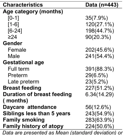
Table 1 describes the demographic characteri-stics of all four hundred forty three enrolled patients. 79.7% of all cases were between the ages of 0 and 2 years. Both genders were almost equally present (45.6% females compared to 54.4% males). The majority of the patients (88.3%) were full term children with history of breast feeding in half of the enrolled population (51.2%) and a mean duration not exceeding 6 months. Unfortunately, it was also noted that more than half of these patients (63%) had at least one family member who was a smoker. Most enrolled patients were previously healthy (85.1%), only (14.9%) had a previous underlying chronic condition [Table 2].
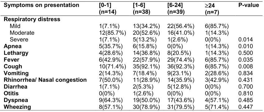
In the study, we divided the RSV positive patients into 4 different subgroups according to age (0-1 month, 1-6 months, 6-24 months, 24 months). The most common presenting symptom was cough (88.7%), followed by fever (64.2%), other symptoms included dyspnea (50%), rhinnorhea (35.7%), lethargy (27.5%), apnea (12.2%), vomiting (20%), diarrhea (8.1%) and one patient had otitis and another had conjunctivitis. A vast majority (75.5%) of RSV positive patients had wheezes on auscultation, and 50% had moderate respiratory distress according to Silverman’s score [Table 3].
Table 1. Demographic characteristics of all enrolled patients
Characteristics Data (n=443)
Age category (months)
[0-1] 35(7.9%)
[1-6] 120(27.1%)
[6-24] 198(44.7%)
≥24 90(20.3%)
Gender
Female 202(45.6%)
Male 241(54.4%)
Gestational age
Full term 391(88.3%)
Preterm 29(6.5%)
Late preterm 23(5.2%)
Breast feeding 227(51.2%)
Duration of breast feeding ( months)
6.34(14.29)
Daycare attendance 56(12.6%)
Siblings less than 5 years 243(54.9%)
Family smoking 283(63.9%)
Family history of atopy 224(50.6%)
Data are presented as Mean (standard deviation) or number of patients (%)
No significant difference in clinical presentation was noted among different age groups except for respiratory distress (P= 0.014) and apnea
(P=0.01). Moderate and severe respiratory distress was noted mainly among those below 6 months of age, while older ages had mild respiratory distress [Table 3].
Table 4 compares the course of hospital stay among the different age groups who are RSV positive. No patients above 2 years of age needed supplemental oxygen, but 50% of those from 0 to 1 month of age did. Higher percentages among age groups (0-1 months) and (1-6 months) needed intensive care management with a highly significant P value (P< 0.0001). No statistically significant difference was detected among the 4 different age groups concerning radiographic findings on chest X-ray, presence of pneumonia, or complications. Only 3 patients required mechanical ventilation (3.06%) and these included one below the age of 1 month and 2 in the 1-6 months group.
Table 2. Underlying conditions among all enrolled patients
Characteristics Data
Hyper reactive airway 112(25.3%)
Chronic lung disease 6(1.4%)
Congenital heart disease 10(2.3%)
Previous NICU* admission 67(15.1%)
Previous Oxygen exposure 50(11.3%)
Other chronic conditions
None 385(86.9%)
Recurrent infections 6(1.4%)
Cow’s milk allergy 8(1.8%)
Neurological diseases 17(3.8%)
Hypothyroidism 3(0.7%)
Gastro esophageal Reflux (GER) 7(1.6%)
Immunodeficiency 1(0.2%)
Neurological and GER 1(0.2%)
Others 15(3.4%)
*NICU: Neonatal Intensive Care Unit
Table 3. Comparison of the clinical presentation between RSV positive patients by age group (months)
Symptoms on presentation [0-1]
(n=14)
[1-6] (n=38)
[6-24] (n=39)
24 (n=7)
P-value
Respiratory distress
Mild 1(7.1%) 13(34.2%) 22(56.4%) 6(85.7%)
Moderate 12(85.7%) 20(52.6%) 16(41.0%) 1(14.3%)
Severe 1(7.1%) 5(13.2%) 1(2.6%) 0(0%) 0.014
Apnea 5(35.7%) 6(15.8%) 0(0%) 1(14.3%) 0.010
Lethargy 4(28.6%) 14(36.8%) 8(20.5%) 1(14.3%) 0.500
Fever 6(42.9%) 22(57.9%) 29(74.4%) 6(85.7%) 0.035
Cough 10(71.4%) 35(92.1%) 36(92.3%) 6(85.7%) 0.008
Vomiting 2(14.3%) 7(18.4%) 9(23.1%) 2(28.6%) 0.834
Rhinorrhea/ Nasal congestion 7(50.0%) 11(28.9%) 14(35.9%) 3(42.9%) 0.431
Diarrhea 1(7.1%) 2(5.3%) 5(12.8%) 0(0%) 0.700
Otitis 0(0%) 1(2.6%) 0(0%) 0(0%) 0.810
Dyspnea 9(64.3%) 19(50.0%) 17(43.6%) 4(57.1%) 0.485
Wheezing 8(57.1%) 30(78.9%) 31(79.5%) 5(71.4%) 0.447
When comparing the management strategies between the two groups, no clinically significant difference was found concerning the type of nebulizer used, antibiotics used, steroids, or the number of chest X-rays done. Normal saline nebulizer was used alone in only 28.9% of RSV-positive patients; all remaining patients needed additional nebulized medications. High flow nasal canula (HFNC) was used in just one RSV-positive patient and 3% of the RSV-RSV-positive patients compared to 0.9% in the RSV-negative group needed mechanical ventilation (P=0.098). Concerning RSV positive patients, out of the 79 (80.6%) who had a chest X-ray done, 53% had normal chest X-ray, 15% had changes of bronchiolitis and 31.6% had a confirmed pneumonia.
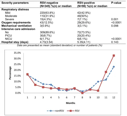
The most compelling differences were observed when comparing the severity parameters between the two groups [Table 6]. Moderate to severe respiratory distress according to Silverman’s score were more prevalent among RSV positive patients (P=0.001). More RSV positive children required oxygen supplementa-tion (29.6%) whether by facemask or nasal canula compared to RSV negative children (12.5%) with a P-value <0.0001.
Intensive care admissions whether to the Neonatal or Pediatric ICUs were more common among RSV positive patients (P<0.0001). Three percent of RSV positive patients required mechanical ventilation compared to only 0.9% in the RSV negative group. The mean duration of hospital stay however was not significantly different (4.73 vs. 5.35, P= 0.143).
Concerning the seasonal variation of Respiratory Syncytial Virus, when compared to other causes of respiratory tract infections, they all had the same pattern. Peak prevalence was found to be in December, followed by November, January and February [Fig. 2].
4. DISCUSSION
Table 4. Comparison of the course in hospital among RSV positive patients according to age group (months)
In-Hospital course [0-1]
(n=14)
[1-6] (n=38)
[6-24] (n=39)
24 (n=7)
P-value
Oxygen requirement 7(50.0%) 13(34.2%) 9(23.1%) 0(0%) 0.140
Intensive care admission
No 4(28.6%) 27(71.1%) 34(87.2%) 7(100.0%)
PICU* 4(28.6%) 11(28.9%) 5(12.8%) 0(0%)
NICU** 6(42.9%) 0(0%) 0(0%) 0(0%) <0.0001
X-ray 13(92.9%) 31(81.6%) 29(74.4%) 6(85.7%) 0.094
Negative 7(53.8%) 17(54.8%) 14(48.3%) 4(57.1%)
Positive 6(46.2%) 14(45.2%) 15(51.7%) 2(28.6%) 0.860
Pneumonia 6(42.9%) 7(18.4%) 10(25.6%) 2(28.6%) 0.440
Complications
None 13(92.9%) 36(94.7%) 36(92.3%) 7(100.0%)
Atelactasis 0(0%) 0(0%) 3(7.7%) 0(0%)
ARDS† 1(7.1%) 2(5.3%) 0(0%) 0.499
Mechanical ventilation 1(7.1%) 2(5.3%) 0(0%) 0(0%) 0.582
*PICU: Pediatric Intensive Care Unit; ** NICU : Neonatal Intensive Care Unit; † ARDS : Acute respiratory distress syndrome
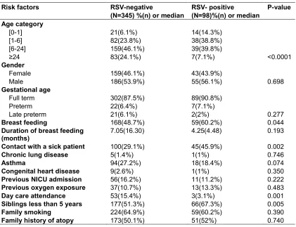
Table 5. Comparison of different risk factors between RSV and non-RSV patients
Risk factors RSV-negative
(N=345) %(n) or median
RSV- positive
(N=98)%(n) or median
P-value
Age category
[0-1] 21(6.1%) 14(14.3%)
[1-6] 82(23.8%) 38(38.8%)
[6-24] 159(46.1%) 39(39.8%)
≥24 83(24.1%) 7(7.1%) <0.0001
Gender
Female 159(46.1%) 43(43.9%)
Male 186(53.9%) 55(56.1%) 0.698
Gestational age
Full term 302(87.5%) 89(90.8%)
Preterm 22(6.4%) 7(7.1%)
Late preterm 21(6.1%) 2(2%) 0.277
Breast feeding 168(48.7%) 59(60.2%) 0.044
Duration of breast feeding (months)
7.05(16.30) 4.25(4.48) 0.193
Contact with a sick patient 100(29.1%) 45(45.9%) 0.002
Chronic lung disease 5(1.4%) 1(1%) 0.746
Asthma 94(27.2%) 18(18.4%) 0.074
Congenital heart disease 9(2.6%) 1(1%) 0.350
Previous NICU admission 56(16.2%) 11(11.2%) 0.222
Previous oxygen exposure 37(10.7%) 13(13.3%) 0.483
Day care attendance 53(15.4%) 3(3.1%) 0.001
Siblings less than 5 years 177(51.3%) 66(67.3%) 0.005
Family smoking 224(64.9%) 59(60.2%) 0.390
Table 6. Comparison of the severity of disease between RSV and non-RSV patients
Severity parameters RSV-negative
(N=345) %(n) or median
RSV-positive
(N=98) %(n) or median
P-value
Respiratory distress
Mild 220(63.8%) 42(42.9%)
Moderate 110(31.9%) 49(50%)
Severe 15(4.3%) 7(7.1%) 0.001
Oxygen requirements 43(12.5%) 29(29.6%) <0.0001
Mechanical ventilation 3(0.9%) 3(3.1%) 0.098
Intensive care admission
No 309(89.6%) 72(73.5%)
PICU 30(8.7%) 20(20.4%)
NICU 6(1.7%) 6(6.1%) <0.0001
Hospital stay (days) 4.73(3.54) 5.35(4.11) 0.143
Data are presented as mean (standard deviation) or number of patients (%)
Fig. 2. Respiratory syncytial virus seasonal variations in Lebanon compared to all other causes of acute respiratory tract infection
Despite the fact that the study included ages up to 13 years, the majority of enrolled patients (79.7%) were below 24 months of age, and this is attributed to the fact that bronchiolitis is a disease confined to this age group as by AAP definition “a constellation of clinical symptoms and signs including a viral upper respiratory prodrome followed by increased respiratory effort and wheezing in children less than 2 years of age” [27]; with RSV being one of its major causes causing symptomatic disease in this age group mainly [10,27]. A wide spectrum of illness is associated with RSV infection, which rarely is asymptomatic. In young children, illness frequently begins with cough, nasal congestion, and fever. Otitis media is common. The primary
manifestations of lower respiratory tract disease in infants are bronchiolitis and pneumonia. These two syndromes may be difficult to distinguish and may occur simultaneously [28]. In our population, 15% of hospitalized patients had bronchiolitis alone and 31.6% had either pneumonia or both. No significant difference in clinical presentation of RSV patients was observed among different age groups with cough (88.7%), wheezing (75.5%) and fever (64.5%) being the most common presenting symptom. Only one case presented with otitis media despite previous reports for being a presenting symptom in up to 17% of cases [10]. Apnea may be the presenting manifestation of RSV disease, particularly in preterm infants [28,29]. Apnea which was seen in
0.0% 5.0% 10.0% 15.0% 20.0% 25.0% 30.0% 35.0%
1 2 3 4 5 6 7 8 9 10 11 12
P
er
ce
n
ta
ge
Months
12.2% of our RSV patients with significant prevalence in the younger groups (P=0.01) has been reported as a complication in 10%-20% of hospitalized infants with RSV infection and is associated with sudden infant death syndrome [10]. It has been postulated that RSV alters the sensitivity of laryngeal chemo receptors and reinforces reflex apnea [28].
The most clinically significant difference in presentation among the compared age groups was the severity of respiratory distress. Ages less than 6 months were more likely to have moderate to severe distress (P= 0.014). This is attributed to the fact that younger ages more commonly present with lower respiratory tract infection including bronchiolitis and pneumonia (45%) compared to only 5% in children more than 2-5 years old [10].
Several environmental and other host factors have been reported to augment the risk of a more severe RSV infection [10]. These include younger age, male gender, gestational age, duration of breastfeeding, daycare attendance, tobacco exposure, presence of young children in the same household, in addition to underlying conditions [10]; all of which were compared between RSV and non-RSV groups here. RSV patients were younger, males, had less duration of breastfeeding, and were more likely to have a sick contact and a young sibling in the household. All other factors were found non contributory.
Limited testing and supportive management has been proposed by the American Academy of Pediatrics in 2006. Our study was conducted several years following the publication of these guidelines and so we sought to determine whether our management was impacted by them. Despite latest recommendations suggesting that only persistent SpO2< 90% is an indication for initiating supplemental oxygen [19], enrolled patients were actually supplemented with O2 when SpO2 dropped below 94% and thus 29.6% of RSV positive patients required oxygen. According to these same guidelines, many of the commonly used management modalities have not been shown to be effective in improving the clinical course of the illness. This includes the routine use of bronchodilators, corticosteroids, antibiotics, and chest radiography [19]. However, almost 70% of the RSV group and 63% of the non-RSV group required either inhaled β-adrenergic or α-adrenergic or both during their hospital stay.
Almost 40% of enrolled patient received antibiotics and 20% received corticosteroids. But these patients included those previously asthmatic or with underlying lung disease and many might have developed secondary bacterial infections later which were not actually documented in our study.
As other studies have previously reported, our study suggests a more severe course for RSV infection when compared to the other causes. RSV positive patients had severe respiratory distress (P=0.001), required more oxygen (P<0.0001), were more likely to be admitted to intensive care unit (P<0.0001) and require mechanical ventilation (P=0.098). But so far, no effective anti-RSV vaccine or therapeutic modality is available. Several studies have focused on the development of RSV entry inhibitors targeting its F protein for the treatment of RSV infection but clinical trials were stopped because of unfavorable pharmaceutical properties of these compounds [29]. For this reason, various efforts have focused on the development of prophylactic RSV monoclonal antibodies. Palivizumab (Synagis®) has been FDA approved since 1998 [19]. The administration of prophylactic RSV antibodies has been shown to decrease severe disease [19]. The AAP recommends that palivizumab be considered for prophylaxis against RSV in children who have chronic lung disease or congenital heart disease that requires medical therapy, and are < 2 years old, as well as for infants born at 32 or fewer weeks’ gestaion. Selected children born between 32 and 35 weeks’ gestation may be considered for prophylaxis if there are other factors that make them more vulnerable to severe RSV disease including either daycare attendance or having a sibling less than 5 years of age [30]. Interestingly, only 3.8% of those who required intensive care were either preterm or had CLD or CHD, while the remaining 98.2% were previously healthy but with possible risk factors. This raises the question to whether the population that deserves prophylaxis should be expanded to include more groups of patients who are previously healthy but with certain risk factors.
start in November, peaks in December but continues throughout the winter and spring season and not ending till the end of July. We report RSV cases throughout the whole year with August being the only month with no reported cases hospitalized at our hospital.
4.1 Limitations of the Study
Despite the sufficient sample obtained during the study period, still it included hospitalized patients in only one tertiary center in Lebanon, and not several medical centers. The first period of the study was retrospective, so data collected at that time might have been incomplete or unavailable, but we relied mostly on calling back patients for missing data when contact was possible. Another setback in the study is that different nasopharyngeal wash swabs were collected by different doctors allowing thus inappropriate sampling techniques affecting the yield of the wash. Samples collected over the weekend might have been conserved for more than 24hrs before transfer to the laboratory, decreasing the yield of the sample. On the other hand, our study included almost 80% of all patients admitted since the availability of RSV rapid antigen detection and nasopharyngeal wash was used for all patients.
5. CONCLUSION
Respiratory Syncytial Virus is a significant burden among patients presenting with pneumonia and bronchiolitis and is associated with a more severe course. Previously healthy children are not at low risk for developing severe disease, so we suggest that immunoprophylaxis guidelines are worth receiving another review. As an effective vaccine against RSV is not yet available, parent education for preventive hygiene measures are more affordable and easy to perform. Awareness should also be raised in parents that what is a minor cold to them may cause a life-threatening illness in a young infant or a high risk child. Clinicians should emphasize on the use of RSV immunoprophylaxis among their patients as recommended by current guidelines keeping in mind each countries’ special seasonal variation, until further updates are available.
6. RECOMMENDATIONS
Further studies are needed to determine a more accurate prevalence of Respiratory Syncytial
Virus in Lebanon using polymerase chain reaction (PCR) for viral detection and including multiple medical centers and thus a larger sample.
COMPETING INTERESTS
Authors have declared that no competing interests exist.
REFERENCES
1. Bryce J, Boschi-Pinto C, Shibuya K, Black RE. WHO estimates of the causes of death in children. Lancet. 2005;365:1147–52. 2. Leader S, Kohlhase K. Recent trends in
severe respiratory syncytial virus (RSV) among US infants, 1997 to 2000. J Pediatr. 2003;143(5 suppl):S127–S132.
3. Leader S, Kohlhase K. Respiratory syncytial virus-coded pediatric hospitalize-tions, 1997 to 1999. Pediatr Infect Dis J. 2002;21(7):629–632.
4. Shay DK, Holman RC, Newman RD, Liu LL, Stout JW, Anderson LJ. Bronchiolitis associated hospitalizations among US children, 1980-1996. JAMA. 1999;282(15): 1440-1446.
5. Boyce TG, Mellen BG, Mitchel EF Jr, Wright PF, Griffin MR. Rates of hospitalization for respiratory syncytial virus infection among children in Medicaid. J Pediatr. 2000;137(6):865–870.
6. Holman RC, Curns AT, Cheek JE, et al. Respiratory syncytial virus hospitalizations among American Indian and alaska native infants and the general United States infant population. Pediatrics. 2004;114(4): e437-44.
7. Stockman LJ, Curns AT, Anderson LJ, Fischer-Langley G. Respiratory syncytial virus-associated hospitalizations among infants and young children in the United States, 1997-2006. Pediatr Infect Dis J. 2012;31(1):5–9.
8. World Health Organization (WHO). Acute respiratory infections.
Available:
http://www.who.int/vaccine_research/disea ses/ari/en/index2.html
9. Stensballe LG, Devasundaram JK, Simoes EA. Respiratory syncytial virus epidemics: The ups and downs of a seasonal virus. Pediatr Infect Dis J. 2003;22:S21-32. 10. Hall CB, Walsh EE. Respiratory syncytial
Demmler-Harrison GJ, Kaplan SL (Eds), saunders, Philadelphia. 2009;2462. 11. Goldmann DA. Transmission of viral
respiratory infections in the home. Pediatr. Infect. Dis. J. 2000;19(10 Suppl):S97-102.
12. King JC Jr, Burke AR, Clemens JD, et al. Respiratory syncytial virus illnesses in human immunodeficiency virus- and noninfected children. Pediatr Infect Dis J. 1993;12:733.
13. Centers for Disease Control and Prevention (CDC). Respiratory syncytial virus activity--United States, 1999-2000 season. MMWR Morb Mortal Wkly Rep. 2000;49:1091.
14. Hall CB, Weinberg GA, Iwane MK, et al. The burden of respiratory syncytial virus infection in young children. N Engl J Med. 2009;360:588.
15. Spence L, Barratt N. Respiratory syncytial virus associated with acute respiratory infections in trinidadian patients. Am J Epidemiol. 1968;88:257.
16. Sung RY, Murray HG, Chan RC, et al. Seasonal patterns of respiratory syncytial virus infection in Hong Kong: A preliminary report. J Infect Dis. 1987;156:527.
17. Cane PA. Molecular epidemiology of respiratory syncytial virus. Rev Med Virol. 2001;11:103.
18. Staat MA, Henrickson K, Elhefni H, et al. Prevalence of respiratory syncytial virus-associated lower respiratory infection and Apnea in infants presenting to the emergency department. Pediatr Infect Dis J. 2013;32:911.
19. McCarthy CA, Hall CB. Respiratory syncytial virus: Concerns and control, Pediatrics in Review. 2003;24;301. DOI: 10.1542/pir.24-9-301.
20. Hall CB, Walsh EE. Respiratory syncytial virus. in: Textbook of pediatric infectious diseases, 6th ed, Feigin RD, Cherry JD, Demmler-Harrison GJ, Kaplan SL (Eds), saunders, Philadelphia. 2009;2462.
21. American Academy of Pediatrics Subcommittee on Diagnosis and Management of Bronchiolitis. Diagnosis and management of bronchiolitis. Pediatrics. 2006;118(4):1774-1793. 22. Gregson D, Lloyd T, Buchan S, Church D.
Comparison of the RSV respi-strip with direct fluorescent-antigen detection for diagnosis of respiratory syncytial virus infection in pediatric patients. J Clin Microbiol. 2005;43:5782-5783.
23. Turkish Neonatal Society (Ankara, Turkey). The seasonal variations of respiratory syncytial virus infections in Turkey: A 2-year epidemiological study. Turk J Pediatr. 2012;54:216-222.
24. El Kholy AA, Mostafa NA, El-Sherbini SA, et al. Morbidity and outcome of severe respiratory syncytial virus infection. Pediatrics International. 2013;55(3):283– 288. DOI: 10.1111/ped.12051.
25. Hirsh S, Hindiyeh M, Kolet L, et al. Epidemiological changes of respiratory syncytial virus (RSV) infections in israel. PLoS ONE. 2014;9(3):e90515.
26. Hall CB, Weinberg GA, Iwane MK. The burden of respiratory syncytial virus infection in Young Children. N Engl J Med. 2009;360(6):588-98.
27. Zorc JJ, Hall CB. Bronchiolitis: Recent evidence on diagnosis and management. Pediatrics. 2010;125:342–349.
28. Lindgren C, Jing L, Graham B, et al. Respiratory syncytial virus infection reinforces reflex apnea in young lambs. Pediatr Res. 1992;31:381.
29. Zhiwu Sun 1, Yanbin Pan 2, Shibo Jiang 1, Lu Lu, Respiratory syncytial virus entry inhibitors targeting the F protein, viruses. 2013;5 :211-225. DOI: 10.3390/v5010211. 30. Committee on Infectious Diseases;
Modified recommendations for Use of palivizumab for prevention of respiratory syncytial virus infections. Pediatrics. 2009;124:1694. DOI: 10.1542/peds.2009-2345.
_________________________________________________________________________________ © 2015 Assaf et al.; This is an Open Access article distributed under the terms of the Creative Commons Attribution License (http://creativecommons.org/licenses/by/4.0), which permits unrestricted use, distribution, and reproduction in any medium, provided the original work is properly cited.
Peer-review history: