www.sciencedomain.org
Evaluation of Outcome of a Rare Presentation of
Appendicitis
Ahmed Makki
1*1
Department of Surgery, King Abdulaziz University, Saudi Arabia.
Author’s contribution
The sole author designed, analyzed and interpreted and prepared the manuscript.
Article Information
DOI: 10.9734/BJMMR/2016/26659
Editor(s):
(1) Franciszek Burdan, Experimental Teratology Unit, Human Anatomy Department, Medical University of Lublin, Poland and Radiology Department, St. John’s Cancer Center, Poland.
Reviewers:
(1) Wagig Mommtaz Ghannam, Mansoura University, Egypt. (2)Ahmed Gado, Misr International Hospital, Egypt. Complete Peer review History:http://sciencedomain.org/review-history/15060
Received 27th April 2016 Accepted 4th June 2016 Published 18th June 2016
ABSTRACT
Introduction: Acute appendicitis is the inflammation of the vermiform appendix and it is the most common intra-abdominal surgical emergency. Therefore, perforated appendicitis is rarely accompanied by pneumoperitoneum and that apparent rarity of association of pneumoperitoneum and acute appendicitis prompted us to discuss this condition.
Materials and Methods: This retrospective study reviewed all cases of acute abdomen with pneumoperitoneum admitted in King Abdulaziz University Hospital from January 2011 to December 2015. Only cases with acute appendicitis were selected to be in the study, Leucocytic count and C-reactive protein, as well as plain X-ray abdomen in erect position and C.T.scan were reviewed preoperatively.
All demographic data (age, gender), as well as the clinical details which included relevant investigations results, in addition to the perioperative data (the type of the performed operative procedure, operative time and the hospital stay with reference to the postoperative complications for each patient), all were collected and analyzed by IBM-SPSS version 22.
Results: Pneumoperitoneum due to perforated appendix was found in 7 out of 131 cases in this study (5.3%), with male predominance 6/7, with a mean age of 29.7 years. The documented postoperative complications were pulmonary complications; intra-abdominal pus collection (pelvic and subphrenic) as well as wound dehiscence, all were found post conventional exploratory laparotomy cases.
Conclusion: The study documented the rarity of pneumoperitoneum due to perforated appendix. Therefore, it should be considered as a cause of acute abdomen with pneumoperitoneum, imaging studies were very essential to visualize these cases, Laparoscopic approach provided better outcome than the conventional laparotomy approach.
Keywords: Acute appendicitis; pneumoperitoneum; acute abdomen; laparotomy; laparoscopy.
1. INTRODUCTION
Acute appendicitis is the inflammation of the vermiform appendix, and it is the most common intra-abdominal surgical emergency requiring surgery. Perforated appendicitis is sometimes accompanied by pneumoperitoneum, often localized beneath the diaphragmatic dome [1]. Pneumoperitoneum is a confusing finding in perforated appendicitis and that may lead to diagnostic error [2].
Although most of the cases of acute abdomen with pneumoperitoneum occur as perforated peptic ulcer, it is important not to miss perforated appendix as a cause for this condition, particularly if exploratory laparotomy shows normal stomach and duodenum. The apparent rarity of the association of free intraperitoneal gas and acute appendicitis prompted us to discuss this condition with its outcome.
2. MATERIALS AND METHODS
This retrospective study reviewed all cases of acute abdomen with pneumoperitoneum admitted in King Abdulaziz University Hospital, from January 2011 to December 2015, among them only cases with acute appendicitis were selected to be involved in the study, these patients presented with abdominal pain, fever and dehydration.
Assessment and resuscitation with intravenous fluids, intravenous antibiotics (Metronidazole, Cephalosporins) and analgesics were carried out by the surgical on-call team. The initial laboratory work up as Leucocytic count and C-Reactive Protein, as well as the radiological investigations as plain X-ray abdomen in erect position which was done for all patients and C.T.scan were reviewed before transfer to the operating theatre, where formal exploratory laparotomy/ laparoscopy was performed with suction of peritoneal exudate and taking bacterial culture swab, then routine exploration of the whole abdomen was performed, appendix was removed and sent to histopathology, finally washing the peritoneal cavity with normal saline and insertion of drains before closure were done.
All demographic data (age, gender), as well as the clinical details which included duration of symptoms, relevant laboratory and imaging investigations results, in addition to the perioperative data as the type of the performed operative procedure, operative time and the hospital stay with reference to the postoperative complications of each patient, were collected and analyzed by IBM-SPSS version 22. Approval of the study was obtained from the Hospital Ethical Committee before start.
3. RESULTS
This retrospective study reviewed 131 cases of acute abdomen with radiologically documented pneumoperitoneum, among them only 7 cases were found to have perforated appendix (5.3%).
morbidity, which were categorized into three groups, pulmonary complications, as atelectasis, pneumonia and pleural effusion, which were found in 3 cases (43%). Intra-abdominal pus collection (abscess) in pelvic and subphrenic spaces which was developed in 3 cases as well (43%), all cases were drained by C.T guided drainage. Wound dehiscence in the form of wound infection was found in 4 cases (57%), which were treated by daily dressing and antibiotics according to the culture till complete healing, therefore incisional hernia had developed in a couple of cases of them, the laparoscopically approached cases were free from those complications in addition to one case with laparotomy had the same outcome.
4. DISCUSSION
The vermiform (‘worm-like’ in Latin) appendix is a small and tubular structure [3]. Acute appendicitis is inflammation of the appendix and it is considered as the most common cause of acute abdomen [4], while pneumoperitoneum is a reliable sign of viscus perforation that is infrequently seen with acute appendicitis [5]. The radiographic finding of intraperitoneal free air secondary to perforation of a acutely inflamed appendix is extremely rare [6,7], but it should not be missed as a cause of pneumoperitoneum.
Acute appendicitis is common in young patients under the age of 30 years, but appendicitis can occur at any age and diagnosis at extremes of age is more difficult. Appendicitis is more common in males than females, that was exactly applied in this study as most of cases were males (6/7) with mean age of 29.7 years.
Perforated appendix is a common complication of acute appendicitis with an estimated incidence of 20%-33% according to Schiaz et al. study. Pneumoperitoneum is rarely found in cases of perforated appendicitis because the appendiceal lumen is usually obliterated central to the perforation, and visceral walling off will tend to localize the process, therefore, perforated appendicitis can be associated with pneumoperitoneum and diffuse peritonitis in rare situation, that accounts about 0-7% of all patients with acute abdomen due to pneumoperitoneum [8], our study showed 5.3% of this condition.
Only few articles have been published in the medical literature about pneumoperitoneum-associated appendicitis since the first case was reported by Gulleurin in 1923, he reported the first two recorded cases, while Greenberg (1961)
reviewed the literature and discovered a total of ten cases, he added two further cases of his own [9], however it seems to be more common than reported in the literatures, that may be due to lack of imaging studies [10,11].
The underlying aetiology of acute appendicitis is uncertain, many theories have been proposed to explain the pathogenesis of acute appendicitis:-1-increased intraluminal pressure and less bulky stool due to low-fiber diet [12]. 2-Anatomical obstruction due to mucosal folds.3-Obstruction of the appendix by a foecolith or swelling of the submucosal lymphoid tissue.
The classic clinical features of acute appendicitis are migrating abdominal pain, which starts centrally (peri-umbilical), then pain shifts down to the right iliac fossa after a few hours, it may be associated with nausea, vomiting and anorexia with mild elevation of temperature.
The classic signs of acute appendicitis are low-grade fever, dry tongue and associated foetor. Localized tenderness, guarding, rebound tenderness in the right iliac fossa, maximally at the site of McBurney’s point, tenderness in the right iliac fossa on palpation of the left iliac fossa may also be present (Rovsing’s sign).Although, its clinical features are straight forward, sometimes they could be confused, particularly, if history taking is difficult or plain X-ray of the abdomen shows rare relevant findings such as pneumoperitonium [13].
In complicated appendicitis with perforation patients usually show a longer period of abdominal pain which becomes more generalized, in addition to vomiting and fever, examination reveals the abdominal tenderness and guarding over the entire abdomen and distension secondary to paralytic ileus, the patient may become hypotensive and dehydrated.
appendicitis with perforation showed very high levels of CRP with a mean of 200.3 mg/dl (Normal value: 1-3 mg/dl) [14].
Imaging studies may be beneficial in the diagnosis of acute complicated appendicitis, though plain radiograph is generally unhelpful in diagnosing appendicitis, but it is a base line investigation in acute abdomen, to exclude other causes of abdominal pain. Abdominal ultrasound is less expensive, readily available tool for patients with uncertain diagnosis, with high sensitivity and specificity [15]. It can exclude other causes of abdominal pain including gynecological pathology (ovarian cyst, pyosalpinx, ectopic pregnancy) and cholecystitis. The disadvantage of ultrasound is that, it is operator dependent and a normal appendix must be visualized to rule out acute appendicitis, which could be harder in patients with a large body habitus or existence of air in the bowel loops which masks the field.
Computerized tomography (CT) has a sensitivity and specificity of over 90% and its use may decrease the rate of negative appendectomy [16]. It is user independent and quick to perform. The main disadvantages of CT include: Exposure to ionizing radiation and cost, in addition to possible iodinated contrast media allergy. CT findings are more obvious in the complicated disease when the appendix is perforated; an appendicular mass, abscess or free air in the peritoneum which can be easily detected with reference to its cause. C.T. scan was done in 6/7 cases in our study, 4 of them referred successfully to the appendix as a source of pneumoperitoneum, that helped in the surgical decision making (2 of them were managed laparoscopically, the other 2 cases had laparotomy), while 2 cases had non-conclusive diagnosis, which was revealed by laparotomy. Magnetic resonance imaging (MRI) may be used in the diagnosis of appendicitis, when CT scan is contraindicated, such as in children or during pregnancy. It is superior to ultrasound in the detection of appendicitis, with sensitivity and specificity of over 95% [17], though it is time consuming, expensive and not a routine practice.
The treatment of acute appendicitis is appendectomy [18], initially patients should be fluid resuscitated and given adequate analgesia, intravenous antibiotics may be administered preoperatively (usually metronidazole and a broad-spectrum cephalosporin). That was our protocol of treatment, being all cases were
complicated appendicitis with pneumoperitonium and consequently peritonitis, continuing the antibiotics postoperatively is advised due to the heavily contaminated field.
In these patients with perforated appendicitis and pneumoperitoneum, a grid-iron incision at the Mcburney’s point may be satisfactory as it provides good access to the appendix, but if the diagnosis is uncertain, midline laparotomy incision may be better as it enables proper exploration and effective peritoneal lavage to be performed followed by removal of the diseased appendix. If bile or foecal stained fluid is identified during laparotomy, a full exploration will be required to get the source of peritoneal contamination.
With improving laparoscopic techniques, the use of diagnostic laparoscopy increased, and laparoscopic appendectomy is becoming more common. It provides less postoperative pain, smooth recovery and lower incidence of wound infection, however there is a prolonged operative time and possibility of intra-abdominal abscess formation [19]. Laparoscopy was exercised in 2 cases in our study, it helped to establish the diagnosis and treat the condition with no documented postoperative complications.
Wound infection is the commonest complication post open appendectomy, with an incidence of 15%. The rate of wound infection is getting low following laparoscopic surgery. The postoperative complications of the condition of pneumoperitoneum–related appendicitis are variables; Wound dehiscence,intra-abdominal abscess collection and pulmonary complications. In our study 4 cases (57%) developed dehiscence of the wound and a purulent discharge, later 2 of them had incisional hernia reported during the long follow-up visits after elimination of infection.
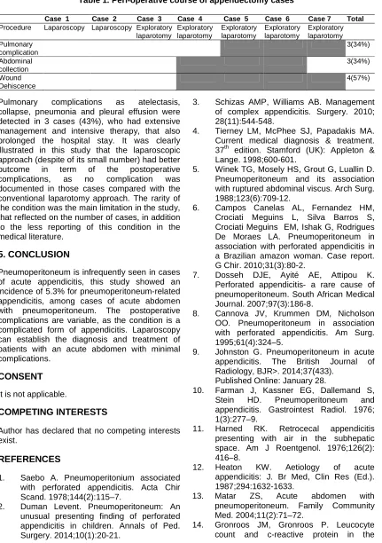
Table 1. Peri-operative course of appendectomy cases
Case 1 Case 2 Case 3 Case 4 Case 5 Case 6 Case 7 Total
Procedure Laparoscopy Laparoscopy Exploratory
laparotomy
Exploratory laparotomy
Exploratory laparotomy
Exploratory laparotomy
Exploratory laparotomy Pulmonary
complication
3(34%)
Abdominal collection
3(34%)
Wound Dehiscence
4(57%)
Pulmonary complications as atelectasis, collapse, pneumonia and pleural effusion were detected in 3 cases (43%), who had extensive management and intensive therapy, that also prolonged the hospital stay. It was clearly illustrated in this study that the laparoscopic approach (despite of its small number) had better outcome in term of the postoperative complications, as no complication was documented in those cases compared with the conventional laparotomy approach. The rarity of the condition was the main limitation in the study, that reflected on the number of cases, in addition to the less reporting of this condition in the medical literature.
5. CONCLUSION
Pneumoperitoneum is infrequently seen in cases of acute appendicitis, this study showed an incidence of 5.3% for pneumoperitoneum-related appendicitis, among cases of acute abdomen with pneumoperitoneum. The postoperative complications are variable, as the condition is a complicated form of appendicitis. Laparoscopy can establish the diagnosis and treatment of patients with an acute abdomen with minimal complications.
CONSENT
It is not applicable.
COMPETING INTERESTS
Author has declared that no competing interests exist.
REFERENCES
1. Saebo A. Pneumoperitonium associated with perforated appendicitis. Acta Chir Scand. 1978;144(2):115–7.
2. Duman Levent. Pneumoperitoneum: An unusual presenting finding of perforated appendicitis in children. Annals of Ped. Surgery. 2014;10(1):20-21.
3. Schizas AMP, Williams AB. Management of complex appendicitis. Surgery. 2010; 28(11):544-548.
4. Tierney LM, McPhee SJ, Papadakis MA. Current medical diagnosis & treatment. 37th edition. Stamford (UK): Appleton & Lange. 1998;600-601.
5. Winek TG, Mosely HS, Grout G, Luallin D. Pneumoperitoneum and its association with ruptured abdominal viscus. Arch Surg. 1988;123(6):709-12.
6. Campos Canelas AL, Fernandez HM, Crociati Meguins L, Silva Barros S, Crociati Meguins EM, Ishak G, Rodrigues De Moraes LA. Pneumoperitoneum in association with perforated appendicitis in a Brazilian amazon woman. Case report. G Chir. 2010;31(3):80-2.
7. Dosseh DJE, Ayité AE, Attipou K. Perforated appendicitis- a rare cause of pneumoperitoneum. South African Medical Journal. 2007;97(3):186-8.
8. Cannova JV, Krummen DM, Nicholson OO. Pneumoperitoneum in association with perforated appendicitis. Am Surg. 1995;61(4):324–5.
9. Johnston G. Pneumoperitoneum in acute appendicitis. The British Journal of Radiology, BJR>. 2014;37(433).
Published Online: January 28.
10. Farman J, Kassner EG, Dallemand S, Stein HD. Pneumoperitoneum and appendicitis. Gastrointest Radiol. 1976; 1(3):277–9.
11. Harned RK. Retrocecal appendicitis presenting with air in the subhepatic space. Am J Roentgenol. 1976;126(2): 416–8.
12. Heaton KW. Aetiology of acute appendicitis: J. Br Med, Clin Res (Ed.). 1987;294:1632-1633.
13. Matar ZS, Acute abdomen with pneumoperitoneum. Family Community Med. 2004;11(2):71–72.
diagnosis of acute appendicitis. Br J Surg. 1999;86:501–504.
15. Puylaert JB, Rutgers PH, Lalisang RI, et al. A prospective study of ultrasonography in the diagnosis of appendicitis. N Engl J Med. 1987;317:666-669.
16. Birnbaum BA, Wilson SR. Appendicitis at the millennium. Radiology. 2000;215:337– 348.
17. Incesu L, Coskun A, Selcuk MB, et al. Acute appendicitis: MR imaging and
sonographic correlation. AJR Am J Roentgenol. 1997;168:669-674.
18. Temple CL, Huchcroft SA, Temple WJ. The natural history of appendicitis in adults. A prospective study. Ann Surg. 1995;221:278–28.
19. Sauerland S, Lefering R, Neugebauer EA. Laparoscopic versus open surgery for suspected appendicitis. Cochrane Database Syst Rev. 2004;4.
_________________________________________________________________________________ © 2016 Makki; This is an Open Access article distributed under the terms of the Creative Commons Attribution License (http://creativecommons.org/licenses/by/4.0), which permits unrestricted use, distribution, and reproduction in any medium, provided the original work is properly cited.
Peer-review history: