_____________________________________________________________________________________________________
*Corresponding author: Email: si.mbagwu@unizik.edu.ng;
www.sciencedomain.org
Histological Study on the Anterior Pituitary Gland of
Monosodium Glutamate-Administered Adult Male
Wistar Rats
Smart I. Mbagwu
1*, Okwudili O. Udemezue
1, Uzozie C. Ofoego
1and Promise E. Ikhine
11
Department of Anatomy, Faculty of Basic Medical Sciences, College of Health Sciences, Nnamdi Azikiwe University, Nnewi, Anambra State, Nigeria.
Authors’ contributions
This work was carried out in collaboration between all authors. Author SIM designed the study, wrote the protocol, managed the experimental process and wrote the first draft of the manuscript. Author OOU proof read and edited the manuscript. Author PEI managed the literature searches, analyses of the study performed the spectroscopy analysis and author UCO reviewed the literatures. All authors read and approved the final manuscript.
Article Information
DOI: 10.9734/BJMMR/2016/16705 Editor(s): (1) Shashank Kumar, Department of Biochemistry, University of Allahabad, Allahabad, India. Reviewers: (1) Martha E. Ruiz Tachiquin, Instituto Mexicanos del Seguro Social, Mexico. (2)Anthony Cemaluk C. Egbuonu, Michael Okpara University of Agriculture Umudike, Nigeria. (3)J. O. Ashaolu, Bowen University, Nigeria. (4)Filip Gabalec, University Hospital Hradec Kralove, Czech Republic. Complete Peer review History:http://sciencedomain.org/review-history/12472
Received 11th February 2015 Accepted 28th May 2015 Published 27th November 2015
ABSTRACT
The routine use of Monosodium glutamate (MSG) as a food additive found in commercial food products has being generating controversy regarding its health effects. This study investigated the effects of MSG administration on the histology of anterior pituitary gland in rats. Twenty adult male rats were used and randomly divided into four groups (n=5, each). The rats in the treated groups received a daily MSG administration through orogastric method at a dose of 100 mg/ kg bw/day, 500 mg/ kg bw/day, 1500 mg/ kg bw/day respectively, while the control rats received 0.5 ml of distilled water. The body weight was measured. After sacrifice, the pituitary gland was harvested and fixed with 10% formalin for routine histological procedures. Results revealed decrease in body weight while the histology of the anterior pituitary gland of the rats showed no observable
pathological alterations. This study indicates no induced histological damage following oral administration of MSG.
Keywords: Monosodium glutamate; anterior pituitary gland; histology.
1. INTRODUCTION
The pituitary gland, or hypophysis is an endocrine gland which protrudes off from the hypothalamus at the base of the brain. It rests upon the hypophysial fossa of the sphenoid bone in the center of the middle cranial fossa where it is covered by a diaphragma sellae and surrounded by sella turcica [1]. The anterior pituitary (adenohypophysis) is a lobe of the gland that regulates several physiological processes such as stress, growth, reproduction, and lactation.
Monosodium glutamate (MSG) is used as a flavor enhancer especially in Chinese, Thainese and Japanese foods [2-4]. There is increase in the production of MSG in many countries today through a natural fermentation process using molasses from sugar cane or sugar beets, as well as starch and corn sugar since its first extraction from seaweed and other plant sources [5].
Ajinomoto is a common brand name for Monosodium glutamate (MSG) in Nigeria and it is commonly used as a food additive and acts as a preservative of palatability [6]. Various seasoning sauce contain significant levels of free glutamate (as MSG), both from natural sources and from added monosodium glutamate [6,7]. Despite its taste stimulation and improved appetite enhancement, reports indicate that MSG is toxic to human and experimental animals [6] producing symptoms such as numbness, weakness, flushing, sweating, dizziness and headaches. [8].
A study by Manal et al. [4] revealed that MSG has the potential of causing an adverse effect on the hepatic and renal functions which might be due to oxidative stress induced by MSG on the liver and renal tissue. MSG has also been associated with retinal degeneration, endocrine disorder [9,10] neurological disorders [11] and has also be found to produce oxygen derived free radicals [12]. It has been reported that MSG causes changes in the liver parenchyma [13].
Studies have shown that the body uses glutamate as a nerve impulse transmitter [11]
and that there are glutamate-responsive tissues all over the body [13], hence an evidence of chest pain, sensation of facial pressure, headaches, burning sensation, excessive fluid retention and sweating has been observed. MSG has been implicated in male infertility [14,15] and has also been shown to cause endocrinological disorders such as reduction in the secretion of growth hormones, leading to stunted growth and irreversibility in obesity [16]. Studies providing the evidence of MSG toxic effects have raised the increasing interest inMSG intake as flavor enhancer. However, the safety of MSG usage has generated such controversial argument locally and internationally [6].Therefore, the aim of this study was to investigate the effects of Monosodium Glutamate on the histology of the anterior pituitary gland.
2. MATERIALS AND METHODS
2.1 Test Materials
Ajinomoto which is one of the trade names of the concentrated forms of Monosodium Glutamate (99% pure MSG) was procured from a local market in Nnewi, Anambra State, Nigeria.
2.2 Animals
Adult male wistar rats weighing 176 grams – 210 grams obtained from the animal house of the Department of Zoology, Nnamdi Azikiwe University, Awka, were used for the experiment. The animals were housed in separate metallic cages and acclimatized for two weeks before the beginning of the study under standard laboratory conditions (temperature: 25±1°C, humidity: 50±5% and lighting: 12 h light/ 12 h dark cycle) in the animal house of the Department of Anatomy, Nnamdi Azikiwe University, Nnewi. The rats were fed a standard chow diet ad libitum with free access to water. This study was approved and registered by the Committee of Animal Investigations, Department of Anatomy, Faculty of Basic Medical Sciences, Nnamdi Azikiwe University.
2.3 Experimental Design
water for three weeks (control), group II,III and IV rats were given MSG for three weeks. In the MSG-treated groups, MSG was administered
daily through orogastric method at a dose of 100 mg/ kg bw/day, 500 mg/ kg bw/day, 1500 mg/ kg bw/day respectively. MSG fresh
solution was prepared every two days and kept in the fridge until the time of use. In the control groups, 0.5 ml distilled water was administered at the same regimen.
2.4 Histological Study
At the end of the experimental periods, the animals were anesthetized with ether fumes and sacrificed by cervical dislocation. The animals were dissected and pituitary glands were carefully harvested and fixed in 10% formol saline. The specimens were then dehydrated in ascending grades of alcohol, cleared in xylene and embedded in paraffin wax. Serial sections (4-5 µm thick) were obtained using a rotatory microtome. The deparaffinized sections were stained with haematoxylin & eosin (H&E) stain. Photomicrographs were obtained using a digital research photographic microscope.
2.5 Statistical Analysis
Data were represented as Mean ± SD. The means were compared for statistical significance using Student's t test for unpaired data. Differences were considered significant at p<0.05. All statistical analyses were performed using SPSS version 17.
3. RESULTS
3.1 Weight Assessment
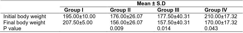
All rats from various groups survived to the end of the study period. The mean body weight for the various groups is presented in Table 1. The mean of the final body weight in group II was significantly lower than that of group I (p<0.05). In group IV, the mean value of the final body
weight was not significantly different from that of group III. A time-dependent steady decrease in body weight was observed in groups III, III and group IV
3.2 Histological Results
Examination of the anterior pituitary gland sections from the rats (groups I-IV, Figs 1-4) showed normal histological features of the anterior pituitary gland which consisted of acidophils,basophils and chromophobes.
4. DISCUSSION
In the present study, the effects of short-term administration of MSG on the histology of the anterior pituitary gland were assessed in rats. In our study, there was a decrease in the body weight of the animals after the administration of MSG. This corresponds to the studies by Miskowiak & Partyka [17] and Kondoh & Torii [19] which suggests that the reduction in weight is likely mediated by increased energy expenditure and not reduced energy intake. Tordoff et al. [18]also reported that 3% MSG in diet or in drinking water did not influence body weight and body fat store in three different species of rodents (Sprague Dawley rat, Obesity-prone Sprague Dawley derivated rat and C57BL\6J mouse)
It has been suggested in several animal studies that MSG is able to induce overweight and obesity as a result of elevation of plasma GLU concentrations to very high values, sufficient enough to penetrate into the brain and exert deleterious effects on the arcuate nucleus neurons, thereby disrupting the hypothalamic signaling cascade of leptin action and resulting in hyperphagia and obesity [19-22]. From our study, we suggest that GLU derived from MSG ingestion was almost entirely catabolised by the enterocyte of intestinal mucosa [23]. Another suggestion to the cause of reduction in weight could partly be as a result of reduced levels of thyroid hormones [24].
Table 1. Descriptive statistics of the mean body weight of the various groups of animals administered monosodium glutamate
Mean ± S.D
Group I Group II Group III Group IV
Initial body weight 195.00±10.00 176.00±26.07 177.50±40.31 210.00±17.32 Final body weight 207.50±5.00 156.00±26.07 157.50±40.31 170.00±17.32
Fig. 1. Photomicrograph of the anterior pituitary gland of animals in group 1 (H&E, x100) This shows normal morphology of the cell with intact interstitial architecture
.
Fig. 3. Photomicrograph of the anterior pituitary gland of animals in group 3 (H&E x100) This shows normal morphology of the cell and interstitial architecture
This study revealed no observable toxic effect on the anterior pituitary gland of the adult wistar rats administered MSG. This is contradictory to the observation by Bojanic et al. [25] that MSG causes hyperplasia of basophilic pituitary cells. Biodun & Biodun [6] reported that ingestion of monosodium glutamate could cause or exacerbate numerous conditions such as asthma, urticaria, atopic dermatitis, ventricular arrhythmia, neuropathy and abdominal discomfort. The disparity in our observation in comparism to other studies further points out the controversy on the safe use of MSG and its inclusion in“Generally Recognized as Safe” (GRAS)-list of foods as opined by Food and Drug Administration (FDA) of the United States.
We can say from our study that though there was no observable deleterious effect of MSG on the histology of the anterior pituitary gland, the observed reduction in body weight of the animals could be a result of reduction in the levels of serum thyroid and growth hormones as mentioned earlier. This therefore implies that short term administration of MSG could exert some effects on substances regulating certain functions in the body without really inducing any observable damage on body tissues. With respect to this, we recommend further investigation on MSG and its effect on anterior pituitary hormones. Long-term studies are also required in order to ascertain the effect of MSG administration on the histology of the anterior pituitary gland.
5. CONCLUSION
There was no observed pathology on the histological section of the anterior pituitary gland non short term administration of Monosodium glutamate.
CONSENT
It is not applicable.
ETHICAL APPROVAL
All authors hereby declare that all experiments have been examined and approved by the ethics committee of Faculty of Basic medical Sciences, Nnamdi Azikiwe University, Nnewi Campus and have therefore been performed in accordance with the ethical standards laid down in the 1964 Declaration of Helsinki.
COMPETING INTERESTS
Authors have declared that no competing interests exist.
REFERENCES
1. Mancall, Elliott L, Brock David G, eds. Cranial Fossae. Gray’s Clinical Anatomy. Elsevier Health Sciences. 2011;154. 2. FDA. Food and drug administration
background for monosodium glutamate; 1995.
3. Ikeda K. On the taste of the salt of glutamic acid. Proceedings of 8th International Congress of Applied Chemistry. 1917;38: 147.
4. Manal Said Tawfik, Nawal AL-Badr. Adverse effects of monosodium glutamate on liver and kidney functions in adult rats and potential protective effect of vitamins C and E. FNS. 2012;3.
5. Fuke S, Shimizu T. Sensory and preference aspects of umami. Journal of Trends in Food Science and Technology. 1977;70(7):879-881.
6. Biodun D, Biodun A. A spice or poison? Is Monosodium glutamate safe for human consumption? Natural Concord Newspaper. 1993;4:5.
7. Rodriquez MC, Obeso JA, Olanow CW. Sub-thalamic nucleus-mediated excitoxicity in Parkinson’s disease: A target for neuroprotection. American Journal of Neurology. 1998;44:174-188.
8. Geha RS, Beiser A, Ren C, Patterson R, Grammar LC, Ditto AM, Harris KE. Review of allergic reaction to monosodium glutamate and outcome of a multicenter double blind placebo-controlled study. Journal of Nutrition. 2001;130:1032S-1038S.
9. Adrienne S. The toxicity safety of MSG A study in suppression of information, Acct. Res. 1999;6(4):259-310.
10. Eweka AO, Adjene JO. Histological studies of the effects of monosodium glutamate on the medial geniculate body of adult Wister rat, Electrone. J. Biomed. 2007;2:9-13. 11. Samuels A. The toxicity/safety of MSG: A
study in suppression of information. Accountability Res. 1999;6:259-310. 12. Singh K, Ahluwalia P. Studies on the effect
mice. J. Nutr. Sci. Vitaminol (Tokyo). 2003;49:145.
13. Bhattacharya T, Bhakta A, Ghosh SK. Long term effect of monosodium glutamate in liver of albino mice after neo-natal exposure. Nepal Medical College Journal. 2011;13(1):11-16.
14. Onakewhor JUE, Oforofuo IAO, Singh SP. Chronic administration of monosodium glutamate induces Oligozoospermia and glycogen administration in wistar rat testes. Africa Journal of Reproduction and Health. 1998;2(2):190-197.
15. Oforofuo IAO, Onakewhor JUE, Idaewor PE. The effects of chronic administration of MSG on the adult wistar rat testes. Journal of Bioscience Research. 1997;9(2):6-15. 16. Eskes TK. Neutral tubes defects, vitamins
and homocysteine. European Journal of Pediatrics. 1998;157:5139-5141.
17. Miskowiak B, Partyka M. Neonatal treatment with Monosodium Glutamate (MSG): Structure of the TSH-immunoreactive pituitary cells. Histology and Histopathology. 2000;15(2):415-9. 18. Tordoff MG, Aleman TR, Murphy MC. No
effects of monosodium glutamate consumption on the body weight or composition of adult rats and mice. Physiol. Behav. 2012;107:338-345. 19. Egbuonu AC, Obidoa O, Ezeokonkwo CA,
Ezeanyika LU, Ejikeme PM. Hepatotoxic effects of low dose oral administration of monosodium glutamate in male albino rats. Afr. J. Biotechnol. 2009;8(13):3031-3035.
20. Diniz SY, Faine LA, Galardhi CM, Rodrigues HG, Ebaid GX, Burneiko RC, Cicogna AC, Novelli EL. Monosodium glutamate in standard and high-fiber diets: metabolic syndrome and oxidative stress in rats. Nutr. 2005;21:749-755.
21. Boutry C, Bos C, Matsumoto H, Even P, Azzout-Marniche D, Tomé D, Blachier F. Effects of monosodium glutamate supplementation on glutamine metabolism in adult rats. Frontiers Biosci. 2011;1(3): 279-90.
22. Kondoh T, Torii K. MSG intake suppresses weight gain, fat deposition, and plasma leptin levels in male Sprague-Dawley rats. Physiol Behav. 2008;95(1-2):135-44. 23. Beyreuther K, Biesalski HK, Fernstrom JD,
Grimm P, Hammes WP, Heinemann U, Kempeski O, Stehle P, Steinhart H, Walker R. Consensus meeting: monosodium glutamate - An update. Eur. J. Clin. Nutr. 2007;61:304-313.
24. Nemeroff CB,Grant LD, Bissette G, Ervin GN, Harrell LE, Pranje AJ. Growth, endocrinological and behavioural deficits after monosodium L- glutamate administration in the neonatal rats: Possible involvement of arcuate dopamine neuron damage. Psychoneuro-endocrinology. 1977;2:179-196.
25. Bojanic VV, Bojanic ZZ, Najman S, et al. Diltiazem prevention of monosodium glutamate toxicity on hypothalamus in Wistar rats. Arch Oncol (Sremska Kamenica). 2004;12:19-20.
© 2016 Mbagwu et al.; This is an Open Access article distributed under the terms of the Creative Commons Attribution License (http://creativecommons.org/licenses/by/4.0), which permits unrestricted use, distribution, and reproduction in any medium, provided the original work is properly cited.
Peer-review history: