_____________________________________________________________________________________________________
www.sciencedomain.org
Retinal Nerve Fiber Layer Thickness in a Subset of
Karachi (Pakistan) Population
Sahrish Mukhtar
1*, Nuzhat Hassan
1, Zafar Dawood
2and Nosheen Zehra
31
Department of Anatomy, Ziauddin University, Karachi, Pakistan.
2
Department of Ophthalmology, Ziauddin University, Karachi, Pakistan.
3
Department of Community Health Sciences, Ziauddin University, Karachi, Pakistan.
Authors’ contributions
This work was carried out in collaboration between all authors. Author SM designed the study, wrote the protocol, and designed the manuscript. Author NH reviewed the whole manuscript, author ZD supervised the sampling and author NZ helped in statistical analysis. All authors read and approved the final manuscript.
Article Information
DOI: 10.9734/BJMMR/2015/20525 Editor(s): (1)Pradeep Venkatesh, Professor, Ophthalmology, All India Institute of Medical Sciences, New Delhi, India.
Reviewers: (1)SK Prabhakar, Ophthalmology, JSS University, India.
(2)Anonymous, University of Eastern Finland, Finland. (3)Gabor Nemeth, University of Debrecen, Debrecen, Hungary. Complete Peer review History:http://sciencedomain.org/review-history/11183
Received 30th July 2015 Accepted 16th August 2015 Published 31st August 2015
ABSTRACT
Aim: To provide the normal range of retinal nerve fiber layer (RNFL) thickness in a subset of
Karachi population by Spectralis OCT and to evaluate the effects of age and gender on it.
Methodology: 300 eyes from 150 healthy subjects aged 40 years and above with no ocular
pathologies were examined using standard protocols by a single examiner. Subjects with high myopia, history of diabetic or hypertensive retinopathy, raised intraocular pressure (> 21 mmHg) and previous intraocular or laser surgery were excluded from the study. The mean retinal nerve fiber layer thickness was calculated and was correlated with age and gender difference.
Results: The mean global retinal nerve fiber layer thickness was found to be 99.02±9.08 µm in our set of population. Out of four quadrants the maximum RNFL thickness was found in inferior quadrant (126.45±16.23 µm) followed by the thickness of 121.50±15.03 µm in superior quadrant, 78.99±13.99 µm in nasal quadrant and 68.90±13.10 µm in temporal quadrant. We found strong negative correlation of RNFL thickness with age (P= 0.001) and not significant relation with gender (P= 0.8).
Conclusions: Keeping in mind the variations in RNFL thickness with ethnic differences, this study
provides the normal values of RNFL thickness according to our set of population. It is concluded that RNFL thickness decreases significantly with increasing age but gender had no significant effect on it.
Keywords: Retinal nerve fiber layer (RNFL); optical coherence tomography (OCT).
1. INTRODUCTION
Optical coherence tomography (OCT) is a technique that allows in vivo imaging of ocular tissue to a very high magnification. It is a non-invasive imaging technique providing high resolution dimensions of retinal nerve fiber layer (RNFL) thickness, macular thickness and optic nerve head measurements [1-4]. Many studies suggest that the RNFL and macular thickness show discrepancy among different ethnic groups [5-7]. A study by Grover et al in Florida suggested the mean RNFL thickness of 166.9±20.9 µm [5]. Whereas, Appukuttan et al from India found mean RNFL thickness of 101.43±8.63 µm in their population [6]. O’ Rese et al did their study in Miami among different ethnic groups and found significant difference in their mean RNFL thickness (p=<0.001). They found 93.9±1.2 µm for Africans, 96.4±1.1 µm for Chinese, 90.1±0.8 µm for Europeans and 95.6±1.4 µm for Hispanics [7]. Therefore, racial difference has to be given importance while diagnosing and following patients from different ethnic groups. Clinicians have to take into account the likelihood of RNFL thickness variation in different races in order to avoid any confusion in the diagnosis.
Several studies have suggested that RNFL varies significantly with age and gender [8-12]. Aging is accompanied by a reduction in the retinal sensitivity and deterioration of the visual function, therefore, it should always be kept in mind while assessing the RNFL thickness [13-15].
The objective of this study was to provide normal values of RNFL thickness in a subset of Karachi population which will help the clinicians in making valid decisions regarding ophthalmic pathologies in our population.
Normally aging precedes ophthalmic problems, so we included those subjects who were 40 years and above. Therefore, by relating the normal data obtained through this study, it will get easier for the clinicians to screen the high risk individuals prior to the onset of diseases involving the retina.
2. METHODOLOGY
This was a cross sectional study recruiting 300 eyes from 150 subjects visiting an ophthalmic OPD in Karachi. Informed consent was obtained from all the subjects. All subjects aged 40 years and above with normotensive eyes and cup disc ratio (CDR) <0.4 were included in the study. Subjects with high myopia, retinal pathologies, history of intraocular surgery or laser therapy and diabetic or hypertensive retinopathy were excluded from the study.
Initially a detailed comprehensive ophthalmic examination was done including testing for refractive error and visual acuity, slit-lamp biomicroscopy, CDR measurement by using direct ophthalmoscope and tonometry. All eyes were normotensive with IOP of 21 mmHg or less with mean value of 14.21±1.26 mmHg.
2.1 OCT Examination
OCT testing was performed by an experienced technician using Spectralis Heidelberg’s OCT after dilating the eyes with 1% tropicamide eye drops. All OCT testing was done by a single operator. During OCT scanning, the cross sectional retinal images along with defined macular margins were produced on the computer screen. Every subject had RNFL thickness scans by fixing his/her gaze at the light source seen through the lens of OCT apparatus. The gaze fixation was to ensure proper positioning of the RNFL with respect to the optic nerve head. After capturing few sequential OCT images, scanning was stopped and the RNFL position was tracked on OCT scan with respect to optic nerve.
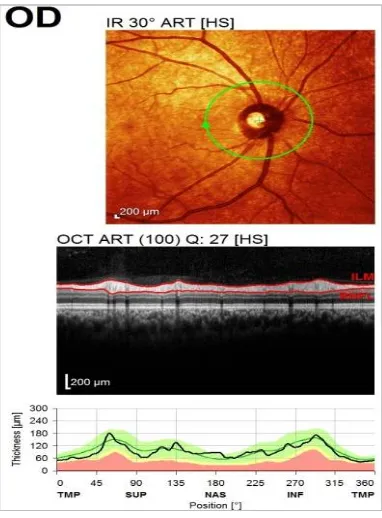
The RNFL examination was under predefined OCT software algorithm which identified the anterior and posterior margins of RNFL and calculated the thickness in different sectors to give an average measurement of RNFL globally and in each quadrant (nasal, temporal, superior and inferior) [Figs. 1a and b].
2.2 Statistical Analysis
mean ± standard deviations were applied. ANOVA and linear regression analysis were done to assess the effect of age. Pooled t-test was applied to assess the thickness variation with gender. P value <0.05 was taken as significant at confidence interval of 95%.
Fig. 1a. RNFL thickness analysis (from anterior margin to posterior margin of RNFL
layer) by using Spectralis software
Fig. 1b. Quadrants of optic disc. G: Globalaverage, S: Superior Quadrant,
I: Inferior Quadrant, N: Nasal Quadrant, T: Temporal Quadrant
3. RESULTS
Three hundred eyes from 150 healthy subjects were clinically examined and were subjected to OCT testing. Mean age of the participants was 57.67±11.42 years. Subjects were divided into 4
groups: 1st group included subjects ranging from 40–49 years of age (n = 102 eyes), 2nd group included subjects ranging from 50–59 years of age (n = 58 eyes), 3rd group included subjects ranging from 60 – 69 years of age (n = 82 eyes) and the 4th group included subjects from 70 years onwards (n = 58 eyes) [Table 1(a)]. A total of 75 females (n = 150 eyes) and 75 males (n = 150 eyes) were recruited in this study [Table 1(b)].
The mean global retinal nerve fiber layer thickness was found to be 99.02±9.08 µm. The superior quadrant showed mean thickness of 121.50±15.03 µm; for inferior quadrant the mean thickness was 126.45±16.23 µm. The nasal quadrant showed mean thickness of 78.99±13.99 µm and the mean thickness for temporal quadrant was found to be 68.90±13.10 µm [Table 2].
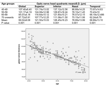
When analyzed by using correlation and regression, the RNFL thickness showed strong negative correlation with age (Beta – 0.8) with P= 0.001 [Fig. 2].
The mean global average retinal thickness for the 1st age group was 107.40±6.40 µm, 2nd age group showed 101.17±4.19 µm, 3rd age group had 95.07±4.18 µm and for the 4th age group it was 87.72±5.91 µm. (P = 0.001) [Table 2].
RNFL variation with gender was not found to be statistically significant in our study group. Mean global retinal thickness in males was 99.10±9.60 µm and in females it was 98.95±8.57 µm with P=0.8 [Table 3].
4. DISCUSSION
To the best of our information this is the first study done in Pakistan using OCT-3 which focuses on the establishment of normal values of RNFL thickness in each quadrant of optic nerve head and its relation to age and gender.
In the present study we determined the normal values of RNFL thickness according to our set of population. By relating this normal data, it will be easier for the clinicians to screen the high risk individuals prior to the onset of various diseases involving the retina especially glaucoma as it is one of the leading causes of irreversible blindness in the world.
In glaucoma there is a gradual loss of retinal ganglion cells (RGCs) leading to reduced retinal
Table 1a. Distribution according to age brackets
Age brackets in years
1 40-49
2 50-59
3 60-69
4
70 onwards
Total (%)
n = no of eyes (%)
102 (34) 58 (19.33) 82 (27.33) 58 (19.33) 300 (100)
Table 1b. Distribution on the basis of gender
Gender Males Females Total (%) n = no of
eyes (%)
150 (50) 150 (50) 300 (100)
thickness [16]. It has been found that before the visual field defect is clinically symptomatic, 30% or more of RCGs are already lost. Significant loss in thickness may lead to visual field defects and optic disc cupping. It has been reported that thinning of retina starts even in the initial stages of glaucoma, therefore, RNFL thickness can be
used as a predictive indicator for glaucomatous damage [17].
Treatment of glaucoma cannot correct the damage but it can prevent the disease to progress, so the sooner the patient is kept on treatment the quicker it can be prevented from causing more harm. Thus, differentiating between healthy eyes and glaucomatous eyes by measuring RNFL thickness may aid in early detection of glaucoma suspects preventing future progression of the disease.
Table 2. RNFL measurements with age using linear regression
Age groups Optic nerve head quadrants mean±S.D. (µm)
Global Superior Inferior Nasal Temporal
40-49 107.40±6.40 131.74±13.02 137.12±12.56 86.88±14.25 73.97±14.63
50-59 101.17±4.19 124.08±12.98 129.87±18.24 78.10±11.45 70.43±10
60-69 95.07±4.18 116.64±10.12 121.60±10.21 76.07±12.15 66.19±12.486
70 onwards 87.72±5.91 107.77±12.25 111.08±11.50 70.15±11.06 62.24±9.79
Mean 99.02±9.08 121.50±15.03 126.45±16.23 78.99±13.99 68.89±13.10
P value 0.001 0.001 0.001 0.001 0.001
Table 3. Comparison of RNFL thickness between male and female by independent T-test
Gender Optic nerve head quadrants mean ± S.D. (µm)
Global Superior Inferior Nasal Temporal
Male 99.10±9.60 120.86±15.21 126.56±15.25 79.88±14.81 69.35±13.41
Female 98.94±8.57 122.14±14.86 126.34±17.21 78.11±13.11 68.43±12.82
P value 0.8 0.4 0.9 0.2 0.5
Our study showed maximum RNFL thickness in inferior quadrant followed by superior, nasal and temporal quadrants which is in accordance with ISNT rule established by Jonas et al in their study conducted in Germany in 1999 [18]. The ISNT rule is an easy way to remember how the optic nerve is supposed to look in a normal eye. Normally the neuro-retinal rim is thinnest temporally and thickest inferiorly. Any deviation from ISNT rule will help the clinicians to detect the optic nerve pathologies at an early stage.
It is stated that aging is accompanied by a reduction of retinal sensitivity and a deterioration of the visual function [13,14] Our results showed strong negative correlation of RNFL thickness with age. This decline in thickness might be due to substantial ganglion cell loss which has been proved by Ooto et al in their study in 2010 [19]. and supported by another study conducted in Germany [20] A Korean study by Song et al in 2010 also suggested that RNFL thickness decreases significantly with increasing age [21].
Gender was not found to have any significant effect on RNFL thickness in our set of population. An Indian study by Rao et al in 2011 also suggested no effect of gender on RNFL thickness [22]. Our results were consistent with the results of other studies done in Florida, England, Korea and Japan [5,11,23,24]. In contrast, study by Kashani et al. in 2010 from Southern California showed significant difference in mean foveal thickness among males and females (201.8±2.7 µm and 186.9±2.6 µm, respectively; P < 0.001) [10]. Another study by Song et al. in 2010 also showed significant correlation between RNFL thickness and gender with the mean thickness of 259.37±23.08 µm in males and 247.90±24.05 µm in females with p=0.009 [21]. This variation in results cannot be explained and its validity needs to be tested in larger sample size.
We came across only one study that was conducted in Lahore, Pakistan by Gondal et al in 2011 in which the normal value for RNFL was obtained in context to the perimetric glaucoma but the sample size was too small to consider it
as a reference value for our set of population. [25].
Our data on RNFL thickness was found to be different with the values previously published for Asians [8] and Caucasians [9]. The mean RNFL global average in our set of population was found to be significantly lesser (p = 0.002) than that obtained by the research from Los Angeles by Alasil et al in 2013 where the mean RNFL thickness for Asians was found to be 100.7±8.5 µm [8]. However in a German study by Bendschneider et al. in 2010, the mean RNFL thickness for Caucasians was found to be 97.2±9.7 µm which is significantly lesser than our values with p= 0.001 [9].
In an Indian study by Appukuttan et al in 2014 the RNFL thickness in each quadrant was calculated and when compared with our values, significant difference was noted [6]. The global average was found to be 101.43±8.63 µm which was significantly higher than our value i.e. 99.02±9.08 µm (p= < 0.001). For the inferior quadrant of Indian population it was found to be 128.34±14.74 µm and for our population it was 126.45±16.23 µm (p = 0.04). The value for superior quadrant of Indian population was 125.27±13.72 µm while our population had different value i.e. 121.50±15.03 µm. (p = < 0.001) For nasal quadrant in Indians it was calculated to be 79.73±12.05 µm and in our population it was 78.99±13.99 µm (p = 0.36). The temporal quadrant showed thickness of 71.95± 7.73 µm in Indian population whereas in our population it was found to be 68.89±13.10 µm (p = <0.001). These contrasting measurements in different regions may be due to diversity in races and differences in technical hands.
Our results are still preliminary but they are promising and require large scale nationwide studies for further strengthening.
5. CONCLUSION
The results of our study have established normal data of RNFL thickness for each quadrant particular to our set of population. This may be used in future by clinicians to screen the high risk individuals prior to the onset of various ophthalmic diseases that involve retina.
The RNFL thickness is significantly correlated with age which shows that for the clinical evaluation of various ophthalmic diseases, age is the important factor to be considered. However, the effect of gender on RNFL is not found to be statistically significant in our study. Assessment of RNFL thickness in various ophthalmic conditions should be interpreted keeping these findings in context.
ETHICAL APPROVAL
Ethical approval was from Ethical Review Committee of Ziauddin University, Karachi, Pakistan.
ACKNOWLEDGEMENT
Ziauddin University for financial assistance. To Akil bin Abdul Qadir Institute of Ophthalmology for allowing me to use their equipments. Mr. Noman Ali for his precious time during my whole research. Dr. Arsalan Manzoor from Ziauddin University, Dr. Tahira Ghazanfar and Miss Hina Mehboob Ali from Akil bin Abdul Qadir Institute of Ophthalmology for their sincere support and help.
COMPETING INTERESTS
Authors have declared that no competing interests exist.
REFERENCES
1. Rao H, Babu J, Addepalli U, Senthil S, Garudadri C. Retinal nerve fiber layer and macular inner retina measurements by spectral domain optical coherence tomograph in Indian eyes with early glaucoma. Eye. 2012;26(1):133-9.
2. Medeiros FA, Zangwill LM, Bowd C, Vessani RM, Susanna R, Weinreb RN.
Evaluation of retinal nerve fiber layer, optic nerve head, and macular thickness measurements for glaucoma detection using optical coherence tomography. American Journal of Ophthalmology. 2005; 139(1):44-55.
3. Wollstein G, Ishikawa H, Wang J, Beaton SA, Schuman JS. Comparison of three optical coherence tomography scanning areas for detection of glaucomatous
damage. American Journal of
Ophthalmology. 2005;139(1):39-43. 4. Seong M, Sung KR, Choi EH, Kang SY,
Cho JW, Um TW, et al. Macular and peripapillary retinal nerve fiber layer measurements by spectral domain optical coherence tomography in normal-tension glaucoma. Investigative Ophthalmology and Visual Science. 2010;51(3):1446-52. 5. Grover S, Murthy RK, Brar VS, Chalam
KV. Comparison of retinal thickness in normal eyes using Stratus and Spectralis optical coherence tomography. Investi-gative Ophthalmology and Visual Science. 2010;51(5):2644-7.
6. Appukuttan B, Giridhar A, Gopalakrishnan M, Sivaprasad S. Normative spectral domain optical coherence tomography data on macular and retinal nerve fiber layer thickness in Indians. Indian Journal of Ophthalmology. 2014;62(3):316.
7. O’ Rese JK, Girkin CA, Budenz DL, Durbin MK, Feuer WJ, Group CONDS. Effect of race, age, and axial length on optic nerve head parameters and retinal nerve fiber layer thickness measured by Cirrus HD-OCT. Archives of Ophthalmology. 2012; 30(3):312-8.
8. Alasil T, Wang K, Keane PA, Lee H, Baniasadi N, de Boer JF, et al. Analysis of normal retinal nerve fiber layer thickness by age, sex, and race using spectral domain optical coherence tomography. Journal of Glaucoma. 2013;22(7):532-41. 9. Bendschneider D, Tornow RP, Horn FK,
Laemmer R, Roessler CW, Juenemann AG, et al. Retinal nerve fiber layer thickness in normals measured by spectral domain OCT. Journal of Glaucoma. 2010; 19(7):475-82.
11. Hirasawa H, Tomidokoro A, Araie M, Konno S, Saito H, Iwase A, et al. Peripapillary retinal nerve fiber layer thickness determined by spectral-domain optical coherence tomography in ophthal-mologically normal eyes. Archives of Ophthalmology. 2010;128(11):1420-6. 12. Ooto S, Hangai M, Tomidokoro A, Saito H,
Araie M, Otani T, et al. Effects of age, sex, and axial length on the three-dimensional profile of normal macular layer structures. Investigative Ophthalmology and Visual Science. 2011;52(12):8769-79.
13. Katz J, Sommer A. Asymmetry and variation in the normal hill of vision. Archives of Ophthalmology. 1986;104(1): 65-8.
14. Heijl A, Lindgren G, Olsson J. Normal variability of static perimetric threshold values across the central visual field. Archives of Ophthalmology. 1987;105(11): 1544-9.
15. Celebi ARC, Mirza GE. Age-related change in retinal nerve fiber layer thickness measured with spectral domain optical coherence tomography. Investi-gative Ophthalmology and Visual Science. 2013;54(13):8095-103.
16. Na JH, Sung KR, Baek S, Kim YJ, Durbin MK, Lee HJ, et al. Detection of glaucoma progression by assessment of segmented macular thickness data obtained using spectral domain optical coherence tomography. Investigative Ophthalmology and Visual Science. 2012;53(7):3817-26. 17. Leung CK-s, Cheung CYL, Weinreb RN,
Qiu K, Liu S, Li H, et al. Evaluation of retinal nerve fiber layer progression in glaucoma: A study on optical coherence tomography guided progression analysis. Investigative Ophthalmology and Visual Science. 2010;51(1):217-22.
18. Jonas JB, Budde WM, Panda-Jonas S. Ophthalmoscopic evaluation of the optic
nerve head. Survey of Ophthalmology. 1999;43(4):293-320.
19. Ooto S, Hangai M, Sakamoto A,
Tomidokoro A, Araie M, Otani T, et al. Three-dimensional profile of macular retinal thickness in normal Japanese eyes. Investigative Ophthalmology and Visual Science. 2010;51(1):465-73.
20. Alamouti B, Funk J. Retinal thickness decreases with age: an OCT study. British Journal of Ophthalmology. 2003;87(7):899-901.
21. Song WK, Lee SC, Lee ES, Kim CY, Kim SS. Macular thickness variations with sex, age, and axial length in healthy subjects: A spectral domain-optical coherence tomography study. Invest Ophthalmol Vis Sci. 2010;51(8):3913-8.
22. Rao HL, Kumar AU, Babu JG, Kumar A, Senthil S, Garudadri CS. Predictors of normal optic nerve head, retinal nerve fiber layer, and macular parameters measured by spectral domain optical coherence tomography. Investigative Ophthalmology and Visual Science. 2011;52(2):1103-10. 23. Sull AC, Vuong LN, Price LL, Srinivasan
VJ, Gorczynska I, Fujimoto JG, et al. Comparison of spectral/Fourier domain optical coherence tomography instruments for assessment of normal macular thickness. Retina (Philadelphia, Pa). 2010; 30(2):235.
24. Kim NR, Kim JH, Lee J, Lee ES, Seong GJ, Kim CY. Determinants of perimacular inner retinal layer thickness in normal eyes measured by Fourier-domain optical coherence tomography. Invest Ophthalmol Vis Sci. 2011;52(6):3413-8.
25. Gondal TM, Qazi ZUA, Jamil AZ, Jamil MH. Accuracy of the retinal nerve fiber layer measurements by stratus optical coherence tomography for perimetric glaucoma. Journal of the College of Physicians and Surgeons Pakistan. 2011; 21(12):749-52.
© 2015 Mukhtar et al.; This is an Open Access article distributed under the terms of the Creative Commons Attribution License (http://creativecommons.org/licenses/by/4.0), which permits unrestricted use, distribution, and reproduction in any medium, provided the original work is properly cited.
Peer-review history:


