_____________________________________________________________________________________________________
*Corresponding author: E-mail: [email protected];
www.sciencedomain.org
Some Features of the Bone Reaction after Implant
Insertion with Rough or Smooth Surface in
Experiment
M. S. Toder
1, A. I. Shevela
2, A. A. Shevela
1,3, P. A. Zheleznyi
3,
A. P. Zheleznaia
3and I. V. Mayborodin
2*1
International Center of Implantology, iDent, Novosibirsk, Russia.
2
Center of New Medical Technologies of Institute of Chemical Biology and Fundamental Medicine of the Siberian Branch of the Russian Academy of Science, 630090, Novosibirsk, Russia.
3
Novosibirsk State Medical University, Russia.
Authors’ contributions
This work was carried out in collaboration between all authors. Author MST designed the study and wrote the protocol. Author AIS designed the study, wrote the protocol and managed the experimental
process.Author AAS managed the experimental process and wrote the first draft of the manuscript.
Author PAZ managed the literature searches. Author APZ managed the experimental process. Author IVM designed the study, wrote the protocol, managed the experimental process, wrote the first draft of the manuscript and performed the analysis of microscopic data. All authors read and approved the final manuscript.
Article Information
DOI: 10.9734/BJMMR/2016/24649 Editor(s): (1) Ashish Anand, Department of Orthopaedic Surgery, GV Montgomery Veteran Affairs Medical Center, Jackson, MS,
USA. Reviewers: (1) Carlos Nelson Elias, Instituto Militar de Engenharia, Brazil. (2)Wagih Mommtaz Ghannam, Mansoura University, Egypt. Complete Peer review History:http://sciencedomain.org/review-history/13486
Received 28th January 2016 Accepted 19th February 2016 Published 27th February 2016
ABSTRACT
The metal implants with helicoid carving and rough or smooth polished surfaces (3S, Israel) were administrated in tibial proximal condyle of not purebred rabbits. At 2 and 6 months after intervention the bone tissues from implantation places were investigated by method of light microscopy. In all cases the edges of bone tissue had minimal scar changes, and both types of metal articles connected to bone densely. Implants were delimited from red and yellow bone marrow structures by thin layer from different types of connective tissue. Morphological symptoms of an inflammation and merge of macrophages in multinuclear cellsweren't detected in one supervision. However the very
small metal fragments were observed in tissues near both types of articles in isolated cases. Considerable microscopic differences of bone tissue condition after implantation of rough or polished metal articles weren't found. Also essential distinctions of condition of tissues around implants between in 2 and 6 months were absent after administration of each article type.
Keywords: Intra bone introduction of metals; delimited of foreign bodies from bone; metal particles in tissues.
1. INTRODUCTION
Tissue response to the intrusion of a foreign body usually includes inflammation, vascular responses and its separation by connective tissue [1-6]. Minimal expression of inflammatory process (low number of leukocytes in the tissue, weak expression of vascular response), absence of giant cells of foreign bodies in metal implants enclosing tissues, insufficient thickness of connective tissue capsule for all terms of research indicates the inertness of such materials for living organism, high compatibility of many metals with biological tissues [7,8].
1.1 The Aim of the Study
To compare morphologic tissue reactions after implantation into the bone of metal implants with rough or smooth surface.
2. MATERIALS AND METHODS
The tests of new articles, their comparison with the applicable articles, testing the new methods of implantation is carried out on experimental animals. The best suitable for this goal are rabbits, specifically proximal condyle of their shin bone, the structure of which corresponds to some human bones very closely. Besides, this part of the extremity of these animals are easy accessible. Due to it, very many researchers use this model for experimental implantation.
Metal articles with various type of surface: rough dental implant 3,75 x 10 mm from Titanium grade 6 having a bullet form and aggressive helicoid carving with grit blasting and acid processing (3S, Israel); and smooth polished article 6 x 10 mm made also of the Titanium grade 6 having a broad-blade helicoid carving for bicortical dental implantation (3S, Israel) were implanted into proximal condyle of shin bone of 10 non-pedigreed rabbits.
All manipulations with the animals were not connected with distress and carried out in compliance with «Regulations on the works using experimental animals». The implantation was
carried out with all aseptic and antiseptic rules in terms of sterile operating room under general anesthesia based on total intravenous anesthesia by propofol. At the initial stage marginal vein of auricle was punctured and catheterized by 24 G catheter, which were fixed by adhesive plaster. The intravenous premedication was carried out: atropine sulphate 0,1% - 0,22-0,27 mg/kg; benadryl 1% - 4,6-5,2 mg/kg; droperidol 0,25% - 1,25 mg; ketorolac trimetamine 1% - 10 mg. Propofol 1%- 15mg/kg was used intravenously as anesthesia induction, propofol 1% - 25-30 mg/kg/hour intravenopusly was also used for support of anesthesia. The infusion therapy was effected by 0,9% sodium chloride solution of 15-25 ml/kg, depending on the expression of blood loss. The respiratory support when necessary was carried out by mask method with 100% oxygen insuflation.
Removal of hair was carried out by surgical scissors on the place of supposed surgical invasion on both extremities at knee joints. After manipulations the skin was processed by alcohol, the operation field was covered with a sterile nap with a hole.
For implantation of articles with rough surface the left extremity was chosen, for implantation of smooth implants – the right extremity was chosen. It is necessary to note that the introduction of each implant into the same extremity of different animals firstly gives an opportunity to accelerate the implantation procedure itself, because there is no need to register what animal and what extremity an article was implanted into; secondly, it makes further observation and examination of animals easier. The sequence of manipulations for implantation:
1. The skin section was made by disposable scalpel along the front line a little bit below the knee joint.
2. The tissues were moved apart to the periosteum by the blunt method (closed branches of the forceps).
4. The hole in chosen part was made by the dentist’s drill up to the “downfall”. 2 bores of increasing diameter are used.
5. The implant was screwed into the made hole by hand and then by the spanner with dynamometric scale, allowing to control the force created. The implant head goes beyond the bone surface for not more than 1 mm.
6. Operative wound was taken in by vicryl 5-0 on each layer.
7. Skin stitches were treated by 5% alcohol-iodine solution.
The animals get rid of stitches by themselves within 1-2 weeks after the operation, there were no visual signs of acute inflammatory process. In most cases for all times the implants were placed on introduction place: in proximal condyle of tibial bone.
For morphologic research of rabbits, the animals were taken out of the experiment 2 or 6 months after the operation by overdose of inhalation ether narcosis. Due to the fact that it was necessary to evaluate the conditions of surrounding tissues and to determine suppurative complications around before removal of foreign bodies, it is reasonable to preliminary remove the entire skin from the extremity from inguinal fold to down to the ankle. In cases, when the implant head was covered by tissues, it was released by the scalpel. Then the spanner with dynamometric scale was put onto it and the implant was screwed out step by step, registering the force at the beginning of its turn. After removal of foreign body a hole with even edges remained in tissues with traces of thread pane.
All manipulations with the object leading to its damage should be better carried out after its fixation to exclude doubts in life-time changes, and further decalcification makes the bone weak and facilitates the excision of the fragment required for further processing, besides, the dehydrogenated bone does not crack. For further morphological research the shin bone after maximal removal of soft tissues was fixed in 4% para-formaldehyde solution on phosphate buffer not less than 24 hours. Then the bone was decalcified by the solution «Biodec R» (Bio Optica Milano, Italy) within 7 days, which damages the tissues less compared with acid decalcification. The duration of processing was determined trying to puncture the bone by dissecting needle far from the hole after removal of the implant. Decalcification was stopped after
the bone became soft and elastic and was easy to cut by scalpel. Further, the bone fragments with the hole were dehydrated constantly mixing up in ethanol with increasing concentration, were clarified in xylene and enclosed into plasticized paraffin. The sections of 5–7 µm thick were stained with hematoxylin and eosin and examined by the light microscope Axioimager M1 (Zeiss, Germany) at magnification to 1200 times.
3. RESULTS
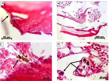
The edge of proximal condyle of shin bone, immediately adjacent to the implant with a rough surface had no notable changes during microscopic research. It can be noted only structures of callus in some parts (Fig. 1а). The formation of a thin strip of compact bone tissue was noted in bone marrow at places of its contact with the implant material (Fig. 1b). But in some cases, the foreign body was separated from bone marrow structures by fibrous connective tissue, closer to metal it was separated by dense and further by friable tissue. The bone marrow itself both red and yellow had no visible pathologic changes. Metal fragments of different size without surrounding inflammatory reaction were found in bone tissue itself, immediately adjacent to the foreign body, and in bone marrow (Fig. 1c, d).
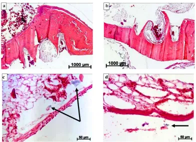
In some cases clear traces from the screw thread edge of the implant in condyle tissue immediately after introduction of the metallic foreign body with a polished surface by little zoom of the light microscope, the bone sides had cicatricle changes, and the cavities formed after removal of the article and were filled in with blood or fibrin. It is necessary to note that thinning of the whole bone in limited parts was found, which can be referred to the installation of the screw-thread at this place (edges of external diameter of the bolt) or with long-term pressure of the foreign body on the bone tissue (Fig. 2a, b). No pathologic changes were found both in red and yellow bone marrow inside the bone at the place of introduction of the foreign body. Along the implant the bone marrow was separated by fibrous connective tissue: closer to metal – by a dense, further – by friable or thin strip of compact bone. Sometimes hemorrhages were present in such connective tissue. In some cases it was practically impossible to differ a dense fibrous connective tissue from a rough fibrous tissue of the bone. Probably, these types of connective tissue separating the foreign body and bone
(Fig. 2c, d). It is also necessary to note the presence of metal particles in connective tissue, separating the bone marrow from the surface of smooth implant, and metal was present close to friable connective tissue and close to rough fibrous tissue, similar to the bone. But the number of such metal fragments and their size was much less than it was after introduction of article with rough surface (Fig. 2c, d).
4. DISCUSSION
The decrease of bone thickness on limited parts at the place of contact with edges of implant thread is probably conditional on atrophy processes firstly and on reorganization of bone tissue, secondly. There are many reports in literature about atrophic and even necrotic changes in tissues contacting with foreign bodies [8-11].
Any foreign body is separated from the organism by connective tissue capsule and this is a normal response of the tissue [1-3]. The most probably is that because of it the founded separation of implants from bone marrow structures occurred by various types of fibrous connective tissue: closer to metal- by dense, further – by friable. Sometimes a thin strip of compact bone was formed there, that is also one of the types of connective tissue. Probably, these types of connective tissue separating the foreign body and bone marrow structures turn into each other in some parts.
At the place of contact of living tissues with the implant, the tissues respond to the foreign body. Probably, the implants immediately after their introduction into the bone firstly initiate acute inflammatory reaction due to surgical trauma and direct interaction of living tissues with the firm
Fig. 1. The morphological results of introduction in a proximal condyle of a rabbit’s tibial bone metal screw implants with a rough surface. Staining by hematoxylin and eosin. a - Structures of a bone callosity in proximal condyle tissue of a tibial bone in 2 months after introduction of
an implant, in certain cases at a bone there are particles of metal (arrow). b - Formation of a thin strip from a compact bone with cicatricial changes in a place of contact of bone marrow
with material of a foreign body in 2 months after implantation. c - Freely located metal fragments of various sizes (arrow) on border between a bone with bone marrow in 2 months after operation. d - The encapsulated particles of metal (arrow) in red bone marrow in 6 months
Fig. 2. The histological changes after implantation of metal screw implants with a polished surface in a rabbit’s tibia. Staining by hematoxylin and eosin. a - 2 months later after implantation in a place of product side contact with a bone of condyle the traces of a screw
thread are located, the bone tissue has cicatricial changes; the bone cavities created after removal of an implant are filled with fibrin or blood. b - In a bone tissue in 6 months after implant introduction there are traces of screw thread sides, the cavities formed after extraction
of a foreign body are filled with fibrin or blood cells. c - Connective tissue with haemorrhage and metal particles (arrow) in marrow in the region of the polished foreign matter in 2 months
after surgical intervention. d - 6 months later after implantation the bone marrow in a tibial condyle is delimited of a foreign body by tissues very similar to a bone or a periosteum, near
such tissues the particles of metal (arrow) are located
non-elastic non-living substance. Acute inflammatory process is gradually changed by chronic one and as it goes out slowly the full union of implant surface with the bone occurs.
By removal of the article, such dense connections of bone tissue with metal break and the cavities in the bone (traces from the edges of screw-thread of the implant) are filled in with blood or fibrin. Because of the same, hemorrhages occurred in connective tissue, separating the foreign body from the bone marrow.
Besides, because even firm foreign bodies destruct by protection systems of human body, the processes of implant degradation go on along the borderline of the implant. Large and
The absence of inflammatory reaction, giant cells of foreign body, connective tissue capsule in surrounding tissues for all times of research give evidence for inertness of the material of articles with rough and with smooth surface for living organism, for compatibility of their materials with tissues. Probably, the material influences on the nature of relationship of the foreign body with living tissues in the first instance, and then the type of surface.
5. CONCLUSION
Thus, considerable microscopic differences of bone tissue condition after implantation of rough or polished metal articles weren't found. Also essential distinctions of condition of tissues around implants between in 2 and 6 months were absent after administration of each article type.
CONSENT
It is not applicable.
ETHICAL APPROVAL
All authors hereby declare that all experiments have been examined and approved by the appropriate ethics committee of Center of New Medical Technologies of Institute of Chemical Biology and Fundamental Medicine and have therefore been performed in accordance with the ethical standards laid down in the 1964 Declaration of Helsinki.
COMPETING INTERESTS
Authors have declared that no competing interests exist.
REFERENCES
1. Maĭborodin IV, Shevela AI, Matveeva VA, et al. Morphological tissue changes after the implantation of elastic lamellar foreign bodies in the experiment. Morfologiia. 2012;141(2):54-60.
2. Maiborodin IV, Kuznetsova IV, Beregovoy EA, et al. Tissue reactions during the
degradation of polylactide implants in the body. Morfologiia. 2013;143(3):59-65. 3. Maĭborodin IV, Shevela AI, Kuznetsova IV,
et al. Tissue responses to silicone materials in the body. Arkh Patol. 2013; 75(4):28-33.
4. Miro-Mur F, Hindié M, Kandhaya-Pillai R, et al. Medical-grade silicone induces release of proinflammatory cytokines in peripheral blood mononuclear cells without activating T cells. J. Biomed. Mater. Res. B Appl. Biomater. 2009;90(2):510-520. 5. Rodriguez A, Anderson JM. Evaluation of
clinical biomaterial surface effects on T lymphocyte activation. J. Biomed. Mater. Res. A. 2010;92(1):214-220.
6. Rodriguez A, Meyerson H, Anderson JM. Quantitative in vivo cytokine analysis at synthetic biomaterial implant sites. J. Biomed. Mater. Res. A. 2009;89(1):152-159.
7. Günter WE, Dambayev GTs, Sysolyatin PG, etc. Medical materials and implants with shape memory. Tomsk: Publishing House Tomsk Un-that. 1998;487.
8. Maĭborodin IV, Iakushenko VK, Maĭborodina VI. Interaction of nickelide-titanium implant with tissues in human. Arkh Patol. 2002;64(2):50-52.
9. Gabriel SE, Woods JE, O'Fallon WM, et al. Complications leading to surgery after breast implantation. N. Engl. J. Med. 1997; 336(10):677-682.
10. Kanhai RC, Hage JJ, Karim RB, Mulder JW. Exceptional presenting conditions and outcome of augmentation mammaplasty in male-to-female transsexuals. Ann. Plast. Surg. 1999;43(5):476-483.
11. Tebbetts JB. A system for breast implant selection based on patient tissue characteristics and implant-soft tissue dynamics. Plast. Reconstr. Surg. 2002; 109(4):1396-1409.
12. Greene WB, Raso DS, Walsh LG, et al. Electron probe microanalysis of silicon and the role of the macrophage in proximal (capsule) and distant sites in augmentation mammaplasty patients. Plast. Reconstr. Surg. 1995;95(3):513-519.
_________________________________________________________________________________ © 2016 Toder et al.; This is an Open Access article distributed under the terms of the Creative Commons Attribution License (http://creativecommons.org/licenses/by/4.0), which permits unrestricted use, distribution, and reproduction in any medium, provided the original work is properly cited.
Peer-review history: