Original Article
Xwig1, a novel putative endoplasmic reticulum
protein expressed during epithelial morphogenesis and
in response to embryonic wounding
PAMELA KLINGBEIL, GIOVANNI FRAZZETTO and TEWIS BOUWMEESTER*
Developmental Biology Programme, European Molecular Biology Laboratory (EMBL), Heidelberg, Germany
ABSTRACT In a subtractive differential screening, we identified a novel gene with interesting characteristics, termed Xenopus wounding induced gene 1 (Xwig1). Xwig1 encodes a novel protein of 912 amino acids containing 13 putative transmembrane segments and an evolutionarily conserved carboxy-terminal domain. Protein localization studies revealed that Xwig1 is anchored in cytoplasmic structures, presumably the endoplasmic reticulum. Expression is largely confined to epithelial cells in regions that undergo morphogenetic processes, such as blastopore closure, hindgut closure, dorsal closure and optic vesicle invagination. Interestingly, Xwig1 transcription is activated in response to embryonic epidermal wounding. The wounding-induced transcription occurs downstream of the transient phosphorylation of extracellular signal-regulated protein kinases and is in part mediated by Elk-1, but independent of dissection-induced FGF signalling. Thus, Xwig1 provides a molecular link between epithelial morphogenesis and wound healing.
KEY WORDS:
Xenopus laevis, wound healing, morphogenesis, endoplasmic reticulum.
0214-6282/2001/$25.00
© UBC Press Printed in Spain
www.ijdb.ehu.es
*Address correspondence to: Dr. T. Bouwmeester. Cellzome GmbH, Meyerhofstrasse 1, D-69117 Heidelberg, Germany. FAX: +49-6221-13757-201. e-mail: [email protected] or [email protected]
Abbreviations used in this paper: BMP, bone morphogenetic protein; ER, endoplasmic reticulum; ERK, extracellular signal-regulated protein kinase; EST, expressed sequence tag; FGF, fibroblast growth factor; GFP, green fluorescent protein; JNK, jun N-terminal kinase; MAPK, mitogen-activated protein kinase; ORF, open reading frame; RACE, rapid amplification of cDNA ends; RT-PCR, reverse transcription polymerase chain reaction; SRE, serum response element; Xwig1, Xenopus wounding induced gene 1.
Introduction
During embryonic development cell-cell communication is of crucial importance for pattern formation and morphogenetic events. The vertebrate embryonic body plan is set-up during gastrulation by intercellular signals, originating in the Spemann-Mangold or gastrula organizer (Harland and Gerhart, 1997; Nieto, 1999). Most of these intercellular signals act as growth factor antago-nists, such as Chordin and Noggin which are BMP inhibitors, Frzb-1 which inactivates Wnt ligands and Cerberus, which acts as a multivalent growth factor antagonist, inhibiting BMP, Wnt and Nodal-related ligands (Nieto, 1999; Bouwmeester, 2000). Through this extracellular inhibitory mechanism cells in all three germ layers acquire their fate and corresponding morphogenetic be-haviour.
Most of the downstream target genes identified so far encode a variety of transcriptional regulators that act as signal-induced effectors. So far, very few structural or other components have been identified that act cell autonomously, downstream of the signal-induced effectors, to co-ordinate cellular processes, such as cell shape changes, cell polarity or cell repair mechanisms.
Here we present the molecular cloning and characterization of a novel putative endoplasmic reticulum (ER) protein, termed
Results
Molecular cloning and characterization of Xenopus wig1 In our continuing effort to isolate novel genes expressed in the Spemann-Mangold organizer we identified in a subtractive differ-ential screening a partial 3’-UTR fragment, that showed an interesting novel expression profile. A full-length cDNA of 2988 bp
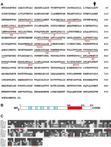
was assembled from a 2.5 kb partial cDNA, isolated from a tailbud cDNA library, and a 5’-RACE extension frag-ment. The resulting contig contains a long open reading frame that is pre-ceded by an in frame termination codon, indicating that it is indeed full-length. This is furthermore supported by North-ern analysis which revealed the pres-ence of a single transcript of ~3.1 kb (data not shown). Based on the finding that this novel gene was activated in response to embryonic wounding we termed it Xenopus wounding induced gene 1 or Xwig1. Xwig1 encodes a putative protein of 912 amino acids with a predicted molecular weight of 99 kDa (Fig. 1A). The deduced amino acid sequence contains 13 hydrophobic seg-ments that are predicted to be trans-membrane domains, suggesting a 13-pass membrane spanning topology (Fig. 1 A,B). Prosite motif predictions indicated the presence of a cleavable signal peptide (cleavage site at posi-tion 58) although this was not con-firmed by SMART analysis (Schultz et al., 2000). Xwig1 contains multiple consensus protein kinase C and ca-sein kinase 2 phosphorylation sites and 13 potential N-myristoylation sites. Besides the hydrophobic segments it does not contain any obvious biologi-cal motif.
Blast searches against the nr data-base revealed weak homology (probabil-ity score 1e-16) to the beta-1,4-exocellulose E6 precursor (celF) from Thermobifida fusca and other bacteria and to the Sua5 gene product from Sac-charomyces cerevisiae (Na et al., 1992). Dbest searches revealed strong homol-ogy to a putative protein encoded by zebrafish EST AW184075 (77% identity from aa 647-836). In addition, weaker, but significant homology was detected to human EST AV652546 (30% identity from aa 651-826) and chick EST AJ394934 (29% identity from aa 643-833) (Fig. 1C). Two other Xenopus EST sequences were also hit, corresponding to distinct, but related genes. EST AW766117 showed
Fig. 1. Predicted amino acid sequence of Xwig1 and homology with putative proteins encoded by different vertebrate ESTs.(A) Amino acid sequence of Xwig1. The black arrow indicates the presump-tive signal peptide cleavage site and underlined in red are the numbered hydrophobic segments. (B) Schematic representation of Xwig1. Indicated are the putative transmembrane regions (blue) and the evolutionarily conserved carboxy-terminal domain (red). (C) Amino acid sequence alignment between the carboxy-terminus of Xwig1 (amino acids 646-814), zebrafish EST AW184075, human EST AV652546, chick EST AJ394934 and Xenopus ESTs AW766117 and BE491372. Underlined in red are conserved amino acids between all proteins. The genebank accession number for Xwig1 is AF310008.
51% identity (from aa 623-813) and EST BE491372 showed only 27% identity (from aa 642-811). Interestingly, human EST AV652546 and chick EST AJ394934 are more closely related to the most divergent Xenopus EST BE491372 than to Xwig1, suggesting that these might constitute an orthologous group (Fig. 1C; grey shading). Blast search against the Drosophila genomic database revealed 25% identity to genomic scaffold 1420000133866047 (section 1 of
B
cyte lysate translation system after addition of microsomal mem-branes, even though other control proteins were efficiently proc-essed (data not shown). Thus, Xwig1 might be a resident endo-plasmic reticulum protein.
Fig. 2. (A) Subcellular localization of Xwig1.GFP fusion protein in animal cap explants. Confocal images showing Xwig1.GFP distribution (green channel), cortical actin visualized by rhodamin phalloidin (red channel), nuclei visualized by DAPI staining (blue channel) and the overlay. Individual channels were scanned and each image is the merge of 4 optical sections. Xwig1.GFP is detected in cytoplasmic structures, presumably the endoplasmic reticulum. (B) RT-PCR analysis of Xwig1 expression during embryonic development (stages according to Nieuwkoop and Faber, 1967). Histone H4 serves as an internal loading control.
52). The identity was again confined to the conserved carboxy-terminal motif from aa 641-857 (data not shown). Thus, Xwig1 is the founding member of a small evolutionarily conserved gene family that is defined by a novel domain of unknown generic function.
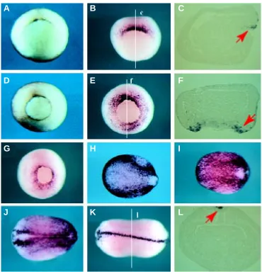
Fig. 3. Xwig1 is expressed in epithelial cells of the Spemann-Mangold organ-izer. Whole-mount in situ hybridization analy-sis was usedto determine the spatio-tem-poral expression of Xwig1 during early (A-C), mid (D-F), late (G) gastrulation and neu-rulation (H-L). A and D are wild-type pigmented embryos to show the blastopore rim. During gastrulation, expression is con-fined to epithelial cells of the blastopore rim (red arrows in C,F). At the end of neurulation expression is observed in a dorsal axial row of cells (red arrow in L). All whole embryos are vegetal views, except H-K which are dorsal views with anterior left.
To gain insights into the biological function of Xwig1 we determined the subcellular localization of a carboxy-terminally-tagged Xwig1.GFP fusion protein. Contrary to Prosite predictions, Xwig1.GFP staining was not detected in the outer cell membrane after ec-topic expression in animal cap explants. Instead however, we found the Xwig1.GFP chimera to be local-ized in intracellular cytoplasmic struc-tures, which presumably correspond to the endoplasmic reticulum (Fig. 2A). Similar results were obtained with the Xwig1.HA chimera (data not shown). In line with the observation that Xwig1 is not secreted, we did not observe proteolytic processing of the Xwig1 precursor in an in vitro rabbit
reticulo-A
B
A
B
C
D
E
F
G
H
I
Xwig1 is differentially expressed during Xenopus
em-bryonic development
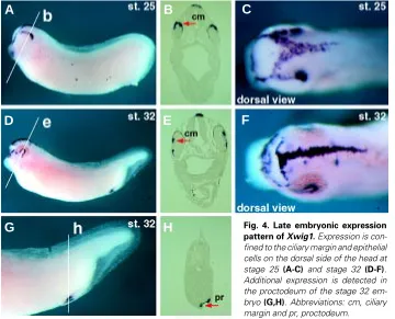
The spatio-temporal expression profile of Xwig1 was deter-mined by RT-PCR analysis and whole mount in situ hybridization. Xwig1 is weakly expressed maternally and transiently activated during embryonic development (Fig. 2B). Zygotic transcription is initiated at the early gastrula stage in superficial epithelial cells of the dorsal blastopore rim of the Spemann-Mangold or gastrula organizer (Fig. 3 A-C). As the blastopore rim extends ventral-laterally Xwig1 expression is likewise extended around the epi-thelial cells of the blastoporal circumference (Fig. 3 D-F). Expres-sion remains strongest on the dorsal side and appears punctuate, presumably nuclear staining reflecting de novo synthesis (Fig. 3E). During the course of gastrulation Xwig1 expression remains associated with epithelial cells of the blastopore rim (Fig. 3G). At the end of gastrulation/onset of neurulation expression increases throughout the entire ectoderm with slightly elevated levels at the non-neural/neural plate border (Fig. 3 H,I). This pattern is ob-served throughout neurulation, although the generic ectodermal staining decreases over time. At the end of neurulation expres-sion is detected only in a single row of dorsal axial cells that seal off the ectoderm/ neural tube along the entire anterior-posterior axis (Fig. 3 K,L). At tailbud stages Xwig1 transcripts are detected in the dorsal aspect of the optic vesicle, where infolding of the retina is initiated, and on the dorsal side of the developing cranium (Fig. 4 A-C). During late eye morphogenesis Xwig1 expression is maintained in the ciliary margin that lines the inner surface of the closing retina and in a stereotypical pattern on the head (Fig. 4 D-F). In addition, expression is maintained in the proctodeum (Fig. 4 G,H). In summary, Xwig1 expression is primarily associated with epithelial cells that form constrictions during different
mor-phogenetic processes, such as blastopore closure and optic vesicle invagination.
Xwig1 expression is activated in
re-sponse to embryonic wounding
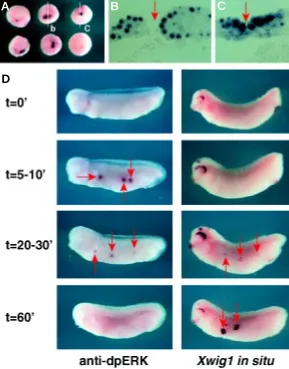
During the course of the whole-mount in situ hybridization analyses we frequently observed ectopic patches of Xwig1 expres-sion in the periphery of ectodermal wounds, which were presumably inflicted by the dechorionation procedure. To further inves-tigate this striking observation we systemati-cally lesioned gastrula animal cap ectoderm with a fine needle and analyzed Xwig1 ex-pression in response to this wounding. In all cases (n=42) we observed that wound inflic-tion in uncommitted gastrula ectoderm re-sulted in weak to strong activation of Xwig1 expression around the periphery of the em-bryonic wound (Fig. 5A). Ectopic activation in many cases was prominent in the nuclei of wounded cells reflecting de novo synthesis (Fig. 5 B,C; red arrow indicates the wound). Embryonic wounding is known to evoke a transient dual phosphorylation of the extra-cellular signal-regulated protein kinases (ERK) (LaBonne and Whitman, 1997; Chris-ten and Slack, 1999). Therefore, we com-pared the temporal kinetics of Xwig1 transactivation to the rapid transient post-translational modification of ERK-1 and –2. To do so, we lesioned differentiated epidermal cells in the abdominal flank of the tailbud embryo and subjected wounded embryos after defined incubation spans to either anti-dpERK immunostaining or Xwig1 in situ hybridization analyses. As previ-ously shown, ERK-1 and –2 are biphosphorylated within 5’-10’ after epidermal wounding in an ephemeral fashion (LaBonne and Whitman, 1997; Christen and Slack, 1999; Fig. 5D; left panel). In contrast, the transcriptional activation of Xwig1 is first observed 20’-30’ after wound infliction and is strongest after 60’, when phosphorylated ERKs are no longer detectable (Fig. 5D; right panel). In summary, the temporal correlation suggests that Xwig1 is an immediate-early component of the transcriptional programme that is acti-vated in response to embryonic epidermal wounding in Xenopus.
Dissection-induced Xwig1 activation is partially
de-pendent on Ets/Elk factors
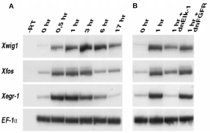
Dissection of animal cap explants results in the spurious activation of immediate-early target genes, such as Xfos and Xegr-1 (Krain and Nordheim, 1999). To compare the expression kinetics of wounding-induced Xwig1 with Xfos and Xegr-1 activa-tion we performed a time course on dissected cap explants and analyzed gene expression by RT-PCR. All three genes are activated within 30’ of incubation (Fig. 6A). Whereas, Xfos and Xegr-1 expression is transient, declining after 3 hr, Xwig1 tran-scripts accumulate till maximum levels after 3 hr and can be detected for a prolonged period till 6 hr before declining. The expression of the wounding-induced immediate-early target genes, Xegr-1 and Xfos is to different extent dependent on Ets/Elk transcription factors that bind to serum response elements (SRE) (Krain and Nordheim, 1999). Ets/Elk transcriptional regulators are
Fig. 4. Late embryonic expression pattern of Xwig1. Expression is con-fined to the ciliary margin and epithelial cells on the dorsal side of the head at stage 25 (A-C) and stage 32 (D-F). Additional expression is detected in the proctodeum of the stage 32 em-bryo (G,H). Abbreviations: cm, ciliary margin and pr, proctodeum.
A
B
C
D
E
F
substrates for the FGF-induced Ras/Raf/ERK signal trans-duction module (Treisman, 1996). To test if Xwig1 like-wise depends on Elk activity and/or on FGF signalling we injected animal blastomeres with mRNA coding for domi-nant negative forms of Elk (dnElk-1) and the FGF receptor (dnFGFR) and subjected explanted animal caps to RT-PCR analysis. Dissection-induced Xwig1 expression is partially downregulated by dnElk-1, to the same extent as ectopic Xfos activation (Fig. 6B). Wounding-induced Xegr-1 transcription is completely abolished to control levels (0 hr), in agreement with data shown by Krain and Nordheim, 1999. The inhibition of FGF signal transduction has no significant effect on the dissection-induced activation of any of the three genes tested, even though FGF-depend-ent mesoderm formation was completely inhibited in con-trol embryos (data not shown).
Discussion
In this study we have presented the molecular cloning and spatio-temporal expression characteristics of a novel multipass membrane spanning protein, termed Xenopus wig1. Xwig1 expression is mainly observed in epithelial cells during diverse morphogenetic processes, such as blastopore closure and optic vesicle invagination. In addi-tion, we have shown that Xwig1 transcription is activated in response to embryonic ectodermal wounding. The wounding-induced activation occurs downstream of ex-tracellular signal-regulated protein kinase (ERK) activa-tion and is partially mediated by Elk/Ets factors.
Xwig1 encodes a novel protein with 13 potential mem-brane-spanning segments and an evolutionarily conserved carboxy-terminal domain. Limited homology is observed with the beta-1,4-exocellulose E6 precursor (celF) from bacteria and SUA5, a Saccharomyces cerevisiae protein required for growth (Na et al., 1992). In addition, several putative proteins encoded by vertebrate ESTs show ex-tensive amino acid sequence identity with the carboxy-terminus. Despite the evolutionary conservation no func-tional link is apparent between these gene products.
What could be the role of Xwig1 during embryonic development and wound healing? Ectopic expression of
down-regulated by dnElk-1. Xwig1 kinetics are slightly different from both Xfos and Xegr-1, as peak levels are first observed between 3-6 hr, when expression of the other two genes is down-regulated. However, the sustained peak expression might in part be due to endogenous up-regulation at the onset of neurulation (see Fig. 3). Xfos expression requires Elk-1 and p38 MAPK-induced effectors for optimal activation in particular stress conditions (Price et al., 1996). This observation suggests that wounding-induced Xwig1 activation requires additional factors too besides an Ets/Elk component. These could be effectors activated by the related JNK/SAPK or p38 MAPK cascades or may be different in nature. Epithelial wounding after physical damage is known to result in Ca2+ influx, which in turn may
trigger alterations in gene expression (Martin, 1997; Grose and Martin, 1999). In contrast to Xwig1 and Xfos, wounding-induced Xegr-1 expression is solely dependent on SRE binding Ets/Elk factors. The endogenous Xegr-1 expression in the marginal zone mesoderm is likewise dependent on Elk-1 activity and is mediated by multiple serum response elements (Panitz et al., 1998). The
endog-Fig. 5. Xwig1 transcription is activated in response to embryonic epithelial wounding.(A) Wounding-induced Xwig1 expression in gastrula embryos. (B,C) Transverse sections of embryos displayed in (A). (D) Time course (0’-60’) of Xwig1 activation compared to the transient phosphorylation of ERK-1 and –2, visualized by anti-dpERK staining, in response to lateral epidermal wounding of stage 27 embryos. Red arrows indicate the embryonic wounds. Xwig1 transcription is activated downstream of ERK activation.
Xwig1 mRNA in different lineages (up to 2 ng/blastomere) has no detectable morphological effect on embryonic development, nei-ther on cell shape of dissected animal cap ectoderm cells (data not shown). Xwig1 appears to be anchored in the endoplasmic reticulum. Xwig1 does not contain a bonafide ER membrane reten-tion motif, but does carry a KKXX/KDEL-like retenreten-tion/retrieval motif (KDLKL) at the extreme carboxy-terminus. Xwig1 could be involved in the secretion of growth factors or other membrane proteins akin to the role of the evolutionarily conserved porcupine gene product in the processing of Wg/Wnt ligands (Tanaka et al., 2000). Porcupine is also a resident ER protein with multipass transmembrane topology. Xwig1 could be involved in the post-translational modification of secreted proteins, although this awaits further biochemical charac-terization. Despite the ER retention in animal cap cells, it is still conceivable that signal peptide processing might be a regulatory step in the biogenesis of Xwig1 protein in different cellular contexts.
How is Xwig1 expression regulated? Dissection-induced Xwig1 up-regulation is similar to Xfos activation, as both are only partially
A
B
C
enous expression patterns of Xwig1 and Xegr-1 bear no similarity also arguing for diverse regulatory control elements.
In future it will be worth dissecting in detail the regulatory network that controls the dissection-induced activation and the endogenous expression pattern of Xwig1 to better understand the common elements between embryonic wound healing and certain morphoge-netic events, such as blastopore closure and optic vesicle invagina-tion in Xenopus. In addiinvagina-tion, morpholino-mediated translainvagina-tional knock-down experiments will allow to determine the function of Xwig1 during embryonic development and during the process of wound healing.
Materials and Methods
Molecular cloning of Xwig1 and derived plasmids
A partial 330 bp Xwig1 fragment was initially identified in a subtractive differential screening for novel organizer genes (unpublished). A full-length cDNA was cloned and assembled from a 2.5 kb partial cDNA, isolated from a directional tailbud cDNA library, and a 0.5 kb 5’-RACE extension fragment (SMART kit, Clontech). Carboxy-terminal tagged pCS2+-Xwig1.GFP and
pCS2+-Xwig1.HA chimeras were generated by in frame fusion of the
PCR-amplified Xwig1 ORF with pCS2+-GFP and pCS2+-HA, respectively.
Align-ments were performed with the DNAMan and DNAStar software packages.
Embryo manipulations, in situ hybridization and microinjection RT-PCR analysis, whole mount in situ hybridization, animal cap assays and histology were done as described (Bouwmeester et al., 1996; Fetka et al., 2000).
Xwig1 was amplified by the following primer pair, (F) 5’-CTATGTTCTAGTGGCTGCTTGC-3’;
(R) 5’-GTGAGTAGTAACAGTGCAGTC-3’, yielding an amplicon of 303 bp; 28 cycles.
Xegr-1 was amplified by:
(F) 5’-GAGATGTTAGCCTTGTATCTGC-3’;
(R) 5’-GTACTGTTGATAGTCTTGAGGTCC-3’ (Panitz et al., 1998)
Xfos was amplified by:
(F) 5’-CTCTGTACACATCAGAATGG-3’;
(R) 5’-AATGTCCTTCAGCATTACAG-3’ (Krain and Nordheim, 1999). Fig. 6. Wounding-induced activation
of Xwig1 is partially dependent on Elk-1, but independent of FGF sig-nalling. (A) Time course of Xwig1, Xfos and Xegr-1 activation in response to ectodermal wounding in dissected gastrula animal cap explants. (B) Xwig1, Xfos and Xegr-1 activation in explanted control caps (0 and 1 hr) and caps injected with dominant-negative Elk-1 (1 hr+dnElk-1) and dnFGFR (1 hr+dnFGFR). EF-1α serves as loading control.
Digoxygenin labelled antisense mRNA was synthesized from a 2.2 kb partial cDNA, pCRII-Xwig1, linearized with NdeI and transcribed with T3. Embryos were wounded with a thin needle at stage 10.5 and stage 27 and fixed after defined time points in MEMFA. Anti-dpERK (Sigma) immunostaining was performed as described (Christen and Slack, 1999). For microinjection, pCS2+-dnELK-1 was linearized with NsiI and
tran-scribed with Sp6 (Panitz et al., 1998), pXFD (dnFGFR) with EcoRI and Sp6 (Amaya et al., 1991) and pCS2+-Xwig1, pCS2+-Xwig1.GFP and
pCS2+-Xwig1.HA with NsiI and Sp6.
Confocal microscopy
Xwig1.GFP mRNA was injected into 2 animal blastomeres of 2-4 cell embryos. Animal caps were explanted at stage 9-10, fixed in 4% PFA/ PBS for 1 hr and processed. Caps were first stained with rhodamin phalloidin (Molecular Probes) at 1:500 dilution for 2-3 h, followed by DAPI staining (Boehringer) at 1:2000 dilution for 5 min. Caps were mounted upside down in Mowiol and fluorescence signals were visualized using a LEICA TCS NT laser-scanning confocal microscope. Single channels were separated by NIH Image 1.60 software and images were processed and assembled in Adope Photoshop 5.5 software. Similar results were obtained with a Xwig1.HA chimera. In this case caps were first incubated with anti-HA (BabCO: 1:1000) in PBSt/10% goat serum O/N at 4oC
followed by 1 h incubation in donkey anti-mouse FITC-conjugated sec-ondary antibody (Jackson ImmunoResearch: 1:250).
Acknowledgements
We are grateful to Tomas Pieler for the dominant-negative ELK-1 construct, André Brändli for the tailbud cDNA library, Jun Wu for assistance with confocal microscopy, Ingrid Fetka for technical assistance and we would like to thank Jochen Wittbrodt for critical comments on the manuscript. This work was supported by the EMBL.
References
AMAYA, E., MUSCI, T.J. and KIRSCHNER, M.W. (1991). Expression of a dominant negative mutant of the FGF receptor disrupts mesoderm formation in Xenopus embryos. Cell 66: 257-270.
BOUWMEESTER, T., KIM, S.-H., SASAI, Y., LU, B. and DE ROBERTIS, E.M. (1996). Cerberus is a head-inducing secreted factor expressed in the anterior endoderm of Spemann’s organizer. Nature 382: 595-601.
BOUWMEESTER, T. (2001). The Spemann-Mangold organizer: the control of fate specification and morphogenetic rearrangements during gastrulation in Xenopus.
Int. J. Dev. Biol. 45: 251-258.
CHRISTEN, B. and SLACK, J.M.W. (1999). Spatial response to fibroblast growth factor signalling in Xenopus embryos. Development 126: 119-125.
FETKA, I., DOEDERLEIN, G. and BOUWMEESTER, T. (2000). Neuroectodermal specification and regionalization of the Spemann organizer in Xenopus. Mech. Dev. 93: 49-58.
GROSE, R. and MARTIN, P. (1999). Parallels between wound repair and morphogen-esis in the embryo. Seminars in Cell & Dev. Biol. 10: 395-404.
HARLAND, R. and GERHART, J. (1997). Formation and function of Spemann’s organizer. Annu. Rev. Cell Dev. Biol. 13: 611-667.
KRAIN, B. and NORDHEIM, A. (1999). Artefactual gene induction during preparation of Xenopus laevis animal cap explants. Int. J. Dev. Biol. 43: 563-566.
LABONNE, C. and WHITMAN, M. (1997). Localization of MAP kinase activity in early Xenopus embryos: implications for endogenous FGF signaling. Dev. Biol. 183: 9-20.
MARTIN, P. (1997). Wound healing-aiming for perfect skin regeneration. Science 276: 75-81.
NA, J.G., PINTO, I. and HAMPSEY, M. (1992). Isolation and characterization of SUA5, a novel gene required for normal growth in Saccharomyces cerevisiae. Genetics 131: 791-801.
NIETO, M.A. (1999). Reorganizing the organizer 75 years on. Cell 98: 417-425.
NIEUWKOOP, P.D. and FABER, J. (1967). Normal Table of Xenopus laevis (Daudin).
North-Holland, Amsterdam.
PANITZ, F., KRAIN, B., HOLLEMANN, T., NORDHEIM, A. and PIELER, T. (1998). The Spemann organizer-expressed gene Xegr-1 responds to the MAP kinase/Ets-SRF signal transduction pathway. EMBO J. 17: 4414-4425.
PRICE, M.A., CRUZALEGUI, F.H. and TREISMAN, R. (1996). The p38 and ERK MAP kinase pathways cooperate to activate Ternary Complex Factors and c-fos tran-scription in response to UV light. EMBO J. 15: 6552-6563.
SCHULTZ, J., COPLEY, R.R., DOERKS, T., PONTING, C.P. and BORK, P. (2000). SMART: A Web-based tool for the study of genetically mobile domains. Nucleic Acids Res. 28: 231-234.
TANAKA, K., OKABAYASHI, K., ASASHIMA, M., PERRIMON, N. and KADOWAKI, T. (2000). The evolutionarily conserved porcupine gene family is involved in the processing of the Wnt family. Eur. J. Biochem. 267: 4300-4311.
TREISMAN, R. (1996). Regulation of transcription by MAP kinase cascades. Curr. Opin. Cell Biol. 8: 205-215.