FORMULATION AND EVALUATION OF CAFFEINE-LOADED SOLID LIPID NANOPARTICLES TO
TREAT CLINICAL MASTITIS
B. SURENDRA, M. NAVEEN KUMAR, PADMINI IRIVENTI*
Department of Pharmaceutics, Dr. K.V. Subba Reddy Institute of Pharmacy, Dupadu, Kurnool, Andhra Pradesh, India. Email: [email protected]
Received: 23 March 2020, Revised and Accepted: 25 April 2020
ABSTRACT
Objective: The objective of the present study was to formulate and evaluate caffeine-loaded solid lipid nanoparticles (SLNs) in the treatment of clinical mastitis.
Methods: These were prepared by homogenization technique using stearic acid, Tween 80, and chloroform as excipients. Pre-formulation studies such as UV spectrophotometry, Fourier transform infrared (FTIR), and differential scanning calorimetry (DSC) were performed for the drug. Entrapment efficiency and in vitro dissolution studies were carried out for prepared SLNs and the optimum formulation (F2) was taken for further studies such as FTIR, DSC, SEM, particle size, and zeta potential analysis.
Results: Obtained results stated that prepared SLNs are roughly spherical in nature and are in nano range. These were incorporated in Carbopol gel and further evaluation studies such as pH, spreadability, viscosity, homogenicity, and in vitro drug diffusion studies were carried out. All the results stated that prepared nanogel has shown sustained release of drug. Antimicrobial study was carried out using Staphylococcus aureus and it was confirmed by the appearance of zone of inhibition.
Conclusion: Nanogel that contains caffeine SLNs with 1:2 ratio drug:lipid has shown good in vitro release. Sustained release of caffeine drug till 12 h was achieved by delivering it in the form of nanogel.
Keywords: Caffeine, Stearic acid, Clinical mastitis, Solid lipid nanoparticles, Antimicrobial activity.
INTRODUCTION
Clinical mastitis is an inflammatory condition of the breast that may occur in the breastfeeding women during the puerperium and is reported in women who continue to breast feed up to 1 year after delivery [1].
Recently, a condition called subclinical mastitis has been described. Subclinical mastitis is diagnosed from the finding of a raised sodium-potassium ratio in the milk and an increased concentration of interleukin-8 (IL-8) when there is no clinical mastitis. Increased sodium and IL-8 levels are thought to indicate that an inflammatory response is occurring despite the absence of clinical signs. Due to this disease, milk production falls below 400 ml/day.
Two principle causes of mastitis are milk stasis and infection. Milk stasis is usually the primary cause which may or may not be accompanied by progress to infection. Intramammary infections caused by Escherichia coli and Staphylococcus aureus (E. coli) are commonly considered to be limited in duration. Sometimes, microorganisms may even be eliminated before or shortly after the onset of clinical symptoms. Therefore, the host defense system appears to eliminate microorganisms efficiently especially when the infection occurs late in lactation [2].
In treating and managing clinical mastitis, culture-based therapy and severity levels play a key role. Antibiotic therapy is strongly recommended for clinical mastitis [3].
The veterinary in clinical practice is often confronted with cases of mastitis that requires systemic antibacterial treatment in addition to local treatment. Furthermore, NSAIDs are most commonly used in treatment. Few examples of drugs used are penicillin’s, oxytetracycline, chloramphenicol, sulfonamides, trimethoprim, etc. [4].
Caffeine is a natural alkaloid found in coffee beans, tea leaves, cocoa beans, cola nuts, and other plants. It is probably the most frequently ingested pharmacologically active substance in the world. It is found in common beverages (coffee, tea, and soft drinks), products containing coca, chocolate, and over counter stimulants. Caffeine is used to treat tiredness and drowsiness and used along with other pain relievers to improve their effect [5]. Caffeine is a methyl xanthine moiety capable to hinder the phosphodiesterase (PDE) enzyme which helps in hydrolysis of cyclic nucleotides resulting in elevated concentration of intracellular cyclic adenosine monophosphate (cAMP). Cell surface receptors inhibition for adenosine is another proposed mechanism. Reduced intracellular cAMP levels are seen in cutaneous leucocytes of patients with psoriasis. Many researchers proposed that as a PDE inhibitor and methyl xanthine, caffeine increases intracellular cAMP levels, which consequently suppress inflammatory pathways [6].
Solid lipid nanoparticles (SLNs) are submicrons colloidal carrier ranging from 50 to 100 nm which are composed of a physiological lipid dispersed in water or in aqueous surface solutions. They consist of macromolecular material, in which the active compound is dissolved, entrapped or to which the active compound is dissolved or attached. SLNs are generally spherical in shape and diameter from 10 to 100 nm. Advantages are it controls or target drug release, improve stability of pharmaceutics, feasibilities of carrying both lipophilic and hydrophilic drugs more affordable [7,8].
Various lipids used in the preparation of SLNs are triglycerides (tristearin), partial glycerides (Imwitor), fatty acids (stearic acid and palmitate acid), steroids (cholesterol), and waxes (acetyl palmitate). Various emulsifiers and their combination (Pluronic F 68, F 127) have been used to stabilize the lipid dispersion. Among them, stearic acid is widely used because of its GRAS status and thus known to be safe and © 2020 The Authors. Published by Innovare Academic Sciences Pvt Ltd. This is an open access article under the CC BY license (http://creativecommons. org/licenses/by/4. 0/) DOI: http://dx.doi.org/10.22159/ajpcr.2020.v13i7.37641
used widely in pharmaceutical and cosmeceutical industries. No skin irritations are reported using this lipid from the reports and sources obtained. Hence, it is used in the present study [9,10].
The topical delivery of drugs is an attractive method for local and systemic treatment and commonly used in the treatment of inflammatory conditions such as musculoskeletal injuries and dermatological diseases. There are many advantages in topical application compared to conventional dosage forms. Especially, some serious systemic and adverse effects are avoided.
When the drug is delivered topically, it can penetrate deeper into skin and hence give better absorption. Topical preparation can be used to prevent the metabolism of drug in the liver. It can be used to avoid the gastrointestinal disorders, risks, inconvenience of intravenous therapy, etc. Furthermore, bioavailability of the drug is increased and targeted action can be achieved.
The topical delivery with gels can increase the time of presence of drug on the skin and improve the delivery and release of the substance [11].
In the present study, an attempt has made to prepare SLNs of caffeine using stearic acid as lipid. The novelty of this work is though caffeine SLNs were prepared earlier by others using them in the treatment of clinical mastitis which was not reported till date. These SLNs help in increasing bioavailability of drug and also provide sustained release of the drug.
MATERIALS AND METHODOLOGY
Caffeine was obtained from Loba Chemie, Mumbai, India. Stearic acid was obtained from Qualikems Pvt. Ltd., Vadodara, India. All other excipients were obtained from Pallav Chemicals & Solvents Pvt., Ltd., Boisar, India. All the reagents used were of analytical grade.
METHODOLOGY
Analytical method for caffeine
Calibration curve in pH 7.4 phosphate buffer
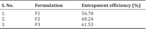
From the standard solution, a stock solution was prepared to give a concentration of 100 μg/ml in 7.4 buffer. Aliquots of 0.5, 1.0, 1.5, 2.0, and 2.5 ml from the stock solution were pipetted out into 10 ml volumetric flasks. The volume was made up to the mark with 7.4 buffer. These dilutions gave 5, 10, 15, 20, and 25 μg/ml concentration of caffeine, respectively. The absorbance of prepared solutions of caffeine in 7.4 buffer was measured at 275 nm spectrophotometrically against 7.4 buffer blank. Standard plot data of caffeine in 7.4 pH buffer are reported in Fig. 1.
Preparation of SLNs Homogenization technique
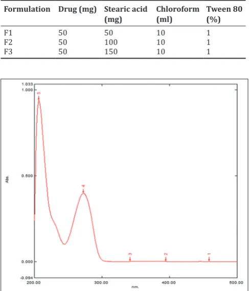
Stearic acid and caffeine were dissolved in 10 ml chloroform in a glass flask (organic phase). Tween 80 was added in 30 ml distilled water and heated up to 75°C using water bath (aqueous phase). Then, the organic phase was added to the aqueous phase under continuous stirring at 1000 rpm using a homogenizer. About 2.5 h later, the flask was removed from the water bath, 10 mL of ice-cold distilled water was added, and stirring of the mixture at 1000 rpm was continued for 2 h. Then, the resulting suspension was washed twice with distilled water by centrifuging at 10,000 rpm for 30 min to remove the supernatant, which contained the free (unreacted) drug. It was followed by freeze-drying that converted it to powder form [12,13]. Three SLN formulations were prepared by varying the drug:lipid ratio. They are given in Table 1.
Characterization and evaluation of SLNs Entrapment efficiency (EE)
To calculate the EE, accurately weighed quantity of 100 mg SLN was taken and dissolved in 7.4 pH buffer. It was stirred for 10 min to break the complex. Then, the solution was filtered and 2 ml was taken from
above solution and diluted up to 10 ml with 7.4 pH buffer [14-16]. It was kept aside for few minutes and absorbance was measured by UV spectrophotometer at 421 nm. It can be calculated using the formula.
EE =Actual drug content in NS Theoretical drug content ×100
In vitro drug diffusion studies
In vitro studies were carried out using cellophane membrane soaked in pH 7.4 buffer overnight. For this studies, Franz diffusion cell was taken and in donor compartment, pH 7.4 buffer was taken. In between donor and receiver compartments, cellophane membrane was placed and tightly held using rubber band. On the membrane, gel was applied. The buffer which passes from donor to receiver compartments through the membrane was collected in receiver compartment using a syringe tube. Sampling was done at regular intervals of 15 and 30 min, 1 h, 2, 3, 4, 5, and 6 h (each time 2 ml of sample was collected and replaced with similar amount of buffer) [14-16]. The obtained samples were analyzed using UV spectrophotometer.
pH buffer is used because human breast milk pH is around 7.4.
Fourier transform infrared (FT-IR) spectroscopic analysis
FT-IR analysis was conducted to verify the interaction between drug and polymer. The sample powder was dispersed in KBr powder and pellets were made by applying 4 kg/cm2 pressure. FT-IR spectra were obtained by powder diffuse reflectance on a FT-IR spectrophotometer type 8400S Shimadzu.
Differential scanning calorimetry (DSC)
DSC was performed on pure drug and its formulations using DSC-60 instrument. Calorimetric measurements were made with empty cell (high purity alpha alumina discs) as the reference. The dynamic scans were taken in nitrogen atmosphere at the heating rate of 10°C min−1. The energy was measured as J/Kcal.
Table 1: Formulation chart of various solid lipid nanoparticle formulations
Formulation Drug (mg) Stearic acid
(mg) Chloroform (ml) Tween 80 (%)
F1 50 50 10 1
F2 50 100 10 1
F3 50 150 10 1
Scanning electron microscopy (SEM)
The surface morphology of formulations was determined using a SEM. Samples were mounted on aluminum mount, using double-sided adhesive tape and sputtered by gold under vacuum and were scanned at an accelerating voltage of 15 KV before observation.
Particle size and zeta potential analysis
The average particle size distribution and charge of the resulting nanoparticles were determined by dynamic light scattering using C:\ Microtrac\FLEX 11.0.0.2 Instruments, United Kingdom. The experiment was performed using clear disposable zeta cell, water as a dispersant which has refractive index – 1.330 and viscosity (cP) – 0.898, and the temperature was kept constant at 25°C. The optimized SLN formulation was further incorporated into topical nanogel prepared.
Nanogel preparation
Appropriate quantity of Carbopol 934 was soaked in water (around 5 ml) for a period of 2 h. Carbopol was then neutralized with triethanolamine (TEA) with stirring. Then, specified amount of SLNs was dissolved in appropriate and pre-weighed amount of propylene glycol. Solvent blend was transferred to Carbopol container and agitated for additional 20 min. The dispersion was then allowed to hydrate and swell for 60 min; finally, the pH was adjusted with 98% TEA until the desired pH value was approximately reached (6.8–7). During pH adjustment, the mixture was stirred gently with a spatula until homogeneous gel was formed. All the samples were allowed to equilibrate for at least 24 h at room temperature before performing rheological measurements [17,18]. Formulation chart of nanogel is given in Table 2.
Evaluation studies pH determination
The pH of the gels was determined using digital pH meter by placing the glass electrode completely into the gel system. The readings were taken for average of 3 times [17,18].
Homogenicity
All developed gels were tested for homogenicity by visual inspection after gels have been set in the container. They were tested for the appearance and presence of any aggregates [17,18].
Spreadability test
Place 0.5 g gel in a pre-marked circle (1 cm diameter) on a glass plate. Another glass plate was then placed over the gel and weight of 500 g was placed over this upper glass plate for 5 min [17,18]. The experiment was carried out in triplicate and spreadability expressed in gm.cm/sec. Spreadability can be calculated using the formula.
S=M.L/T
Where, S=Spreadability M=Weight tied to upper slide L=Length of glass slide
T=Time taken to separate the glass slide completely from each other.
Rheological studies
The rheological measurements were performed on the Brookfield viscometer. All measurements were carried out using parallel plates measuring systems having 50 mm diameter and 1 mm gap at 25°C. The rheological properties of the nanogel were studied at different shear rates (rpm) and the viscosity was measured in cP [17,18].
In vitro drug diffusion studies
The caffeine SLN-loaded nanogel was permeated through dialysis bag. Optimized formulation was selected for these studies. A 0.5 g of nanogel was placed in the bag and is placed in a beaker containing 150 ml of phosphate buffer of pH 7.4 and constantly stirred with a small magnetic bead. During the experiment, temperature was maintained
at 37± 0.5°C to simulate the human skin condition. A 5 ml of samples were withdrawn at 0.5, 1, 2, 6, and 12 h and replaced with fresh receptor solution [17,18]. The samples withdrawn were analyzed spectrophotometrically at 235 nm. The amount of drug released was calculated and the percentage drug released was plotted against time.
Similarly, it was done for marketed topical antibiotic gel.
Antimicrobial test
Organisms used: S. aureus (Gram-positive bacteria).
Procedure
Nutrient agar medium was prepared in aseptic condition (2.8 g of agar powder in 100 ml distilled water). Prepared agar medium was poured in sterilized Petri plates and allowed to solidify. The above-mentioned organism was collected from the culture tube with a prior sterilized metal loop and streaked on solidified agar plates. Then, bores or cups were made in the agar Petri plates with a sterilized metal borer and the sample was placed in the cups. The Petri plates were placed in the incubator for 36 h [19].
RESULTS AND DISCUSSION • EE
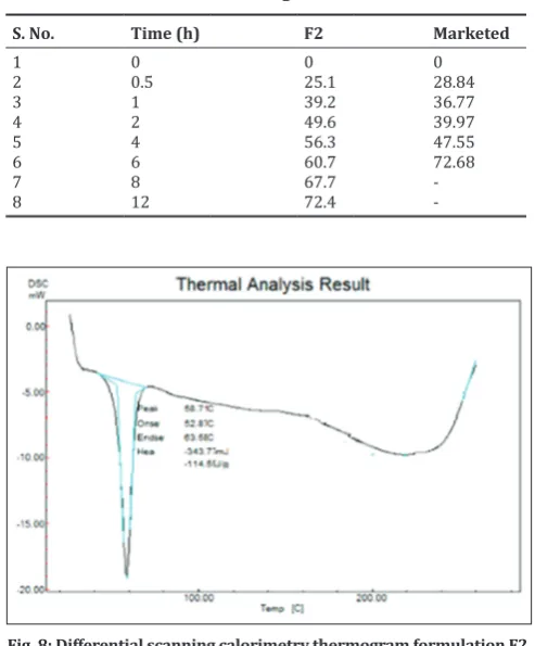
EE studies were carried out for F1-F3 formulations. All the EEs were between 55% and 69%. For F2, EE was 68.24% which is higher than F1 and F3. It is because, in case of F1 (1:1 ratio), equal amount of drug and lipid was present for interaction. Hence, the release rate was reduced due to no extra drug molecules present. In case of F3 (1:3 ratio) formulations, excess lipid molecules were present in the formulation and these molecules formed a thick sheet around the drug leading to delay in drug release. In case of F2 (1:2) formulation, drug and lipid ratio was satisfactory. Although lipid molecule was in excess, they only formed a thin sheet around the drug and hence high % release was observed.
The drug EEs noted for different F1 formulation are given in Table 3.
• In vitro drug diffusion studies
The SLN formulations were subjected to in vitro release studies. The results obtained in in vitro release studies were plotted in percentage cumulative drug release versus time and shown in Fig. 2. It was found that formulation containing drug:lipid in 1:2 ratio has shown maximum in vitro drug release, as compared to other formulations. This could be due to poor loading seen in F1 due to insufficient lipid and in case of F3, it could be able to inability of drug to enter into the larger network of lipid. In vitro drug diffusion studies of all the formulation are given in Table 4.
• SEM
From the results, obtained SLNs were roughly spherical. It shows formation of nanoparticles resembling spheres. SEM image of F2 formulation is given in Fig. 3.
Table 2: Formulation of nanogel
Table 3: Entrapment efficiencies of F1-F6 formulations
S. No. Formulation Entrapment efficiency (%)
1. F1 56.78
2. F2 68.24
• FT-IR spectroscopy (FTIR)
Major peaks of caffeine, i.e., amines, amides, and carboxylic groups with bond frequency of 3300–3000, 3500–3000, and 1710–1650, respectively, are seen in pure caffeine at 3112, 1599 and 3510, and 1661 and 1698, respectively. FTIR spectrum of pure caffeine is shown in Fig. 5.
Similarly, in case of F2, where stearic acid has been taken as lipid, peaks pertaining to amines, amides were observed at 1625 and 2917, respectively. Major peaks such as amines, amides, and carboxylic groups disappeared.
This shows that though pure drug is present, it is entrapped in the lipid system. This is also confirmed with blunt peaks obtained in FTIR spectrum. FTIR spectra of F2 formulation are shown in Fig. 6, respectively.
• DSC
DSC endograms of pure drug and F2 formulation are given in Figs. 7 and 8, respectively. Melting point of pure caffeine was observed at 242°C. In case of F2 formulation, an endothermic peak was seen at 58°C that resembles melting point of stearic acid. Drug peak was observed at around 230°C but was not sharp. The absence of sharp peak pertaining to drug indicates the presence of drug but embedded in lipid system showing drug-lipid compatibility.
Evaluation of nanogels • pH determination
pH of obtained gel was found to be 7.4 which is near to neutral pH. This shows that prepared gel does not cause any skin irritation as it is near to skin pH.
• Homogenicity
All the gels prepared clear and transparent. It shows that no aggregates were present.
• Spreadability
Prepared gel was spread on skin and was found to spread easily. It shows that prepared gel has good viscosity.
• Rheological studies:
Viscosity for prepared gel was found to be good. It shows that obtained results were optimum which helped in good spreadability. All the above results are given in Table 5.
• In vitro drug diffusion studies
The nanogel containing F2 formulation and normal antibiotic gel was subjected to in vitro diffusion studies. The results obtained were plotted in percent cumulative drug release versus time in Fig. 9. It was found that F2 nanogel has shown maximum in vitro drug diffusion of 72.4% and ordinary antibiotic gel release was 72.68%. Furthermore, sustained release up to 12 h was found in former, but it was limited to 6 h in later. This could be due to entrapment and slow release of drug from polymer complex in nanogels, whereas due to availability of free drug, quick release was seen in marketed formulation. In vitro % drug diffusion results of F2 and marketed gel are given in Table 6.
Antimicrobial test
Zone of inhibition was observed and is shown in Fig. 10. From the results, it can be stated that prepared SLN formulation has antimicrobial activity.
Table 4: In vitro drug diffusion studies (F1-F3)
Time (h) F1 F2 F3
• Particle size analysis and zeta potential and measurement Particle size of solid lipid nanoparticles was found between 100 and 1000 nm (steric acid). Average particle size was 181.5 nm in stearic acid. Obtained results state that prepared nanoparticles were in nanosize which is one of the objectives of the study. Particle size distribution of F2 formulation is given in Fig. 4.
Zeta potential is the major function which determines the interaction of formulation with biological system. It determines the charge type present on the nanoparticle surface. Zeta potential of the prepared
SLNs was found as −15.2 mv. It shows decrease in particle size has led
to increase in surface area that resulted in higher zeta potential.
Fig. 3: Scanning electron microscopy photograph of F2 formulation Fig. 2: In vitro drug diffusion studies (F1-F3)
Fig. 5: Fourier transform infrared spectrum of pure caffeine
CONCLUSION
Nanogel that contains caffeine SLNs with 1:2 ratio drug:lipid has shown good in vitro release. Sustained release of caffeine drug till 12 h was achieved by delivering it in the form of nanogel.
ACKNOWLEDGMENT
Authors are thankful to the Principal, Dr. K.V. Subba Reddy Institute of Pharmacy, Kurnool, for providing necessary facilities to carry out the work.
AUTHORS’ CONTRIBUTIONS
All the authors contributed equally in preparation of manuscript.
CONFLICTS OF INTEREST Authors have none to declare.
AUTHORS’ FUNDING
Authors did not receive any funding for the present work.
REFERENCES
1. Barbara B, Hogg MD. Puerperal mastitis. Glob Libr Womens Med 2008. Doi: 10.3843/glowm.10142.
2. Dopfer D, Barkema MW, Lam TJ, Schukken YM, Gaastra W. Recurrent clinical mastitis caused by Escherichia coli in dairy cows. J Dairy Sci 1999;82:80-5.
3. Constable PD, Morin DE. Treatment of clinical mastitis. Using antimicrobial susceptibility profiles for treatment decisions. Vet Clin North Am Food Anim Pract 2003;19:139-55.
4. Diarmid SC. Antibacterial drugs used against mastitis in cattle by the systemic route. N Z Vet J 1978;26:290-5.
5. Nawrot P, Jordan S, Rotstein J, Hugehottz A, Feeley M. Effects of caffeine on human health. Food Addit Contam 2003;20:1-30.
6. Alireza B, Alaleh R, Mohammad MF, Saeed S, Marzieh A, Ahmed N. The role of caffeine in pain management: A brief literature review. Anesth Pain Med 2016;6:e33193.
7. Boushey HA. Bronchodilators and other agents used in asthma. In:
Katzung BG, editor. Basic and Clinical Pharmacology. 7th ed. Los
Altos, CA: Appletion and Lange; 1998. p. 330-7.
8. Alashqar M, Goldstein N. Caffeine in the treatment of atopic dermatitis and psoriasis; A review polo. In: Psoriasis. Landon: From Gene to Clinic International Congress; 2017. p. 77.
9. Ratwat MK, Jain A, Singh S. Studies on binary lipid matrix based solid lipid nanoparticles of repaglinide: In vitro and in vivo evaluation. J Pharm Sci 2011;6:2366-78.
10. Anu M, Satvinder K. Design, formulation and characterization of steric acid-based solid lipid nanoparticles of candesartan cilexetis to augment its oral bioavailability. Asian J Pharm Clin Res 2018;11:344-50. 11. Milla GB, Silvioalan GB, Camilamahara DD, Lassissa AR, Pedro RN,
Carvalho FO, et al. Development and evaluation of stability of a gel formulation containing the monoterpene borneol. Sci World J 2016;2016:7394685.
12. Chandra BP, Nagaraju R, Saritha D, Sailakshmi B, Srikanth R. Formulation and evaluation of lansoprazole loaded nanosponges. Turk J Pharm Sci 2016;13:304-10.
13. Phatak AA, Chaudhari PD. Development and evaluation of nanogel as a carried for transdermal delivery of aceclofenac. Asian J Pharm Tech 2012;2:125-32.
14. Bakhtiary J, Barar A, Aghanejad AA, Saei E, Nemati J, Ezzati ND, et al. Microparticles containing erlotinib-loaded solid lipid nanoparticles for treatment of non-small cell lung cancer. Drug Dev Ind Pharm 2017;43:1244-53.
15. Mona MA, Amira MM. Solid lipid nanoparticles and nanostructured
Table 6: In vitro % drug diffusion studies
S. No. Time (h) F2 Marketed
1 0 0 0
Table 5: Evaluation parameters of F2 nanogel
S. No. Evaluation parameter Results
Fig. 9: In vitro % drug diffusion studies
lipid carriers of tolnaftate: Design, optimization and in vitro evaluation. Int J Pharm Pharm Sci 2016;8:380-5.
16. Yasmin BM, Prathyusha RG. Formulation and evaluation of dasatinib loaded solid lipid nanoparticles. Int J Pharm Pharm Sci 2018;10:14-20. 17. Sheikh SR, Abdul BA. Development and evaluation of mucoadhesive
nanogel of nevirapine for vaginal application. Int J Appl Pharm
2019;11:144-9.
18. Mahendra AG, Rasika DB. Formulation and evaluation of topical anti-inflammatory herbal gel. Asian J Pharm Clin Res 2019;12:252-5. 19. Halima S, Rachida A, Fatima ZE. Antioxidant and antibacterial