Anusha Narayan
In Partial Fulfillment of the Requirements
for the Degree of
Doctor of Philosophy
California Institute of Technology
Pasadena, California
2010
c
2010
Anusha Narayan
Acknowledgements
I’d like to begin by thanking my two advisors. I hope I can absorb a fraction of Paul
Sternberg’s unique blend of energetic enthusiasm and level-headed pragmatism, and I
very much appreciate his way of making sure I always had the support and resources
I needed. Earning my experimental spurs in the laboratory of Gilles Laurent was
a formative experience, and I learned much from his approach to science and his
knowledge of physiology. Together, they made sure I always had a safety net even
as I was out on a limb, sawing madly; when the going was rough their support and
concern was extraordinary.
Thanks are also due to my committee: Shuki Bruck, who was supportive and
encouraging from my earliest days at Caltech; Erin Schuman who was always
inter-ested, and never short of ideas and suggestions; and Thanos Siapas, who was always
available, concerned and supportive.
Having two advisors meant that I was lucky enough to work with two great groups
of people: the Sternberg and Laurent labs, who blended to a nicety inspiring science
and goofy fun. In particular, I would like to thank Vivek Jayaraman and Glenn Turner
for their friendship, for being always generous with their time and counsel, and for
respec-nique, was one of the scientific highlights of my time in grad school. Watching Cindy
Chiu take on and conquer tasks of incredible complexity was always inspirational, as
was Eric Mosser’s ability to maintain an even keel: I would like to thank them both for
their support and friendship. I tremendously enjoyed cahooting with Melanie Pribisko
Yen and the Eating Club, combining our collective passion for talking and eating. I
would like to thank Ofer Mazor and Benjamin Rubin for innumerable highly
enjoy-able conversations running the gamut from slow microprocessors to water-buffaloes
bellowing across a swamp. I would also like to thank the many good friends from the
early weeks of international student orientation who have enriched my life at Caltech.
In particular, I would like to thank Rogier Braakman, for his support, his calm good
sense, his cheerful patience, and a number of other things. And finally, I would like
to thank my parents. I would not be at Caltech without them, and I will always be
Abstract
The nematode C.elegans, with its 302 neurons and abundance of genetic, laser
ab-lation, electrophysiological and imaging tools, is a compact and attractive system
for neural circuit analysis. An understanding of the functional dynamics of neural
computation requires physiological analyses. We undertook the first characterization
of transfer at central synapses in C.elegans. To achieve this we employed optical
stimulation techniques using channelrhodopsin-2, and combined this with whole-cell
patch clamp electrophysiological recording techniques. We show that the synapse
be-tween AFD and AIY, the first stage in the thermotactic circuit, exhibits excitatory,
tonic and graded release. The gain at the synapse was low (<0.1), and release was frequency independent, showing no signs of facilitation or depression. The AFD-AIY
synapse thus seems designed for robust and reliable transmission of a scaled-down
temperature signal from AFD to AIY, enabling AIY to continuously monitor
temper-ature information and integrate it with other incoming sensory information. We also
investigated the synapse between ASER, a chemosensory neuron, and AIY, and found
that the synaptic response was small and inconsistent. The combination of optical
stimulation tools with neural recording techniques is a powerful way to analyze
Contents
Acknowledgements iv
Abstract vi
List of Figures xi
1 Introduction 1
1.1 The anatomy and nervous system of the nematode C. elegans . . . . 1
1.2 Worm behavior . . . 3
1.3 The worm toolkit . . . 4
1.4 Remote optical control of neural circuitry . . . 4
1.5 Electrophysiology in the worm . . . 7
1.6 Functional versus Static connectivity maps . . . 8
1.7 Outline of Thesis . . . 9
2 Transfer at a thermosensory synapse in C.elegans 11 2.1 The AFD-AIY synapse and the circuit for thermotaxis . . . 11
2.2 Experimental Procedures . . . 15
4.2.1 Activation of ASER using ChR2 . . . 54
4.2.2 Response in AIY to ASER activation . . . 56
4.3 Discussion . . . 58
4.4 Contributions . . . 60
5 Conclusions and future directions 62 5.1 Characteristics of transfer at worm synapses . . . 62
5.2 Future directions . . . 63
List of Figures
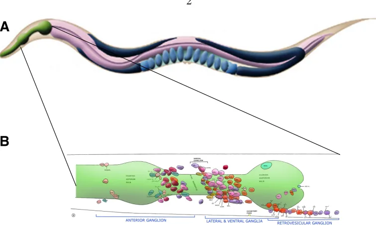
1.1 The worm nervous system . . . 2
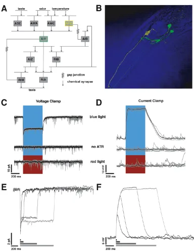
2.1 Using chR2 to stimulate AFD. . . 13
2.2 ChR2 stimulation and recording setup. . . 17
2.3 Calibrating AFD response to Blue Light. . . 20
2.4 Synaptic Response in AIY is tonic. . . 22
2.5 Spontaneous events in AFD and ASER. . . 25
2.6 AIY synaptic response is graded and reverses between -20 mV and 0 mV. 28 2.7 AFD and AIY response to different pulse stimulation protocols: no evi-dence for facilitation and depression. . . 30
3.1 Reversing the AFD-mediated synaptic current in AIY . . . 41
3.2 I-V curves for synaptic current . . . 43
3.3 Effect of Cs+ on synaptic current . . . . 45
3.4 Native currents in the AIY membrane . . . 46
3.5 Effect of substituting Cs+ for K+ on the IV curve . . . . 47
3.6 Spontaneous events in ASER . . . 49
3.7 Spontaneous events in AFD . . . 49
4.1 Activating ASER using ChR2 . . . 55
4.2 AIY voltage response to ASER stimulation . . . 56
4.3 AIY current response to ASER stimulation . . . 57
Chapter 1
Introduction
From the perspective of systems neuroscience, a small nervous system with access to
a multiplicity of tools to selectively perturb and analyze the activity of ensembles of
neurons has tremendous appeal. C. elegans, with its three-hundred odd neurons, is
one such system.
1.1
The anatomy and nervous system of the
ne-matode
C. elegans
C. elegans is a roundworm, one of around 30,000 species belonging to the Phylum
nematoda. It is a free-living soil nematode feeding on bacteria, about 2-3 mm in
length when full grown, and has fewer than a thousand cells in total. Of these, 302
are neurons in the hermaphrodite (there are both males and hermaphrodites in C.
elegans). The male has 79 additional neurons, nearly all of which are involved in
control of mating. It takes around three days for a worm to grow from egg to adult,
sinusoidal movement of the worm, are studded along the ventral midline. There are
two nerve cords - dorsal and ventral, that carry the processes of most neurons from the
posterior part of the worm as they project to the nerve ring. There are approximately
6400 chemical synapses, 900 gap junctions and 1500 neuromuscular junctions (Altun
et al., 2002-2009).
1.2
Worm behavior
For its small size and compact nervous system, the worm exhibits a range of behaviors
(de Bono & Maricq, 2005). In addition to executing functions such as locomotion,
mating and egg-laying, a worm will exhibit preferences in temperature, the valence
and concentration of chemicals, and mechanosensory environment. When placed
in a shallow thermal, chemical, or electrical gradient, worms will migrate towards
preferred zones consistently and reproducibly, based on initial conditions and past
history (Hedgecock & Russell, 1975, Ward, 1973, Sukul & Croll, 1978). A worm will
respond to negative stimuli such as a harsh mechanical touch, or aversive chemicals
by backing away rapidly. Worms also display social feeding behavior (de Bono, 2003)
as well as basic forms of learning such as habituation and paired conditioning (Giles
the possible uses of remote control of cellular signaling.
The use of optical stimulation techniques has opened up many exciting possibilities
for the control and selective manipulation of neural circuits, and in combination with
electrophysiology can be used to address fundamental questions of neural coding.
1.5
Electrophysiology in the worm
The earliest electrophysiology from a nematode was an analysis of the locomotor
cir-cuit in the worm Ascaris lumbricoidis (Walrond et al., 1985, Walrond & Stretton,
1985a,b, Davis & Stretton, 1989a,b). Neurons in Ascaris are large - around 80-100
µm in diameter. Motor neurons in Ascaris were found to exhibit graded active and synaptic responses, and tonic synaptic release (Davis & Stretton, 1989a). Through
simultaneous recordings of motor neurons and the muscles they innervate, the
lo-comotor circuit was found to employ reciprocal inhibition to produce a sequence
of alternating contractions of the worm body, enabling its characteristic sinusoidal
movement pattern (Walrond & Stretton, 1985b). Given the striking anatomical
sim-ilarities between the locomotory circuit inAscaris and C. elegans, these studies were
very informative in assessing the excitatory and inhibitory function of specific motor
neurons in C. elegans.
aver-what order would open up an enormous range of interesting questions. In order to
understand the dynamic functional connectivity, an understanding of the mechanisms
of transfer and gain control are called for. Electrophysiological analyses, and to some
extent, calcium imaging of neural activity are the tools that are most appropriate to
convert our static connectivity maps into functional ones, by allowing us to monitor
neural activity over different timescales, and during behavior.
1.7
Outline of Thesis
This thesis describes an attempt to characterize transfer at C. elegans synapses by
combining electrophysiological and optogenetic techniques, by expressing ChR2 in
pre-synaptic neurons and using whole-cell patch clamp recording techniques to
mon-itor the post-synaptic neuron. Our attempt is the first functional characterization of
the dynamics at central synapses in the worm. We begin by describing a prominent
thermosensory synapse, between the sensory neuron AFD and the interneuron AIY,
in Chapter Two. We find that this synapse is excitatory, has low gain, and exhibits
graded and tonic release. Chapter Three describes a further voltage-clamp
character-ization of the AFD-AIY synapse and additionally, describes native voltage activated
currents in AFD, AIY and ASER, a chemosensory neuron. In Chapter Four, we
the synaptic response to be weak and unreliable. Finally, in Chapter Five, we present
Chapter 2
Transfer at a thermosensory
synapse in
C.elegans
2.1
The AFD-AIY synapse and the circuit for
ther-motaxis
C.elegans combines several useful features as a model organism for neural circuit
anal-ysis: a compact nervous system, detailed anatomical data from electron microscopy
(EM), and access to system analysis tools such as precise genetic manipulations,
fo-cal laser ablations of individual neurons, and real-time monitoring of neural activity
using calcium imaging and electrophysiology. In addition, having a small nervous
system does not preclude the worm from exhibiting a range of behaviors that can be
described quantitatively (de Bono & Maricq, 2005, Hobert, 2003). One such behavior
is thermotaxis. When placed on a temperature gradient, worms aggregate to the
temperature at which they were cultivated (Tcult) and track isotherms as narrow as
0.05◦C near it (Hedgecock & Russell, 1975). Worms can track isotherms near Tcult
the basic features of synaptic transfer at a central chemical synapse inC.elegans.
2.2
Experimental Procedures
2.2.1
Strains
To identify AFD and AIY we used the strains PY1322 and OH98 expressing gfp
in AFD and AIY respectively. We obtained the strain PY1322 oyIs18 [gcy-8::gfp]
from the Caenorhabditis Genetics Center (CGC). OH98 (mgIs32 [ttx-3::gfp,
lin-15(+)];him-5) was a gift from Oliver Hobert; we outcrossed it to N2 (wildtype) to
eliminate the him-5 mutation. We obtained the strain OH3192 (ntIs1 [gcy-5::gfp])
from the CGC and used it to identify ASER. We performed recordings on synaptic
transmission mutants deficient inunc-13, a syntaxin binding protein, using the strain
BC168 unc-13(s69) which was a gift from Anne Hart. We constructed a strain
ex-pressing chR2 in AFD, PS5755 (syIs218 [gcy-8::chR2::yfp, pax-2::gfp, lin-15(+)]), by
injectinggcy-8::chR2::yfp plasmid DNA into MT1642lin-15(n765ts) worms with the
co-injection marker pHC294.1, apax-2::gfp plasmid with GFP expression in the vulva
and tail (a gift from Helen Chamberlin). We integrated the line by subjecting it to
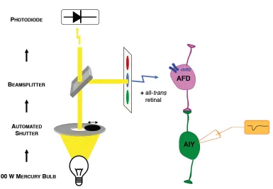
Figure 2.2: chR2 stimulation and recording setup. A 100W Mercury lamp provides light. A computerized shutter controls the shape of the light pulse. A beamsplitter sends a fraction of the light to a photodiode for real-time monitoring of the light stimulus, and the bulk of the light through a gfp filter to excite the preparation with blue light.
achieve a holding potential of around -65 mV (mean: -67 mV, median -66.8 mV,
interquartile distance 3 mV, n=66 cells).
Light stimulus was provided using a 100W mercury lamp. A Sutter SmartShutter
was used to control timing. The opening and closing latencies of the shutter were
8-12 ms. A liquid light guide was used to make the field of view uniform. The light
was filtered using an Olympus gfp filter cube to provide blue light within the
450-490nm wavelength range. A high-speed silicon photodetector (Det100A, Thor Labs)
was mounted on a beam splitter at the light source to continuously monitor the light
stimulus waveform. Figure 2.2 shows a schematic of the setup. The peak intensity of
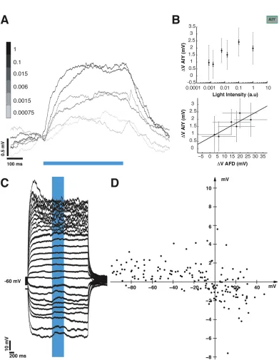
ically, these cells tend to be more depolarized than spiking neurons. It is important to
take this into account, since the value of resting potential that we attempt to mimic
with our holding potential will affect our transfer function, as the membrane may have
different regimes of behavior varying with potential. In the absence of true estimates
of resting potential, we attempted to assess the effect of holding potential by injecting
current to clamp AFD at different voltage values and assessing the evoked
depolar-ization. We clamped the cell from +40 mV to -100 mV and measured the response
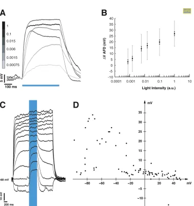
to a fixed light pulse at each step. We note that the light evoked depolarization in
AFD varies as a function of holding potential (Figure 2.3C shows an example set of
traces from one neuron, the V-V curve in Figure 2.3D shows the variation of evoked
potential with holding potential, 7 trials from 6 neurons), and over the range of -30
to -70 mV, the evoked depolarization is in the range of 20-40 mV. This is comparable
to the depolarization we evoke in our experiments with a holding potential of ∼-65 mV.
2.3.2
Depolarization of AIY by presynaptic, light-evoked
de-polarization
Having established a calibration of our presynaptic light-evoked depolarization, we
If the synapse between AFD and AIY is chemical, it must have an associated ionic
conductance and thus, reversal potential. To assess the value of this putative
rever-sal potential, we imposed a presynaptic light-evoked depolarization while AIY was
held at holding potentials between -100 and +40 mV (Figure 2.6C; 7 trials from 4
neurons). The synaptic potential appears to reverse between -20 mV and 0 mV
(Fig-ure 2.6D). The synaptic depolarization becomes more pronounced at hyperpolarized
potentials, although the amplitude variation was less dramatic. At more depolarized
holding potentials, the membrane response became noisier. This is probably due to
the opening of other voltage-activated conductances, which might explain the greater
variability in evoked response size.
We attempted the same experiment in voltage clamp; however due to the small
size of evoked currents it was difficult to resolve changes with holding potential. Data
from those experiments are presented and discussed in Chapter 3.1.
2.3.6
No evidence of facilitation or depression at the
AFD-AIY synapse: frequency independent
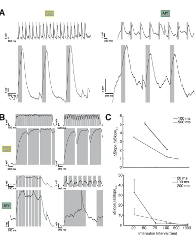
We stimulated the synapse with trains of pulses ranging in width and intervals from
20 ms to 1 s. We observed no significant change in the size of the synaptic response.
we found no clear trend. We also computed a similar metric: ratio of the slope of the
rising phase of the first pulse to the average of the succeeding ones; here, we found
a decreasing trend that was mirrored both pre and post-synaptically (Figure 2.7C).
We conclude that there is no frequency dependence in the evoked potential, and no
obvious facilitation or depression, at least within this range.
2.4
Discussion
Previous ablation and imaging studies show that the neuron AFD responds to
warm-ing (Kimura et al., 2004) and that it senses temperature at the distal end of its
den-drite (Chung et al., 2006). Our stimulation of this synapse substitutes light-mediated
ChR2 activity for temperature and, indeed, is the first study to do so. How relevant
are our stimulation levels to those that AFD experiences in vivo?
Electrophysiolog-ical data (Ramot et al., 2008) indicate that AFD responds to changes in ambient
temperature with inward currents of ∼10 pA and depolarizations of ∼40 mV when T>Tcult. These values are comparable to our ChR2 mediated currents and voltages
in AFD. With stimulation in this range, the present study shows that the AFD-AIY
synapse is graded and tonic, with a faithful rendering of ’∆T’ from AFD to AIY.
The first graded central synapses to be characterized were in the metathoracic
lineariz-showing a response that is time- and frequency- invariant; the low (<0.1) gain may
facilitate processing in AIY by rescaling its input to stay within its dynamic range.
The use of optical stimulation techniques in combination with physiology can serve
as a powerful tool in our efforts to understand how this compact nervous system
Chapter 3
Further characterization of
synaptic and native currents in
AFD, AIY and ASER
In the previous chapter, we characterized the first synapse in the thermosensory
pathway inC. elegans. The synapse was graded and tonic with low gain. The evoked
currents and potentials were small ( <0.5 pA and ∼2-4 mV). In this chapter, we describe additional features of the synaptic response, and provide a preliminary
char-acterization of the native voltage-dependent currents exhibited in AIY. In addition,
we document the spontaneous psps we recorded from a variety of neurons.
3.1
Reversal of the AFD-mediated synaptic
cur-rent in AIY
We previously characterized the synaptic potential evoked in AIY in response to
stim-ulating the presynaptic neuron AFD with blue light (see Figure 2.6C). Additionally,
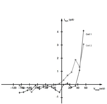
Figure 3.2: Effect of holding potential on the AFD-mediated synaptic current in AIY.Evoked synaptic current, Isyn as a function of holding potential, Vhold. I-V curves
synap-Figure 3.6: Spontaneous events in ASER The ASER membrane exhibits a range of voltage dependent behavior in response to current injections ranging from +5 pA to -5 pA from holding current (the current needed to keep the cell at -74 mV.) The presence of small psp-like events is notable.
Figure 3.8: Spontaneous events in AIYin response to current injections ranging from +5 pA to -5 pA from holding current
reduces other voltage-activated currents, revealing these events. As the membrane is
depolarized, the synaptic potentials should start to reverse. We cannot discern clear
reversals. One possible explanation could be the fact that at depolarizing potentials
other voltage gated currents are activated, making it challenging to isolate individual
synaptic events. To test if these events are synaptic in origin, we recorded from the
same neurons in an unc-13 (s69) mutant background. Unc-13 is a syntaxin-binding
protein and unc-13 mutants have significantly reduced synaptic transmission
(Rich-mond et al., 1999). We found significant reduction in frequency of such events (see
Figure 2.5) in ASER. We conclude that at least a fraction of these psp-like events are
synaptic in origin.
Such events were seen in AFD as well (see Figure 3.7) , and the AFD membrane
3.5
Discussion
The synaptic current evoked in AIY through light-evoked depolarization of AFD was
small and appeared to reverse in the range of -20 mV to 0 mV; this is consistent
with a current carried by a mixed-cationic conductance. The synaptic current was
either abolished or unchanged when Cs+ was substituted for K+in the patch pipette,
leaving the effect of Cs+ upon the synaptic current unresolved. However, voltage
dependent outward currents in AIY were reduced fourfold when Cs+ was substituted
for K+, indicating that a large fraction of the voltage activated outward current
appears to be K+ mediated. The membrane of AFD, AIY and ASER all exhibit a
variety of voltage dependent potentials, and a high incidence of psp like events, at
least a fraction of which are synaptic in origin. We conclude that the membrane of
these neurons in C. elegans exhibit rich dynamics that help shape their response to
Chapter 4
Transfer at a chemosensory
synapse in
C. elegans
The worm is known to navigate towards certain cues and away from others; this
behavior presumably allows it to find food and mates. Sensory information from
the external environment is transduced through a set of sensory neurons, processed
through layers of interneurons, and finally affect the behavior of the worm by directing
movement toward or away from the stimulus. In Chapter 2 we described one form
of sensory response exhibited by the worm, thermotaxis; in this chapter we discuss
another: the ability to navigate chemical gradients, chemotaxis.
4.1
The circuit for chemotaxis and the ASER-AIY
synapse
Chemical sensation is an important mechanism by which C. elegans explores and
navigates its environment. Worms can chemotax towards peak of gradients of ions
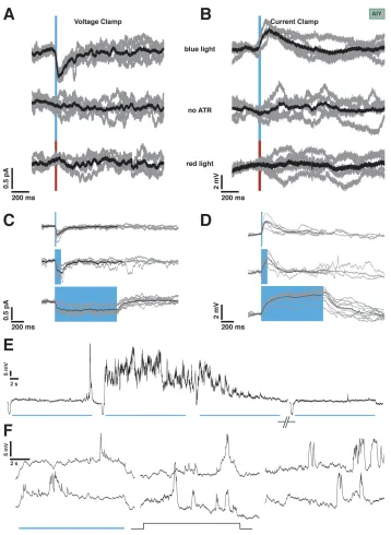
Figure 4.1: Activating ASER using ChR2.Voltage clamp recordings from ASER. A 500 ms pulse of blue light, and not red, causes inward current in ASER expressing functional chR2. All-trans retinal is required. 10 trials from a single neuron in gray, average in black. VHold = -65 mV. D. Current Clamp recordings from AFD in situations identical to (C).
Figure 4.2: AIY voltage response to ASER stimulationA. Current clamp recordings from AIY in response to a 1 second long pulse of blue light. 5 example trials from one neuron, top (black), overlay of 10 trials from same neuron (gray) with average (black), bottom. VHold =∼-70 mV. B. Current clamp recordings from AIY in response to red light,
5 individual trials, top, 7 trials (gray) overlaid with average (black), bottom. VHold = ∼
-65 mV.
evoke currents up to 10 pA and depolarizations up to 40 mV (Figure 4.1A). Control
experiments with red light or using worms fed no ATR showed no response (Figure
4.1B). The response was reliable and reversible.
4.2.2
Response in AIY to ASER activation
Having ensured that our light stimulation does indeed depolarize ASER, we next
recorded responses from AIY to blue light stimulation. With 1 s pulses of blue light,
we occasionally saw a small depolarization in AIY. Figure 4.2A shows example traces
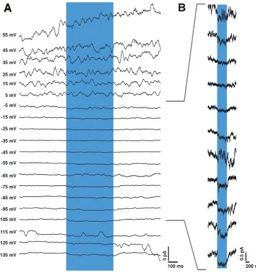
Figure 4.3: AIY current response to ASER stimulation. A. Voltage clamp record-ings from AIY in response to a 200 ms long pulse of blue light. 5 example trials from one neuron, top (black), overlay of 20 trials from same neuron, bottom (gray), average of 20 trials, bottom (black). VHold = -65 mV. B. Voltage clamp recordings from AIY in response
Figure 4.4: AIY response to ASER stimulation is not consistent. Voltage clamp recordings from AIY in response to 200 ms pulse of blue light. 20-trial averages from 3 different cells. VHold = -65 mV.
of the same neuron to red light pulses, which produce no depolarization. We repeated
the experiment in voltage clamp with 200 ms light pulses, to similar effect (Figure
4.3). A small current of < 0.1 pA was occasionally observed. This response was very variable, however, and in most of our recordings, we were not able to identify it
clearly. Figure 4.4 shows the average current evoked by a 200 ms blue light pulse in
three different cells, with the time courses and amplitudes of evoked current varying
widely. Most of our recordings showed no response.
4.3
Discussion
The ASER-AIY synapse is a prominent one: both behaviorally, given its role in the
chemotaxis circuit, and structurally, given the large number of points of synaptic
contact (determined by electron microscope studies by White et al. (1986)) between
un-Chapter 5
Conclusions and future directions
Armed with a static wiring diagram, genetic and laser ablation tools and multiple
ways to stimulate and read out neural activity including electrophysiology, and optical
imaging, theC. elegans nervous system is an attractive one for the study of neuronal
communication. To truly understand the neural processing and control of behavior
we need a functional wiring diagram. Characterizing transfer at individual synapses
is a necessary first step before understanding functional transformation inC. elegans
neural circuits. We attempted to characterize transfer at two sensory synapses in the
worm, and found a combination of optogenetic stimulation techniques with
physiolog-ical recordings a useful technique, allowing us to control individual neurons with high
specificity. Our study was the first of its kind in attempting to characterize transfer
at central synapses in C.elegans.
5.1
Characteristics of transfer at worm synapses
We found that the thermosensory synapse between AFD-AIY exhibits excitatory,
several tens of seconds, making it a highly reliable mechanism for conveying
temper-ature difference information from AFD to AIY. We expect each C. elegans synapse
to be different; in addition to characterizing a thermosensory synapse we also
investi-gated the chemosensory synapse from ASER to AIY. We found it difficult to reliably
evoke a synaptic response in AIY by ASER stimulation. The response, when it did
occur, was variable and small, despite the fact that the wiring diagram suggests a
high degree of connectivity between ASER and AIY. It is possible that release at
the ASER-AIY synapse is more stochastic; it is possible that neuromodulatory or
conjunctive sensory input is necessary for AIY to respond to ASER. Much therefore
remains to be done to understand the functional activation of neural pathways in
C.elegans.
5.2
Future directions
Our results from the study of the AFD-AIY synapse suggest some intriguing questions
regarding the mechanisms of information integration at AIY. It will be interesting to
track the flow of thermosensory information downstream, to other neurons in the
circuit such as AIZ and RIA. Additionally, the role of neuromodulation and
concomi-tant synaptic input in causing neurotransmitter release in AIY bears investigation.
these questions, as might taking neurons out of the circuit using halorhodopsin.
Our study is the first that combines optogenetic stimulation with
electrophysi-ology to analyse real-time functioning of neural circuits in C. elegans. Some of our
results - the absence of a consistent response at the ASER-AIY synapse, and the
weakness of the response at the AFD-AIY, run counter to what one might expect by
estimating synaptic weights from an inspection of the static connectivity maps. We
think therefore that this approach is very promising, and that the analyses of other
synapses in the worm using similar techniques will help illuminate the general
prin-ciples by which this small neural system encodes information and shapes a worm’s
Bibliography
Airan, R. D., Thompson, K. R., Fenno, L. E., Bernstein, H., & Deisseroth, K. (2009).
Temporally precise in vivo control of intracellular signalling. Nature, 458, 1025–
1029.
Altun, Z., Herndon, L., Crocker, C., Lints, R., & Hall, D. (2002-2009). Wormatlas.
http://www.wormatlas.org.
Altun, Z. F. & Hall, D. G. (2008). Handbook of c. elegans anatomy. In Wormatlas.
http://www.wormatlas.org/hermaphrodite/hermaphroditehomepage.htm.
Arenkiel, B. R., Peca, J., Davison, I. G., Feliciano, C., Deisseroth, K., Augustine,
G. J., Ehlers, M. D., & Feng, G. (2007). In vivo light-induced activation of neural
circuitry in transgenic mice expressing channelrhodopsin-2. Neuron, 54, 205–218.
Attwell, D., Borges, S., Wu, S. M., & Wilson, M. (1987). Signal clipping by the rod
output synapse. Nature, 328, 522–4.
Bargmann, C. I. & Horvitz, H. R. (1991). Chemosensory neurons with overlapping
functions direct chemotaxis to multiple chemicals in c. elegans. Neuron, 7, 729–742.
Chen, B. L., Hall, D. H., & Chklovskii, D. B. (2006). Wiring optimization can relate
neuronal structure and function. Proc Natl Acad Sci U S A, 103, 4723–8.
Chi, C. A., Clark, D. A., Lee, S., Biron, D., Luo, L., Gabel, C. V., Brown, J.,
Sengupta, P., & Samuel, A. D. (2007). Temperature and food mediate long-term
thermotactic behavioral plasticity by association-independent mechanisms in c.
el-egans. J Exp Biol, 210, 4043–52.
Chung, S. H., Clark, D. A., Gabel, C. V., Mazur, E., & Samuel, A. D. (2006). The
role of the afd neuron in c. elegans thermotaxis analyzed using femtosecond laser
ablation. BMC Neurosci, 7, 30.
Clark, D. A., Biron, D., Sengupta, P., & Samuel, A. D. (2006). The afd sensory
neu-rons encode multiple functions underlying thermotactic behavior in caenorhabditis
elegans. J Neurosci, 26, 7444–51.
Clark, D. A., Gabel, C. V., Gabel, H., & Samuel, A. D. (2007). Temporal activity
patterns in thermosensory neurons of freely moving caenorhabditis elegans encode
spatial thermal gradients. J Neurosci, 27, 6083–90.
Colosimo, M. E., Brown, A., Mukhopadhyay, S., Gabel, C., Lanjuin, A. E., Samuel,
A. D., & Sengupta, P. (2004). Identification of thermosensory and olfactory
Pathway-Francis, M. M., Mellem, J. E., & Maricq, A. V. (2003). Bridging the gap between
genes and behavior: recent advances in the electrophysiological analysis of neural
function in caenorhabditis elegans. Trends Neurosci, 26, 90–9.
Giles, A. C., Rose, J. K., & Rankin, C. H. (2006). Investigations of learning and
memory in caenorhabditis elegans. Int. Rev. Neurobiol., 69, 37–71.
Goodman, M. B., Hall, D. H., Avery, L., & Lockery, S. R. (1998). Active currents
regulate sensitivity and dynamic range in c. elegans neurons. Neuron, 20, 763–72.
Goodman, M. B. & Lockery, S. R. (2000). Pressure polishing: a method for re-shaping
patch pipettes during fire polishing. J Neurosci Methods, 100, 13–5.
Graubard, K. (1978). Synaptic transmission without action potentials: input-output
properties of a nonspiking presynaptic neuron. J Neurophysiol, 41, 1014–25.
Graubard, K., Raper, J. A., & Hartline, D. K. (1980). Graded synaptic transmission
between spiking neurons. Proc Natl Acad Sci U S A, 77, 3733–5.
Griesinger, C. B., Richards, C. D., & Ashmore, J. F. (2005). Fast vesicle replenishment
allows indefatigable signalling at the first auditory synapse. Nature, 435, 212–5.
Hamill, O. P., Marty, A., Neher, E., Sakmann, B., & Sigworth, F. J. (1981). Improved
patch-clamp techniques for high-resolution current recording from cells and cell-free
Kusano, K. (1968). Further study of relationship between pre- and postsynaptic
potentials in squid giant synapse. Journal of General Physiology, 52, 326–&.
Laughlin, S. B., Howard, J., & Blakeslee, B. (1987). Synaptic limitations to contrast
coding in the retina of the blowfly calliphora. Proc R Soc Lond B Biol Sci, 231,
437–67.
Laurent, G. (1993). A dendritic gain control mechanism in axonless neurons of the
locust, schistocerca americana. J Physiol, 470, 45–54.
Liewald, J. F., Brauner, M., Stephens, G. J., Bouhours, M., Schultheis, C., Zhen, M.,
& Gottschalk, A. (2008). Optogenetic analysis of synaptic function. Nat Methods,
5, 895–902.
Liu, Q., Hollopeter, G., & Jorgensen, E. M. (2009). Graded synaptic transmission
at the caenorhabditis elegans neuromuscular junction. Proc Natl Acad Sci U S A,
106, 10823–8.
Llinas, R., Steinberg, I. Z., & Walton, K. (1981). Relationship between presynaptic
calcium current and postsynaptic potential in squid giant synapse. Biophys J, 33,
323–51.
Nagel, G., Szellas, T., Huhn, W., Kateriya, S., Adeishvili, N., Berthold, P., Ollig, D.,
Hegemann, P., & Bamberg, E. (2003). Channelrhodopsin-2, a directly light-gated
cation-selective membrane channel. Proc Natl Acad Sci U S A, 100, 13940–5.
Petreanu, L., Huber, D., Sobczyk, A., & Svoboda, K. (2007).
Channelrhodopsin-2-assisted circuit mapping of long-range callosal projections. Nat. Neurosci., 10,
663–668.
Pierce-Shimomura, J. T., Faumont, S., Gaston, M. R., Pearson, B. J., & Lockery,
S. R. (2001). The homeobox gene lim-6 is required for distinct chemosensory
rep-resentations in c. elegans. Nature, 410, 694–8.
Raizen, D. M. & Avery, L. (1994). Electrical activity and behavior in the pharynx of
Caenorhabditis elegans. Neuron, 12, 483–495.
Ramot, D., Johnson, B. E., Berry, T. L., J., Carnell, L., & Goodman, M. B. (2008).
The parallel worm tracker: a platform for measuring average speed and
drug-induced paralysis in nematodes. PLoS ONE, 3, e2208.
Reigl, M., Alon, U., & Chklovskii, D. B. (2004). Search for computational modules
in the C. elegans brain. BMC Biol., 2, 25.
Richmond, J. E., Davis, W. S., & Jorgensen, E. M. (1999). Unc-13 is required for
Simmons, P. J. (1999). The performance of synapses that convey discrete graded
potentials in an insect visual pathway. J Neurosci, 19, 10584–94.
Sterling, P. & Matthews, G. (2005). Structure and function of ribbon synapses.
Trends Neurosci, 28, 20–9.
Sukul, N. C. & Croll, N. A. (1978). Influence of Potential Difference and Current on
the Electrotaxis of Caenorhaditis elegans. J. Nematol., 10, 314–317.
Suzuki, H., Thiele, T. R., Faumont, S., Ezcurra, M., Lockery, S. R., & Schafer,
W. R. (2008). Functional asymmetry in caenorhabditis elegans taste neurons and
its computational role in chemotaxis. Nature, 454, 114–7.
Tsalik, E. L. & Hobert, O. (2003). Functional mapping of neurons that control
locomotory behavior in Caenorhabditis elegans. J. Neurobiol., 56, 178–197.
Walrond, J. P., Kass, I. S., Stretton, A. O., & Donmoyer, J. E. (1985). Identification
of excitatory and inhibitory motoneurons in the nematode Ascaris by
electrophys-iological techniques. J. Neurosci., 5, 1–8.
Walrond, J. P. & Stretton, A. O. (1985a). Excitatory and inhibitory activity in
the dorsal musculature of the nematode ascaris evoked by single dorsal excitatory