28
In Brief
Diabetic Ketoacidosis and Hyperglycemic
Hyperosmolar Syndrome
The two most common life-threaten-ing complications of diabetes mellitus include diabetic ketoacidosis (DKA) and hyperglycemic hyperosmolar syn-drome (HHS). Although there are important differences in their patho-genesis, the basic underlying mecha-nism for both disorders is a reduction in the net effective concentration of circulating insulin coupled with a concomitant elevation of counterreg-ulatory hormones (glucagon, cate-cholamines, cortisol, and growth hor-mone).
These hyperglycemic emergencies continue to be important causes of morbidity and mortality among patients with diabetes. DKA is report-ed to be responsible for more than 100,000 hospital admissions per year
in the United States1and accounts for
4–9% of all hospital discharge
sum-maries among patients with diabetes.1
The incidence of HHS is lower than DKA and accounts for <1% of all
pri-mary diabetic admissions.1
Most patients with DKA have type 1 diabetes; however, patients with type 2 diabetes are also at risk during
the catabolic stress of acute illness.2
Contrary to popular belief, DKA is more common in adults than in
chil-dren.1 In community-based studies,
more than 40% of African-American patients with DKA were >40 years of age and more than 20% were >55
years of age.3 Many of these adult
patients with DKA were classified as
having type 2 diabetes because 29% of patients were obese, had measur-able insulin secretion, and had a low prevalence of autoimmune markers of
-cell destruction.4
Treatment of patients with DKA and HHS utilizes significant health care resources. Recently, it was esti-mated that treatment of DKA episodes accounts for more than one of every four health care dollars spent on direct medical care for adults with type 1 diabetes, and for one of every two dol-lars for those patients experiencing
multiple episodes of DKA.5
Despite major advances in their management, recent series have reported a mortality rate of 2–5% for
DKA, and ~15% for HHS.1,2 DKA is
the most common cause of death in children and adolescents with type 1 diabetes and accounts for half of all deaths in diabetic patients <24 years
of age.6
The cause of death in patients with DKA and HHS rarely results from the metabolic complications of hyper-glycemia or metabolic acidosis but rather relates to the underlying med-ical illness that precipitated the meta-bolic decompensation. Thus, success-ful treatment requires a prompt and careful search for the precipitating cause(s).
This review discusses the pathogen-esis, clinical presentation, complica-tions, and recommendations for treat-ment of DKA and HHS.
Guillermo E. Umpierrez, MD, FACP; Mary Beth Murphy, RN, MS, CDE, MBA; and Abbas E. Kitabchi, PhD, MD, FACP, FACE
Diabetic ketoacidosis (DKA) and hyperosmolar hyperglycemic syndrome (HHS) are two acute complications of diabetes that can result in increased morbidity and mortality if not efficiently and effectively treated. Mortality rates are 2–5% for DKA and 15% for HHS, and mortality is usually a conse-quence of the underlying precipitating cause(s) rather than a result of the metabolic changes of hyperglycemia. Effective standardized treatment proto-cols, as well as prompt identification and treatment of the precipitating cause, are important factors affecting outcome.
29
Fr
om Resear
c
h
to Practice
/
Acute Care of P
atients With Diabetes
PATHOGENESIS
DKA is characterized by hyper-glycemia, metabolic acidosis, and increased circulating total body ketone concentration. Ketoacidosis results from the lack of, or ineffective-ness of, insulin with concomitant ele-vation of counterregulatory hormones (glucagon, catecholamines, cortisol,
and growth hormone).7,8 The
associa-tion of insulin deficiency and increased counterregulatory hormones leads to altered glucose production and disposal and to increased lipolysis and production of ketone bodies. Hyperglycemia results from increased hepatic and renal glucose production (gluconeogenesis and glycogenolysis) and impaired glucose utilization in
peripheral tissues.7 Increased
gluco-neogenesis results from the high avail-ability of noncarbohydrate substrates (alanine, lactate, and glycerol in the
liver and glutamine in the kidney)9
and from the increased activity of glu-coneogenic enzymes (phosphoenol pyruvate carboxykinase [PEPCK], fructose-1,6-bisphosphatase, and pyruvate carboxylase). From a quanti-tative standpoint, increased hepatic glucose production represents the major pathogenic disturbance respon-sible for hyperglycemia in patients
with DKA.7 In addition, both
hyper-glycemia and high ketone levels cause osmotic diuresis that leads to hypo-volemia and decreased glomerular fil-tration rate. The latter further aggra-vates hyperglycemia.
The mechanisms that underlie the increased production of ketones have
recently been reviewed.8The
combina-tion of insulin deficiency and increased concentration of counterregulatory hormones causes the activation of hor-mone-sensitive lipase in adipose tissue. The increased activity of tissue lipase causes breakdown of triglyceride into glycerol and free fatty acids (FFA). While glycerol becomes an important substrate for gluconeogenesis in the liver, the massive release of FFA assumes pathophysiological predomi-nance, as they are the hepatic precur-sors of the ketoacids. In the liver, FFA are oxidized to ketone bodies, a process predominantly stimulated by glucagon. Increased concentration of glucagon lowers the hepatic levels of malonyl coenzyme A (CoA) by block-ing the conversion of pyruvate to acetyl CoA through inhibition of acetyl CoA carboxylase, the first rate-limiting enzyme in de novo fatty acid
synthesis. Malonyl CoA inhibits carni-tine palmitoyl-transferase I (CPT I), the rate-limiting enzyme for transester-ification of fatty acyl CoA to fatty acyl carnitine, allowing oxidation of fatty acids to ketone bodies. CPT I is required for movement of FFA into the mitochondria where fatty acid oxida-tion takes place. The increased activity of fatty acyl CoA and CPT I in DKA
leads to accelerated ketogenesis.8
Studies in animals and humans with diabetes have shown that lower insulin levels are needed for antilipoly-sis than for peripheral glucose
uptake.10,11 HHS is characterized by a
relative deficiency of insulin concen-tration to maintain normoglycemia but adequate levels to prevent lipolysis
and ketogenesis.11 To date, very few
studies have been performed compar-ing differences in counterregulatory response in DKA versus HHS. Patients with HHS have been reported to have higher insulin concentration (demonstrated by basal and
stimulat-ed C-peptide levels),12 and reduced
concentrations of FFA, cortisol, growth hormone, and glucagon
com-pared to patients with DKA.12–14
However, one study reported similar levels of FFA in patients with DKA
and HHS,12 indicating that further
studies are needed to characterize metabolic responses in such patients.
PRECIPITATING CAUSES
DKA is the initial manifestation of
dia-betes in 20% of adult patients1 and
30–40% of children15,16 with type 1
diabetes. In patients with established diabetes, precipitating factors for DKA include infections, intercurrent illness-es, psychological stress, and poor com-pliance with therapy. Infection is the most common precipitating factor for DKA, occurring in 30–50% of cases. Urinary tract infection and pneumonia account for the majority of infections. Other acute conditions that may pre-cipitate DKA include cerebrovascular accident, alcohol/drug abuse, pancre-atitis, pulmonary embolism, myocar-dial infarction, and trauma. Drugs that affect carbohydrate metabolism, such as corticosteroids, thiazides, sympath-omimetic agents, and pentamidine, may also precipitate the development of DKA.
Recent studies have emphasized the importance of noncompliance and psychological factors in the incidence of DKA. In a survey of 341 female
patients with type 1 diabetes,17it was
reported that psychological problems complicated by eating disorders were a contributing factor in 20% of recur-rent ketoacidosis in young women. More recently, it was reported that up to one-third of young women with type 1 diabetes have eating
distur-bances,18 which affect the
manage-ment of diabetes and increase the risk of microvascular complications. Factors that may lead to insulin omis-sion in young subjects included fear of gaining weight with good metabolic control, fear of hypoglycemia, rebel-lion from authority, and diabetes-related stress. Noncompliance with therapy has also been reported to be a major precipitating cause for DKA in urban black and medically indigent patients. A recent study reported that in urban black patients, poor compli-ance with insulin accounted for more than 50% of DKA cases admitted to a
major urban hospital.3
Most patients with HHS have type 2 diabetes. HHS is the initial manifes-tation of diabetes in 7–17% of
patients.1,3 Infection is the major
pre-cipitating factor, occurring in 30–60% of patients, with urinary tract infections and pneumonia being
the most common infections.19 In
many instances, an acute illness, such as cerebrovascular accident or myocardial infarction, provokes the release of counterregulatory hor-mones, resulting in hyperglycemia. Furthermore, in many cases, the patient or caregiver is unaware of the signs and symptoms of decompensat-ed diabetes, or the patient is unable to treat the progressive dehydration. Certain medications that cause DKA may also precipitate the development of HHS, including glucocorticoids,
thiazide diuretics, dilantin, and
-blockers.
DIAGNOSIS Symptoms and Signs
The clinical presentation of DKA usu-ally develops rapidly, over a period of <24 hours. Polyuria, polydipsia, and weight loss may be present for several days before the development of ketoacidosis, and vomiting and abdominal pain are frequently the presenting symptoms. Abdominal pain, sometimes mimicking an acute abdomen, is reported in 40–75% of
cases of DKA.20In our institution, we
have observed that the presence of abdominal pain is associated with a more severe metabolic acidosis and
30
with a history of alcohol or cocaine abuse, but not with the severity of hyperglycemia or dehydration. Although the potential of an acute abdominal problem requiring surgical intervention should not be over-looked, in the majority of patients, the abdominal pain spontaneously resolves after correction of the meta-bolic disturbance.
Physical examination reveals signs of dehydration, including loss of skin turgor, dry mucous membranes, tachycardia, and hypotension. Mental status can vary from full alertness to profound lethargy; however, <20% of patients are hospitalized with loss of
consciousness.2,21 Most patients are
normothermic or even hypothermic at presentation. Acetone on breath and labored Kussmaul respiration may also be present on admission, particu-larly in patients with severe metabolic acidosis.
Typical patients with HHS have undiagnosed diabetes, are between 55 and 70 years of age, and frequently are nursing home residents. Most patients who develop HHS do so over days to weeks during which they experience polyuria, polydipsia, and progressive decline in the level of con-sciousness. The most common clinical presentation for patients with HHS is
altered sensorium.19
Physical examination reveals signs of volume depletion. Fever due to underlying infection is common, and signs of acidosis (Kussmaul respira-tion, acetone breath) are usually absent. Gastrointestinal manifesta-tions (abdominal pain, vomiting) fre-quently reported in patients with DKA are not typically present in HHS. Thus, the presence of abdomi-nal pain in patients without signifi-cant metabolic acidosis needs to be investigated. In some patients, focal neurological signs (hemiparesis, hemi-anopsia) and seizures (partial motor seizures more common than general-ized) may be the dominant clinical features, resulting in a common
misdi-agnosis of stroke.21 Despite the focal
nature of neurological findings, these manifestations often reverse complete-ly after correction of the metabolic disorder.
Laboratory Findings
Although the diagnoses of DKA and HHS can be suspected on clinical grounds, confirmation is based on laboratory tests. The syndrome of DKA consists of the triad of
hyper-glycemia, hyperketonemia, and meta-bolic acidosis.
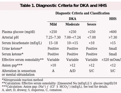
In the past, the most widely used diagnostic criteria for DKA included a blood glucose level >250 mg/dl, a moderate degree of ketonemia, serum bicarbonate <15 mEq/l, arterial pH <7.3, and an increased anion gap metabolic acidosis. Although these criteria served well for research pur-poses, they have significant limitations in clinical practice because the majori-ty of patients with DKA present with mild metabolic acidosis despite
elevat-ed serum glucose and
-hydroxybu-tyrate concentrations. Thus, the bio-chemical criteria for diagnosis were
recently modified.2 Table 1
summa-rizes the biochemical criteria for the diagnosis and empirical subclassifica-tion of DKA and HHS.
The diagnostic criteria for HHS include a plasma glucose concentra-tion >600 mg/dl, a serum osmolality >320 mOsm/kg of water, and the absence of significant ketoacidosis. Although by definition patients with HHS have a serum pH >7.3, a serum bicarbonate >18 mEq/l, and negative ketone bodies in urine and plasma, mild ketonemia may be present. Approximately 50% of patients with HHS have an increased anion gap metabolic acidosis as the result of con-comitant ketoacidosis and/or an
increase in serum lactate levels.2
The assessment of ketonemia, the key diagnostic feature of ketoacidosis, is usually performed by the nitroprus-side reaction. However, clinicians should be aware that the nitroprusside
reaction provides a semiquantitative estimation of acetoacetate and acetone levels but does not recognize the
pres-ence of -hydroxybutyrate, which is
the main ketoacid in DKA. Therefore, this test may underestimate the level of
ketosis. Direct measurement of
-hydroxybutyrate is now available by fingerstick method, which is a more accurate indicator of ketoacidosis.
Common Laboratory Pitfalls
Patients with DKA frequently present with leukocytosis in the absence of infection. However, a leukocyte count
>25,000 mm3 or the presence of
>10% neutrophil bands is seldom seen in the absence of bacterial
infec-tion.22The admission serum sodium is
usually low because of the osmotic flux of water from the intracellular to the extracellular space in the presence of hyperglycemia. To assess the severi-ty of sodium and water deficit, serum sodium may be corrected by adding 1.6 mg/dl to the measured serum sodi-um for each 100 mg/dl of glucose above 100 mg/dl. An increase in serum sodium concentration in the presence of hyperglycemia indicates a rather profound degree of water loss. Extreme hypertriglyceridemia, which may be present during DKA due to impaired lipoprotein lipase activity, may cause lipemic serum with spuri-ous lowering of serum glucose
(pseudonormoglycemia)23 and serum
sodium (pseudohyponatremia)24 in
laboratories still using volumetric test-ing or dilution of samples with ion-specific electrodes.
Diagnostic Criteria and Classification
DKA HHS
Mild Moderate Severe
Plasma glucose (mg/dl) >250 >250 >250 >600 Arterial pH 7.25–7.30 7.00–<7.24 <7.00 >7.30 Serum bicarbonate (mEq/L) 15–18 10–<15 <10 >15 Urine ketone* Positive Positive Positive Small Serum ketone* Positive Positive Positive Small Effective serum osmolality** Variable Variable Variable >320 mOsm/kg
Anion gap*** >10 >12 >12 <12
Alteration in sensorium A A/D S/C S/C
or mental obtundation *Nitroprusside reaction method
**Calculation: Effective serum osmolality: 2[measured Na (mEq/L)] glucose (mg/dl)/18 ***Calculation: Anion gap: (Na) (ClHCO
3) (mEq/L). See text for details. A, alert; D, drowsy; S, stuporous, C, comatose.
31
Fr
om Resear
c
h
to Practice
/
Acute Care of P
atients With Diabetes
The admission serum potassium concentration is usually elevated in
patients with DKA. In a recent series,3
the mean serum potassium in patients with DKA and those with HHS was 5.6 and 5.7 mEq/l, respectively. These high levels occur because of a shift of potassium from the intracellular to the extracellular space due to acidemia, insulin deficiency, and hypertonicity. Similarly, the admission serum phosphate level may be normal or elevated because of metabolic aci-dosis. Dehydration also can lead to increases in total serum protein, albu-min, amylase, and creatine phospho-kinase concentration in patients with acute diabetic decompensation. Finally, serum creatinine, which is measured by a colorimetric method, may be falsely elevated as a result of interference by blood acetoacetate
lev-els.25
Clinicians should remember that not all patients who present with
ketoacidosis have DKA. Patients with chronic ethanol abuse with a recent binge culminating in nausea, vomit-ing, and acute starvation may present with alcoholic ketoacidosis. The key diagnostic feature that differentiates diabetic and alcohol-induced ketoaci-dosis is the concentration of blood
glucose.26While DKA is characterized
by severe hyperglycemia, the presence of ketoacidosis without hyperglycemia in an alcoholic patient is virtually diagnostic of alcoholic ketoacidosis. In addition, some patients with decreased food intake (<500 kcal/day) for several days may present with starvation ketosis. However, a healthy subject is able to adapt to prolonged fasting by increasing ketone clearance by peripheral tissue (brain and mus-cle) and by enhancing the kidney’s ability to excrete ammonia to com-pensate for the increased acid produc-tion. Therefore, a patient with starva-tion ketosis rarely presents with a
serum bicarbonate concentration <18 mEq/l.
TREATMENT
Figures 1 and 2 show the recommend-ed algorithm suggestrecommend-ed by the recent American Diabetes Association posi-tion statement on treatment of DKA
and HHS.27 In general, treatment of
DKA and HHS requires frequent monitoring of patients, correction of hypovolemia and hyperglycemia, replacement of electrolyte losses, and careful search for the precipitating cause(s). A flow sheet is invaluable for recording vital signs, volume and rate of fluid administration, insulin dosage, and urine output and to assess the efficacy of medical therapy. In addition, frequent laboratory moni-toring is important to assess response to treatment and to document resolu-tion of hyperglycemia and/or metabol-ic acidosis. Serial laboratory measure-ments include glucose and electrolytes
Figure 1. Protocol for Management of Adult Patients with Diabetic Ketoacidosis
*Serum Na+ should be corrected for hyperglycemia (for each 100 mg/dl glucose above 100 mg/dl, add 1.6 mEq to sodium value for corrected serum sodium value).
**Upper limits for serum potassium may vary by laboratory. Adapted with permission from reference 27.
32
and, in patients with DKA, venous pH, bicarbonate, and anion gap val-ues until resolution of hyperglycemia and metabolic acidosis.
Fluid Therapy
Patients with DKA and HHS are invariably volume depleted, with an estimated water deficit of ~100 ml/kg
of body weight.28 The initial fluid
therapy is directed toward expansion of intravascular volume and restora-tion of renal perfusion. Isotonic saline (0.9% NaCl) infused at a rate of 500–1,000 mL/h during the first 2 h is usually adequate, but in patients with hypovolemic shock, a third or fourth liter of isotonic saline may be needed to restore normal blood pres-sure and tissue perfusion.
After intravascular volume deple-tion has been corrected, the rate of normal saline infusion should be reduced to 250 mL/h or changed to
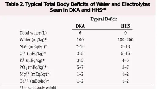
0.45% saline (250–500 mL/h) depending on the serum sodium con-centration and state of hydration. The goal is to replace half of the estimated water deficit over a period of 12–24 h. See Table 2 for typical total body deficits of water and electrolytes in DKA and HHS.
Once the plasma glucose reaches 250 mg/dl in DKA and 300 mg/dl in HHS, replacement fluids should con-tain 5–10% dextrose to allow contin-ued insulin administration until ketonemia is controlled while
avoid-ing hypoglycemia.2 An additional
important aspect of fluid management in hyperglycemic states is to replace the volume of urinary losses. Failure to adjust fluid replacement for urinary losses may delay correction of elec-trolytes and water deficit.
Insulin Therapy
The cornerstone of DKA and HHS
management is insulin therapy. Prospective randomized studies have clearly established the superiority of low-dose insulin therapy in that small-er doses of insulin result in less
hypo-glycemia and hypokalemia.29,30 Insulin
increases peripheral glucose utilization and decreases hepatic glucose produc-tion, thereby lowering blood glucose concentration. In addition, insulin therapy inhibits the release of FFAs from adipose tissue and decreases ketogenesis, both of which lead to the reversal of ketogenesis.
In critically ill and mentally obtunded patients, regular insulin given intravenously by continuous infusion is the treatment of choice. Such patients should be admitted to an intensive care unit or to a step down unit where adequate nursing care and quick turnaround of labora-tory tests results are available. An ini-tial intravenous bolus of regular
Figure 2. Management of Adult Patients with Hyperosmolar Hyperglycemic Syndrome
*This protocol is for patients admitted with mental status change or severe dehydration who require admission to an ICU. **Effective serum osmolality calculation: 2[measured Na (mEq/l)] glucose (mg/dl)/18
***Serum Na+ should be corrected for hyperglycemia (for each 100 mg/dl glucose above 100 mg/dl, add 1.6 mEq to sodium value for corrected serum sodium value).
†Upper limits for serum potassium may vary by laboratory. Adapted with permission from reference 27.
33
Fr
om Resear
c
h
to Practice
/
Acute Care of P
atients With Diabetes
insulin of 0.15 unit/kg of body weight, followed by a continuous infusion of regular insulin at a dose of 0.1 unit/kg/h (5–10 unit/h) should be administered. This will result in a fair-ly predictable decrease in plasma glu-cose concentration at a rate of 65–125
mg/h.31
When plasma glucose levels reach 250 mg/dl in DKA or 300 mg/dl in HHS, the insulin infusion rate is reduced to 0.05 unit/kg/h (3–5 units/h), and dextrose (5–10%) should be added to intravenous fluids. Thereafter, the rate of insulin adminis-tration may need to be adjusted to maintain the above glucose values until ketoacidosis or mental obtunda-tion and hyperosmolality are resolved. During therapy, capillary blood glu-cose should be determined every 1–2 hours at the bedside using a glucose oxidase reagent strip. Blood should be drawn every 2–4 h for determination of serum electrolytes, glucose, blood urea nitrogen, creatinine, magnesium, phosphorus, and venous pH.
A conscious patient with mild DKA could be admitted to a general hospi-tal ward. In such patients, the admin-istration of regular insulin every 1–2 h by subcutaneous or intramuscular route has been shown to be as effec-tive in lowering blood glucose and ketone bodies concentration as giving the entire insulin dose by intravenous
infusion.32,33 Furthermore, it has been
shown that the addition of albumin in the infusate was not necessary to pre-vent adsorption of insulin to the IV
tubing or bag.33 Such patients should
receive the recommended hydrating solution and an initial “priming” dose of regular insulin of 0.4 unit/kg of body weight, given half as intravenous bolus and half as a subcutaneous or
intramuscular injection (Figures 1 and 2). The effectiveness of intramuscular or subcutaneous administration has been shown to be similar; however, subcutaneous injections are easier and less painful.
Potassium
Despite a total body potassium deficit of ~3–5 mEq/kg of body weight, most patients with DKA have a serum potassium level at or above the upper
limits of normal.2 These high levels
occur because of a shift of potassium from the intracellular to the extracellu-lar space due to acidemia, insulin defi-ciency, and hypertonicity. Both insulin therapy and correction of acidosis decrease serum potassium levels by stimulating cellular potassium uptake in peripheral tissues. Therefore, to pre-vent hypokalemia, most patients require intravenous potassium during the course of DKA therapy. Replace-ment with intravenous potassium (two-thirds as potassium chloride [KCl] and one-third as potassium
phosphate [KPO4]) should be initiated
as soon as the serum potassium con-centration is below 5.0 mEq/L. The treatment goal is to maintain serum potassium levels within the normal range of 4–5 mEq/L.
In some hyperglycemic patients with severe potassium deficiency, insulin administration may precipitate
profound hypokalemia,34 which can
induce life-threatening arrhythmias and respiratory muscle weakness. Thus, if the initial serum potassium is lower than 3.3 mEq/L, potassium replacement should begin immediately by an infusion of KCl at a rate of 40 mEq/h, and insulin therapy should be
delayed until serum potassium is ≥3.3
mEq/L (Figure 1).
Bicarbonate
Bicarbonate administration in patients with DKA remains controversial. Severe metabolic acidosis can lead to impaired myocardial contractility, cerebral vasodilatation and coma, and several gastrointestinal complications. However, rapid alkalinization may result in hypokalemia, paradoxical central nervous system acidosis, and worsened intracellular acidosis (as a result of increased carbon dioxide production) with resultant alkalosis. Controlled studies have failed to show any benefit from bicarbonate therapy in patients with DKA with an arterial
pH between 6.9 and 7.1.35 However,
most experts in the field recommend bicarbonate replacement in patients with a pH <7.0. In patients with DKA
with arterial pH ≥7.0, or in patients
with HHS, bicarbonate therapy is not
recommended.2 See Figures 1 and 2
for dosing guidelines.
Phosphate
Total body phosphate deficiency is universally present in patients with DKA, but its clinical relevance and benefits of replacement therapy remain uncertain. Several studies have failed to show any beneficial effect of phos-phate replacement on clinical
out-come.36Furthermore, aggressive
phos-phate therapy is potentially hazardous, as indicated in case reports of children with DKA who developed hypocal-cemia and tetany secondary to
intra-venous phosphate administration.37
Theoretical advantages of phosphate therapy include prevention of respira-tory depression and generation of ery-throcyte 2,3-diphosphoglycerate.
Because of these potential benefits, careful phosphate replacement may be indicated in patients with cardiac dys-function, anemia, respiratory depres-sion, and in those with serum phos-phate concentration lower than 1.0–1.5 mg/dl. If phosphate replace-ment is needed, it should be adminis-tered as a potassium salt, by giving
half as KPO4and half as KCl. In such
patients, because of the risk of hypocalcemia, serum calcium and phosphate levels must be monitored during phosphate infusion.
TRANSITION TO
SUBCUTANEOUS INSULIN
Patients with moderate to severe DKA should be treated with continuous intravenous insulin until ketoacidosis is resolved. Criteria for resolution of ketoacidosis include a blood glucose
Typical Deficit DKA HHS Total water (L) 6 9 Water (ml/kg)* 100 100–200 Na(mEq/kg)* 7–10 5–13 Cl(mEq/kg)* 3–5 5–15 K(mEq/kg)* 3–5 4–6 PO4(mEq/kg)* 5–7 3–7 Mg(mEq/kg)* 1–2 1–2 Ca(mEq/kg)* 1–2 1–2
*Per kg of body weight
Table 2. Typical Total Body Deficits of Water and Electrolytes Seen in DKA and HHS28
34
<200 mg/dl, a serum bicarbonate level
≥18 mEq/L, a venous pH >7.3, and a
calculated anion gap ≤12 mEq/L. The
criteria for resolution of HHS include improvement of mental status, blood glucose <300 mg/dL, and a serum osmolality of <320 mOsm/kg.
When these levels are reached, sub-cutaneous insulin therapy can be started. If patients are able to eat, split-dose therapy with both regular (short-acting) and intermediate-acting insulin may be given. It is easier to make this transition in the morning before breakfast or at dinnertime.
Patients with known diabetes may be given insulin at the dosage they were receiving before the onset of DKA. In patients with newly diag-nosed diabetes, an initial insulin dose of 0.6 unit/kg/day is usually sufficient to achieve and maintain metabolic control. Two-thirds of this total daily dose should be given in the morning and one-third in the evening as a split-mixed dose. If patients are not able to eat, intravenous insulin should be continued while an infusion of 5% dextrose in half-normal saline is given at a rate of 100–200 mL/h.
A critical element to avoid recur-rence of hyperglycemia or ketoacido-sis during the transition period to sub-cutaneous insulin is to allow a 1- or 2-h overlap of intravenous insulin infu-sion during the initiation of subcuta-neous regular insulin to ensure ade-quate plasma insulin levels.
COMPLICATIONS
Hypoglycemia is the most common complication during insulin infusion. Despite the use of low-dose insulin protocols, hypoglycemia is still
report-ed in 10–25% of patients with DKA.3
The failure to reduce insulin infusion rate and/or to use dextrose-containing solutions when blood glucose levels reach 250 mg/dl is the most important risk factor associated with hypo-glycemia during insulin infusion.
Frequent blood glucose monitoring (every 1–2 h) is mandatory to recog-nize hypoglycemia and serious compli-cations. Many patients with hyper-glycemic crises who experience hypo-glycemia during treatment do not expe-rience adrenergic manifestations of sweating, nervousness, fatigue, hunger, and tachycardia despite low blood glu-cose levels (GEU, unpublished observa-tions). Clinicians should be aware that recurrent episodes of hypoglycemia might be associated with a state of hypoglycemia unawareness (loss of
perception of warning symptoms of developing hypoglycemia), which may complicate diabetes management after resolution of hyperglycemic crises.
Hypoglycemia is not frequently observed in patients with HHS. Blood glucose values <60 mg/dl have been reported in <5% of HHS patients
dur-ing intravenous insulin therapy.3
Although the admission serum potassium concentration is commonly elevated in patients with DKA and HHS, during treatment, plasma con-centration of potassium will invariably decrease. Both insulin therapy and cor-rection of acidosis decrease serum potassium levels by stimulating cellular potassium uptake in peripheral tissues. Thus, to prevent hypokalemia, replace-ment with intravenous potassium as soon as the serum potassium
concen-tration is ≤5.0 mEq/L is indicated
(upper limits may vary by laboratory). In patients admitted with normal or reduced serum potassium, insulin administration may precipitate
pro-found hypokalemia.34Thus, if the
ini-tial serum potassium is <3.3 mEq/L, intravenous potassium replacement should begin immediately, and insulin therapy should be held until serum
potassium is ≥3.3 mEq/L (see Figures
1 and 2).
Cerebral edema is a rare but seri-ous complication of DKA. It occurs in ~1% of episodes of DKA in chil-dren38,39and is associated with a
mor-tality rate of 40–90%.40 Clinically,
cerebral edema is characterized by a decreasing level of consciousness and headache, followed by seizures, sphincter incontinence, pupillary changes, papilledema, bradycardia, and respiratory arrest.
It has been hypothesized that cere-bral edema in children with DKA may be caused by the rapid shift in extra-cellular and intraextra-cellular fluids and changes in osmolality due to accumu-lation of osmolytes in brain cells
exposed to hyperosmolar conditions.41
A rapid decrease in extracellular osmolality during treatment would then result in osmotically mediated swelling of the brain. Although osmotic factors and other mechanisms may play a part in the development of cerebral edema, recent data suggest that cerebral edema in children with
DKA is related to brain ischemia.42In
children with DKA, both hypocapnia (which causes cerebral vasoconstric-tion) and extreme dehydration (as determined by a high initial serum urea nitrogen concentration) were
associated with increased risk for cere-bral edema. Hyperglycemia superim-posed on an ischemic insult increases the extent of neurological damage, blood-brain barrier dysfunction, and edema formation.
In addition, it has been shown that a lower serum sodium concentration that does not resolve during therapy may be associated with increased risk
of cerebral edema.42,43The more
fre-quent occurrence of cerebral edema in children than in adults may be explained in part by the fact that chil-dren’s brains have higher oxygen requirements than those of adults and are thus more susceptible to ischemia.
Measures that may decrease the risk of cerebral edema in high-risk patients are gradual replacement of sodium and water deficits in patients with high serum osmolality (maximal reduction in osmolality 3 mOsm/kg/h) and the addition of dextrose to the hydrating solutions once blood glu-cose reaches 250 mg/dl in DKA and
300 mg/dl in HHS.2
Patients with cerebral edema should be transferred to an intensive care unit setting. If signs of increased intracranial pressure or brain hernia-tion are present, only 7–14% of patients recover without permanent significant neurological disabilities.
Treatment includes the immediate
use of intravenous mannitol,40
reduc-tion of fluid administrareduc-tion rate, and possible mechanical ventilation to help
reduce brain swelling.44Corticosteroid
and diuretic therapy have no proven benefit over the immediate use of
intravenous mannitol.40
PREVENTION
The financial burden of DKA and HHS is estimated to exceed $1 billion per year. The most common precipi-tating causes of DKA and HHS include infection, intercurrent illness, psychological stress, and noncompli-ance with therapy. Many episodes could be prevented through better and novel approaches to patient education and effective outpatient treatment programs.
Paramount in this effort is improved education regarding day management. Education on sick-day management should review: • the importance of early contact
with the health care provider • the importance of insulin during an
illness and the reasons never to dis-continue insulin without contacting the health care team
35
Fr
om Resear
c
h
to Practice
/
Acute Care of P
atients With Diabetes
• blood glucose goals and the use of supplemental short- or rapid-acting insulin
• availability of medications to sup-press a fever and treat an infection • initiation of an easily digestible
li-quid diet containing carbohydrates and salt when nauseated
• information for family members on sick-day management and record keeping, including assessing and documenting temperature, respira-tion and pulse, blood glucose and urine/blood ketones, insulin taken, oral intake, and weight
• information for primary care providers and school personnel on the signs and symptoms of new-onset and decompensated diabetes.
Approximately 50% of DKA admissions may be preventable with improved outpatient treatment pro-grams and better adherence to self-care. Outpatient management is more cost effective and can minimize missed days of school or work for patients with diabetes and their family
mem-bers.45 The frequency of
hospitaliza-tions for DKA have been reduced fol-lowing diabetes education programs, improved follow-up care, and access
to medical advice.15,45,46
Additionally, an alarming rise in insulin discontinuation because of economic reasons as the precipitating cause for DKA in urban African Americans illustrates the need for health care legislation guaranteeing reimbursement for medications to treat diabetes.
Novel approaches to patient educa-tion incorporating a variety of health care beliefs and socioeconomic issues are critical to an effective prevention program.
Home blood ketone monitoring
systems, which measure
-hydroxybu-tyrate levels on a fingerstick blood specimen, are now commercially
available.47These systems measure
-hydroxybutyrate levels in 30 seconds with a detection range of 0–6 mmol/L. Clinical studies have shown that
ele-vations of -hydroxybutyrate levels
are extremely common in patients with poorly controlled diabetes, even in the absence of positive urinary
ketones.48 The use of home
glucose-ketone meters may allow early recog-nition of impending ketoacidosis, which may help to guide insulin thera-py at home and may possibly prevent hospitalization for DKA.
HHS occurs frequently in elderly or debilitated patients who do not
recog-nize or cannot treat the symptoms of diabetes and dehydration or, in many cases, who have caregivers who are not knowledgeable about the signs and symptoms of diabetes and the conditions, procedures, and medica-tions that can lead to decompensa-tion. Therefore, additional education as well as the use of glucose and ketone monitoring may decrease the incidence and severity of HHS in this susceptible group.
References
1Graves EJ, Gillium BS: Detailed diagnosis and
procedures: National Discharge Survey, 1995. National Center for Health Statistics. Vital Health Stat 13 (no. 133), 1997
2Kitabchi AE, Umpierrez GE, Murphy MB,
Barrett EJ, Kreisberg RA, Malone JI, Wall BM: Management of hyperglycemic crises in patients with diabetes. Diabetes Care 24:31–53, 2001
3Umpierrez GE, Kelly JP, Navarrete JE, Casals
MMC, Kitabchi AE: Hyperglycemic crises in urban Blacks. Arch Int Med 157:669–675, 1997
4Umpierrez GE, Woo W, Hagopian WA:
Immunogenetic analysis suggests different patho-genesis for obese and lean African-Americans with diabetic ketoacidosis. Diabetes Care 22:1517–1523, 1999
5Javor KA, Kotsanos JG, McDonald RC, Baron
AD, Kesterson JG, Tierney WM: Diabetic ketoacidosis charges relative to medical charges of adult patients with type I diabetes. Diabetes Care 20:349–354, 1997
6Basu A, Close CF, Jenkins D, Krentz AJ,
Nattrass M, Wright AD: Persisting mortality in diabetic ketoacidosis. Diabet Med 10:282–289, 1992
7Gerich JE, Lorenzi M, Bier DM, Tsalikian E,
Schneider V, Karam JH, Forsham PH: Effects of physiologic levels of glucagon and growth hor-mone on human carbohydrate and lipid metabo-lism: studies involving administration of exoge-nous hormone during suppression of endogeexoge-nous hormone secretion with somatostatin. J Clin Invest 57:875–884, 1976
8McGarry JD: Regulation of ketogenesis and the
renaissance of carnitine palmitoyltransferase. Diabetes Metab Rev 5:271–284, 1989
9Felig P, Wahren J: Influence of endogenous
insulin secretion on splanchnic glucose and amino acid metabolism in man. J Clin Invest 50:1702–1711, 1971
10Yu SS, Kitabchi AE: Biological activity of
proinsulin and related polypeptides in the fat tis-sue. J Biol Chem 248:3753–3761, 1973
11Shade DS, Eaton RP: Dose response to insulin
in man: differential effects on glucose and ketone body regulation. J Clin Endocrinol Metab 44:1038–1053, 1977
12Chupin M, Charbonnel B, Chupin F: C-peptide
blood levels in ketoacidosis and in hyperosmolar non-ketotic diabetic coma. Acta Diabet 18:123–128, 1981
13Gerich JE, Martin MM, Recant LL: Clinical
and metabolic characteristics of hyperosmolar nonketotic coma. Diabetes 20:228–238, 1971
14Lindsey CA, Falooma GR, Unger RH: Plasma
glucagon in nonketotic hyperosmolar coma. JAMA 229:1771–1773, 1974
15Kaufman FR, Halvorson M: The treatment and
prevention of diabetic ketoacidosis in children and adolescents with type 1 diabetes mellitus. Pediatr Ann 28:576–582, 1999
16Kauffman FR, Halvorson M: Strategies to
pre-vent diabetic ketoacidosis in children with known type 1 diabetes. Clin Diabetes 15:236–239, 1997
17Polonsky WH, Anderson BJ, Lohrer PA,
Aponte JE, Jacobson AM, Cole CF: Insulin omis-sion in women with IDDM. Diabetes Care 17:1178–1185, 1994
18Rydall AC, Rodin GM, Olmsted MP, Devenyi
RG, Daneman D: Disordered eating behavior and microvascular complications in young women with insulin-dependent diabetes mellitus. N Engl J Med 336:1849–1854, 1997
19Wachtel TJ, Tetu-Mouradjain LM, Goldman
DL, Ellis SE, O’Sullivan PS: Hyperosmolality and acidosis in diabetes mellitus: a three-year experi-ence in Rhode Island. J Gen Int Med 6:495–502, 1991
20Campbell IW, Duncan LJ, Innes JA, MacCuish
AC, Munro JF: Abdominal pain in diabetic metabolic decompensation: clinical significance. JAMA 233:166–168, 1975
21Guisado R, Arieff AI: Neurologic
manifesta-tions of diabetic comas: correlation with bio-chemical alterations in the brain. Metabolism 24:665–669, 1975
22Slovis CM, Mark VG, Slovis RJ, Bain RP:
Diabetic ketoacidosis and infection: leukocyte count and differential as early predictors of infec-tion. Am J Emerg Med 5:1–5, 1987
23Rumbak MJ, Hughes TA, Kitabchi AE:
Pseudonormoglycaemia in diabetic ketoacidosis with elevated triglycerides. Am J Emerg Med 9:61–63, 1991
24Kaminska ES, Pourmatabbed G: Spurious
labo-ratory values in diabetic ketoacidosis and hyper-lipidemia. Am J Emerg Med 11:77–80, 1993
25Assadi FK, John EG, Formell L, Rosenthal IM:
Falsely elevated serum creatinine concentration in ketoacidosis. J Pediatr 107:562–564, 1985
26Umpierrez GE, DiGirolamo M, Tuvlin JA,
Issacs SD, Bhoolasm SM, Kokko JP: Differences in metabolic and hormonal milieu in diabetic-and alcohol-induced ketoacidosis. J Crit Care 15:52–59, 2000
27American Diabetes Association: Hyperglycemic
crises in patients with diabetes mellitus (Position Statement). Diabetes Care 24:1988–1996, 2001
28Ennis ED, Stahl EJVB, Kreisburg RA: The
hyperosmolar hyperglycemic syndrome. Diabetes Rev 2:115–126, 1994
29Kitabchi AE, Fisher JN, Murphy MB, Rumbak
MJ: Diabetic ketoacidosis and the hyperglycemic hyperosmolar nonketotic state. In Joslin’s Diabetes Mellitus 13th ed. Kahn CR, Weir GC, Eds. Philadelphia, Pa., Lea & Febiger, 1994, p. 738–770
30Kitabchi AE, Ayyagari V, Guerra SMO,
36
versus conventional therapy of insulin for treat-ment of diabetic ketoacidosis. Ann Int Med 84:633–638, 1976
31Morris LR, Kitabchi AE: Efficacy of low-dose
insulin therapy for severely obtunded patients in diabetic ketoacidosis. Diabetes Care 3:53–56, 1980
32Fisher JN, Shahshahani MN, Kitabchi AE:
Diabetic ketoacidosis: low-dose insulin therapy by various routes. N Engl J Med 297:238–241, 1977
33Sacks HS, Shahshahani M, Kitabchi AE, Fisher
JN, Young RT: Similar responsiveness of diabetic ketoacidosis to low-dose insulin by intramuscu-lar injection and albumin-preinfusion. Ann Int Med 90:36–42, 1979
34Abramson E, Arky R: Diabetic acidosis with
initial hypokalemia: therapeutic implications. JAMA 196:401–403, 1966
35Morris LR, Murphy MB, Kitabchi AE:
Bicarbonate therapy in severe diabetic ketoacido-sis. Ann Intern Med 105:836–840, 1986
36Fisher JN, Kitabchi AE: A randomized study of
phosphate therapy in the treatment of diabetic ketoacidosis. J Clin Endocrinol Metab 57:177–180, 1983
37Zipf MB, Bacon GE, Spencer ML, Kelch RP,
Hopwood NJ, Hawker CD: Hypocalcemia, hypomagnesemia, and transient hypoparathy-roidism during therapy with potassium
phos-phate in diabetic ketoacidosis. Diabetes Care 2:265–268, 1979
38Bello FA, Sotos JF: Cerebral oedema in diabetic
ketoacidosis in children. Lancet 336:64, 1990
39Duck SC, Wyatt DT: Factors associated with
brain herniation in the treatment of diabetic ketoacidosis. J Pediatr 113:10–14, 1988
40Rosenbloom AL: Intracerebral crises during
treatment of diabetic ketoacidosis. Diabetes Care 13:22–33, 1990
41Finberg L: Why do patients with diabetic
ketoacidosis have cerebral swelling, and why does treatment sometimes make it worse? Arch Pediatr Adolesc Med 150:785–786, 1996
42Glaser N, Barnett P, McCaslin I, Nelson D,
Trainor J, Louie J, Kaufman F, Quayle K, Roback M: Risk factors for cerebral edema in children with diabetic ketoacidosis. The Pediatric Emergency Medicine Collaborative Research Committee of the American Academy of Pediatrics. N Engl J Med 344:264–269, 2001
43Silver SM, Clark EC, Schroeder BM, Sterns
RH: Pathogenesis of cerebral edema after treat-ment of diabetic ketoacidosis. Kidney Int 51:1237–1244, 1997
44White NH: Diabetic ketoacidosis in children.
Endocrinol Metab Clin North Am 29:657–682, 2000
45Laffel LM, Brachett J, Kaufman F, Ho J,
Anderson BJ: Changing the process of diabetes care improves metabolic outcomes and reduces hospitalizations. Qual Manag Health Care 6:53–62, 1998
46Runyan JW: The Memphis chronic disease
pro-gram: comparisons in outcome and the nurse’s extended role. JAMA 231:264–267, 1975
47Byrne HA, Tieszen KL, Hollis S, Dornan TL,
New JP: Evaluation of an electrochemical sensor for measuring blood ketones. Diabetes Care 23:500–503, 2000
48MacGillivray MH, Li PK, Lee JT, Mills BJ,
Vourhess ML, Putnam TI, Schaeffer PA: Elevated plasma beta-hydroxybutyrate concen-trations without ketonuria in healthy insulin-dependent diabetic patients. J Clin Endocrinol Metab 54:665–668, 1982
Guillermo E. Umpierrez, MD, FACP, is an associate professor of medicine, Mary Beth Murphy, RN, MS, CDE, MBA, is research nurse director, and Abbas E. Kitabchi, PhD, MD, FACP, FACE, is a professor of medicine and director of the Division of
Endocrinology, Diabetes, and Metabolism at the University of Tennessee Health Science Center in Memphis.